Current Knowledge on the Multifactorial Regulation of Corpora Lutea Lifespan: The Rabbit Model - MDPI
←
→
Page content transcription
If your browser does not render page correctly, please read the page content below
animals
Review
Current Knowledge on the Multifactorial Regulation of
Corpora Lutea Lifespan: The Rabbit Model
Massimo Zerani                , Angela Polisca *, Cristiano Boiti and Margherita Maranesi

                                          Dipartimento di Medicina veterinaria, Università di Perugia, via San Costanzo 4, 06126 Perugia, Italy;
                                          massimo.zerani@unipg.it (M.Z.); boiti.cristiano@gmail.com (C.B.); margherita.maranesi@unipg.it (M.M.)
                                          * Correspondence: angela.polisca@unipg.it

                                          Simple Summary: Corpora lutea (CL) are temporary endocrine structures that secrete progesterone,
                                          which is essential for maintaining a healthy pregnancy. A variety of regulatory factors come into play
                                          in modulating the functional lifespan of CL, with luteotropic and luteolytic effects. Many aspects of
                                          luteal phase physiology have been clarified, yet many others have not yet been determined, including
                                          the molecular and/or cellular mechanisms that maintain the CL from the beginning of luteolysis
                                          during early CL development. This paper summarizes our current knowledge of the endocrine and
                                          cellular mechanisms involved in multifactorial CL lifespan regulation, using the pseudopregnant
                                          rabbit model.

                                          Abstract: Our research group studied the biological regulatory mechanisms of the corpora lutea (CL),
                                          paying particular attention to the pseudopregnant rabbit model, which has the advantage that the
                                          relative luteal age following ovulation is induced by the gonadotrophin-releasing hormone (GnRH).
                                CL are temporary endocrine structures that secrete progesterone, which is essential for maintaining

                                          a healthy pregnancy. It is now clear that, besides the classical regulatory mechanism exerted by
Citation: Zerani, M.; Polisca, A.;        prostaglandin E2 (luteotropic) and prostaglandin F2α (luteolytic), a considerable number of other
Boiti, C.; Maranesi, M. Current
                                          effectors assist in the regulation of CL. The aim of this paper is to summarize our current knowledge
Knowledge on the Multifactorial
                                          of the multifactorial mechanisms regulating CL lifespan in rabbits. Given the essential role of CL in
Regulation of Corpora Lutea
                                          reproductive success, a deeper understanding of the regulatory mechanisms will provide us with
Lifespan: The Rabbit Model. Animals
                                          valuable insights on various reproductive issues that hinder fertility in this and other mammalian
2021, 11, 296. https://doi.org/
10.3390/ani11020296
                                          species, allowing to overcome the challenges for new and more efficient breeding strategies.

Academic Editor: Rosa María               Keywords: rabbit; corpus luteum; reproduction
García-García and Maria Arias Alvarez
Received: 30 November 2020
Accepted: 21 January 2021
Published: 25 January 2021                1. Introduction
                                                Corpora lutea (CL) are temporary endocrine structures that secrete progesterone,
Publisher’s Note: MDPI stays neutral      which is essential for a healthy pregnancy in most species. In rabbits, the CL develop
with regard to jurisdictional claims in   rapidly following ovulation and reach their maximum size and functional capacity within
published maps and institutional affil-
                                          nine to ten days. This process shows the intense angiogenesis and active granulosa or theca
iations.
                                          cell luteinization of preovulatory follicles, due to the effects of several local angiogenic
                                          growth factors, gonadotropins and other hormones [1,2]. In pregnant rabbits, the CL
                                          lifespan lasts for about 30 days [3]; however, if pregnancy does not occur, the lifespan of
                                          the CL is much shorter, and luteal regression starts around day 12 and ends 16 days after
Copyright: © 2021 by the authors.         ovulation when the peripheral plasma progesterone concentrations drop to the baseline
Licensee MDPI, Basel, Switzerland.        values [4,5]. Therefore, the absence of embryonic signals or the end of gestation activates
This article is an open access article    luteolysis, a comprehensive regressive process that leads to total functional and structural
distributed under the terms and
                                          CL demise, in which prostaglandin (PG) F2 α (PGF2α) plays a central role [6].
conditions of the Creative Commons
                                                Many regulatory factors, including cytokines, growth factors, prostaglandin E2 (PGE2)
Attribution (CC BY) license (https://
                                          and PGF2α released by different CL cell types, including endothelial and local immune
creativecommons.org/licenses/by/
                                          cells and fibroblasts, as well as progesterone and 17β-estradiol released by luteal and
4.0/).

Animals 2021, 11, 296. https://doi.org/10.3390/ani11020296                                                  https://www.mdpi.com/journal/animals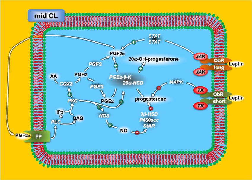
Animals 2021, 11, 296                                                                                          2 of 18

                        follicular cells and hormones, control the functional lifespan of the CL, with luteotrophic
                        and luteolytic effects [7]. However, the overall balance between these contrasting actions
                        varies considerably with the age of the CL and/or in the presence/absence of an embryo [8].
                        Many facets of luteal physiology have been clarified, but others are still poorly understood,
                        including the molecular and/or cellular mechanisms that protect the CL from luteolysis
                        from the early luteal phase. Moreover, the mechanisms that are induced by the adminis-
                        tration of exogenous PGF2α have been extensively investigated in rabbits [9–11] in order
                        to evaluate PG paracrine and/or autocrine functions and other possible regulators that
                        switch on (luteotropic)/off (luteolyic) progesterone production by the CL at a specific stage
                        of its life cycle. However, there are few data on the mechanisms that protect the developing
                        CL from functional luteolysis in the early luteal phase, which starts on day six of a pseu-
                        dopregnancy, when the luteal cells acquire the ability to respond to the luteolytic effects
                        of exogenous PGF2α (luteolytic capacity) [9]. Luteolysis is a key event in reproduction
                        for spontaneously ovulating species, as well as for rabbits, whose mating activity triggers
                        a neuroendocrine reflex, which, combined with GnRH or exogenous human chorionic
                        gonadotropin (hCG) exogenous administration, induces ovulation [12,13].
                              This paper provides a summary of our current knowledge on the endocrine and
                        cellular mechanisms of multifactorial CL lifespan regulation, acquired using the pseudo-
                        pregnant rabbit model, which was able to determine the relative luteal phase following
                        GnRH-induced ovulation. Most of the mechanisms described in this review were observed
                        during our studies on the progressive age-dependent response of the CL to PGF2α con-
                        ducted over a 20-year period [9]. A better understanding of these mechanisms may provide
                        us with valuable insights in the challenge to find more efficient breeding strategies for
                        rabbits, as well as for other species.

                        2. Prostaglandins
                              Prostaglandins (PGs) play a key regulatory role in CL function and the lifespan: PGF2α
                        is the main luteolytic agent produced by the uterine endometrium of numerous mammals,
                        including rabbits, but not by primates [14–16], while PGE2 plays a crucial luteoprotective
                        role, with luteotrophic and/or antiluteolytic effects [6]. In some species, PGF2α and PGE2
                        are produced by the CL [17–21].
                              An essential step in PG biosynthesis is the cyclooxygenase (COX) 1 (COX1) and/or
                        COX2 enzymatic conversion of arachidonic acid (AA)—produced by phospholipase A2
                        (PLA2) activity—into PGH2 [22–24]. This latter PG is then transformed into four struc-
                        turally active PGs (PGE2, PGF2α, PGD2 and PGI2) by specific PG synthases [25]. PGF2α
                        biosynthesis is particular, since three specific ketoreductases catalyze this PG from PGH2,
                        PGD2 or PGE2, respectively [26]. PGE2-9-ketoreductase (PGE2-9-K) is present in the rabbit
                        ovary [27] and CL [28]. This ketoreductase also converts progesterone into its inactive
                        metabolite through its 20α-hydroxysteroid dehydrogenase (HSD) catalytic activity.
                              We previously reported [21] that, in rabbits, intra-luteal PGF2α activates luteoly-
                        sis with an auto-amplification loop: during the mid- and late-luteal phases, it activates
                        COX2 and PGE2-9-K; the former converts AA into PGH2, which is then transformed into
                        PGF2α and PGE2, while the latter is converted into PGF2α through PGE2-9-K activation.
                        Moreover, this enzyme significantly reduces PGF2α-induced progesterone through its
                        20α-hydroxysteroid dehydrogenase (20α-HSD) activity that converts progesterone into
                        20α-OH-progesterone. Late-luteal phase PGE2 production plays another essential role:
                        PGE2-9-K enzymatic activity make this PG the main source of PGF2α synthesis.
                              Arosh et al. [29] suggested that CL PG biosynthesis is mainly directed toward PGE2
                        production rather than PGF2α. In fact, PGH2 conversion into PGE2 (PGE synthase) is
                        150-fold higher than that of PGH2 into PGF2α (PGF synthase) [30]. These results [29,30],
                        combined with our data [21,31], allow us to hypothesize [31] that rabbit CL in the early and
                        mid-luteal phases use the same cellular enzymatic pathways (PLA2/AA/COX2/PGH2/PGE
                        synthase/PGE2) to produce an initial PGE2 amount, while the final luteal production of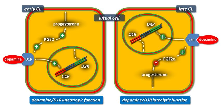
Animals 2021, 11, x 3 of 17
Animals 2021, 11, 296 fold higher than that of PGH2 into PGF2α (PGF synthase) [30]. These results 3[29,30], of 18
combined with our data [21,31], allow us to hypothesize [31] that rabbit CL in the early
and mid-luteal phases use the same cellular enzymatic pathways
(PLA2/AA/COX2/PGH2/PGE synthase/PGE2) to produce an initial PGE2 amount, while
PGE2 (early
the final CL) or
luteal PGF2α (mid-CL)
production of PGE2 is(early
regulated
CL) orbyPGF2α
PGE2-9-K inactivation
(mid-CL) or activation,
is regulated by PGE2-
respectively (Figure 1, upper, functional luteolysis).
9-K inactivation or activation, respectively (Figure 1, upper, functional luteolysis).
Figure
Figure 1. 1. Schematic
Schematic model
model reporting
reporting thethe functional
functional (upper)
(upper) and and structural
structural (lower)
(lower) luteolytic
luteolytic pathways
pathways induced by prostaglandin F2 α (PGF2α) in rabbit mid-corpora lutea (CL) (day 9 of
induced by prostaglandin F2 α (PGF2α) in rabbit mid-corpora lutea (CL) (day 9 of pseudopregnancy).
pseudopregnancy). Since prostaglandin E2 (PGE2)-9-K and 20α-hydroxysteroid dehydrogenase
Since prostaglandin E2 (PGE2)-9-K and 20α-hydroxysteroid dehydrogenase (HSD) represent two dif-
(HSD) represent two different activities of a single enzyme, they are joined. Figure from the study
ferent activities et
by Maranesi ofal.
a single enzyme,
2010 [31]. they are joined.
For acronyms, Figure
see the from
list of the study by
abbreviations inMaranesi
the text. et al. 2010 [31].
For acronyms, see the list of abbreviations in the text.
Several studies have investigated the possible factors involved in PGF2α-induced
Several studies have investigated the possible factors involved in PGF2α-induced
luteolytic capacity
luteolytic capacity during
during the
the mid-luteal
mid-luteal phase
phase [7,9,32–36].
[7,9,32–36]. Interleukin
Interleukin 1 (IL1),
1 (IL1), with
with other
other
cytokines that are normally present in rabbit luteal cells [32,33], are locally involved in thethe
cytokines that are normally present in rabbit luteal cells [32,33], are locally involved in
CLCL function
function control
control leading
leading toto apoptosis
apoptosis asas proinflammatory
proinflammatory mediators[34].
mediators [34].Moreover,
Moreover,
locallyacting
locally acting hormones
hormones andandpro-pro-
andand antiapoptotic
antiapoptotic intra-luteal
intra-luteal factors
factors may interact
may interact dy-
namically. 17β-Estradiol is one of the main luteotropic effectors, since its absence leadsleads
dynamically. 17β-Estradiol is one of the main luteotropic effectors, since its absence to
to luteolysis
luteolysis through
through apoptosis
apoptosis activation
activation [7]. [7]. Nitric
Nitric oxide
oxide synthase
synthase (NOS)
(NOS) andanditsits product
product
nitricoxide
nitric oxide(NO)
(NO)are arealso
alsoknown
knownto to have
have pro-
pro- andand antiapoptotic
antiapoptoticproperties
propertiesthat
thatmodulate
modu-
late various intracellular pathways—in particular B-cell CLL/lymphoma 2 (BCL2)-like 1 1
various intracellular pathways—in particular B-cell CLL/lymphoma 2 (BCL2)-like
(BCL2L1)and
(BCL2L1) and tumor
tumor protein
protein p53
p53 (TP53)
(TP53) proteins
proteins [35].
[35]. InIn rabbits,
rabbits, NOS
NOS luteal
luteal inhibition
inhibition
favors
favors apoptosis
apoptosis [36].
[36].
Our study [31] on the key protein-encoding genes involved in apoptotic mechanism
control revealed that PGF2α induces luteolysis in luteal cells with an acquired luteolytic
capacity through the upregulation of luteal IL1B and TP53 gene transcripts and the down-
regulation of the estrogen receptor 1 (ESR1) and BCL2L1 receptors. This PGF2α-induced
CL regression seems to be the result of two distinct mechanisms: the steroidogenic pathway,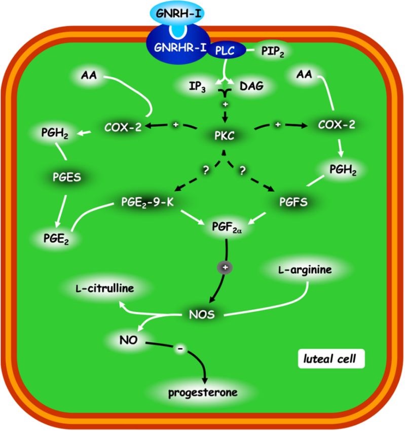
Animals 2021, 11, 296                                                                                           4 of 18

                        by ESR1 downregulation, and the apoptotic pathway, by the dynamic changes of the TP53
                        and BCL2L1 proteins and gene transcripts (Figure 1, lower, structural luteolysis). Finally,
                        aglepristone (RU534), an antiprogestinic, increases progesterone release in rabbit mid- and
                        late-CL, whereas this antiprogestinic reduces PGF2α and enhances PGE2 only during the
                        late-luteal stage [37].

                        3. Nitric Oxide
                              Nitric oxide is a potent vasodilator factor involved in several biological processes,
                        such as neurotransmissions and cytotoxicity, under both physiological and pathological
                        conditions [38,39]. NO is produced by the enzymatic action of NOS, which converts
                        L-arginine into NO and L-citrulline. There are three forms of NOS: two constitutive
                        Ca2+ -dependent forms neuronal NOS (nNOS) and endothelial NOS (eNOS) and an in-
                        ducible Ca2+ -independent form (iNOS) [38,40]. With the exception of neuronal and en-
                        dothelial cells, constitutive eNOS and nNOS are normally expressed in various cell types
                        and produce low levels of NO. Contrastingly, the inducible form only produces large
                        quantities of NO when the expression is activated [38,40]. NOS is present in both ovar-
                        ian stroma and follicular granulosa cells of several mammalian species, including rabbit
                        ovaries, where it regulates steroidogenesis [17,41–44]. The NO/NOS system present in
                        rabbit, rat and mare ovaries is also involved in ovulation [43–49]. All of these studies
                        suggest that NO regulates the key mechanisms of ovarian physiology.
                              In rabbits, NO has a direct antisteroidogenic effect at the luteal level. Numerous
                        in vivo and in vitro experiments have found that NO and NOS are the main targets of
                        PGF2α and effectors of PGF2α-induced luteolysis in competent CL [10,11,17,18,33,50].
                        Ovarian NO is known to be a mediator of the luteolytic action induced by PGF2α in
                        rabbits and other mammalian species [17,51–55]. Ovarian NO might also control the CL
                        lifespan by regulating 17β-estradiol and progesterone concentrations. However, in contrast
                        to earlier findings in rat and human in vitro cultured CL [41,56], NO did not affect the
                        total androgens and 17β-estradiol production in rabbit CL [17]. Contrastingly, in rabbit
                        CL cultured in vitro, the NO donor, sodium nitroprusside, greatly reduced progesterone
                        secretion in all luteal developmental stages [17]. Luteal NOS activity decreases between
                        the early-to mid-luteal phases with elevated steroidogenesis levels [17,57], which increase
                        again in late-CL when the progesterone levels drop and natural luteolysis initiates [5,57].

                        4. Leptin
                             Leptin is a cytokine secreted mainly by adipocytes and encoded by the obese gene [58].
                        Leptin regulates the hypothalamic centers of satiety and energy metabolism through the
                        modulation of various neurotransmitters [59,60].
                             The leptin receptor (ObR) has six isoforms (a–f) resulting from mRNA splice vari-
                        ants [61,62]. ObRa–d and ObRf have identical extracellular and transmembrane do-
                        mains [62,63]. A long intracellular domain of ObRb activates the Janus kinase (JAK)/signal
                        transducer with the subsequent signal transducer and activator of transcription (STAT)
                        phosphorylation [64]. Contrastingly, the short intracellular domain of ObRa, ObRc,d and
                        ObRf activates the mitogen-activated protein kinase (MAPK) pathway [61,65].
                             Several studies have found that various key mammalian reproductive processes
                        are modulated by leptin [66], including steroidogenesis [67,68], ovulation [69,70], preg-
                        nancy [71,72] and menstrual cycles [73,74]. Moreover, leptin is the crucial link between
                        adipose tissue and the reproductive system, since it provides information on whether
                        energy reserves are adequate for normal reproductive function [75].
                             Leptin receptors are present in several tissues of the hypothalamic–pituitary–gonadal
                        (HPG) axis and in pituitary [76], granulosa, theca and interstitial ovary cells [77]. Various
                        studies have reported that leptin directly inhibits steroidogenesis in intracellular signaling
                        pathways in theca, granulosa and luteinized granulosa cells of rodents, bovines and
                        primates [67,68,77–79].
als 2021, 11, x 5 of 17
Leptin receptors are present in several tissues of the hypothalamic–pituitary–gonadal
Animals 2021, 11, 296(HPG)axis and in pituitary [76], granulosa, theca and interstitial ovary cells [77]. Various 5 of 18
studies have reported that leptin directly inhibits steroidogenesis in intracellular signaling
pathways in theca, granulosa and luteinized granulosa cells of rodents, bovines and
primates [67,68,77–79].
Our studies Our studiesCL
on rabbit on [80]
rabbit CL [80]
show thatshow that
leptin leptinprogesterone
affects affects progesterone and PGF2α release
and PGF2α
with different intracellular signaling pathways through different receptors
release with different intracellular signaling pathways through different receptors (long (long ObR and
short ObR). More specifically, leptin inhibits progesterone release through the MAPK
ObR and short ObR). More specifically, leptin inhibits progesterone release through the
cascade (short ObR) and stimulates PGF2α release through the JAK pathway (long ObR)
MAPK cascade (short ObR) and stimulates PGF2α release through the JAK pathway (long
(Figure 2).
ObR) (Figure 2).
Figure 2. Schematic representation of the leptin mechanisms regulating progesterone release in
Figure 2. Schematic representation of the leptin mechanisms regulating progesterone release in rabbit mid-CL. For acronyms,
rabbit mid-CL. For acronyms, see the list of abbreviations in the text.
see the list of abbreviations in the text.
5. Gonadotropin-Releasing Hormone (GnRH)
5. Gonadotropin-Releasing Hormone (GnRH)
Gonadotropin-Releasing Hormone (GnRH) is a hypothalamic-releasing decapeptide
Gonadotropin-Releasing Hormone (GnRH) is a hypothalamic-releasing decapeptide
and a key regulator
and a keyof the mammalian
regulator of the reproductive system. GnRH
mammalian reproductive regulatory
system. GnRH action on
regulatory action on
the reproductive functions is exerted
the reproductive largely
functions via luteinizing
is exerted largely viahormone
luteinizing(LH) and follicle-
hormone (LH) and follicle-
stimulating hormone
stimulating (FSH) secretion,
hormone (FSH)which also affect
secretion, whichsteroidogenesis and germ cell
also affect steroidogenesis and germ cell
developmentdevelopment
[81]. Although the hypothalamus and pituitary gland
[81]. Although the hypothalamus and pituitary gland are the main are
GnRHthe main GnRH
synthesis andsynthesis
action sites,
andseveral studies
action sites, have studies
several reportedhavean extra-hypothalamic presence
reported an extra-hypothalamic presence
of GnRH andofits cognate
GnRH andreceptor (GnRHR)
its cognate receptorin (GnRHR)
numerousinperipheral
numeroustissues, including
peripheral tissues, including
reproductive reproductive
organs such as the gonads,
organs such asprostate,
the gonads,uterine tube, uterine
prostate, placenta andplacenta
tube, mammary and mammary
glands [82]. Previous
glands [82]. Previous studies have highlighted that GnRH regulatessteroid
studies have highlighted that GnRH regulates the ovarian the ovarian steroid
hormones [82]. In rabbit[82].
hormones CL, InGnRH
rabbitadministration was found towas
CL, GnRH administration be associated
found to be with CL
associated with CL
regression with decreased levels of serum progesterone [83]. Contrastingly,
regression with decreased levels of serum progesterone [83]. Contrastingly, no GnRH no GnRH
effects were observed on ovarian
effects were observedtissue steroidtissue
on ovarian production
steroidby other authors
production [84]. authors [84].
by other
The studies conducted
The studies in conducted
our laboratory [85]
in our highlighted
laboratory [85]that the autocrine,
highlighted paracrine
that the autocrine, paracrine
and/or endocrine
and/or roles of GnRH
endocrine typeofI GnRH
roles (GnRH-I)typedirectly diminished
I (GnRH-I) directlythe progesterone
diminished the progesterone
secretion in rabbit
secretion in rabbit CL that had acquired luteolytic competence (Figure 3): via
CL that had acquired luteolytic competence (Figure 3): GnRH-I acts GnRH-I acts via
GnRHR-I by activating
GnRHR-I by phospholipase C (PLC) and stimulating
activating phospholipase C (PLC) andthe inositol trisphosphate
stimulating the inositol trisphosphate
(IP3) and diacylglycerol (DAG) pathways.
(IP3) and diacylglycerol (DAG)Through
pathways. the Through
activationthe of activation
protein kinase C
of protein kinase C
(PKC), these two intracellular messengers stimulate COX2 activity and PGF2α release. This
PG induces (via paracrine, autocrine and/or intracrine mechanisms) an increase in NOS
activity and NO levels [11], which downregulates the progesterone levels [18,31] (Figure 1,
upper, functional luteolysis).Animals 2021, 11, x 6 of 17
(PKC), these two intracellular messengers stimulate COX2 activity and PGF2α release.
Animals 2021, 11, 296 This PG induces (via paracrine, autocrine and/or intracrine mechanisms) an increase
6 of 18in
NOS activity and NO levels [11], which downregulates the progesterone levels [18,31]
(Figure 1, upper, functional luteolysis).
Figure3.3.Schematic
Figure Schematic representation
representation ofof the
the post-receptorial
post-receptorialmechanism
mechanismofofGnRH-I
GnRH-Iregulating thethe
regulating
progesterone release in rabbit CL. The other possible protein kinase C (PKC) targets are
progesterone release in rabbit CL. The other possible protein kinase C (PKC) targets are represented
represented by hatched lines. Figure from the study by Zerani et al. 2010 [85]. For acronyms, see
by hatched lines. Figure from the study by Zerani et al. 2010 [85]. For acronyms, see the list of
the list of abbreviations in the text.
abbreviations in the text.
6.6.Endothelin
Endothelin1 1
Endothelin1 1(ET1),
Endothelin (ET1),a a21-amino
21-aminoacidacidpeptide,
peptide,isisa apotent
potentvasoconstrictor
vasoconstrictorsecreted
secretedbyby
vascular endothelial cells [86,87]. Many tissues other than the vascular
vascular endothelial cells [86,87]. Many tissues other than the vascular endothelium are endothelium are
known to express ET1, including follicular granulosa
known to express ET1, including follicular granulosa cells [88–92]. cells [88–92].
InInrabbit
rabbitCL,CL,ET1
ET1receptors
receptorsareareexpressed
expressedininthethevascular
vascularcompartments
compartmentsand andluteal
luteal
cells,thus
cells, thusevidencing
evidencingthat thatthetheET1
ET1system
systemisisrelated
relatedtotoovarian
ovarianblood
bloodflow flowand
andsteroid
steroid
hormoneproduction
hormone production[91,92].
[91,92]. Moreover,
Moreover, ET1-induced
ET1-induced luteolysis
luteolysis inin rabbits
rabbitson onday
daynine
nineof
ofthe
thepseudopregnancies
pseudopregnancieswas was prevented
prevented by administering
administering captopril,
captopril, the theangiotensin-
angiotensin-
convertingenzyme
converting enzymeinhibitor
inhibitor(ACE).
(ACE).ItItisisimportant
importanttotonote
notethat
thatPGF2α-induced
PGF2α-inducedluteolysis
luteolysis
was not influenced by captopril. These findings indicate that
was not influenced by captopril. These findings indicate that the cascade mechanism the cascade mechanism
triggeredbybyPGF2α
triggered PGF2αdoes doesnotnotrequire
requirethe
therenin–angiotensin
renin–angiotensinsystem systemfor
forinducing
inducingluteolysis
luteolysis
ininrabbits
rabbits[92],[92],which
whichisisiningoodgoodagreement
agreementwith withthe
thedata
dataobtained
obtainedfor forcows
cows[93].
[93].Strict
Strict
cooperationbetween
cooperation betweenendothelin
endothelinand andNO NOisisrequired
requiredfor forendothelial
endothelialcellcellmigration
migrationand and
angiogenesis[94].
angiogenesis [94].ET1
ET1was wasfound
foundtotostimulate
stimulateendothelial
endothelialNOS NOSunder
underdifferent
differentphysio-
physio-
pathological
pathologicalconditions
conditions[95],[95],while
whileNO/NOS
NO/NOSisisaarecognized
recognizedsystemsysteminvolved
involvedininbothboth
PGF2α [11] and ET1 [96]-induced luteal
PGF2α [11] and ET1 [96]-induced luteal regression. regression.
7.7.Adrenocorticotropic
AdrenocorticotropicHormone
Hormone
Adrenocorticotropic
Adrenocorticotropichormone
hormone(ACTH)
(ACTH)isisaamajor
majorcomponent
componentofofthe
thehypothalamic–
hypothalamic–
pituitary–adrenal
pituitary–adrenal (HPA) axis, which is synthesized and secreted by theanterior
(HPA) axis, which is synthesized and secreted by the anteriorpituitary
pituitary
gland
glandininresponse
responsetotostress.
stress.This
Thisresponse
responseisisactivated
activatedby
bythe
thehypothalamic
hypothalamiccorticotropin-
corticotropin-
releasing hormone (CRH), which stimulates pituitary ACTH release, with subsequent
glucocorticoid secretion from the adrenal glands.
There is strong evidence that female reproduction can be impaired by stress [97].
In fact, CRH, ACTH and glucocorticoid negatively affect fertility by targeting the hy-
pothalamic GnRH neurons [98], as well as pituitary LH and/or FSH production andAnimals 2021, 11, x 7 of 17
releasing hormone (CRH), which stimulates pituitary ACTH release, with subsequent
Animals 2021, 11, 296 glucocorticoid secretion from the adrenal glands. 7 of 18
There is strong evidence that female reproduction can be impaired by stress [97]. In
fact, CRH, ACTH and glucocorticoid negatively affect fertility by targeting the
hypothalamic GnRH neurons [98], as well as pituitary LH and/or FSH production and sex
sex steroid
steroid synthesis
synthesis bybyovarian
ovarianfollicles
folliclesand
and CL.
CL. However,
However, the the mechanisms
mechanismsby bywhich
which
hormones
hormones released during stress may inhibit reproductive mechanisms have yettotobebe
released during stress may inhibit reproductive mechanisms have yet
clarified;
clarified;however,
however, any
anydirect
directaction
actionofofACTH
ACTHononovarian
ovarianfunctions
functionsrequires
requiresthe theactivation
activation
ofofmelanocortin receptor 2 (MC2R) [99], while any indirect action requires glucocorticoid
melanocortin receptor 2 (MC2R) [99], while any indirect action requires glucocorticoid
receptor
receptor(GR)
(GR)activation.
activation.
The presence
The presence of of
ACTH
ACTH andand
glucocorticoid
glucocorticoidreceptors in theinluteal
receptors cells ofcells
the luteal rabbit
of CL [100]
rabbit CL
supports the hypothesis that ACTH affects ovarian functions both directly
[100] supports the hypothesis that ACTH affects ovarian functions both directly and and indirectly.
During the early
indirectly. and mid-luteal
During the early phases (days fourphases
and mid-luteal and nine of thefour
(days pseudopregnancies),
and nine of the
ACTH increased the in vitro progesterone and PGE2
pseudopregnancies), ACTH increased the in vitro progesterone andreleases but reduced
PGE2 the PGF2α
releases but
release. Contrastingly, ACTH increased the in vivo plasmatic cortisol levels within four
reduced the PGF2α release. Contrastingly, ACTH increased the in vivo plasmatic cortisol
hours, while the progesterone levels dropped 24 h later and for the following 48 h. Daily
levels within four hours, while the progesterone levels dropped 24 h later and for the
injections of ACTH did not affect the progesterone profile following ovulation. Taken
following 48 h. Daily injections of ACTH did not affect the progesterone profile following
together, these findings indicate that ACTH directly induces the upregulation of luteal
ovulation. Taken together, these findings indicate that ACTH directly induces the
progesterone synthesis through MC2R (Figure 4), while it indirectly blocks CL functions
upregulation of luteal progesterone synthesis through MC2R (Figure 4), while it indirectly
through the cortisol/GR system.
blocks CL functions through the cortisol/GR system.
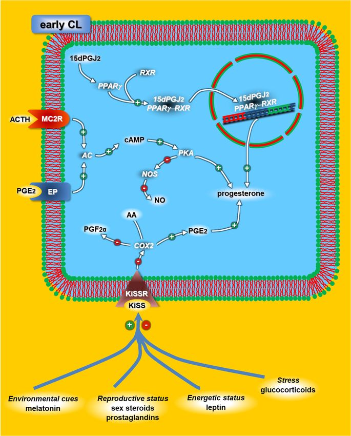
Figure 4. Schematic diagram of the adrenocorticotropic hormone (ACTH), kisspeptins (KiSS) and
peroxisome proliferator-activated receptor (PPAR) mechanisms modulating progesterone release in
early rabbit CL. The effectors that could directly modulate the KiSS/KiSSR (receptor) system at the
CL level are represented by blue lines. For acronyms, see the list of abbreviations in the text.Animals 2021, 11, 296 8 of 18
8. Immunity Mediators
It is now widely accepted that luteolysis is an event mediated by immune effectors
in rabbits and other species, as demonstrated by the presence of immune cells during
spontaneous luteal regression [32]. Luteal immune cells are key modulators of CL activity,
affecting the luteal, endothelial and stromal cells through several cytokines, including
IL1, tumor necrosis factor (TNF)α, monocyte chemoattractant protein-1 (MCP1) and in-
terleukin 2 (IL2) [33,101,102]. In rabbits, during spontaneous luteolysis, the expression
levels of MCP1 and IL1β increased on day 15 of the pseudopregnancies [33]. These find-
ings show the greater influx of macrophages and immune cells observed during luteal
regression [103]. The IL2 transcript increases earlier (day 13 of the pseudopregnancies)
than the other cytokines [33]; in fact, T lymphocytes were detected in rabbit CL before the
macrophages [103].
The IL-1 cytokine is present in the ovaries of various species, including rabbits [104,105].
IL1β has various effects on the ovaries [106]: it inhibits progesterone production, increases
PG synthesis and PGF2 receptor expression, it inhibits COX2 mRNA degradation [107], en-
hances NO production and induces the activation of constitutive and inducible NOS [108].
Our studies report [21] that injecting pseudopregnant rabbits with PGF2α markedly
upregulated COX2 and IL1β mRNA expression and increased PGF2α release and COX2
activity only in CL with acquired luteolytic capacity [31]. These data suggest that IL1β en-
hances intra-luteal PGF2α synthesis by upregulating the luteal function of COX2 and NOS,
thus promoting functional regression in luteal cells that have achieved luteolytic capacity.
9. Peroxisome Proliferator-Activated Receptor
The peroxisome proliferator-activated receptors (PPARs) include a family of three
(a, d and c) nuclear receptor/transcription factors, which regulate steroidogenesis, angio-
genesis, tissue remodeling, cell cycle and apoptosis [109], which are all essential processes
for normal ovarian function [110]. All three PPARs have been detected in the ovaries
of numerous species [111], including rats [110,112], mice [113], pigs [114], sheeps [115],
cows [116–118], rabbits [119] and humans [120,121].
Komar [110] reported that PPARc activation affected the progesterone synthesis in
ovarian cells. In particular, an endogenous activator of PPARc 15d-PGJ2 inhibited both
the basal and gonadotropin-induced production of progesterone in human granulosa
cells [122], while 15d-PGJ2 and ciglitazone, a synthetic PPARc activator, increased pro-
gesterone production by granulosa cells in equine chorionic gonadotropin (eCG)-primed
immature rats [123]. PPARc activation by 15d-PGJ2, ciglitazone or another synthetic activa-
tor, troglitazone, also increased progesterone release by porcine theca and bovine luteal
cells [114,124]. Taken together, these findings indicate that the cell type, stage of cell differ-
entiation, stage of the ovarian cycle and/or animal species influence the effects of PPARc
on progesterone production [110].
Our study [125,126] suggests that PPARc may play a luteotropic role in rabbit CL
through a mechanism that upregulates 3β-hydroxysteroid dehydrogenase (3β-HSD) and
increases progesterone while it downregulates PGF2α and its correlated enzyme COX2 [21]
(Figure 4). Moreover, the significant decrease in PPARc in the luteal cell nucleus during
the late-luteal stage supports the aforementioned mechanism, thus suggesting that this
reduction may be required for luteolysis to take place.
10. Dopamine
The catecholamine dopamine (DA) is a neurotransmitter widely distributed in the
brain and in various peripheral organs of numerous species [127]. DA exerts its physiolog-
ical actions by binding to specific receptors (DR). In mammals, there are five dopamine
receptor subtypes, which are grouped into the D1R-like and D2R-like receptor superfami-
lies [127,128].
D1R-like receptors stimulate the production of the second messenger cyclic adenosine
monophosphate (cAMP); contrastingly, D2R-like receptors inhibit cAMP synthesis, whichD1R-like receptors stimulate the production of the second messenger cyclic
adenosine monophosphate (cAMP); contrastingly, D2R-like receptors inhibit cAMP
synthesis, which decreases the protein kinase A (PKA) activity [128]. In mammals,
dopamine receptors are widely expressed in many organs and tissues, including the
reproductive system [128]. D1R has been detected in the luteal cells of humans [129,130],
Animals 2021, 11, 296 9 of 18
horses [131], rats [132], cows [118] and rabbits [133], suggesting that DA might be directly
involved in the physiological pathways regulating the CL function.
Our studies [133] provide evidence that CL produce DA and that the DA/D1R-D3R
system regulates
decreases the protein thekinase
CL lifespan
A (PKA) byactivity
exerting either
[128]. luteotrophic
In mammals, or luteolytic
dopamine actions
receptors are
depending on the luteal stage. In fact, the DA/D1R-D3R system stimulated
widely expressed in many organs and tissues, including the reproductive system [128]. D1R PGE2 and
progesterone
has synthesis
been detected in the by early
luteal CL,
cells of while
humans it increased
[129,130], PGF2α production
horses [131], andcows
rats [132], decreased
[118]
progesterone
and production
rabbits [133], by mid-
suggesting thatand
DAlate-CL
might (Figure 5). involved in the physiological
be directly
A multi-synaptic
pathways regulating the neural pathway connects the ovaries to the central nervous system
CL function.
in mammals [134]. Moreover,
Our studies [133] provide evidence the ovarian thatinterstitial
CL produce stroma
DA andis composed of many
that the DA/D1R-
different
D3R cellregulates
system types, including neuron-like
the CL lifespan by exertingor neuroendocrine cellsor[135].
either luteotrophic These
luteolytic data
actions
suggest thaton
depending extrinsic andstage.
the luteal intrinsic neurons
In fact, are another paracrine
the DA/D1R-D3R system source of DA
stimulated thatand
PGE2 can
bind its cognate
progesterone receptors
synthesis D1R and
by early D3R init the
CL, while CL, thus
increased supporting
PGF2α the hypothesis
production that
and decreased
the DA/DR system
progesterone plays by
production a physiological
mid- and late-CL role in regulating
(Figure 5). the CL lifespan and functions.
5. Schematic representation of the dopamine receptor-dependent mechanism modulating progesterone
Figure 5.
Figure progesterone production
production
in early (Day 4) and late (Day 9) rabbit CL. D1R: dopamine receptor subtype 1 (cAMP increase) and D3R:
in early (Day 4) and late (Day 9) rabbit CL. D1R: dopamine receptor subtype 1 (cAMP increase) and D3R: dopamine dopamine
receptor
receptor subtype 3 (cAMP decrease). Italic: D1R and D3R genes. Figure from the study by Parillo et al.
subtype 3 (cAMP decrease). Italic: D1R and D3R genes. Figure from the study by Parillo et al. 2014 [132]. 2014 [132].
11. Kisspeptin
A multi-synaptic neural pathway connects the ovaries to the central nervous system
in mammals [134]. Moreover,
The hypothalamic the ovarian interstitial
neuropeptide kisspeptins stroma is composed
(KiSS) of many
are greatly different
involved in
cell types, including
mammalian neuron-like
reproduction. In fact,orthey
neuroendocrine
regulate the cells [135].and
synthesis These data suggest
production that
of GnRH
extrinsic and intrinsic
that are required neurons
to initiate are another
puberty paracrine
and sustain source
normal of DA that can
reproductive bind its
function cognate
[136].
receptors D1R and D3R in the CL, thus supporting the hypothesis that the DA/DR
KiSS and its receptor KiSS1R are expressed in various ovarian structures, including system
plays a physiological role in regulating the CL lifespan and functions.
the CL of several mammalian species [137–139], supporting the hypothesis that these
neuropeptides can regulate the CL lifespan by modulating the steroidogenic enzymes
11. Kisspeptin
The hypothalamic neuropeptide kisspeptins (KiSS) are greatly involved in mammalian
reproduction. In fact, they regulate the synthesis and production of GnRH that are required
to initiate puberty and sustain normal reproductive function [136].
KiSS and its receptor KiSS1R are expressed in various ovarian structures, including
the CL of several mammalian species [137–139], supporting the hypothesis that these
neuropeptides can regulate the CL lifespan by modulating the steroidogenic enzymes
controlling progesterone synthesis. Moreover, Laoharatchatathanin et al. [140] suggested
that KiSS is involved in the luteinization of rat granulosa cells.
Based on data obtained in our laboratory [141], we hypothesize that, besides the well-
known hypothalamic mechanism, the KiSS/KiSS1R system may also directly control the
rabbit CL lifespan via local mechanisms. In fact, KiSS was found to exert a luteotrophic ac-
tion by increasing luteal progesterone synthesis, likely through autocrine and/or paracrine
mechanisms that simultaneously reduce PGF2α production and increase PGE2 production
by blocking COX2 activity (Figure 4). The lack of KiSS1R expression in late-CL suggestsAnimals 2021, 11, 296 10 of 18
that the functional activity of the KiSS/KiSS1R system is mainly regulated by the gene
and/or protein expression of the receptor.
Interestingly, there is sufficient evidence to suggest that the hypothalamic KiSS-1
gene expression is regulated by several factors, including melatonin, gonadal steroids
and leptin, which convey environmental cues and reproductive and metabolic conditions,
respectively [142,143]. The theory that these factors could modulate the luteal KiSS/KiSS1R
system cannot be ruled out (Figure 4).
12. Nerve Growth Factor
The nerve growth factor (NGF), together with brain-derived growth factor and other
neurotrophins, belong to the neurotrophin family [144]. These neurotrophins maintain
normal physiological functions in the central and peripheral nervous systems, including
neural development, differentiation and synaptic plasticity [145,146]. NGF and its receptors
neurotrophic receptor tyrosine kinase 1 (NTRK1) and nerve growth factor receptor (NGFR)
have been found in rabbit ovaries [147,148]. In particular, our studies [149] have evidenced
that NGF from seminal plasma supports the neuroendocrine ovulatory reflex induced
by mating and/or vaginal stimulation through a novel mechanism exerted on the uterus
and/or cervix.
Although there is sufficient experimental evidence suggesting that seminal plasma
NGF is able to induce ovulation in rabbits [147], its potential role in regulating the CL lifes-
pan has not yet been thoroughly explored. To date, we only know that NGF and its cognate
receptor NTRK1 are expressed in rabbit CL at various stages of a pseudopregnancy [149].
Contrastingly, using purified NGF obtained from seminal plasma, Silva et al. [150,151] ob-
served that, in llamas, CL increased vascularization, upregulated cytochrome P450, family
11, subfamily A, member 1/P450 side chain cleavage and steroidogenic acute regulatory
protein transcripts and increased progesterone secretion. All of these findings support the
hypothesis that NGF positively affects CL development. Tribulo et al. [152] and Stewart
et al. [153] obtained similar results in heifers; however, no luteotrophic effect was observed
in alpaca CL using recombinant human NGF [154,155].
13. Conclusions
In conclusion, it is now well-documented that the progressive acquisition of luteolytic
competence by rabbit CL is not only due to their increased sensitivity to PGF2 induced
by the upregulation of PGF2α and its receptors and to the decrease of the luteotropic
factors (E2, PGE2 and ACTH), but it is also caused by several antisteroidogenic factors.
These include, among others, GnRH, ET1 and leptin, which influence the inflammatory,
vascular and apoptotic processes involved in the luteolytic process through interaction with
PGF2α and the NO/NOS system. During PGF2α-induced CL regression with luteolytic
competence, all these factors concomitantly induce the upregulation of NOS, COX2 and
PGE2-9-K activities and gene transcripts for ETI, COX2, IL1B and TP53, as well as the
downregulation of several other transcripts, including ESR1 and BCLXL. Therefore, the
luteolytic effect of PGF2α is the result of its influence on distinct processes involving the
regulation of vasoactive peptides, steroidogenic pathways and apoptotic pathways. How-
ever, despite the increased knowledge on the physiology of rabbit CL, it is recommended
that further research should be undertaken in the near future by a younger generation of
researchers who will be able to apply these new discoveries in the challenge for new rabbit
breeding strategies.
Author Contributions: Conceptualization, C.B., M.M. and M.Z.; infographic, M.Z.; writing—original
draft preparation, M.M. and A.P. and writing—review and editing, C.B. and M.Z. All authors have
read and agreed to the published version of the manuscript.
Funding: This research was partially funded by the BC Red-water (Perugia, Italy), PFZM Kitchen-
brown (Matelica, Italy) and MMZM Bighead (Settevalli, PG, Italy) trusts.Animals 2021, 11, 296 11 of 18
Acknowledgments: In memory of Francesco Parillo (1964–2018), friend and colleague, who substan-
tially contributed to the results of our research group.
Conflicts of Interest: The authors declare no conflict of interest.
Abbreviations
3β-HSD 3β-hydroxysteroid dehydrogenase
15d-PGJ2 15-deoxy-∆12,14-prostaglandin J2
20α-HSD 20α-hydroxysteroid dehydrogenase
AA arachidonic acid
ACE angiotensin converting enzyme
ACTH adrenocorticotropic hormone
BAX BCL2-associated X protein
BCL2L1 B-cell CLL/lymphoma 2 (BCL2)-like 1
cAMP cyclic adenosine monophosphate
CL corpora lutea; COX1
COX1 cyclooxygenase 1
COX2 cyclooxygenase 2
CRH corticotropin-releasing hormone
DA dopamine
DAG diacylglycerol
DR dopamine receptor
eCG equine chorionic gonadotropin
eNOS endothelial NOS
ESR1 estrogen receptor subtype-1
ET1 endothelin 1
GnRH gonadotropin-releasing hormone
GnRH-I gonadotropin-releasing hormone type I
GnRHR gonadotropin-releasing hormone receptor
GR glucocorticoid receptor
hCG human chorionic gonadotropin
HPA hypothalamic-pituitary-adrenal
HPG hypothalamic–pituitary–gonadal
IL1B interleukin 1 Beta
iNOS inducible NOS
IP3 inositol trisphosphate
JAK Janus kinase
KiSS kisspeptin
KiSSR kisspeptin receptor
MAPK mitogen-activated protein kinase
MC2R melanocortin receptor type 2
MCP1 monocyte chemoattractant protein-1
nNOS neuronal NOS
NGF nerve growth factor
NGFR nerve growth factor receptor
NO nitric oxide
NOS nitric oxide synthase
NTRK1 neurotrophic receptor tyrosine kinase 1
ObR leptin (obesity) receptor
P450scc P450 side-chain cleavage
PG prostaglandin
PGD2 prostaglandin D2
PGE2-9-K PGE2-9-ketoreductaseAnimals 2021, 11, 296 12 of 18
PGE2 prostaglandin E2
PGF2α prostaglandin F2α
PGH2 prostaglandin H2
PGI2 prostaglandin I2
PKA protein kinase A
PKC protein kinase C
PLA2 phospholipase A2
PLC phospholipase C
PPAR peroxisome proliferator-activated receptor
RXR retinoid X receptor
StAR steroidogenic acute regulatory protein
STAT signal transducer and activator of transcription
TK tyrosine kinase
TNFα tumor necrosis factor α
TP53 tumor protein p53
References
1. Webb, R.; Woad, K.J.; Armstrong, D.G. Corpus luteum (CL) function: Local control mechanisms. Domest. Anim. Endocrinol. 2002,
23, 277–285. [CrossRef]
2. Acosta, T.J.; Miyamoto, A. Vascular control of ovarian function: Ovulation, corpus luteum formation and regression. Anim.
Reprod. Sci. 2004, 82–83, 127–140. [CrossRef] [PubMed]
3. Spies, H.G.; Hilliard, J.; Sawyer, C.H. Maintenance of corpora lutea and pregnancy in hypophysectomized rabbits. Endocrinology
1968, 83, 354–356. [CrossRef] [PubMed]
4. Scott, R.S.; Rennie, P.I. Factors controlling the life-span of the corpora lutea in the pseudopregnant rabbit. Reprod. Fertil. 1970, 23,
415–422. [CrossRef] [PubMed]
5. Browning, J.Y.; Keyes, P.F.; Wolf, R.C. Comparison of serum progesterone, 20α-dihydroprogesterone, and estradiol-17β in
pregnant and pseudopregnant rabbits: Evidence for postimplantation recognition of pregnancy. Biol. Reprod. 1980, 23, 1014–1019.
[CrossRef]
6. Niswender, G.D.; Juengel, J.L.; Silva, P.J.; Rollyson, M.K.; McIntush, E.W. Mechanisms controlling the function and life span of the
corpus luteum. Physiol. Rev. 2000, 80, 1–29. [CrossRef] [PubMed]
7. Goodman, S.B.; Kugu, K.; Chen, S.H.; Preutthipan, S.; Tilly, K.I.; Tilly, J.L.; Dharmarajan, A.M. Estradiol-mediated suppression of
apoptosis in the rabbit corpus luteum with a shift in expression of Bcl-2 family members favoring cellular survival. Biol. Reprod.
1998, 59, 820–827. [CrossRef]
8. Stocco, C.; Telleria, C.; Gibori, G. The molecular control of corpus luteum formation, function, and regression. Endocr. Rev. 2007,
28, 117–149. [CrossRef]
9. Boiti, C.; Canali, C.; Zerani, M.; Gobbetti, A. Changes in refractoriness of rabbit Corpora lueta to a Prostaglandin F2α analogue,
alfaprostol, during pseudopregnancy. Prostag. Other Lipid. Mediat. 1998, 56, 255–264. [CrossRef]
10. Boiti, C.; Zampini, D.; Zerani, M.; Guelfi, G.; Gobbetti, A. Prostaglandin receptors and role of G protein-activated pathways on
corpora lutea of pseudopregnant rabbit in vitro. J. Endocrinol. 2001, 168, 141–151. [CrossRef]
11. Boiti, C.; Guelfi, G.; Zampini, D.; Brecchia, G.; Gobbetti, A.; Zerani, M. Regulation of nitric-oxide synthase isoforms and role of
nitric oxide during prostaglandin F2α -induced luteolysis in rabbits. Reproduction 2003, 125, 807–816. [CrossRef] [PubMed]
12. Mehaisen, G.M.; Vicente, J.S.; Lavara, R.; Viudes-de-Castro, M.P. Effect of eCG dose and ovulation induction treatments on
embryo recovery and in vitro development post-vitrification in two selected lines of rabbit does. Anim. Reprod. Sci. 2005, 90,
175–184. [CrossRef] [PubMed]
13. Dal Bosco, A.; Rebollar, P.G.; Boiti, C.; Zerani, M.; Castellini, C. Ovulation induction in rabbit does: Current knowledge and
perspectives. Anim. Reprod. Sci. 2011, 129, 106–117. [CrossRef] [PubMed]
14. O’Grady, J.P.; Caldwell, B.V.; Auletta, F.J.; Speroff, L. The effects of an inhibitor of prostaglandin synthesis (indomethacin) on
ovulation, pregnancy, and pseudopregnancy in the rabbit. Prostaglandins 1972, 1, 97–106. [CrossRef]
15. Keyes, P.L.; Bullock, D.W. Effects of prostaglandin F2α on ectopic and ovarian corpora lutea of the rabbit. Biol. Reprod. 1974, 10,
519–525. [CrossRef]
16. Lytton, F.D.; Poyser, N.L. Prostaglandin production by the rabbit uterus and placenta in vitro. J. Reprod. Fertil. 1982, 66, 591–599.
[CrossRef]
17. Gobbetti, A.; Boiti, C.; Canali, C.; Zerani, M. Nitric oxide synthase acutely regulates progesterone production by in vitro cultured
rabbit corpora lutea. J. Endocrinol. 1999, 160, 275–283. [CrossRef]
18. Boiti, C.; Zerani, M.; Zampini, D.; Gobbetti, A. Nitric oxide synthase activity and progesterone release by isolated corpora lutea of
rabbits in early- and mid-luteal phase of pseudopregnancy are differently modulated by prostaglandin E-2 and prostaglandin
F-2α via adenylate cyclase and phospholipase C. J. Endocrinol. 2000, 164, 179–186. [CrossRef]Animals 2021, 11, 296 13 of 18
19. Parillo, F.; Catone, G.; Maranesi, M.; Gobbetti, A.; Gasparrini, B.; Russo, M.; Boiti, C.; Zerani, M. Immunolocalization, gene expres-
sion, and enzymatic activity of cyclooxygenases, prostaglandin E2 -9-ketoreductase, and nitric oxide synthases in Mediterranean
buffalo (Bubalus bubalis) corpora lutea during diestrus. Microsc. Res. Tech. 2012, 75, 1682–1690. [CrossRef]
20. Diaz, F.J.; Anderson, L.E.; Wu, Y.L.; Rabot, A.; Tsai, S.J.; Wiltbank, M.C. Regulation of progesterone and prostaglandin F2α
production in the CL. Mol. Cell Endocrinol. 2002, 191, 65–80. [CrossRef]
21. Zerani, M.; Dall’Aglio, C.; Maranesi, M.; Gobbetti, A.; Brecchia, G.; Mercati, F.; Boiti, C. Intraluteal regulation of prostaglandin
F2α -induced prostaglandin biosynthesis in pseudopregnant rabbits. Reproduction 2007, 133, 1005–1116. [CrossRef] [PubMed]
22. Smith, W.L.; Garavito, R.M.; De Witt, D.L. Prostaglandin endoperoxide H synthase (cyclooxygenase)-1 and -2. J. Biol. Chem. 1996,
271, 33157–33160. [CrossRef] [PubMed]
23. Sakurai, T.; Tamura, K.; Okamoto, S.; Hara, T.; Kogo, H. Possible role of cyclooxygenase 2 in the acquisition of ovarian luteal
function in rodents. Biol. Reprod. 2003, 69, 835–842. [CrossRef] [PubMed]
24. Simmons, D.L.; Botting, R.M.; Hla, T. Cyclooxygenase isozymes: The biology of prostaglandin synthesis and inhibition. Pharmacol.
Rev. 2004, 56, 387–437. [CrossRef]
25. Helliwell, R.J.A.; Adams, L.F.; Mitchell, M.D. Prostaglandin synthases: Recent developments and a novel hypothesis.
Prostaglandins Leukot. Essent. Fat. Acids 2004, 70, 101–113. [CrossRef]
26. Watanabe, K. Prostaglandin F synthase. Prostaglandins Other Lipid Mediat. 2002, 68–69, 401–407. [CrossRef]
27. Schlegel, W.; Daniels, D.; Kruger, S. Partial purification of prostaglandin E2 -9 ketoreductase and prostaglandin-15-
hydroxydehydrogenase from ovarian tissues of rabbits. Clin. Physiol. Biochem. 1987, 5, 336–342.
28. Wintergalen, N.; Thole, H.H.; Galla, H.J.; Schlegel, W. Prostaglandin-E2 -9-reductase from corpus-luteum of pseudopregnant
rabbit is a member of the aldo-keto reductase superfamily featuring 20-alphahydroxysteroid dehydrogenase-activity. Eur. J.
Biochem. 1995, 234, 264–270. [CrossRef]
29. Arosh, J.A.; Banu, S.K.; Chapdelaine, P.; Madore, E.; Sirois, J.; Fortier, M.A. Prostaglandin biosynthesis, transport and signaling in
corpus luteum: A basis for autoregulation of luteal function. Endocrinology 2004, 145, 2551–2560. [CrossRef]
30. Madore, E.; Harvey, N.; Parent, J.; Chapdelaine, P.; Arosh, J.A.; Fortier, M.A. An aldose reductase with 20a-hydroxysteroid
dehydrogenase activity is most likely the enzyme responsible for the production of prostaglandin F2a in the bovine endometrium.
J. Biol. Chem. 2003, 278, 11205–11212. [CrossRef]
31. Maranesi, M.; Zerani, M.; Lilli, L.; Dall’Aglio, C.; Brecchia, G.; Gobbetti, A.; Boiti, C. Expression of luteal estrogen receptor,
interleukin-1, and apoptosis-associated genes after PGF2α administration in rabbits at different stages of pseudopregnancy.
Domest. Anim. Endocrinol. 2010, 39, 116–130. [CrossRef] [PubMed]
32. Krusche, C.A.; Vloet, T.D.; Herrier, A.; Black, S.; Beier, H.M. Functional and structural regression of the rabbit corpus luteum is
associated with altered luteal immune cell phenotypes and cytokine expression patterns. Histochem. Cell Biol. 2002, 118, 479–489.
[CrossRef] [PubMed]
33. Boiti, C.; Guelfi, G.; Zerani, M.; Zampini, D.; Brecchia, G.; Gobbetti, A. Expression patterns of cytokines, p53, and nitric oxide
synthase isoenzymes in corpora lutea of pseudopregnant rabbits during spontaneous luteolysis. Reproduction 2004, 127, 229–238.
[CrossRef] [PubMed]
34. Del Vecchio, R.P.; Sutherland, W.D. Prostaglandin and progesterone production by bovine luteal cells incubated in the presence
or absence of the accessory cells of the corpus luteum and treated with interleukin-1beta, indomethacin and luteinizing hormone.
Reprod. Fertil. Dev. 1997, 9, 651–658. [CrossRef]
35. Leon, L.; Jeannin, J.F.; Bettaieb, A. Post-translational modifications induced by nitric oxide (NO): Implication in cancer cells
apoptosis. Nitric Oxide 2008, 19, 77–83. [CrossRef]
36. Preutthipan, S.; Chen, S.H.; Tilly, J.L.; Kugu, K.; Lareu, R.R.; Dharmarajan, A.M. Inhibition of nitric oxide synthesis potentiates
apoptosis in the rabbit corpus luteum. Reprod. Biomed. Online 2004, 9, 264–270. [CrossRef]
37. Parillo, F.; Dall’Aglio, C.; Brecchia, G.; Maranesi, M.; Polisca, A.; Boiti, C.; Zerani, M. Aglepristone (RU534) effects on luteal
function of pseudopregnant rabbits: Steroid receptors, enzymatic activities, and hormone productions in corpus luteum and
uterus. Anim. Reprod. Sci. 2013, 138, 118–132. [CrossRef]
38. Moncada, S.; Palmer, R.M.J.; Higgs, E.A. Nitric oxide: Physiology, pathophysiology, and pharmacology. Pharmacol. Rev. 1991, 42,
109–142.
39. Schmidt, H.H.; Walter, U. NO at work. Cell 1994, 78, 919–925. [CrossRef]
40. Xie, Q.W.; Nathan, C. The high-output nitric oxide pathway: Role and regulation. J. Leukoc. Biol. 1994, 56, 576–582. [CrossRef]
41. Van Voorhis, B.J.; Dunn, M.S.; Snyder, G.D.; Weiner, C.P. Nitric oxide: An autocrine regulator of human granulosa-luteal cell
steroidogenesis. Endocrinology 1994, 135, 1799–1806. [CrossRef] [PubMed]
42. Chatterjee, S.; Gangula, P.R.; Dong, Y.L.; Yallampalli, C. Immunocytochemical localization of nitric oxide synthase-III in
reproductive organs of female rats during the oestrous cycle. Histochem. J. 1996, 28, 715–723. [CrossRef] [PubMed]
43. Hesla, J.S.; Preutthipan, S.; Maguire, M.P.; Chang, T.S.; Wallach, E.E.; Dharmarajan, A.M. Nitric oxide modulates human chorionic
gonadotropin-induced ovulation in the rabbit. Fertil. Steril. 1997, 67, 548–552. [CrossRef]
44. Jablonka-Shariff, A.; Olson, L.M. Hormonal regulation of nitric oxide synthases and their cell-specific expression during follicular
development in the rat ovary. Endocrinology 1997, 138, 460–468. [CrossRef] [PubMed]
45. Yamauchi, J.; Miyazaki, T.; Iwasaki, S.; Kishi, I.; Kuroshima, M.; Tei, C.; Yoshimura, Y. Effects of nitric oxide on ovulation and
ovarian steroidogenesis and prostaglandin production in the rabbit. Endocrinology 1997, 138, 3630–3637. [CrossRef]Animals 2021, 11, 296 14 of 18
46. Pinto, C.R.F.; Paccamonti, D.L.; Eilts, B.E.; Short, C.R.; Godke, R.A. Effect of nitric oxide synthase inhibitors on ovulation in hCG
stimulated mares. Theriogenology 2002, 58, 1017–1026. [CrossRef]
47. Shukovski, L.; Tsafriri, T. The involvement of nitric oxide in the ovulatory process in the rat. Endocrinology 1995, 135, 2287–2290.
[CrossRef]
48. Bonello, N.; McKie, K.; Jasper, M.; Andrew, L.; Ross, N.; Braybon, E.; Brannstrom, M.; Norman, R.J. Inhibition of nitric oxide:
Effects on interleukin-1á-enhanced ovulation rate, steroid hormone, and ovarian leukocyte distribution at ovulation in the rat.
Biol. Reprod. 1996, 54, 436–445. [CrossRef]
49. Zackrisson, U.; Mikuni, M.; Wallin, A.; Delbro, D.; Hedin, L.; Brannstrom, M. Cell-specific localization of nitric oxide synthases
(NOS) in the rat ovary during follicular development, ovulation and luteal formation. Hum. Reprod. 1996, 11, 2667–2673.
[CrossRef]
50. Boiti, C.; Zampini, D.; Guelfi, G.; Paolocci, F.; Zerani, M.; Gobbetti, A. Expression patterns of endothelial and inducible isoforms
in corpora lutea of pseudopregnant rabbits at different luteal stages. J. Endocrinol. 2002, 173, 285–296. [CrossRef]
51. Korzekwa, A.; Woclawek-Potocka, I.; Okuda, K.; Acosta, T.J.; Skarzynski, D.J. Nitric oxide in bovine corpus luteum: Possible
mechanisms of action in luteolysis. J. Anim. Sci. 2007, 78, 233–242. [CrossRef]
52. Korzekwa, A.; Jaroszewski, J.J.; Bogacki, M.; Deptula, K.M.; Maslanka, T.S.; Acosta, T.J.; Okuda, K.; Skarzynski, D.J. Effects of
prostaglandin F2α and nitric oxide on the secretory function of bovine luteal cells. J. Reprod. Dev. 2004, 50, 411–417. [CrossRef]
[PubMed]
53. Jaroszewski, J.J.; Skarzynski, D.J.; Hansel, W. Nitric oxide as a local mediator of prostaglandin F2-induced regression in bovine
corpus luteum: An in vivo study. Exp. Biol. Med. 2003, 228, 1057–1062. [CrossRef] [PubMed]
54. Roberto da Costa, R.P.; Costa, A.S.; Korzekwa, A.; Platek, R.; Siemieniuch, M.; Galvão, A.; Redmer, D.A.; Robalo Silva, J.;
Skarzynski, D.J.; Ferreira-Dias, G. Actions of a nitric oxide donor on prostaglandins production and angiogenic activity in the
equine endometrium. Reprod. Fertil. Dev. 2008, 20, 674–683. [CrossRef]
55. Ferreira-Dias, G.; Costa, A.S.; Mateus, L.; Korzekwa, A.J.; Galvão, A.; Redmer, D.A.; Lukasik, K.; Szóstek, A.Z.; Woclawek-Potocka,
I.; Skarzynski, D.J. Nitric oxide stimulates progesterone and prostaglandin E2 secretion as well as angiogenic activity in the
equine corpus luteum. Domest. Anim. Endocrinol. 2010, 40, 1–9. [CrossRef]
56. Olson, L.M.; Jones-Burton, C.M.; Jablonka-Shariff, A. Nitric oxide decreases estradiol synthesis of rat luteinized ovarian cells:
Possible role for nitric oxide in functional luteal regression. Endocrinology 1996, 137, 3531–3539. [CrossRef]
57. Holt, J.A. Regulation of progesterone production in the rabbit corpus luteum. Biol. Reprod. 1989, 40, 201–208. [CrossRef]
58. Zhang, Y.; Proenca, R.; Maffei, M.; Barone, M.; Leopold, L.; Friedman, J.M. Positional cloning of the mouse obese gene and its
human homologue. Nature 1994, 372, 425–432. [CrossRef]
59. Morash, B.; Li, A.; Murphy, P.R.; Wilkinson, M.; Ur, E. Leptin gene expression in the brain and pituitary gland. Endocrinology 1999,
140, 5995–5998. [CrossRef]
60. Harris, R.B. Leptin–much more than a satiety signal. Annu. Rev. Nutr. 2000, 20, 45–75. [CrossRef]
61. Houseknecht, K.L.; Portocarrero, C.P. Leptin and its receptors: Regulators of whole-body energy homeostasis. Domest. Anim.
Endocrinol. 1998, 15, 457–475. [CrossRef]
62. Sweeney, G. Leptin signalling. Cell. Signal. 2002, 14, 655–663. [CrossRef]
63. Zabeau, L.; Lavens, D.; Peelman, F.; Eycherman, S.; Vandekerckhove, J.; Tavernier, J. The ins and out of leptin receptor activation.
FEBS Lett. 2003, 546, 45–50. [CrossRef]
64. Baumann, H.; Morella, K.K.; White, D.W.; Dembski, M.; Bailon, P.S.; Kim, H.; Lai, C.F.; Tartaglia, L.A. The full-length leptin
receptor has signaling capabilities of interleukin 6-type cytokine receptors. Proc. Nat. Acad. Sci. USA 1996, 9, 8374–8378.
[CrossRef] [PubMed]
65. Bjørbæk, A.S.; Uotani, S.; da Silva, B.; Flier, J.S. Divergent signaling capacities of the long and short isoforms of the leptin receptor.
J. Biol. Chem. 1997, 272, 32686–32695. [CrossRef] [PubMed]
66. Mantzoros, C.S. Role of leptin in reproduction. Ann. N. Y. Acad. Sci. 2000, 900, 174–183. [CrossRef]
67. Spicer, L.J.; Francisco, C.C. The adipose obese gene product, leptin: Evidence of a direct inhibitory role in ovarian function.
Endocrinology 1997, 138, 3374–3379. [CrossRef]
68. Agarwal, S.K.; Vogel, K.; Weitsman, S.R.; Magoffin, D.A. Leptin antagonizes the insulin-like growth factor-I augmentation of
steroidogenesis in granulosa and theca cells of the human ovary. J. Clin. Endocrinol. Metab. 1999, 84, 1072–1076. [CrossRef]
69. Cunningham, M.J.; Clifton, D.K.; Steiner, R.A. Leptin’s actions on the reproductive axis: Perspectives and mechanisms. Biol.
Reprod. 1999, 60, 216–222. [CrossRef]
70. Ryan, N.K.; Woodhouse, C.M.; Van Der Hoeck, K.H.; Gilchrist, R.B.; Armstrong, D.T.; Norman, R.J. Expression of leptin and its
receptor in the murine ovary: Possible role in the regulation of oocyte maturation. Biol. Reprod. 2002, 66, 1548–1554. [CrossRef]
71. Mounzih, K.; Qiu, J.; Ewart-Toland, A.; Chehab, F.F. Leptin is not necessary for gestation and parturition but regulates maternal
nutrition via a leptin resistance state. Endocrinology 1998, 139, 5259–5262. [CrossRef] [PubMed]
72. Mukherjea, R.; Castonguay, T.W.; Douglass, L.W.; Moser-Veillon, P. Elevated leptin concentrations in pregnancy and lactation:
Possible role as a modulator of substrate utilization. Life Sci. 1999, 65, 1183–1193. [CrossRef]
73. Quinton, N.D.; Laird, S.M.; Kocon, M.A.; Li, T.C.; Smith, R.F.; Ross, R.J.; Blakemore, A.I. Serum leptin levels during the menstrual
cycle of healthy fertile women. Br. J. Biomed. Sci. 1999, 56, 16–19. [PubMed]You can also read