The DNA-helicase HELLS drives ALK ALCL proliferation by the transcriptional control of a cytokinesis-related program
←
→
Page content transcription
If your browser does not render page correctly, please read the page content below
Tameni et al. Cell Death and Disease (2021)12:130
                                    https://doi.org/10.1038/s41419-021-03425-0                                                                                                             Cell Death & Disease

                                     ARTICLE                                                                                                                                                    Open Access

                                    The DNA-helicase HELLS drives ALK− ALCL
                                    proliferation by the transcriptional control of a
                                    cytokinesis-related program
                                    Annalisa Tameni1,2, Elisabetta Sauta 2,3, Valentina Mularoni 2, Federica Torricelli2, Gloria Manzotti2, Giorgio Inghirami4,
                                    Riccardo Bellazzi3, Valentina Fragliasso 2 and Alessia Ciarrocchi 2

                                      Abstract
                                      Deregulation of chromatin modifiers, including DNA helicases, is emerging as one of the mechanisms underlying the
                                      transformation of anaplastic lymphoma kinase negative (ALK−) anaplastic large cell lymphoma (ALCL). We recently
                                      identified the DNA-helicase HELLS as central for proficient ALK−ALCL proliferation and progression. Here we assessed
                                      in detail its function by performing RNA-sequencing profiling coupled with bioinformatic prediction to identify HELLS
                                      targets and transcriptional cooperators. We demonstrated that HELLS, together with the transcription factor YY1,
                                      contributes to an appropriate cytokinesis via the transcriptional regulation of genes involved in cleavage furrow
                                      regulation. Binding target promoters, HELLS primes YY1 recruitment and transcriptional activation of cytoskeleton
                                      genes including the small GTPases RhoA and RhoU and their effector kinase Pak2. Single or multiple knockdowns of
                                      these genes reveal that RhoA and RhoU mediate HELLS effects on cell proliferation and cell division of ALK−ALCLs.
                                      Collectively, our work demonstrates the transcriptional role of HELLS in orchestrating a complex transcriptional
                  1234567890():,;
1234567890():,;
                  1234567890():,;
1234567890():,;

                                      program sustaining neoplastic features of ALK−ALCL.

                                    Introduction                                                                                        of the biological complexity of this disease and of its
                                      Anaplastic large cell lymphomas (ALCLs) are a group of                                            relative rarity that reduces the possibility of extensive
                                    neoplasms arising from the transformation of mature T-                                              profiling7–11. Understanding the mechanisms that
                                    cell1. The presence of chromosomal rearrangements                                                   underline the development and evolution of ALK−ALCLs
                                    involving the ALK gene stratifies ALCLs in ALK+ and                                                  is crucial to define the molecular vulnerabilities of these
                                    ALK− identifying two distinct diseases with different                                               lymphomas and to develop specific therapeutic strategies.
                                    clinical behavior and prognosis2–4. ALK− are known to be                                              DNA helicases are a class of enzymes whose primary
                                    the most aggressive subtype of ALCL and the life expec-                                             function is to unpack DNA. Considered as molecular
                                    tancy of affected patients is significantly reduced by the                                           motors, these proteins unwind the DNA exploiting ATP
                                    lack of effective therapies4–6. The molecular bases of                                              hydrolysis, thus facilitating replication and transcrip-
                                    ALK−ALCLs remain largely unknown as a consequence                                                   tion12–14. For their importance in DNA maintenance,
                                                                                                                                        repair, and chromosomal segregation, helicases are con-
                                                                                                                                        sidered guardian of the genomic stability15,16. Thus, it is
                                    Correspondence: Valentina Fragliasso (valentina.fragliasso@ausl.re.it) or                           not surprising that genetic or transcriptional alterations in
                                    Alessia Ciarrocchi (alessia.ciarrocchi@ausl.re.it)
                                    1                                                                                                   many members of this family have been linked to different
                                     Clinical and Experimental Medicine PhD Program, University of Modena and
                                    Reggio Emilia, Modena 41125, Italy                                                                  disease conditions including predisposition to cancer17–20.
                                    2
                                     Laboratory of Translational Research, Azienda USL-IRCCS di Reggio Emilia,                          Besides, the relevance of these enzymes in promoting
                                    Reggio Emilia 42123, Italy
                                                                                                                                        transcription initiation and cancer progression, has
                                    Full list of author information is available at the end of the article
                                    These authors contributed equally: Valentina Fragliasso, Alessia Ciarrocchi                         recently started to emerge as essential mechanism to
                                    Edited by I. Amelio

                                    © The Author(s) 2021
                                                        Open Access This article is licensed under a Creative Commons Attribution 4.0 International License, which permits use, sharing, adaptation, distribution and reproduction
                                                        in any medium or format, as long as you give appropriate credit to the original author(s) and the source, provide a link to the Creative Commons license, and indicate if
                                    changes were made. The images or other third party material in this article are included in the article’s Creative Commons license, unless indicated otherwise in a credit line to the material. If
                                    material is not included in the article’s Creative Commons license and your intended use is not permitted by statutory regulation or exceeds the permitted use, you will need to obtain
                                    permission directly from the copyright holder. To view a copy of this license, visit http://creativecommons.org/licenses/by/4.0/.

                                    Official journal of the Cell Death Differentiation Association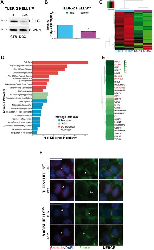
Tameni et al. Cell Death and Disease (2021)12:130                                                             Page 2 of 13

explain their contribution to cell biology17,21,22. In virtue   siRNA transfection
of their centrality in this fundamental mechanism, heli-          MAC2A and TLBR-2 cells (1×10^6) were transfected
cases are currently counted among the most appealing            with 30 nM siRNA concentration for single KD. SiRNA
targets for cancer therapies.                                   transfections were performed using the Cell Line
  We recently demonstrated that HELLS, a DNA helicase           Nucleofector Kit SF and Amaxa 4D Nucleofector (pro-
of the SWI/SNF2 family, is required for proficient               gram DS-130 for TLBR-2, FI115 for MAC2A). Twenty-
ALK−ALCLs proliferation. We showed that HELLS is a              four hours after transfection, cells were harvested and
downstream target of STAT3 and that its expression is           plated 2.5 × 10⁵ cells/ml. For siRNA scramble, we used a
controlled by the ALK−ALCLs specific lncRNA Black-               Silencer Select negative control (Ambion, Life Technol-
Mamba. Besides, we demonstrated that BlackMamba                 ogies). For PAK2 and RHOA we used a Silencer Select
interacts with HELLS driving its positioning on target          Validated siRNAs, ID:s10022 and ID:s759, respectively
promoters, suggesting that the role of this helicase in this    (Ambion, Life Technologies). For RHOU we used two
tumor setting may rely on its transcriptional activity8.        different Silencer Selected Pre-designed siRNAs
  In this work, we explored in detail the molecular             ID:224502, ID: s33826 (Ambion, Life Technologies). For
function of HELLS investigating the transcriptional pro-        YY1, we used a Silencer Select Validated siRNAs: ID:
gram through which this helicase supports ALK−ALCLs.            s14958 (Ambion, Life Technologies)
We provided evidence that HELLS coordinates the
expression of a program of genes involved in cytoskeleton       RNA extraction and quantitative PCR (qRT-PCR)
organization and cytokinesis thus orchestrating timing of         Total RNA was extracted by TRIzol (Thermo Fisher
cell division. We also showed that YY1 is a central partner     Scientific) according to the manufacture’s instructions.
of HELLS in supporting this program.                            One microgram of total RNA was retrotranscribed using
                                                                the iScript cDNA kit, (Biorad). The amplified transcript
Materials and methods                                           level of each specific gene was normalized on CHMP2A
Cell culture and treatments                                     housekeeping. ΔΔCt quantification method was used for
  The human ALK−ALCL cell line MAC2A was a kind                 RT-qPCR analyses. The list of primers used is provided in
gift of Dr. Giorgio Inghirami. The human Breast                 Supplementary Table 2.
Implanted Associated (BIA)-ALCL cell line TLBR-2 was a
kind gift of Dr. Alain Epstein. Cell identity was deter-        Western blot
mined yearly. All cell lines were genotyped and routinely         Western blot analysis was performed using standard
tested for Mycoplasma contamination. Cell lines were            techniques8.
cultured in RPMI-1640 medium (Gibco) supplemented                 The primary antibodies were: HELLS (Rabbit
with 10% FBS at 37 °C in an atmosphere of 5% CO2.               mAb#7998, 1:1000 Cell Signaling Technology), γ PAK2 (E-
TLBR-2 cells were supplemented with IL2 (20U/ml).               9 Mouse mAb sc-373740, 1:1000 Santa Cruz Biotechnol-
  Doxycycline hyclate was purchased from Sigma and              ogy, Inc), RHOA (26C4 Mouse mAb sc-418, 1:1000 Santa
dissolved in H2O.                                               Cruz Biotechnology, Inc), RHOU (Rabbit, PA5-69128,
                                                                1:500 Invitrogen), YY1 (Rabbit, D3D4Q, 1:1000, Cell Sig-
Cell growth and cell division                                   naling Technology), β-tubulin antibody (sc-23949, 1:100,
  For cell growth assays, cells were washed with                Santa Cruz Biotechnology, Inc) and GAPDH (Rabbit
phosphate-buffered saline, seeded at 2.5×10⁵ cells/ml and       mAb#2118, 1:2000, Cell Signaling Technology)
treated with 100 nM doxycycline. Viable cells were                All secondary antibodies (rabbit and mouse) were HRP-
counted by trypan blue exclusion.                               conjugated (GE Healthcare) and diluted 1:3000.
                                                                  Densitometric analysis was performed using the ImageJ
Plasmids and viral infections                                   software.
  pLKO Tet-On vectors expressing shRNAs against
HELLS and lncRNA BlackMamba were generated by                   Immunofluorescence (IF)
cloning synthetic double-stranded oligonucleotides into           Cells were spotted on glass slides using Cytospin
pLKO Tet-On vector (Addgene #21915). Vectors were               (Thermo Scientific), fixed with 4% paraformaldehyde for
packaged into lentiviral particles HEK 293T-cell line and       10 min and permeabilized with 0.1% Triton X-100 for
used for infection of low passages MAC2A or TLBR-2 at           3 min. Dots were blocked in 1% PBS-BSA solution for
multiplicity of infection. Cells were selected with 0.5 or      40 min at room temperature and incubated with phalloi-
1 μg/ml of puromycin (MAC2A and TLBR-2 respectively)            din (Alexa Fluor® 488, Thermo Fisher) for actin staining
for 3 days.                                                     for 50 min. Dots were washed in PBS for three times and
  The list of shRNAs sequences is provided in Supple-           nuclei were stained with DAPI. For microtubules staining
mentary Table 1.                                                we used β-tubulin antibody (sc-23949, 1:100, Santa Cruz

Official journal of the Cell Death Differentiation Association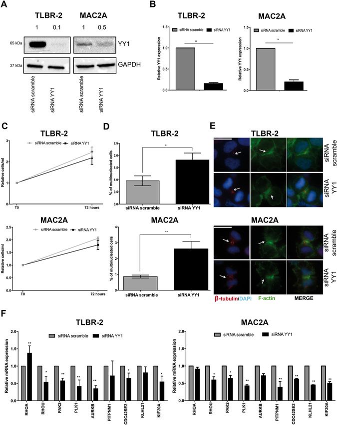
Tameni et al. Cell Death and Disease (2021)12:130                                                              Page 3 of 13

Biotechnology, Inc). Immunofluorescences were detected           preparation protocol. Sequencing was performed using
with Nikon Eclipse (Ni) microscope using 60X.                   Illumina NEXSeq high-output cartridge (double-stranded,
                                                                reads length 75bp-2 ×75).
Chromatin immunoprecipitation (ChIP)                               A sequencing depth of at least 60 million reads for each
  ChIP was performed as previously described8. Chro-            sample was guaranteed.
matin was precipitated with antibodies against HELLS               Sequencing quality was assessed using the FastQC
(4ug, Rabbit Polyclonal, orb178580, Biorbyt), YY1               v0.11.8 software (www.bioinformatics.babraham.ac.uk/
(D3D4Q, 1:100, Cell Signaling Technolgy), or IgG-isotype        projects/fastqc/), showing on average a Phred score per
control (#66362, Cell Signaling Technology). Each qRT-          base >34 in each sample. Raw sequences were then aligned
PCR value was normalized over the appropriate input             to the human reference transcriptome (GRCh38, Gencode
control and reported in graphs as a relative fold on IgG.       release 30) using STAR version 2.723 and gene abundances
  The list of primers used is provided in Supplementary         were estimated with RSEM algorithm (v1.3.1)24. Differential
Table 2.                                                        expression analysis was performed using DESeq2 R pack-
                                                                age25, considering a False Discovery Rate (FDR) of 10% and
Co-Immunoprecipitation (Co-IP)                                  excluding genes with low read counts. Heatmap repre-
  Cells were collected, crosslinked with 1% formaldehyde        sentation and unsupervised hierarchical clustering with a
for 10 min, treated with 1.25 M glycine for 5 min and           complete linkage method were exploited to graphically
resuspended in Buffer A (10 mM HEPES pH 7.9, 1.5 mM             depict differentially expressed genes (FRD < 0.1).
MgCl2, 10 mM KCl, 0.5% NP-40) supplemented with                    Significant genes underwent enrichment analysis, per-
protease inhibitor for 8 min in ice. After centrifugation at    formed on Gene Ontology biological processes, KEGG
3000 rpm for 2 min, the supernatant was collected as the        and Reactome pathways databases via enrichR package26,
cytoplasmic fraction and used as the quality control of the     using a significance threshold of 0.05 on p-value adjusted
experiment. The pellet was washed two times with BUF-           by Benjamini–Hochberg correction for multiple testing.
FER B (10 mM HEPES pH7.9, 1.5 mM MgCl2, 10 mM
KCl), centrifuged at 3000 rpm for 2 min. Nuclei were            Transcriptional factors motif enrichment
resuspended in lysis buffer (50 mM Tris-HCl pH 7.4,               For transcriptional factor motif search, JASPAR 2020
150 mM NaCl, 1 mM EDTA, 1% Triton X-100) supple-                and PROMO (version 3.0.2) software tools were used. A
mented with protease inhibitor and kept for 1 h in rota-        motif similarity threshold of 80% and a dissimilarity level
tion at 4°C. Nuclei extracts were sonicated using a             of 15% were respectively applied for JASPAR 2020 and
Bioruptor® Pico sonicator (Diagenode) and centrifuged at        PROMO prediction results.
16,000 g for 10 min. Supernatant was kept and was
quantified with Bradford. For each experiment, 4 mg of           Statistical analysis
nuclei extracts was used for immunoprecipitation and              Statistical analyses were performed using the GraphPad
150ɥg was kept as input control. Precoating step was            Prism Software (GraphPad). Statistical significance was
performed using Protein A-Sepharose® CL-4B beads (GE            determined using Student’s t test. Each experiment was
Healthcare, Sigma Aldrich), HELLS antibody (Rabbit              replicated multiple time (>3 up to 6).
Polyclonal, orb178580, Biorbyt). Preclearing step was
performed using total nuclear lysate and Protein A-             Results
Sepharose beads for aspecific removal for 1 h in rotation        HELLS controls ALK−ALCL proliferation by transcriptionally
at 4°C. After centrifugation at 500 g for 5 min, we com-        coordinating a panel of cytoskeleton related genes
bined the beads from precoating and supernatant from            involved in cytokinesis
preclearing steps and kept in rotation overnight at 4°C.          To get insight into the transcriptional regulation of
After centrifugation of 500 g for 5 min, the supernatant        HELLS, we performed an RNA-sequencing profiling in
was discarded and washed four times with TBS 1X                 TLBR-2 cells which represent the ALK−ALCL subtype
(50 mM Tris-HCl pH 7.4, 150 mM NaCl). Laemmli                   known as Breast Implanted Associated (BIA)-ALCL8,27.
Sample Buffer 4x (Biorad) was added to the immuno-              We generated inducible HELLS knockdown (KD) lines
precipitated and samples were boiled for 20 min. Co-IP          (TLBR-2 HELLSKD) using doxycycline (DOX)-inducible
was detected by western blot using the secondary anti-          shRNA. HELLS KD was assessed by WB and qRT-PCR
body mouse anti-rabbit IgG HRP conjugate (L27A9)                (Fig. 1A, B) and functionally validated by the reduction in
(#5127, 1:2000, Cell Signaling Technology).                     the expression of already described HELLS-downstream
                                                                targets28 (Supplementary Fig. 1A). After DOX induction,
Library preparation and RNA-sequencing                          the gene expression profile of TLBR-2 HELLSKD cells was
  RNA seq libraries were obtained starting from 500 ng of       analyzed and compared to the one obtained from
total RNA following Illumina TruSeq Stranded TotalRNA           untreated cells used as control. Transcriptional changes

Official journal of the Cell Death Differentiation Association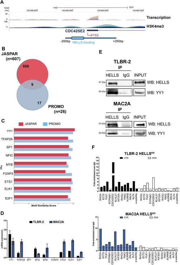
Tameni et al. Cell Death and Disease (2021)12:130                                                                                   Page 4 of 13

  Fig. 1 HELLS transcriptionally controls cytokinesis. A Western blot shows HELLS expression in TLBR-2 HELLSKD cells after 48 h of doxycycline
  (DOX) induction. GAPDH was used as housekeeping gene. B qRT-PCR analysis of HELLS expression in TLBR-2 HELLSKD cells after 48 h of doxycycline
  induction. The values represent mean ± SEM (n = 3) *p < 0.05; **p < 0.01. C The heatmap depicts hierarchical clustering based on the 728
  differentially expressed genes, whose read counts are Z-score normalized. Unsupervised hierarchical clustering was performed between DOX and
  CTRL samples (as indicated by the colored bar on columns) with a complete linkage method. Color intensity for each gene shows Z-score values
  ranging from red for upregulation and green for downregulation D Most significant enriched pathways (adjusted p-value
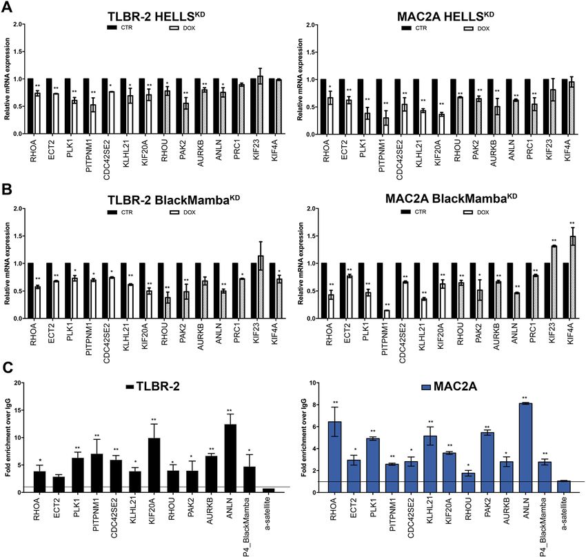
Tameni et al. Cell Death and Disease (2021)12:130                                                                  Page 5 of 13

observed after HELLSKD revealed 728 differentially              BlackMamba and HELLS and further indicating the
expressed genes. 413 were downregulated and 315 were            cytoskeleton related genes as a central node of the tran-
upregulated upon HELLSKD with a FDR < 0.1 (Fig. 1C).            scriptional program supported by this axis in ALK−ALCL.
Gene ontology analysis of HELLS-target genes revealed             To further investigate the direct role of HELLS in the
the enrichment of several categories including cell cycle,      regulation of these genes, we performed ChIP experi-
DNA damage, histone modification, and chromatin                  ments to assess the binding of HELLS on their promoters.
organization. Noticeably, top scoring in this list, there       We observed a significant and specific enrichment of
were multiple Rho GTPases and cytoskeleton related              HELLS binding on 10 out of 11 promoter regions tested in
categories, including cytoskeleton regulation by Rho            both MAC2A and TLBR-2 cells (Fig. 2C). These data
GTPases and Rho GTPases signaling (Fig. 1D, E). This            confirm that HELLS acts as a transcriptional activator of
was particularly interesting since we previously reported       these genes.
that the reduced cellular proliferation displayed by
ALK−ALCL upon HELLS loss is associated with defects in          YY1 is a transcriptional partner of HELLS in ALK−ALCL
cytokinesis and a marked increase in multi-nucleated cells        Little is still known on the transcriptional function of
of which Rho-GTPases are major players8.                        helicases. Thus, to explore how HELLS controls the
  To give a phenotypic readout to these results, we eval-       expression of its target genes we searched for putative
uated the organization of cytoskeleton in T-cells29 per-        transcriptional factors (TFs) able to cooperate with
forming immunostaining for β-tubulin (as a principal            HELLS in ALK−ALCL. A region spanning 500 bp around
constituent of microtubules) and for F-actin upon               the transcriptional starting site (TSS) of each HELLS-
HELLSKD in TLBR-2 and in an additional cell line                target gene was selected (Fig. 3A). A prediction search for
representing the systemic ALK−ALCL subtype (MAC2A)              TF binding sites enriched in these regions was performed
(Supplementary Fig. 1B, C). Although we did not observe         using JASPAR30 and PROMO31 tools. 607 TFs and 26 TFs
differences in the quantity of β-tubulin (Supplementary         were significantly identified by JASPAR and PROMO,
Fig. 1D), a less organized localization of β-tubulin in the     respectively. Merge of these lists resulted in a final list of 9
midzone of central spindle was detected in HELLSKD cells        top scoring TFs and HELLS potential co-factors (pre-
as compared to control cells (Fig. 1F). By contrast, we         dicted to bind up to 90% of the promoters regions used in
observed a profound reorganization of the F-actin with a        this search): Yin Yang 1(YY1), Transcription Factor AP-2
reduced alignment and compaction in cleavage furrow             Alpha (TFAP2A), Sp1 transcriptional factor (SP1),
structure. This phenomenon resulted in an incomplete            Nuclear Factor 1C (NFIC), MYB Proto-Oncogene Tran-
actomyosin ring formation in HELLSKD cells as compared          scription Factor (MYB), Forkhead Box P3 (FOXP3), ETS
to control cells (Fig. 1F), in agreement with the multi-        Proto-Oncogene 1 (ETS1), ETS Transcription Factor
nucleated phenotype previously reported8.                       ELK1 (ELK1) and E2F Transcription Factor 1(E2F1)
  Together, these results indicate that, in ALK−ALCL,           (Fig. 3B, C).
HELLS contributes to an appropriate cytokinesis via the           To validate this analysis, we first tested the basal
transcriptional control of genes involved in contractile        expression of each of these TFs in ALK−ALCL cell lines. 7
ring regulation.                                                out of 9 (78%) TFs were expressed in all tested cell lines
                                                                even if with variable levels. By contrast, no expression was
HELLS controls cytokinesis by directly binding to target        observed for FOXP3 and SP1 (Fig. 3D).
gene promoters                                                    Next, we investigated the potential interaction of these
  Using RT-qPCR, we validated a representative set of           factors with HELLS by co-immunoprecipitation experi-
altered genes involved in cytoskeleton and cytokinesis          ments in nuclear extracts. Among all the TFs investigated,
confirming the RNA-sequencing results and the effect of          only YY1 was found to interact with HELLS in both
HELLS KD on these processes (Fig. 2A, Supplementary             TLBR-2 and MAC2A cells (Fig. 3E), although the levels of
Fig. 2A). Since we showed that HELLS cooperates with            YY1 in MAC2A cells are significantly lower than in
the lncRNA BlackMamba for transcriptional activity in           TLBR2. To enforce the idea of cooperation between
ALK−ALCL and concurs to the lncRNA BlackMamba                   HELLS and YY1, we performed ChIP assay to investigate
pro-oncogenic role in these cells8, we investigated whe-        the binding of YY1 on the promoter regions of HELLS-
ther lncRNA BlackMamba was involved in the HELLS-               target genes both in MAC2A and TLBR-2 cell lines. We
dependent regulation of these genes. Taking advance of          showed that 80% of the tested promoter (9/11) were
the previously generated TLBR-2 and MAC2A Black-                simultaneously bound by both factors supporting the
MambaKD inducible cell lines8, we showed that silencing         hypothesis of a functional cooperation between these two
of this lncRNA resulted in an important reduction of all        factors (Fig. 3F). To establish a hierarchy within their
tested HELLS-target genes (Fig. 2B, Supplementary Fig.          relationship, we analyzed how HELLS KD affects YY1
2B, C) confirming the functional synergy between                 binding on target promoters. Noticeably, YY1 was

Official journal of the Cell Death Differentiation Association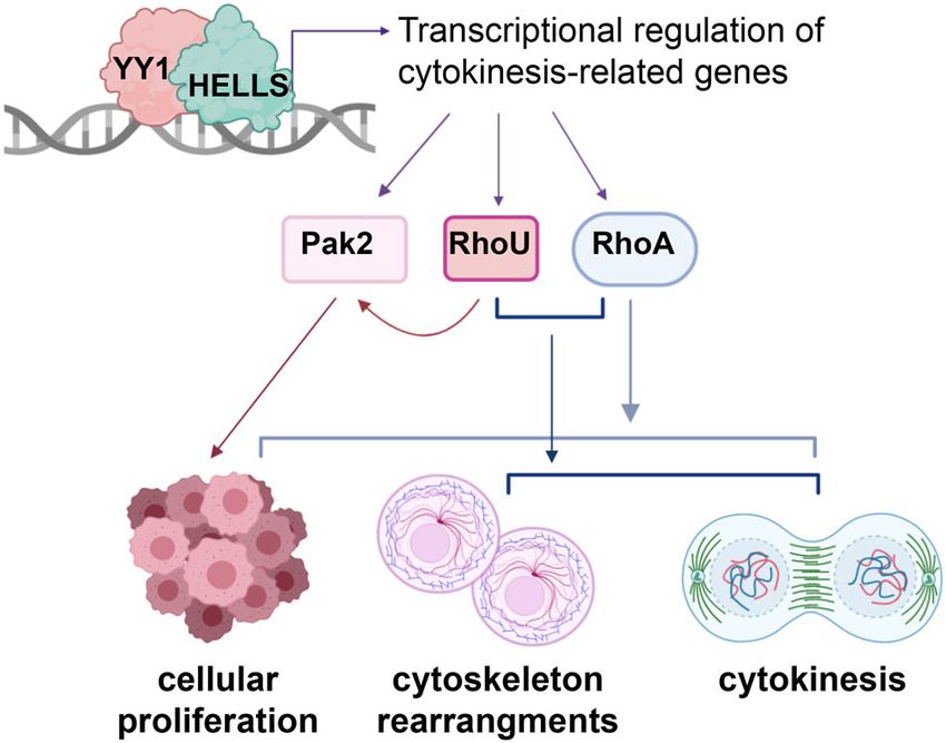
Tameni et al. Cell Death and Disease (2021)12:130                                                                                      Page 6 of 13

  Fig. 2 HELLS binds target gene promoters. A qRT-PCR validation of significantly downregulated genes obtained from RNA-sequencing. White bars
  represent the most significantly downregulated genes (FDR < 0.1) and gray bars represent less significant downregulated genes (FDR > 0.1) in TLBR-2
  HELLSKD cells and MAC2A HELLSKD cells after 48 h of doxycycline (DOX) induction. Each data represent mean ± SEM (n = 3). Two-tailed t-test. *p <
  0.05; **p < 0.01. B qRT-PCR validation of significantly downregulated genes obtained from RNA-sequencing. White bars represent the most
  significantly downregulated genes (FDR < 0.1) and gray bars represent less significant downregulated genes (FDR > 0.1) in TLBR-2 BlackMambaKD cells
  and MAC2A BlackMambaKD cells after 6 days of doxycycline (DOX) induction. Each data represent mean ± SEM (n = 3). Two-tailed t-test. *p < 0.05;
  **p < 0.01. C ChIP qRT-PCR detection of HELLS antibody in a panel of target gene promoters in TLBR-2 and MAC2A.P4_BlackMamba and α-satellite
  were used as positive and negative controls, respectively. The values represent the relative fold enrichment over IgG and are indicated as mean ±
  SEM (n = 3). Two-tailed t-test. *p < 0.05; **p < 0.01.

displaced by these regions in the absence of HELLS while                    partner in the transcriptional program of ALK−ALCL, we
no perturbation of YY1 expression levels was detected                       silenced YY1 with specific siRNA. WB analyses and qRT-
upon HELLS KD (Fig. 3F, Supplementary Fig. 3A) indi-                        PCR confirmed the efficiency of the silencing (Fig. 4A, B).
cating that HELLS is crucial in positioning YY1 on spe-                     Parallel analysis demonstrated that previously reported
cific target genes in the context of ALK−ALCL.                               YY1 target genes33–35 were coherently altered upon its
                                                                            KD (Supplementary Fig. 3B, C). Proliferation analysis of
YY1 cooperates with HELLS to foster the transcription of                    TLBR2 and MAC2A transfected with either siRNA
cytokinesis-related genes                                                   against YY1 or scramble oligos as control did not evidence
  YY1 is an ubiquitous transcriptional factor known to                      a significant effect on cell proliferation (Fig. 4C). However,
have a fundamental role in normal and cancer-related                        we observed a significant increase in the number of multi-
processes32. Thus, to consolidate its role as HELLS                         nucleated cells in YY1KD (Fig. 4D, E).

Official journal of the Cell Death Differentiation Association
Tameni et al. Cell Death and Disease (2021)12:130 Page 7 of 13 Fig. 3 HELLS interacts with the transcriptional factor YY1. A Schematic representation of CDC42SE2 genomic locus showing the position, the level of transcription and the enrichment of H3K4me3 on its putative promoter. B The Venn diagram represents the intersection between transcriptional factors (TFs) identified by JASPAR (n = 607) and PROMO (n = 26) TFs binding prediction tools. The intersection shows the number of HELLS-predicted and common TFs (n = 9). C JASPAR and PROMO similarity scores are represented for top-ranked enriched transcription factors (TFs). D qRT-PCR analysis of top scoring TFs in TLBR-2 and MAC2A cell lines. Each data represent mean ± SEM (n = 3). E Nuclear extract from TLBR-2 and MAC2A cells were tested for the presence of a multi-protein complex. Co-immunoprecipitation experiments were performed to evaluate binding of HELLS to YY1. Western blots are representative of two independent experiments. F ChIP qRT-PCR detection of YY1 in TLBR-2 HELLSKD and MAC2A HELLSKD after 48 h of doxycycline (DOX) induction. KLF4 and α-satellite were used as positive and negative controls, respectively. The values represent the fold enrichment over IgG (representative of three independent experiments). Official journal of the Cell Death Differentiation Association
Tameni et al. Cell Death and Disease (2021)12:130                                                                                        Page 8 of 13

  Fig. 4 YY1 cooperates with HELLS to regulate the multi-nucleated phenotype. A Western Blot shows the knockdown of YY1 36 h post-
  nucleofection with a specific siRNA in TLBR-2 and MAC2A. GAPDH was used as housekeeping gene. B qRT-PCR analysis of YY1 expression after
  siRNA in TLBR-2 and MAC2A cell lines (36 h post-nucleofection). Each data represent mean ± SEM (n = 3). Two-tailed t-test. **p < 0.01. C The
  graph represents the relative growth curve of TLBR-2 and MAC2A 72 h post-nucleofection with YY1 specific siRNA. Data were normalized on
  siRNA scramble values. Each data point represents the mean ± SEM (n = 3). Two-tailed t-test. D. The panels show the percentage of multi-
  nucleated cells in at least 500 cells stained with β-tubulin and F-actin antibodies (48 h post-nucleofection). Each data point represents the mean ±
  SEM (n = 3). Two-tailed t-test. *p < 0.05; **p < 0.01. E Panels show representative immunofluorescences of TLBR-2 and MAC2A stained with DAPI,
  F-actin, and β-tubulin antibodies 48 h post-nucleofection. The scale bar represents 10 μm. F qRT-PCR analysis of a panel of selected HELLS-target
  genes in TLBR-2 and MAC2A 36 h post-nucleofection with specific YY1 siRNA. The values represent mean ± SEM (n = 3). Two-tailed t-test. *p <
  0.05; **p < 0.01.

Official journal of the Cell Death Differentiation Association
Tameni et al. Cell Death and Disease (2021)12:130 Page 9 of 13
Coherently, silencing of YY1 was associated with Immunofluorescences showed that both RhoAKD and
repression of RHOU, PAK2, PLK1, AURKB, PITPNM1, RhoUKD affected β-tubulin organization at central spin-
CDC42SE2, KLHL21, and KIF20A (Fig. 4F). This was in dles or midbody structures. The proper F-actin organi-
line with a positive cooperation of YY1 and HELLS in the zation at contractile ring was also dramatically affected
regulation of cytokinesis genes and with the observed resulting in an abnormal formation of cleavage furrow
increase in multi-nuclei. Intriguingly, YY1 did not affect (Fig. 5E). As expected, combined RhoAKD and RhoUKD
RHOA expression suggesting that other factors partici- resulted in a more pronounced phenotype close to the one
pate to HELLS transcriptional regulation of ALK−ALCLs. obtained with HELLSKD (Fig. 5E).
The involvement of other transcriptional partners likely The analysis of cell proliferation was coherent with
accounts for the fact that YY1KD only partially recapitu- these observations. Single silencing of RhoU and Pak2 did
lates HELLSKD phenotype. not affect significantly cell growth while a significant
decrease in cell proliferation was observed in RhoAKD in
Downstream effects of HELLS are mediated by multiple both cell models. Combined RhoUKD/RhoAKD resulted in
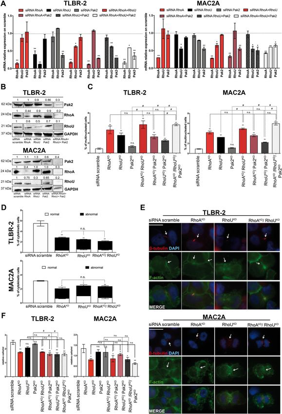
Rho-GTPases and effectors a cell proliferation reduction similarly to single RhoAKD
Rho-GTPases are a family of small GTPase proteins whereas combined RhoUKD/Pak2KD enhanced the effects
involved in many aspects of intracellular actin dynamics of single RhoUKD and Pak2KD but only in TLBR-2.
including cell division. During the cytokinesis, Rho- Notably, triple KD resulted in a significant decrease in cell
GTPases trigger the initiation of the cleavage furrow proliferation, but this reduction was similar to RhoAKD or
from which cytosol division begins36–39. RhoUKD/Pak2KD (Fig. 5F). Collectively, these data
Rho-GTPases are also known for their role in the reg- demonstrated that RhoA and RhoU mediate HELLS
ulation of cell growth and proliferation and cytoskeleton effects on cell proliferation and cell division of
rearrangements in T-lymphomas40. Thus, to consolidate ALK−ALCLs and that RhoA has a prominent role in this
the relevance of the transcriptional program associated process.
with HELLS, we investigated the relevance of these genes
in ALK−ALCL. To this end, we knocked down the Discussion
expression of RhoA, RhoU, and Pak2 with specific siR- Aberrant expression of epigenetic modifiers fostering
NAs. Since Rho-GTPases are known to work in a highly the transcriptional program of neoplastic T-cells is
cooperative manner41–43, we also combined siRNAs to emerging as a common feature and potential vulnerability
simultaneously knockdown two or more of these proteins. of ALK− ALCLs45,46.
Figures 5A and 5B show the efficiency of the silencing at In line with this evidence, here we showed that the
the mRNA and protein level in both MAC2A and TLBR-2 DNA-helicase HELLS supports ALK−ALCL proliferation
cells. by controlling a gene expression program that is func-
We noticed that while RhoAKD or Pak2KD did not alter tional for the execution of cytokinesis and therefore for
the expression of the others, RhoU KD exerted a sig- proficient cell division (Fig. 6).
nificant effect on RhoA expression, suggesting a tight HELLS is a multifunctional protein proved to play,
interplay between these two proteins. among the others, critical roles in DNA methylation,
Morphologically, RhoAKD and RhoUKD led to the chromatin packaging, and development of lymphoid tis-
increase in the percentage of multi-nucleated cells relative sue47. Known also as Lymphoid-specific helicase (Lsh)
to siRNA scramble. By contrast, no such effect was HELLS is required for normal development and survival
observed for Pak2 silencing. This phenomenon slightly of lymphoid and other tissues via chromatin organiza-
increased when RhoAKD and RhoUKD were combined. On tion4849, promotion of DNA double-strand break
the contrary, combined RhoUKD/Pak2KD and RhoAKD/ repair50,51 and chromatin accessibility modification52,53.
Pak2KD did not increase the percentage of multi- In cancer, HELLS is deregulated in several settings i.e.
nucleated cells relative to RhoUKD and RhoAKD, respec- gliomas54, retinoblastoma55,56, prostate28, breast carcino-
tively. Consistently, triple KD resulted in an induction of mas57,58, medulloblastoma59, leukemia60 where it pro-
multinuclei comparable to combined RhoAKD/RhoUKD or motes cellular proliferation and stemness. A significant
RhoAKD (Fig. 5C). To assess if the multi-nucleated phe- part of HELLS activity in these processes is mediated by
notype resulted from cytokinesis failure44, we quantified its transcriptional function. The way through which
cytokinetic cells by immunofluorescence using F-actin HELLS controls gene expression is still partially unde-
and β-tubulin staining. A significant decrease in the per- fined. Its interaction with epigenetic silencers including
centage of total cytokinetic cells and a relative increase in G9a61 and DNMTs62 as well as with transcriptional fac-
abnormal cytokinetic cells were observed in single and tors like E2F328,56 and c-Myc54 has been described. Here,
combined RhoAKD/RhoUKD compared to siRNA scram- we added an additional part of information showing that
ble (Fig. 5D). in the specific context of ALK−ALCL, HELLS interacts
Official journal of the Cell Death Differentiation AssociationTameni et al. Cell Death and Disease (2021)12:130 Page 10 of 13 Fig. 5 (See legend on next page.) and functionally cooperates with the transcription factor two factors physically interact and co-occupy the same YY1. We demonstrate that DNA binding motif for YY1 is regions at the level of target promoters to ensure tran- enriched within the HELLS binding sites and that these scription of a large set of cytokinesis-related genes. Official journal of the Cell Death Differentiation Association
Tameni et al. Cell Death and Disease (2021)12:130                                                                                          Page 11 of 13

  (see figure on previous page)
  Fig. 5 Rho-GTPases mediate HELLS-downstream effects. A qRT-PCR analysis of RhoA, RhoU and Pak2 expression after single or combined siRNAs
  in TLBR-2 and MAC2A cell lines (36 h post-nucleofection). Each data represent mean ± SEM (n = 3). Two-tailed t-test. *p < 0.05, **p < 0.01. B Western
  blots show knockdown of RhoA, RhoU, or Pak2 36 h post-nucleofection with specific siRNAs in TLBR-2 and MAC2A. GAPDH was used as
  housekeeping gene. C The histograms show the percentage of multi-nucleated cells in at least 500 cells TLBR-2 and MAC2A stained with DAPI, F-
  actin, and β-tubulin antibodies (48 h post-nucleofection). Cells were nucleofected with single and combined specific siRNAs against RhoA, RhoU, and
  Pak2. Each data point represents the mean ± SEM (n = 3). Two-tailed t-test. *p < 0.05 and **p < 0.01 relative to siRNA scramble. # p < 0.05 relative to
  RhoAKD, RhoUKD, or Pak2KD. D The histograms show the percentage of cytokinetic cells in at least 500 cells TLBR-2 and MAC2A stained with DAPI, F-
  actin, and β-tubulin antibodies (48 h post-nucleofection). The term “abnormal” refers to cytokinetic cells with defects in cleavage furrow and/or
  microtubules structures. Cells were nucleofected with single and combined specific siRNAs against RhoA and RhoU. Each data point represents the
  mean ± SEM (n = 3). Two-tailed t-test. *p < 0.05 and **p < 0.01 relative to siRNA scramble. E Immunofluorescence of TLBR-2 and MAC2A nucleofected
  with siRNA scramble and single and combined RhoA and RhoU siRNAs (48 h post-nucleofection). Cells were stained with DAPI, F-actin, and β-tubulin
  antibodies. The scale bar represents 10 μm. F Relative growth curve of TLBR-2 and MAC2A nucleofected with siRNA scramble and single or combined
  RhoA and RhoU and Pak2 siRNAs (72 h post-nucleofection). Each data were normalized to siRNA scramble and represent the mean ± SEM (n = 3).
  Two-tailed t-test. *p < 0.05 and **p < 0.01 relative to siiRNA scramble. # p < 0.05 relative to RhoAKD, RhoUKD, or Pak2KD.

                                                                                  By RNA-sequencing profiling we showed that a sig-
                                                                               nificant part of the HELLS transcriptional program in
                                                                               ALK−ALCL converges on the regulation of cytoskeleton
                                                                               and cytokinesis. To the best of our knowledge this is the
                                                                               first time that the transcriptional activity of HELLS is
                                                                               linked to these biological processes.
                                                                                  Cytoskeleton is the structure responsible for cell shape
                                                                               maintenance and organization. It also confers mechanical
                                                                               support to every cellular process from proliferation and
                                                                               division to cell migration, adhesion, and interaction with
                                                                               the surrounding microenvironment.
                                                                                  Of note, recent genetic and molecular profiling studies
                                                                               have unveiled a critical role of cytoskeleton during
                                                                               transformation and progression of T-cells68,69. Although
                                                                               we still lack of a definitive overall view of how cytoske-
                                                                               leton change during lymphomagenesis, the emerging
  Fig. 6 Proposed molecular mechanism. Schematic representation                picture suggests that the cytoskeleton transcends the
  of HELLS role in the regulation of ALK−ALCLs cytokinesis-related             maintenance of cell morphology and polarity providing a
  program (created with BioRender.com).
                                                                               more complex support to T-cells in the response to
                                                                               intrinsic and environmental clues.
                                                                                  Among HELLS-downstream effectors, we identified
   YY1 is an ubiquitously expressed transcriptional factor                     several Rho-GTPases and their related proteins including
with a fundamental role in embryogenesis, adult hema-                          RhoA, RhoU, and Pak2. Single and combined KDs of
topoiesis, differentiation, replication, and cellular pro-                     these proteins highlight that RhoA and RhoU are key
liferation32,63,64. YY1 ensures the proper completion of                       effectors of HELLS program controlling both cell pro-
mitosis and its knockdown leads to defects in cytokinesis                      liferation and cell division.
and accumulation of multi-nucleated cells65. YY1 is often                         RhoA is a key player in T-cell processes70 and its
deregulated in hematopoietic malignancies where it con-                        deregulation has emerged as a central issue of T-
trols the survival and growth of neoplastic cells66,67.                        lymphoma biology68,70–73. Coherently, our results
   Coherently, here we reported that in ALK−ALCL loss of                       demonstrate that RhoA is a fundamental effector of
YY1 is associated with accumulation of multi-nucleated                         HELLS-dependent oncogenic program. At the transcrip-
cells likely attributable to incomplete cytokinesis.                           tional level, RhoA results regulated by HELLS but not by
   The functional relationship between HELLS and YY1                           YY1 suggesting a more complex regulation of this Rho-
has never been described before. Interestingly, we showed                      GTPase in this setting. Limited information are available
that the loss of HELLS leads to YY1 displacement from                          on the regulation of RhoA74, and given its centrality,
DNA. This seems to indicate that HELLS, by altering                            additionally studies are needed to better clarify this point.
chromatin accessibility, works by priming the binding of                          As RhoU can mediate the effects of WNT and
specific transcription factors to target promoters therefore                    STAT3 signaling pathways in regulating cell morphology,
regulating specific set of context specific genes.                               cytoskeletal organization, and proliferation75,76, our data

Official journal of the Cell Death Differentiation Association
Tameni et al. Cell Death and Disease (2021)12:130 Page 12 of 13
provide a new layer of complexity demonstrating a new 2. Morris, S. W. et al. Fusion of a kinase gene, ALK, to a nucleolar protein gene,
role of RhoU in cytokinesis via the STAT3-BlackMamba- NPM, in non-Hodgkin’s lymphoma. Science 263, 1281–1284 (1994).
3. Hapgood, G. & Savage, K. J. The biology and management of systemic ana-
HELLS axis. plastic large cell lymphoma. Blood 126, 17–25 (2015).
The cooperative role of Rho-GTPases in the execution 4. Parrilla Castellar, E. R. et al. ALK-negative anaplastic large cell lymphoma is a
of neoplastic program driven by HELLS is in agreement genetically heterogeneous disease with widely disparate clinical outcomes.
Blood 124, 1473–1480 (2014).
with their fundamental role in the regulation of cell 5. Ferreri, A. J., Govi, S., Pileri, S. A. & Savage, K. J. Anaplastic large cell lymphoma,
proliferation, cell division, and actin polymerization in ALK-negative. Crit. Rev. Oncol. Hematol. 85, 206–215 (2013).
cancer77. 6. Savage, K. J. et al. ALK- anaplastic large-cell lymphoma is clinically and
immunophenotypically different from both ALK+ ALCL and peripheral T-cell
Collectively, our data provide novel insights into the lymphoma, not otherwise specified: report from the International Peripheral T-
mechanism sustaining the progression of ALK−ALCL via Cell Lymphoma Project. Blood 111, 5496–5504 (2008).
the untapped role of HELLS as transcriptional regulator of 7. Crescenzo, R. et al. Convergent mutations and kinase fusions lead to onco-
genic STAT3 activation in anaplastic large cell lymphoma. Cancer Cell 27,
cytokinesis. Since HELLS is expressed in many tumors 516–532 (2015).
and plays a relevant role in the transcription and genomic 8. Fragliasso, V. et al. The novel lncRNA BlackMamba controls the neoplastic
stability of cancers, its pharmacological inhibition may phenotype of ALK(-) anaplastic large cell lymphoma by regulating the DNA
helicase HELLS. Leukemia 34, 2964–2980 (2020).
represent a promising therapeutic strategy in lymphomas 9. Scarfo, I. et al. Identification of a new subclass of ALK-negative ALCL expres-
and in other human neoplasms. sing aberrant levels of ERBB4 transcripts. Blood 127, 221–232 (2016).
10. Hassler, M. R. et al. Insights into the pathogenesis of anaplastic large-cell
Acknowledgements lymphoma through genome-wide DNA methylation profiling. Cell Rep. 17,
The authors are grateful to Marina Grassi for technical help. 596–608 (2016).
11. Luchtel, R. A. et al. Recurrent MSC (E116K) mutations in ALK-negative ana-
Author details plastic large cell lymphoma. Blood 133, 2776–2789 (2019).
1
Clinical and Experimental Medicine PhD Program, University of Modena and 12. Singleton, M. R., Dillingham, M. S. & Wigley, D. B. Structure and mechanism of
Reggio Emilia, Modena 41125, Italy. 2Laboratory of Translational Research, helicases and nucleic acid translocases. Annu. Rev. Biochem. 76, 23–50 (2007).
Azienda USL-IRCCS di Reggio Emilia, Reggio Emilia 42123, Italy. 3Department of 13. Abdel-Monem, M. & Hoffmann-Berling, H. Enzymic unwinding of DNA. 1.
Electrical, Computer and Biomedical Engineering, University of Pavia, Via Purification and characterization of a DNA-dependent ATPase from Escher-
Ferrata 5, 27100 Pavia, Italy. 4Department of Pathology and Laboratory ichia coli. Eur. J. Biochem. 65, 431–440 (1976).
Medicine, Weill Cornell Medicine, New York, NY 10065, USA 14. Lohman, T. M., Tomko, E. J. & Wu, C. G. Non-hexameric DNA helicases and
translocases: mechanisms and regulation. Nat. Rev. Mol. Cell Biol. 9, 391–401 (2008).
15. Larsen, N. B. & Hickson, I. D. RecQ helicases: conserved guardians of genomic
Author contributions
integrity. Adv. Exp. Med. Biol. 767, 161–184 (2013).
A.T. designed research and performed experiments, E.S. performed
16. Brosh, R. M. Jr. DNA helicases involved in DNA repair and their roles in cancer.
bioinformatics analysis, V.M. and G.M. analyzed data, F.T. prepared samples for
Nat. Rev. Cancer 13, 542–558 (2013).
sequencing, R.B. supervised bioinformatics analyses, G.I. contributed to data
17. Chan, E. M. et al. WRN helicase is a synthetic lethal target in microsatellite
analyses, V.F. and A.C. designed research, supervised experiments, and wrote
unstable cancers. Nature 568, 551–556 (2019).
the manuscript.
18. Deans, A. J. & West, S. C. DNA interstrand crosslink repair and cancer. Nat. Rev.
Cancer 11, 467–480 (2011).
Data availability 19. Levran, O. et al. The BRCA1-interacting helicase BRIP1 is deficient in Fanconi
Raw data files of RNA-sequencing have been deposited in EMBL-EBI anemia. Nat. Genet. 37, 931–933 (2005).
ArrayExpress and are accessible through the accession number E-MTAB-9918. 20. Thijssen, P. E. et al. Mutations in CDCA7 and HELLS cause immunodeficiency-
centromeric instability-facial anomalies syndrome. Nat. Commun. 6, 7870 (2015).
Ethics statement 21. Nimonkar, A. V. et al. BLM-DNA2-RPA-MRN and EXO1-BLM-RPA-MRN con-
This research does not involve animal or other human subjects. stitute two DNA end resection machineries for human DNA break repair.
Genes Dev. 25, 350–362 (2011).
Funding 22. Perez-Calero, C. et al. UAP56/DDX39B is a major cotranscriptional RNA-DNA
This work was funded by Ministero della Salute (Ricerca Finalizzata helicase that unwinds harmful R loops genome-wide. Genes Dev. 34, 898–912
No. GR-2016-02364298, V.F.). (2020).
23. Dobin, A. et al. STAR: ultrafast universal RNA-seq aligner. Bioinformatics 29,
Conflict of interest 15–21 (2013).
The authors declare that they have no conflict of interest. 24. Li, B. & Dewey, C. N. RSEM: accurate transcript quantification from RNA-Seq
data with or without a reference genome. BMC Bioinform. 12, 323 (2011).
Publisher’s note 25. Love, M. I., Huber, W. & Anders, S. Moderated estimation of fold change and
Springer Nature remains neutral with regard to jurisdictional claims in dispersion for RNA-seq data with DESeq2. Genome Biol. 15, 550 (2014).
published maps and institutional affiliations. 26. Kuleshov, M. V. et al. Enrichr: a comprehensive gene set enrichment analysis
web server 2016 update. Nucleic Acids Res. 44, W90–W97 (2016).
Supplementary information The online version contains supplementary 27. Lechner, M. G. et al. Survival signals and targets for therapy in breast implant-
material available at https://doi.org/10.1038/s41419-021-03425-0. associated ALK-anaplastic large cell lymphoma. Clin. Cancer Res. 18, 4549–4559
(2012).
28. von Eyss, B. et al. The SNF2-like helicase HELLS mediates E2F3-dependent
Received: 23 October 2020 Revised: 11 January 2021 Accepted: 12 January transcription and cellular transformation. EMBO J. 31, 972–985 (2012).
2021 29. Billadeau, D. D., Nolz, J. C. & Gomez, T. S. Regulation of T-cell activation by the
cytoskeleton. Nat. Rev. Immunol. 7, 131–143 (2007).
30. Fornes, O. et al. JASPAR 2020: update of the open-access database of tran-
scription factor binding profiles. Nucleic Acids Res. 48, D87–D92 (2020).
31. Messeguer, X., Escudero, R., Farre, D., Nunez, O., Martinez, J. & Alba, M. M.
References PROMO: detection of known transcription regulatory elements using species-
1. Swerdlow, S. H. et al. The 2016 revision of the World Health Organization tailored searches. Bioinformatics 18, 333–334 (2002).
classification of lymphoid neoplasms. Blood 127, 2375–2390 (2016).
Official journal of the Cell Death Differentiation AssociationTameni et al. Cell Death and Disease (2021)12:130 Page 13 of 13
32. Lu, Z. et al. Polycomb Group Protein YY1 is an essential regulator of hema- 55. Benavente, C. A., Finkelstein, D., Johnson, D. A., Marine, J. C., Ashery-Padan, R. &
topoietic stem cell quiescence. Cell Rep. 22, 1545–1559 (2018). Dyer, M. A. Chromatin remodelers HELLS and UHRF1 mediate the epigenetic
33. Morales-Martinez, M. et al. Regulation of Kruppel-Like Factor 4 (KLF4) expres- deregulation of genes that drive retinoblastoma tumor progression. Onco-
sion through the transcription factor Yin-Yang 1 (YY1) in non-Hodgkin B-cell target 5, 9594–9608 (2014).
lymphoma. Oncotarget 10, 2173–2188 (2019). 56. Zocchi, L. et al. Chromatin remodeling protein HELLS is critical for retino-
34. Riggs, K. J. et al. Yin-yang 1 activates the c-myc promoter. Mol. Cell Biol. 13, blastoma tumor initiation and progression. Oncogenesis 9, 25 (2020).
7487–7495 (1993). 57. Tao, Y., Liu, S., Briones, V., Geiman, T. M. & Muegge, K. Treatment of breast
35. Lin, J., He, Y., Chen, J., Zeng, Z., Yang, B. & Ou, Q. A critical role of transcription cancer cells with DNA demethylating agents leads to a release of Pol II stalling
factor YY1 in rheumatoid arthritis by regulation of interleukin-6. J. Autoimmun. at genes with DNA-hypermethylated regions upstream of TSS. Nucleic Acids
77, 67–75 (2017). Res. 39, 9508–9520 (2011).
36. Liu, Z. & Weiner, O. D. Positioning the cleavage furrow: All you need is Rho. J. 58. He, X. et al. Chromatin remodeling factor LSH drives cancer progression by
Cell Biol. 213, 605–607 (2016). suppressing the activity of fumarate hydratase. Cancer Res. 76, 5743–5755
37. Chircop, M. Rho GTPases as regulators of mitosis and cytokinesis in mam- (2016).
malian cells. Small GTPases 5, e29770 (2014). 59. Robinson, M. H. et al. Upregulation of the chromatin remodeler HELLS is
38. Miller, A. L. & Bement, W. M. Regulation of cytokinesis by Rho GTPase flux. Nat. mediated by YAP1 in Sonic Hedgehog Medulloblastoma. Sci. Rep. 9, 13611
Cell Biol. 11, 71–77 (2009). (2019).
39. Piekny, A., Werner, M. & Glotzer, M. Cytokinesis: welcome to the Rho zone. 60. Lee, D. W. et al. Proliferation-associated SNF2-like gene (PASG): a SNF2 family
Trends Cell Biol. 15, 651–658 (2005). member altered in leukemia. Cancer Res. 60, 3612–3622 (2000).
40. Voena, C. & Chiarle, R. RHO family GTPases in the biology of lymphoma. Cells 8, 61. Myant, K. et al. LSH and G9a/GLP complex are required for developmentally
646 (2019). programmed DNA methylation. Genome Res. 21, 83–94 (2011).
41. Guo, F., Cancelas, J. A., Hildeman, D., Williams, D. A. & Zheng, Y. Rac GTPase 62. Myant, K. & Stancheva, I. LSH cooperates with DNA methyltransferases to
isoforms Rac1 and Rac2 play a redundant and crucial role in T-cell develop- repress transcription. Mol. Cell Biol. 28, 215–226 (2008).
ment. Blood 112, 1767–1775 (2008). 63. Gordon, S., Akopyan, G., Garban, H. & Bonavida, B. Transcription factor YY1:
42. Choudhari, R. et al. Redundant and nonredundant roles for Cdc42 and Rac1 in structure, function, and therapeutic implications in cancer biology. Oncogene
lymphomas developed in NPM-ALK transgenic mice. Blood 127, 1297–1306 25, 1125–1142 (2006).
(2016). 64. Kleiman, E., Jia, H., Loguercio, S., Su, A. I. & Feeney, A. J. YY1 plays an essential
43. Dumont, C. et al. Rac GTPases play critical roles in early T-cell development. role at all stages of B-cell differentiation. Proc. Natl Acad. Sci. USA 113,
Blood 113, 3990–3998 (2009). E3911–E3920 (2016).
44. Lens, S. M. A. & Medema, R. H. Cytokinesis defects and cancer. Nat. Rev. Cancer 65. Affar el, B. et al. Essential dosage-dependent functions of the transcription
19, 32–45 (2019). factor yin yang 1 in late embryonic development and cell cycle progression.
45. Fiore, D., Cappelli, L. V., Broccoli, A., Zinzani, P. L., Chan, W. C. & Inghirami, G. Mol. Cell Biol. 26, 3565–3581 (2006).
Peripheral T cell lymphomas: from the bench to the clinic. Nat. Rev. Cancer 20, 66. Potluri, V. et al. Transcriptional repression of Bim by a novel YY1-RelA complex
323–342 (2020). is essential for the survival and growth of Multiple Myeloma. PLoS ONE 8,
46. Laurent, C. et al. Gene alterations in epigenetic modifiers and JAK-STAT sig- e66121 (2013).
naling are frequent in breast implant-associated ALCL. Blood 135, 360–370 67. Antonio-Andres, G. et al. Role of Yin Yang-1 (YY1) in the transcription reg-
(2020). ulation of the multi-drug resistance (MDR1) gene. Leuk. Lymphoma 59,
47. Geiman, T. M. & Muegge, K. Lsh an SNF2/helicase family member, is required 2628–2638 (2018).
for proliferation of mature T lymphocytes. Proc. Natl Acad. Sci. USA 97, 68. Cortes, J. R. et al. RHOA G17V induces T follicular helper cell specification and
4772–4777 (2000). promotes lymphomagenesis. Cancer Cell 33, 259–273 (2018). e7.
48. Zhu, H. et al. Lsh is involved in de novo methylation of DNA. EMBO J. 25, 69. Abate, F. et al. Activating mutations and translocations in the guanine
335–345 (2006). exchange factor VAV1 in peripheral T-cell lymphomas. Proc. Natl Acad. Sci. USA
49. Baumann, C., Ma, W., Wang, X., Kandasamy, M. K., Viveiros, M. M. & De La 114, 764–769 (2017).
Fuente, R. Helicase LSH/Hells regulates kinetochore function, histone H3/Thr3 70. Bros, M., Haas, K., Moll, L. & Grabbe, S. RhoA as a key regulator of innate and
phosphorylation and centromere transcription during oocyte meiosis. Nat. adaptive immunity. Cells 8, 733 (2019).
Commun. 11, 4486 (2020). 71. Cools, J. RHOA mutations in peripheral T cell lymphoma. Nat. Genet. 46,
50. He, Y. et al. Lsh/HELLS is required for B lymphocyte development and 320–321 (2014).
immunoglobulin class switch recombination. Proc. Natl Acad. Sci. USA 117, 72. Nagata, Y. et al. Variegated RHOA mutations in adult T-cell leukemia/lym-
20100–20108 (2020). phoma. Blood 127, 596–604 (2016).
51. Spruce, C. et al. HELLS and PRDM9 form a pioneer complex to open 73. Palomero, T. et al. Recurrent mutations in epigenetic regulators, RHOA and
chromatin at meiotic recombination hot spots. Genes Dev. 34, 398–412 FYN kinase in peripheral T cell lymphomas. Nat. Genet. 46, 166–170 (2014).
(2020). 74. Chan, C. H. et al. Deciphering the transcriptional complex critical for RhoA
52. Law, C. T. et al. HELLS regulates chromatin remodeling and epigenetic gene expression and cancer metastasis. Nat. Cell Biol. 12, 457–467 (2010).
silencing of multiple tumor suppressor genes in human hepatocellular car- 75. Canovas Nunes, S. et al. The small GTPase RhoU lays downstream of JAK/STAT
cinoma. Hepatology 69, 2013–2030 (2019). signaling and mediates cell migration in multiple myeloma. Blood. Cancer J. 8,
53. Ren, J., Finney, R., Ni, K., Cam, M. & Muegge, K. The chromatin remodeling 20 (2018).
protein Lsh alters nucleosome occupancy at putative enhancers and mod- 76. Schiavone, D., Dewilde, S., Vallania, F., Turkson, J., Di Cunto, F. & Poli, V. The
ulates binding of lineage specific transcription factors. Epigenetics 14, 277–293 RhoU/Wrch1 Rho GTPase gene is a common transcriptional target of both the
(2019). gp130/STAT3 and Wnt-1 pathways. Biochem. J. 421, 283–292 (2009).
54. Zhang, G. et al. Chromatin remodeler HELLS maintains glioma stem cells 77. Svensmark, J. H. & Brakebusch, C. Rho GTPases in cancer: friend or foe?
through E2F3 and MYC. JCI Insight 4, e126140 (2019). Oncogene 38, 7447–7456 (2019).
Official journal of the Cell Death Differentiation AssociationYou can also read