Hippo Signaling Regulates Microprocessor and Links Cell-Density-Dependent miRNA Biogenesis to Cancer
←
→
Page content transcription
If your browser does not render page correctly, please read the page content below
Hippo Signaling Regulates Microprocessor
and Links Cell-Density-Dependent
miRNA Biogenesis to Cancer
Masaki Mori,1,2,3,4 Robinson Triboulet,1,3,4 Morvarid Mohseni,1,2,4 Karin Schlegelmilch,1,2,4 Kriti Shrestha,1,2,4
Fernando D. Camargo,1,2,4,* and Richard I. Gregory1,3,4,*
1Stem   Cell Program, Boston Children’s Hospital, MA 02115, USA
2Department    of Stem Cell and Regenerative Biology, Harvard University, Cambridge, MA 02138, USA
3Department of Biological Chemistry and Molecular Pharmacology and Department of Pediatrics, Harvard Medical School, Boston,

MA 02115, USA
4Harvard Stem Cell Institute, Boston, MA 02115, USA

*Correspondence: fernando.camargo@childrens.harvard.edu (F.D.C.), rgregory@enders.tch.harvard.edu (R.I.G.)
 http://dx.doi.org/10.1016/j.cell.2013.12.043

SUMMARY                                                           the 50 and 30 flanking segments to generate pre-miRNA. Various
                                                                  cofactors can associate with Microprocessor (Fukuda et al.,
Global downregulation of microRNAs (miRNAs) is                    2007; Gregory et al., 2004; Siomi and Siomi, 2010). These regu-
commonly observed in human cancers and can                        latory proteins include hnRNP A1 (Guil and Cáceres, 2007), p68
have a causative role in tumorigenesis. The mecha-                and p72 (DDX5 and DDX17, respectively) (Fukuda et al., 2007),
nisms responsible for this phenomenon remain                      Smad (Davis et al., 2008), KHSRP (Trabucchi et al., 2009),
poorly understood. Here, we show that YAP, the                    BRCA1 (Kawai and Amano, 2012), and FUS/TLS (Morlando
                                                                  et al., 2012). Microprocessor can also be modulated by inhibitory
downstream target of the tumor-suppressive Hippo-
                                                                  factors, including Lin28A/B (Piskounova et al., 2011), Musashi
signaling pathway regulates miRNA biogenesis in                   homolog 2 (MSI2), and Hu antigen R (HuR) (Choudhury et al.,
a cell-density-dependent manner. At low cell density,             2013), and NF90-NF45 (Sakamoto et al., 2009) binding to distinct
nuclear YAP binds and sequesters p72 (DDX17),                     subsets of pri-miRNAs. Pre-miRNAs are exported to the
a regulatory component of the miRNA-process-                      cell cytoplasm by exportin 5 (XPO5), where they are further
ing machinery. At high cell density, Hippo-mediated               cleaved by a complex of the ribonuclease DICER and the dou-
cytoplasmic retention of YAP facilitates p72 associa-             ble-stranded RNA-binding protein TRBP2, generating mature
tion with Microprocessor and binding to a specific                miRNA duplexes (Chendrimada et al., 2005). The 50 or 30 miRNA
sequence motif in pri-miRNAs. Inactivation of the                 is selected and loaded into the RNA-induced silencing complex
Hippo pathway or expression of constitutively active              (RISC) that recognizes sites in the 30 untranslated region (UTR) of
YAP causes widespread miRNA suppression in cells                  target mRNAs to repress protein expression (Bartel, 2009).
                                                                     Altered miRNA expression is a hallmark of cancer, and individ-
and tumors and a corresponding posttranscrip-
                                                                  ual miRNAs can have either tumor-suppressive or oncogenic
tional induction of MYC expression. Thus, the Hippo               functions. Furthermore, a prevailing feature observed in human
pathway links contact-inhibition regulation to miRNA              cancers is the global decrease in miRNA expression compared
biogenesis and may be responsible for the wide-                   to the corresponding normal tissue (Lee et al., 2008; Lu et al.,
spread miRNA repression observed in cancer.                       2005; Maillot et al., 2009; Ozen et al., 2008; Thomson et al.,
                                                                  2006). This miRNA suppression has a causative role in tumori-
INTRODUCTION                                                      genesis (Chang et al., 2008; Kumar et al., 2007, 2009), implying
                                                                  its potential as a therapeutic target, but the underlying mecha-
MicroRNAs (miRNAs) comprise a large family of regulatory RNAs     nism is unknown. Importantly, widespread miRNA repression
that repress expression of target messenger RNAs (mRNAs) and      in cancers is likely a result of defective miRNA processing, as evi-
have important roles in development and disease. Processing to    denced by the accumulation of pri-miRNAs and the correspond-
the mature 22 nucleotide miRNA is executed by the stepwise       ing depletion of mature miRNAs (Lee et al., 2008; Thomson et al.,
cleavage of long primary miRNAs (pri-miRNAs) by the Micropro-     2006). Although rare mutations in Dicer (Hill et al., 2009), TRBP2
cessor and Dicer complexes (Figure S1A available online).         (Melo et al., 2009), and XPO5 (Melo et al., 2010) have been
Microprocessor minimally comprises the ribonuclease DROSHA        reported, the pathways and mechanisms controlling miRNA
and its double-stranded RNA-binding partner DGCR8 (Denli          expression remain poorly understood.
et al., 2004; Gregory et al., 2004). Microprocessor recognizes       To investigate how miRNA expression might be dysregulated
pri-miRNA through the stem loop (Zeng et al., 2005) and the       in tumors, we focused on the report that miRNA biogenesis is
stem-loop-ssRNA junction (Han et al., 2006) and cleaves both      affected by cell density (Hwang et al., 2009). These observations

                                                                       Cell 156, 893–906, February 27, 2014 ª2014 Elsevier Inc. 893
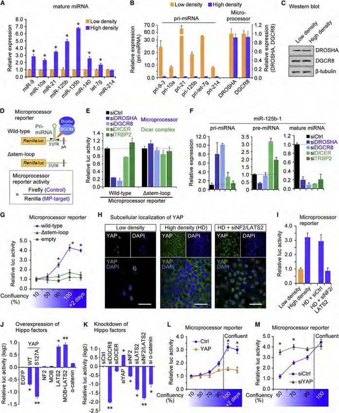
Figure 1. YAP Regulates Microprocessor Activity in a Cell-Density-Dependent Manner
(A–M) (A) qRT-PCR analysis of miRNA expression in HaCaT cells. Data were normalized to U6. *p < 0.05, Student’s t test. (B) Relative expression of pri-miRNAs
and DROSHA and DGCR8 at low and high densities. qRT-PCR data normalized to GAPDH. (C) Western blot analysis. (D) Schematic representation of the
                                                                                                                          (legend continued on next page)

894 Cell 156, 893–906, February 27, 2014 ª2014 Elsevier Inc.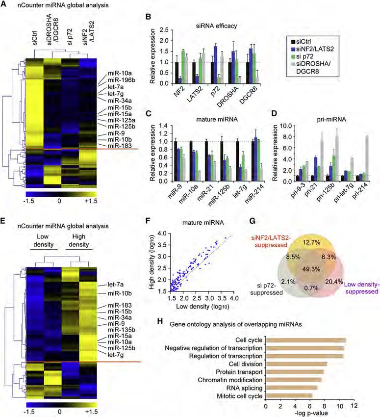
are especially relevant considering that loss of cell contact inhi-               the nontransformed human keratinocyte HaCaT cell line. Consis-
bition is a common feature of tumor cells. Connecting these pre-                  tent with published data, we observed elevated miRNA expres-
viously reported phenomena, we postulated that the observed                       sion at high cell density (Figures 1A and S1B, Hwang et al., 2009).
global miRNA repression in tumors might be related to the cell-                   To avoid the selective loss of miRNAs with low GC content that
density-dependent regulation of miRNA biogenesis. We focused                      reportedly occurs when extracting RNA from a small number of
on the Hippo-signaling pathway as a potential regulator of                        cells (Kim et al., 2012), we plated similar numbers of cells onto
cell-density-dependent miRNA biogenesis because (1) Hippo                         plates of different sizes. This then allowed us to culture cells at
pathway activity is highly sensitive to cell density and cell-cell                varying confluence without introducing the technical artifact
junctions (Kim et al., 2011; Schlegelmilch et al., 2011; Silvis                   caused by different RNA yields. The corresponding pri-miRNAs
et al., 2011; Zhao et al., 2007); (2) the Hippo pathway regulates                 were upregulated at lower cell density (Figure 1B), implying a
the balance between differentiation and renewal of multiple                       general blockade of miRNA processing at lower cell density.
stem and progenitor cell types (Camargo et al., 2007; Lian                        Expression of Microprocessor components DROSHA and
et al., 2010; Schlegelmilch et al., 2011); and (3) misregulation of               DGCR8 was not altered by cell density (Figures 1B and 1C), sug-
Hippo signaling is a common feature of human solid tumors                         gesting that the activity, not the quantity, of Microprocessor may
(Harvey et al., 2013; Zhao et al., 2010). The Hippo cascade is                    underlie the altered miRNA biogenesis.
emerging as an essential pathway for the regulation of tissue                        To assess Microprocessor activity in cells, we engineered a
homeostasis and organ size (Ramos and Camargo, 2012) and                          luciferase reporter that utilizes portions of pri-miR-125b-1 or
is characterized by responsiveness to physiological cues such                     pri-miR-205 embedded in the 30 UTR of the Renilla luciferase
as cellular crowding (Zhao et al., 2007), activation of G-protein-                gene (Figure 1D). A similar approach to monitor Microprocessor
coupled receptors (Yu et al., 2012), cell shape (Wada et al.,                     activity has been described (Tsutsui et al., 2008). Cleavage by
2011), and mechanical forces (Dupont et al., 2011; Halder                         Microprocessor is expected to destabilize the Renilla luciferase
et al., 2012). These cues culminate in differential subcellular                   mRNA and to lead to decreased Renilla luminescence. We
localization of the transcriptional coactivator YAP. At low cell                  measured Microprocessor activity by normalizing to the control
density, Hippo signaling is suppressed, and YAP localizes in                      Firefly luciferase value so that the calculated values positively
the nucleus, where it promotes cellular proliferation through tran-               correlated with the endogenous Microprocessor activity (Fig-
scriptional mechanisms. As cellular crowding increases, cell-cell                 ure 1D). To validate these reporters, we measured response to
contacts form and YAP is phosphorylated and sequestered in                        DROSHA or DGCR8 knockdown in HaCaT cells (Figures 1E
the cytoplasm by adherens junction proteins E-cadherin (Kim                       and S1C), where pri-miR-125b, but not pre-miR-125b, accumu-
et al., 2011) and a-catenin (Schlegelmilch et al., 2011; Silvis                   lates and mature miR-125b is suppressed (Figure 1F). Validation
et al., 2011). Nuclear YAP induces the reversible overgrowth of                   was also performed using Dgcr8 knockout mouse embryonic
multiple organs and tumorigenesis in mice (Camargo et al.,                        stem cells (Figures S1D and S1E). The reporter was not affected
2007; Dong et al., 2007). Additionally, deregulation of the Hippo                 by knockdown of DICER or TRBP2 (Figure 1E). To further confirm
pathway has been reported at a high frequency in a broad range                    the specificity, we generated a control construct in which the
of different human carcinomas, and it often correlates with poor                  pre-miRNA stem loop was deleted (Figure 1D). Expression of
patient prognosis (Harvey et al., 2013).                                          this reporter was unresponsive to depletion of Microprocessor
   Here, we identify the Hippo-signaling pathway as a regulator                   (Figure 1E). Altogether, these data verify that the reporter serves
of Microprocessor activity. We show that YAP regulates miRNA                      as a sensitive readout of Microprocessor activity in cells.
biogenesis through sequestering the Microprocessor compo-                            Using the reporter system, we found that Microprocessor
nent p72 in a cell-density-dependent manner. We furthermore                       activity was enhanced at higher cell densities compared to
find that perturbation of Hippo signaling causes widespread                       lower-cell confluency (Figures 1G and S1F). To explore how
miRNA suppression in cells and tumors and may underlie the                        this cell-density-dependent Microprocessor activity could be
widespread miRNA repression in human tumors.                                      regulated, we focused on the Hippo-signaling pathway. YAP
                                                                                  localizes in the nucleus at low confluency and translocates to
RESULTS                                                                           the cytoplasm at high density (Figure 1H). This localization is
                                                                                  dependent on the upstream kinase LATS2 and other upstream
Hippo Pathway Component YAP Regulates                                             negative regulatory molecules such as the tumor suppressor
Microprocessor Activity in a Cell-Density-Dependent                               NF2. Inactivation of NF2 and LATS2 abrogated the cytoplasmic
Manner                                                                            sequestration of YAP at higher density (Figure 1H, ‘‘HD + siNF2/
To investigate the potential mechanism of cell-density-depen-                     LATS2’’). Using the Microprocessor reporter, we found that
dent miRNA biogenesis and gain insight into global miRNA sup-                     knockdown of NF2 and LATS2 abrogated the enhanced Micro-
pression in tumors, we first characterized miRNA regulation in                    processor activity observed at high density (Figure 1I), implying

Microprocessor (MP) reporter. The Dstem-loop mutant lacks the pre-miRNA stem loop crucial for the recognition by Microprocessor. (E) Microprocessor reporter
assays. (F) Expression levels of pri-, pre-, and mature miR-125b after indicated siRNA-mediated knockdown, normalized to GAPDH for the pri-miRNA and to U6
for the pre- and mature miRNA. (G) Microprocessor reporter activity at different cell densities. *p < 0.05 versus empty, Student’s t test. (H) Immunocytochemistry
analysis of YAP localization. YAP nuclear translocation was induced by knockdown of NF2 and LATS2. Scale bar, 30 mm. (I–M) Microprocessor reporter assays.
*p < 0.05, **p < 0.01, Student’s t test.
Data are represented as mean ± SEM. See also Figure S1.

                                                                                       Cell 156, 893–906, February 27, 2014 ª2014 Elsevier Inc. 895
that the Hippo pathway regulates Microprocessor activity. Addi-      the interaction between p72 and DROSHA/DGCR8 complex
tionally, forced expression of either YAP or a nuclear-targeted      was significantly decreased at lower density and instead p72
phospho mutant YAP S127A repressed Microprocessor activity,          was associated with YAP (Figure 2C). To gain deeper insight
whereas overexpression of LATS2 resulted in enhanced reporter        into these cell-density-dependent interactions, we fractionated
activity, presumably through YAP phosphorylation and cyto-           cell lysates collected at low and high densities using a gel-filtra-
plasmic retention (Figure 1J). Individual knockdown of NF2,          tion column. At high density, p72 eluted in the same fractions as
LATS2, or a-catenin had the reciprocal effect on Microprocessor      DROSHA and DGCR8, implying p72 association with the Micro-
activity (Figure 1K). We also tested Lats1- and Lats2-deficient      processor complex (Figure 2D, ‘‘High density’’). Remarkably, at
mouse embryonic fibroblasts (MEFs) generated by transducing          low density, p72 was not detected in the same fractions as
Lats1/;Lats2fl/fl MEFs with Cre-expressing adenovirus (Kim         DROSHA but was in the lower molecular weight fractions where
et al., 2013, Figure S1G) and observed suppressed Micro-             YAP was also eluted, implying the interaction of p72 and YAP at
processor reporter activity (Figure S1H) and lowered miRNA           low cell density (Figure 2D, ‘‘Low density’’).
expression (Figures S1I and S1J). We further examined the con-          This dynamic cell-density-dependent association of p72 with
sequences of YAP overexpression or knockdown at different cell       Microprocessor raised the possibility that nuclear YAP might
densities. Both forced YAP expression (Figure 1L) and YAP            inhibit Microprocessor activity at low cell density by binding
knockdown (Figure 1M) abrogated the cell density dependency          and sequestering p72 from DROSHA and DGCR8. Overexpres-
of Microprocessor activity. Altogether, these results reveal that    sion of the constitutively active YAP S127A mutant led to a
the Hippo-signaling pathway and its downstream component             reduction in the relative amount of p72 associated with DROSHA
YAP regulate Microprocessor activity.                                in co-IPs (Figures 2E–2G). Further analyses indicated that YAP
                                                                     WW domain 1 (W177–W199) and p72 C-terminal proline-rich
YAP Sequesters p72 from Microprocessor in a Cell-                    sequence were essential for that interaction, whereas YAP WW
Density-Dependent Manner                                             domain 2 and p72 K50 residue, which is required for HDAC1
We next interrogated how YAP could control Microprocessor ac-        interaction (Mooney et al., 2010), were not (Figures 2H and 2I).
tivity. Because YAP has an established role as a transcriptional     We then tested YAP mutants with the Microprocessor reporter
coactivator, we first focused on the possible transcriptional        system. YAP WW domain mutant 1 (WW1) failed to inhibit Micro-
role of YAP (Yagi et al., 1999). Microarray analyses showed          processor activity (Figure S2D). We knocked down p72 and
that none of the Microprocessor-related genes were significantly     found that the density-dependent enhancement of Micropro-
affected by YAP activation (Figure S2A). To further rule out a       cessor activity was abrogated in the Microprocessor reporter
transcriptional role for YAP in Microprocessor regulation, we        system in a similar fashion as knockdown of NF2/LATS2 (Fig-
made use of a YAP S94A mutant unable to bind the TEAD-family         ure S2E). Combinatorial knockdown did not show any additive
of transcription factors (Schlegelmilch et al., 2011; Zhao et al.,   effect. qRT-PCR analyses of pri-, pre-, and mature miR-125b
2008). YAP S94A and its nuclear-targeted version YAP S94A/           corroborated these results (Figure S2F). Taken together, the as-
5SA suppressed Microprocessor activity to a similar extent as        sociation of p72 with Microprocessor is cell density dependent,
their wild-type counterparts (Figure S2B). These results imply       and nuclear YAP sequesters p72 through its WW1 domain at low
that YAP regulates Microprocessor activity independent of its        cell density to suppress Microprocessor activity.
transcriptional activity. We therefore focused on the possibility       We further explored whether the TEAD proteins were also part
that YAP might regulate Microprocessor posttranscriptionally.        of the protein complex containing YAP and p72. Co-IPs revealed
   We tested whether YAP might physically interact with Micro-       that the YAP/p72 complex does not contain TEAD1 (Figure S2G).
processor components. We did not detect an association be-           Additionally, we also observed interaction of TAZ, a YAP paralog,
tween YAP and DROSHA or DGCR8 in coimmunoprecipitation               with p72 (Figure S2H) (Dupont et al., 2011; Halder et al., 2012).
(co-IP) assays (Figure 2A). We next considered that YAP might        Forced expression of TAZ lowered mature miRNA expression
associate with the Microprocessor accessory proteins p68             (Figure S2I), which was accompanied with increased pri-miRNA
(DDX5) and p72 (DDX17), DEAD (Asp-Glu-Ala-Asp)-box RNA               expression (Figure S2J). Simultaneous knockdown of YAP and
helicases that are components of a large DROSHA-containing           TAZ had an additive effect on Microprocessor reporter activity
complex that is required for processing of a large subset of         (Figures S2K and S2L). Thus, our results suggest a similar role
miRNAs (Fukuda et al., 2007; Gregory et al., 2004). It is emerging   of TAZ in miRNA biogenesis and further implicate another
that several different cellular signaling pathways use the p68       Hippo-signaling molecule in the regulation of miRNA processing.
and/or p72 association with Microprocessor to effect regulation
of pri-miRNA processing (Newman and Hammond, 2010; Siomi             Inhibition of the Hippo-Signaling Pathway Suppresses
and Siomi, 2010). Co-IPs indicated that p72, but not the structur-   Microprocessor Activity
ally similar p68, specifically associates with endogenous YAP        We next examined the significance of p72 and Hippo signaling
protein (Figure 2A).                                                 for Microprocessor function using a Microprocessor biochem-
   Immunocytochemistry showed that YAP and p72 colocalize in         ical assay (Figure 3A). We depleted DGCR8, p72, or NF2 and
the nucleus of HaCaT cells at low density (Figure 2B) and not at     LATS2 in a stable HEK293T cell line expressing Flag-DROSHA
high density (Figure S2C). We assessed the impact of cell density    (Figure 3B) and affinity-purified DROSHA-containing complexes
on this interaction by co-IPs with endogenous p72 protein.           (Figure 3C). When NF2 and LATS2 were depleted (Figure 3B),
At higher density, p72 interacted with DROSHA and DGCR8,             the amount of p72 associated with DROSHA was lowered
consistent with its role in pri-miRNA processing. Interestingly,     (Figure 3C), consistent with our findings in HaCaT cells. We

896 Cell 156, 893–906, February 27, 2014 ª2014 Elsevier Inc.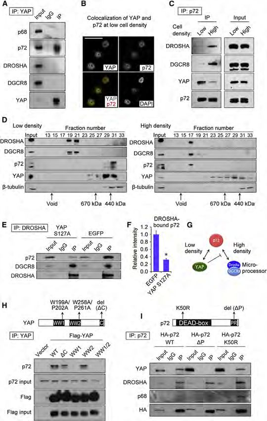
Figure 2. YAP Sequesters p72 from Micro-
                                                                                      processor Complex in a Cell-Density-
                                                                                      Dependent Manner
                                                                                      (A–I) (A) Coimmunoprecipitation assays (Co-IPs)
                                                                                      with endogenous YAP in HaCaT cells. (B) Immu-
                                                                                      nocytochemistry of YAP and p72 in HaCaT cells at
                                                                                      low density. Nuclei were stained with DAPI. Scale
                                                                                      bar, 30 mm. (C) Co-IP with HA-p72 in HaCaT cells
                                                                                      at low and high density. (D) Western blot analysis
                                                                                      of Superose 6 gel-filtration fractions. Whole-cell
                                                                                      lysates from HaCaT cells cultured at low and high
                                                                                      densities were fractionated. b-tubulin served as a
                                                                                      control. (E) Co-IP with Flag-DROSHA in HaCaT
                                                                                      cells transfected with YAP or control EGFP. (F)
                                                                                      Densitometry measurement for the amount of p72
                                                                                      bound by DROSHA in the HaCaT cells transfected
                                                                                      with YAP or control EGFP (n = 3). *p < 0.05, Stu-
                                                                                      dent’s t test. (G) The scheme of interactions
                                                                                      among YAP, p72, and Microprocessor. (H) Co-IP
                                                                                      with Flag-YAP and YAP mutants. Mutations in YAP
                                                                                      are represented in the top panel. WT, wild-type. (I)
                                                                                      Co-IP with HA-p72 and p72 mutants. Mutations in
                                                                                      p72 are represented in the top panel.
                                                                                      See also Figure S2.

                                                                                       Global Impact of Hippo Pathway on
                                                                                       miRNA Biogenesis
                                                                                       Our data above would predict that Hippo
                                                                                       signaling inactivation and consequent
                                                                                       YAP nuclear translocation would result in
                                                                                       general miRNA suppression. We sought
                                                                                       to examine this by utilizing nCounter tech-
                                                                                       nology to profile >600 different miRNAs.
                                                                                       Indeed, NF2/LATS2 knockdown at high
                                                                                       cell density lowered (
Figure 3. Hippo Pathway and p72 Regulate
                                                                                                 Pri-miRNA Processing Efficiency of Micro-
                                                                                                 processor
                                                                                                 (A–D) (A) Scheme of the Microprocessor assay.
                                                                                                 IVT, in vitro-transcribed. (B) Western blot analysis
                                                                                                 showing the siRNA efficacies and expression of
                                                                                                 Flag-DROSHA. (C) Western blot of purified protein
                                                                                                 complexes. (D) Microprocessor assays with in-vi-
                                                                                                 tro-transcribed pri-miR-125b. The numbers for
                                                                                                 Microprocessor indicate the relative amounts of
                                                                                                 Flag-IP products used for western blot (C) and
                                                                                                 Microprocessor assay (D).

                                                                                                   stem-loop structure (Figure 5C). The D50
                                                                                                   mutant showed slightly impaired interac-
                                                                                                   tion, but deletion of the 30 flanking
                                                                                                   segment almost totally abolished the
                                                                                                   interaction, suggesting that p72 interacts
                                                                                                   through the 30 FS of pri-miR-21 (Fig-
                                                                                                   ure 5C). Sequential shortening of the
                                                                                                   30 FS suggested that the distal part of
                                                                                0
(Hwang et al., 2009), HaCaT cells also showed widespread                 the 3 FS was dispensable (+81, +108, Figure 5D), though the
variation of miRNA expression in a cell-density-dependent                interaction was minimally impaired with the shortest mutant
manner. At lower cell density, 57.3% of miRNAs were sup-                 (+55, Figure 5D). To test whether p72 recognizes a specific
pressed (
Figure 4. Global Impact of Hippo Pathway on miRNA Biogenesis
(A–G) (A) Global miRNA expression analysis of HaCaT with indicated knockdown. The miRNAs with a relative expression change of 1.2-fold
compared to the control (siCtrl) were analyzed by hierarchical clustering. (B) The efficacy of siRNAs used in (A). The expression values were normalized to GAPDH.
(C) qPCR-quantification of mature miRNAs normalized to U6. (D) Pri-miRNA expression levels measured by qRT-PCR. Data were normalized to GAPDH. (E)
miRNA expression analysis in RNA samples from low- and high-density HaCaT cells. miRNAs with a relative expression change of 1.2-fold between
the low- and high-density conditions were analyzed by hierarchical clustering. (F) Scatter plot of miRNA expression levels (log10) in the low and the high densities.
(G) Venn diagram showing the overlap of miRNAs repressed by siNF2/LATS2, si p72, or low density. (H) Gene ontology analysis of the overlapping miRNAs in (G).
Bonferroni-corrected p values were indicated. Data are represented as mean ± SEM. See also Figure S3.
to alanine in addition to S127, and it displays enhanced nuclear expression (Figures 6D and S5A). Among the growth-related
localization (Zhao et al., 2007). After Dox treatment for 4 days, proteins tested, we found enhanced expression of MYC protein
YAP and YAP target genes CTGF, CYR61, and AMOTL2 were (Figure 6E) after YAP induction. MYC induction was also
induced without affecting the mRNA levels of MYC (Figure 6A). observed in HaCaT cell lines inducibly expressing YAP S94A
Numerous mature miRNAs were repressed (Figures 6B and and YAP S94A/5SA, suggesting that MYC was posttranscrip-
6C) with a corresponding accumulation or sustained pri-miRNA tionally induced (Figures 6F and S5B).
Cell 156, 893–906, February 27, 2014 ª2014 Elsevier Inc. 899Figure 5. p72 DEAD Box RNA Helicase Binds to a Sequence Motif in the 30 Flanking Segment of Pri-miRNA (A–H) (A–D) EMSA with recombinant p72 protein and in-vitro-transcribed pri-miR-21, pri-miR-125b-1, or deletion mutants of pri-miR-21. Stem loop (Dstem-loop), 50 flanking segment (D50 ) or 30 flanking segment (D30 ) were deleted. (E) Identification of a sequence motif in the miRNAs repressed by knockdown of both NF2/ LATS2 and p72. (F) Schematics showing the motif in the 30 flanking segments of pri-miR-21 and pri-miR-125b-1. (G) Pri-miR-21 schematic indicating the motif mutations introduced and the control mutant. Arrowheads indicate cleavage sites by the Microprocessor. (H) EMSA with recombinant p72 protein and the +55 mutant, the control mutant, and the motif mutant of the pri-miR-21. See also Figure S4. To further examine the YAP-mediated posttranscriptional S94A/5SA, also strongly activated the luciferase reporter, rein- induction of MYC, we investigated the relevance of the MYC forcing the role of YAP in the posttranscriptional regulation of 30 UTR, which contains potential targeting sites for several MYC expression (Figure 6G). The activity of Luc-MYC 30 UTR miRNAs that were repressed by YAP overexpression and p72 was also suppressed at higher cell density and correlated with depletion (Figure S5C). Among them, let-7 (Kumar et al., 2007) the accumulation of miRNAs targeting MYC (Figure 6H). Knock- and miR-34a (Christoffersen et al., 2010) were reported to target down of p72 at higher cell density rescued the repression of lucif- MYC 30 UTR. We utilized a luciferase gene harboring the MYC erase activity, suggesting that cell-density-dependent regulation 30 UTR (Kumar et al., 2007) and compared luciferase activity to of MYC 30 UTR was mediated by p72 (Figure 6H). Collectively, a control plasmid. YAP 5SA overexpression induced luciferase the posttranscriptional induction of MYC protein is a func- activity >10-fold compared to a control EGFP (Figure 6G). tional outcome of YAP-mediated cell-density-dependent global TEAD-binding-deficient YAP mutants, YAP S94A and YAP miRNA repression. 900 Cell 156, 893–906, February 27, 2014 ª2014 Elsevier Inc.
Figure 6. YAP-Regulated miRNAs Repress MYC Expression
(A–H) (A) qRT-PCR analysis with data normalized to GAPDH. (B) miRNA northern blot performed with spike-in of luciferase siRNA for normalization. (C) qRT-PCR
analysis of mature miRNA levels normalized to U6. *p < 0.05, Student’s t test. (D) Relative pri-miRNA expression measured by qRT-PCR normalized to GAPDH.
(E and F) Western blot analysis using indicated antibodies. (G and H) Luciferase assays with a MYC 30 UTR reporter. HaCaT cells were cotransfected with the
luciferase and the expression plasmids for YAP or control EGFP. Luciferase activity was normalized to that of pRL-Tk.
**p < 0.01, Student’s t test. n.s., not significant. Data are represented as mean ± SEM. See also Figure S5.

YAP Mediates Global miRNA Suppression in Tumors                                   The liver tumor model relied on hepatocyte-specific deletion
A large number of solid human cancers demonstrate impaired                     of Nf2 in adult mice (Figures 7G and 7H). This resulted in
Hippo signaling and exhibit constitutive nuclear YAP localization              hepatomegaly, cholangiocarcinoma-like tumor formation (Fig-
(Harvey et al., 2013). Additionally, YAP activation leads to rapid             ure S6A–S6C), and induction of YAP target genes (Figure 7I).
tumor development in mice (Benhamouche et al., 2010;                           The expression of mature miRNAs was repressed in the liver
Camargo et al., 2007; Dong et al., 2007; Schlegelmilch et al.,                 tumors compared to control tissue (Figure 7J), which was
2011; Zhang et al., 2010). Our findings in vitro imply that YAP-               accompanied by the accumulation of pri-miRNAs in the tumors
driven tumors might exhibit global miRNA repression. We tested                 (Figure 7K). The miRNA global analysis revealed that 61.0% of
this prediction in mouse models of YAP-induced tumorigenesis.                  miRNAs were repressed in the liver tumors as compared to
We evaluated this in two distinct contexts of YAP activation:                  normal tissue (Figure 7L). To explore the relevance of p72 in
acute (8 days) YAP induction in the epidermis (Figure 7A) and                  the context of YAP-induced tumorigenesis, we examined the as-
chronic (50 weeks) YAP activation in the liver (Figure 7G).                    sociation of p72 with Microprocessor and YAP. Co-IP revealed
Short-term expression of a transgenic YAP S127A in the Kera-                   that the interaction between p72 and Microprocessor observed
tin-14-positive (K14+) epidermal progenitor cells induces in situ              in normal livers was significantly decreased in tumor tissues,
squamous cell carcinoma-like tumors in mice, which can pro-                    whereas the association between p72 and YAP was increased
duce invasive growth upon transplantation into nude mice                       in Nf2-deficient tumors (Figure 7M). The interaction between
(Schlegelmilch et al., 2011). Gene expression analyses of the                  p72 and YAP was also observed in the skin tumors (Figure S6D).
oncogenic epidermal cells revealed potent induction of trans-                  Overall these results demonstrate that YAP-driven tumorigen-
genic YAP S127A (hYAP, Figure 7B) and YAP target genes (Fig-                   esis is associated with widespread miRNA suppression and
ure 7C). As predicted, YAP induction led to the repression of                  that YAP activation promotes the dissociation of p72 from Micro-
numerous mature miRNAs (Figure 7D) and the accumulation of                     processor complex in tumor cells.
pri-miRNAs (Figure 7E), recapitulating our in vitro findings.                     Finally, we examined whether this transcription-independent
Global analysis revealed that 52.5% of miRNAs were sup-                        function of YAP plays a causative role in cellular growth. We
pressed at least 0.8-fold in the tumorigenic cells as compared                 chose to study the consequences of expressing the TEAD-bind-
to the normal epidermal cells (Figure 7F).                                     ing defective mutant YAP S94A/5SA, given that current dogma

                                                                                    Cell 156, 893–906, February 27, 2014 ª2014 Elsevier Inc. 901
(legend on next page) 902 Cell 156, 893–906, February 27, 2014 ª2014 Elsevier Inc.
suggests that most of YAP’s effects are mediated by tran-                            variants or for Microprocessor-binding variants of human pri-
scription through the TEAD proteins. YAP S94A/5SA repressed                          miRNAs identified a CNNC motif in the 30 FS conserved among
miRNAs with the p72-binding motifs (Figure S6E) and also                             vertebrates for a large subset of human pri-miRNAs. This func-
showed significant acceleration of cell growth in HaCaT cells,                       tional motif, located 16–20 nt downstream of the Drosha
though this effect was less potent than that of YAP 5SA. Coex-                       cleavage site is required for efficient pri-miRNA processing and
pression of p72 in this cellular context fully counteracted                          overlaps with the p72-binding motif that we identified. In that
the effect of YAP S94A/5SA (Figure S6F). A similar effect was                        study, the authors identified SRp20/SRSF3 as a factor that binds
observed in HepG2 human hepatocellular carcinoma cells,                              the CNNC motif. Although the relevance of this splicing regulator
where YAP S94A/5SA expression significantly promoted their                           in miRNA biogenesis was not tested, it is possible that multiple
anchorage-independent growth in a p72-dependant manner                               different factors may converge at this 30 FS site to mediate pri-
(Figures S6G and S6H). Our results here support a functional                         miRNA processing. It will be interesting to examine the interplay
role for YAP in mediating cellular proliferation independent of                      between p72 and other possible regulators in miRNA biogenesis
its canonical transcriptional partners and dependent on the                          (Auyeung et al., 2013).
Microprocessor component p72.                                                           The mechanism that we characterized represents a unique
                                                                                     transcription-independent function of the YAP protein. YAP
DISCUSSION                                                                           has another transcription-independent role in the growth control
                                                                                     of intestinal stem cells, where YAP sequesters Dishevelled pro-
Here, we uncover an unexpected role for the Hippo-signaling                          tein in the cytoplasm, thereby repressing Wnt signaling (Barry
pathway in the regulation of miRNA biogenesis. Our results pro-                      et al., 2013). The major transcriptional role of YAP is mediated
vide mechanistic understanding for two unexplained phenom-                           through TEAD DNA-binding proteins (Zhao et al., 2008), and
ena: cell-density-dependent activation of miRNA biogenesis                           therefore YAP S94A, which is deficient in TEAD binding, has a
and widespread decrease in miRNA expression in tumors                                deficit in the transcription of crucial target genes. The finding
(Hwang et al., 2009; Lu et al., 2005). We found that YAP, the                        that YAP S94A and its nuclear-targeted version YAP S94A/5SA
downstream Hippo-signaling transducer, induces widespread                            repressed Microprocessor activity provides evidence that YAP
miRNA repression by sequestering p72 from the Microprocessor                         represses miRNAs independent of its transcriptional activity.
in a cell-density-dependent manner (Figure 7N). At low cell den-                     Furthermore, YAP can induce cellular proliferation independent
sity, YAP is nuclear, promotes cell proliferation, and represses                     of TEAD and can be rescued by p72. MYC globally suppresses
miRNA biogenesis. At higher cell density, YAP is inactivated                         miRNA through transcription (Chang et al., 2008). Our findings
by exclusion from the cell nucleus, thereby allowing p72 to                          cannot be explained by the transcriptional repression of miRNA
associate with Microprocessor and pri-miRNAs, resulting in                           by MYC because we observe accumulation of pri-miRNAs and
enhanced miRNA biogenesis. The association of the related pro-                       corresponding decrease in mature miRNAs upon manipulation
tein, p68 (DDX5), with the Microprocessor is also dynamically                        of the YAP/p72/Microprocessor pathway. Also, this posttran-
regulated. p68 is directly phosphorylated by MAPK-activated                          scriptional control of miRNA biogenesis corresponds well with
protein kinase 2 (MK2), and p68 phosphorylation is necessary                         the reported widespread blockade of pri-miRNA processing
for its nuclear localization (Hong et al., 2013). Therefore, an                      observed in various human cancers.
emerging theme for controlling Microprocessor activity is                               Cell proliferation and differentiation need to be coordinated for
through the accessibility of these related cofactors either by                       the dynamic control of organ growth and repair. The molecular
phosphorylation-dependent control of nuclear localization (for                       and cellular mechanisms responsible for integrating these pro-
p68) or through the sequestration of p72 in the nucleus by                           cesses remain poorly understood. Because the Hippo-signaling
YAP. Cell signaling pathways can impact other components of                          pathway plays an important role in organ size control, it will be of
the miRNA biogenesis machinery, including the mitogen-acti-                          interest to examine the relevance of miRNA expression changes
vated protein kinase (MAPK) Erk-mediated phosphorylation                             in that context. Elevated miRNA expression likely serves to
of TRBP (Paroo et al., 2009) and the epidermal growth factor                         repress cell proliferation and promote cell differentiation (Kanel-
receptor (EGFR)-mediated phosphorylation of Ago2 (Shen                               lopoulou et al., 2005; Yi et al., 2009). Failure of this switching may
et al., 2013).                                                                       lead to uncontrolled cell expansion and widespread repression
   p72 enhances pri-miRNA processing by the Microprocessor                           of miRNAs, which are hallmarks of tumors. Our findings that
and recognizes a VCAUCH sequence motif in the pri-miRNA 30                           the Hippo pathway synchronizes cellular expansion and miRNA
flanking region (30 FS). A recent in vitro selection and high-                       biogenesis illuminate the potential for new therapeutics that
throughput sequencing approach for functional pri-miRNA                              target miRNA biogenesis for the treatment of human cancers.

Figure 7. YAP Mediates the Global Repression of miRNA Biogenesis in Tumors
(A–N) (A) Mouse model of YAP-induced skin tumorigenesis. (B) Expression of exogenous human YAP S127A and endogenous mouse Yap normalized to Hprt1 in
isolated epidermal cells. (C) Expression levels of YAP target genes normalized to Hprt1. (D) Mature miRNA expression levels normalized to sno142. *p < 0.05,
Student’s t test. (E) Expression levels of the pri-miRNAs normalized to Hprt1. (F) Global miRNA analysis. (G) Mouse model of YAP-induced tumorigenesis in the
liver. (H) Expression levels of mouse Yap normalized to Hprt1. (I) The expression levels of YAP target genes normalized to Hprt1. (J) Mature miRNA expression
levels normalized to sno142. *p < 0.05, Student’s t test. (K) Relative expression levels of the pri-miRNAs normalized to Hprt1. (L) Global miRNA analysis in the liver
tissues. (M) Co-IP with p72 in the normal tissues and tumors from the mouse livers. (N) Proposed model.
Data are represented as mean ± SEM. See also Figure S6.

                                                                                          Cell 156, 893–906, February 27, 2014 ª2014 Elsevier Inc. 903
EXPERIMENTAL PROCEDURES Fractionation of Protein Complexes
Whole-cell lysates of HaCaT cells cultured at the low and high densities were
Cell Lines fractionated with Superose 6 gel filtration column as described previously
HaCaT cells were cultured in DMEM + 10% FBS. At low density, cells existed (Gregory et al., 2004). Fractions from the gel-filtration chromatography were
as single cells or small colonies. For high-density conditions, similar numbers concentrated with Amicon Ultra centrifugal filters (Millipore) and analyzed by
of cells were seeded in a smaller culture dish than the low-density condition SDS-PAGE and western blot.
and were cultured to reach confluency. Percent confluence was estimated
by microscopic observation. pInducer20 (Meerbrey et al., 2011) -YAP 5SA, Immunocytochemistry
-YAP S94A, or -YAP S94A/5SA was transduced to generate doxycycline HaCaT cells were fixed with 4% paraformaldehyde for 10 min at RT, permea-
(Dox)-inducible HaCaT and HepG2 cell lines. The transduced cells were bilized with 0.1% Triton X-100 for 1 min at RT, blocked with 2% FBS, and incu-
selected with G418 (400 ng/ml) for 2 weeks. For YAP induction, Dox was bated with antibody against YAP (1:200, Cell Signaling, #4912) and p72 (1:200,
added at 1000 ng/ml for 4 days. HEK293T-Flag-DROSHA cells (Gregory Bethyl Laboratories, A300-509A) at 4 C overnight. After washing PBS, cells
et al., 2004) were cultured in DMEM + 10% FBS with puromycin (2 mg/ml). were incubated with anti-mouse-Alexa Fluor 488 and anti-rabbit-Alexa Fluor
For proliferation assays, cells were plated at 1.5 3 105 cells/ml in triplicate in 546 (1:1,000, Invitrogen). Nuclei were stained with DAP (Invitrogen).
6-well plates and were counted at the indicated time points. SV40 LT-immor-
talized Lats1/;Lats2fl/fl MEFs were described previously (Kim et al., 2013). Microprocessor Assay
For Lats2 deletion, Ad5-CMV-Cre (Gene Transfer Vector Core, University of Microprocessor was purified from HEK293T-Flag-DROSHA stable cell line
Iowa) was infected. 96 hr after transfection of siRNA for NF2, LATS2, p72, or DGCR8, or negative
control. In vitro transcription of pri-miR-21 and miR-125b-1 and Micropro-
Plasmids cessor assays, using affinity-purified Flag-Drosha complexes was performed
Plasmids for YAP, YAP S127A, WW1-, WW2-, WW1/WW2 mutants, DC, and as described previously (Gregory et al., 2004).
a-catenin were described previously (Schlegelmilch et al., 2011). Plasmids
for YAP S94A, S94A/5SA (Zhao et al., 2008), HA-p72-WT, and K50R (Mooney miRNA Global Expression Analysis
et al., 2010) were kindly provided. pRL-MYC-30 UTR (Kumar et al., 2007) was nCounter miRNA assay (nanoString, Geiss et al., 2008) was used for global
from Addgene (Plasmid 14806). Luciferase assays were performed using miRNA analysis. miRNAs with normalized expression levels more than those
dual-luciferase reporter system (Promega). Lipofectamine 2000 (Invitrogen) of negative control probes were analyzed. The miRNA expression was normal-
was used for transfections. ized to all miRNAs except for liver analysis, which was normalized with the top
100 genes. For hierarchical clustering analysis, the normalized values for each
Microprocessor Reporter miRNA were z transformed and Multiple Experiment Viewer (Saeed et al.,
For the Microprocessor reporter, the human pre-miR-125b stem loop with the 2006) was used for computing the complete linkage hierarchical clustering
flanking upstream and downstream sequences were inserted to the 30 UTR of algorithm with the Pearson correlation metric. The gene ontology enrichment
Renilla luciferase gene in psiCHECK2 plasmid (Table S1). For the mutated con- analyses for miRNAs were performed with starBase (Yang et al., 2011).
trol (Dstem loop), the stem loop was deleted (Table S1). For the motif deletions, Bonferroni-corrected p values were presented.
GCATCC (+16 to +21 in the 30 FS, ‘‘Dmotif’’) or the proximal sequence of 30 FS
(+1 to +65 in the 30 FS, ‘‘Dproximal’’) was deleted. Motif Analysis
For discovering potential p72 recognition sites in the pri-miRNA, the
Gene Expression Analysis pre-miRNA sequences with flanking regions 55 nt upstream and 55 nt
For miRNA, RNA extraction was with TRIzol (Invitrogen). TaqMan miRNA downstream were obtained from the Ensemble database. The sequences
assays (Applied Biosystems) were used to quantify mature miRNA expres- were analyzed with Improbizer (http://users.soe.ucsc.edu/kent/improbizer/
sion. Pri-miRNA levels were analyzed by qPCR using Fast SYBR Green improbizer.html) sequence logos were generated using WebLogo (Crooks
Master Mix (Applied Biosystems). For pre-miRNA quantification, small et al., 2004).
RNAs were enriched using mirVana (Ambion). Primers used for qPCR
are listed in Table S2. TaqMan probes (Applied Biosystems) were used Recombinant p72 Protein Purification and EMSA
for mRNA quantification. For knockdown experiments, Lipofectamine His-tagged p72 was expressed and purified from BL21-CodonPlus Compe-
RNAiMAX (Invitrogen) was used to transfect siRNAs (sequences in Table tent bacteria (Stratagene). EMSA with internally labeled pri-miR-125b, pri-
S3) at 10 nM. miR-21, or mutated pri-miR-21 (sequences in Table S5) was performed in
binding buffer (50 mM Tris [pH 7.5], 100 mM NaCl, 10 mM 2-mercaptoethanol,
Northern Blot 20 U RNasin [Promega], 1 mM ATP) with 1 nM pri-miRNA and incubating for
Total RNA was isolated from 5 3 105 cell HaCaT cells cultured at either low 45 min at RT. Bound complexes were resolved on native 3.5% polyacrylamide
or high densities. 100 fmoles of control RNA (GL2 siRNA, 50 - CGUACGCG gels and visualized by radiography.
GAAUACUUCG-30 ) was spiked into cell lysates. Northern blots were per-
formed as described (Gregory et al., 2004). Soft Agar Assay
HepG2 cell lines were suspended in DMEM with 10% FBS, 0.3% SeaPlaque
Immunoprecipitation and Western Blot agarose (Lonza, #50101), and 1,000 ng/ml Dox and were plated at 3,000
Cells were lysed with NETN buffer (100 mM NaCl, 20 mM Tris-Cl (pH 8.0), cells/well in a 6-well culture dish on a layer of 0.6% agar containing the
0.5 mM EDTA, 0.5% Nonidet P-40). After centrifugation at 20,000 3 g at same medium. DMEM with 10% FBS and 1000 ng/ml Dox was added on
4 C for 5 min, lysates were pretreated with Protein A/G Sepharose beads the gels. Cell colonies were stained with crystal violet after 14 days in culture
(Sigma) and incubated with antibodies at 4 C overnight. The protein-antibody and quantified with Image J.
complexes were incubated with protein A/G sepharose at 4 C for 1 hr. For the
IP with Flag or HA tag, the pretreated lysates were incubated with anti-Flag Mouse Models
M2 Affinity Gel (A2220, Sigma-Aldrich), EZview Red Anti-HA Affinity Gel Mouse experiments were approved by the BCH Animal Care and Use Com-
(Sigma), or control IgG AC (Santa Cruz) at 4 C for 1 hr. The beads were mittee and were performed in accordance with all relevant guidelines and
washed three times with NETN 200 buffer (200 mM NaCl, 20 mM Tris-Cl regulations.
[pH 8.0], 0.5 mM EDTA, and 0.5% Nonidet P-40). The sample buffer was
added and incubated at 95 C for 5 min. After centrifugation at 20,000 3 g Skin Tumorigenesis Model
for 1 min, the supernatants were collected for western blot analysis using Adult R26stoprtTA/+ Col-tetO-YAPS127A/+ K14-Cre (‘‘+Cre’’ group) and
antibodies in Table S4. R26stoprtTA/+ Col-tetO-YAPS127A/+ (‘‘-Cre’’ control group, n = 6) were treated
904 Cell 156, 893–906, February 27, 2014 ª2014 Elsevier Inc.for 8 days with Dox (1 mg/ml) administered in drinking water. The epidermal plex to Ago2 for microRNA processing and gene silencing. Nature 436,
cells, which are enriched for the skin progenitor cells, were collected as 740–744.
described (Schlegelmilch et al., 2011). Choudhury, N.R., de Lima Alves, F., de Andrés-Aguayo, L., Graf, T., Cáceres,
J.F., Rappsilber, J., and Michlewski, G. (2013). Tissue-specific control of brain-
Liver Tumorigenesis Model enriched miR-7 biogenesis. Genes Dev. 27, 24–38.
Nf2fl/fl (Benhamouche et al., 2010) female mice (n = 3) were administered
Christoffersen, N.R., Shalgi, R., Frankel, L.B., Leucci, E., Lees, M., Klausen,
PBS or AAV2/8-Cre (AV-8-PV1091, University of Pennsylvania Vector Core,
M., Pilpel, Y., Nielsen, F.C., Oren, M., and Lund, A.H. (2010). p53-independent
MOI = 1011) to induce hepatocyte-specific deletion of Nf2 gene. Livers were
upregulation of miR-34a during oncogene-induced senescence represses
inspected after 50 weeks tumors were collected for analysis.
MYC. Cell Death Differ. 17, 236–245.
Statistical Analysis Davis, B.N., Hilyard, A.C., Lagna, G., and Hata, A. (2008). SMAD proteins
For all quantified data, mean ± SEM is presented. Statistical significance be- control DROSHA-mediated microRNA maturation. Nature 454, 56–61.
tween two experimental groups is indicated by an asterisk, and comparisons Denli, A.M., Tops, B.B.J., Plasterk, R.H.A., Ketting, R.F., and Hannon, G.J.
were made using the Student’s t test. p values less than 0.05 were considered (2004). Processing of primary microRNAs by the Microprocessor complex.
significant. Nature 432, 231–235.
Dong, J., Feldmann, G., Huang, J., Wu, S., Zhang, N., Comerford, S.A.,
ACCESSION NUMBERS Gayyed, M.F., Anders, R.A., Maitra, A., and Pan, D. (2007). Elucidation of a
universal size-control mechanism in Drosophila and mammals. Cell 130,
The GEO accession numbers for nCounter and microarray analyses are 1120–1133.
GSE52276 and GSE49384.
Dupont, S., Morsut, L., Aragona, M., Enzo, E., Giulitti, S., Cordenonsi, M.,
Zanconato, F., Le Digabel, J., Forcato, M., Bicciato, S., et al. (2011). Role of
SUPPLEMENTAL INFORMATION YAP/TAZ in mechanotransduction. Nature 474, 179–183.
Fukuda, T., Yamagata, K., Fujiyama, S., Matsumoto, T., Koshida, I., Yoshi-
Supplemental Information includes six figures and five tables and can be found
mura, K., Mihara, M., Naitou, M., Endoh, H., Nakamura, T., et al. (2007).
with this article online at http://dx.doi.org/10.1016/j.cell.2013.12.043.
DEAD-box RNA helicase subunits of the Drosha complex are required for
processing of rRNA and a subset of microRNAs. Nat. Cell Biol. 9, 604–611.
ACKNOWLEDGMENTS
Gregory, R.I., Yan, K.P., Amuthan, G., Chendrimada, T., Doratotaj, B., Cooch,
Thanks to Ralf Janknecht for HA-p72 plasmid, Tyler Jacks for pRL MYC 30 UTR N., and Shiekhattar, R. (2004). The Microprocessor complex mediates the
plasmid, Kun-Liang Guan for YAP S94A and S94A/5SA plasmids, and Dae-Sik genesis of microRNAs. Nature 432, 235–240.
Lim for Lats1/2 knockout MEFs. M.M is supported by Japan Heart Foundation/ Guil, S., and Cáceres, J.F. (2007). The multifunctional RNA-binding protein
Bayer Yakuhin Research Grant Abroad. R.T. was supported by a Simeon Burt hnRNP A1 is required for processing of miR-18a. Nat. Struct. Mol. Biol. 14,
Wolbach Fellowship from Boston Children’s Hospital. R.I.G is supported by a 591–596.
grant from the US National Institute of General Medical Sciences (NIGMS)
Halder, G., Dupont, S., and Piccolo, S. (2012). Transduction of mechanical and
(R01GM086386). F.D.C is supported by awards from Stand Up to Cancer-
cytoskeletal cues by YAP and TAZ. Nat. Rev. Mol. Cell Biol. 13, 591–600.
AACR initiative, NIH grant R01 CA131426, and Department of Defense (DOD
W81XWH-09). F.D.C. is a Pew Scholar in the Biomedical Sciences. Han, J., Lee, Y., Yeom, K.H., Nam, J.W., Heo, I., Rhee, J.-K., Sohn, S.Y., Cho,
Y., Zhang, B.T., and Kim, V.N. (2006). Molecular basis for the recognition of
Received: August 7, 2013 primary microRNAs by the Drosha-DGCR8 complex. Cell 125, 887–901.
Revised: November 21, 2013 Harvey, K.F., Zhang, X., and Thomas, D.M. (2013). The Hippo pathway and
Accepted: December 31, 2013 human cancer. Nat. Rev. Cancer 13, 246–257.
Published: February 27, 2014
Hill, D.A., Ivanovich, J., Priest, J.R., Gurnett, C.A., Dehner, L.P., Desruisseau,
D., Jarzembowski, J.A., Wikenheiser-Brokamp, K.A., Suarez, B.K., Whelan,
REFERENCES
A.J., et al. (2009). DICER1 mutations in familial pleuropulmonary blastoma.
Science 325, 965.
Auyeung, V.C., Ulitsky, I., McGeary, S.E., and Bartel, D.P. (2013). Beyond sec-
ondary structure: primary-sequence determinants license pri-miRNA hairpins Hong, S., Noh, H., Chen, H., Padia, R., Pan, Z.K., Su, S.B., Jing, Q., Ding, H.F.,
for processing. Cell 152, 844–858. and Huang, S. (2013). Signaling by p38 MAPK stimulates nuclear localization
of the microprocessor component p68 for processing of selected primary
Barry, E.R., Morikawa, T., Butler, B.L., Shrestha, K., de la Rosa, R., Yan, K.S.,
microRNAs. Sci. Signal. 6, ra16.
Fuchs, C.S., Magness, S.T., Smits, R., Ogino, S., et al. (2013). Restriction of
intestinal stem cell expansion and the regenerative response by YAP. Nature Hwang, H.W., Wentzel, E.A., and Mendell, J.T. (2009). Cell-cell contact
493, 106–110. globally activates microRNA biogenesis. Proc. Natl. Acad. Sci. USA 106,
7016–7021.
Bartel, D.P. (2009). MicroRNAs: target recognition and regulatory functions.
Cell 136, 215–233. Kanellopoulou, C., Muljo, S.A., Kung, A.L., Ganesan, S., Drapkin, R., Jenu-
Benhamouche, S., Curto, M., Saotome, I., Gladden, A.B., Liu, C.H., Giovan- wein, T., Livingston, D.M., and Rajewsky, K. (2005). Dicer-deficient mouse
nini, M., and McClatchey, A.I. (2010). Nf2/Merlin controls progenitor homeo- embryonic stem cells are defective in differentiation and centromeric silencing.
stasis and tumorigenesis in the liver. Genes Dev. 24, 1718–1730. Genes Dev. 19, 489–501.
Camargo, F.D., Gokhale, S., Johnnidis, J.B., Fu, D., Bell, G.W., Jaenisch, R., Kawai, S., and Amano, A. (2012). BRCA1 regulates microRNA biogenesis via
and Brummelkamp, T.R. (2007). YAP1 increases organ size and expands the DROSHA microprocessor complex. J. Cell Biol. 197, 201–208.
undifferentiated progenitor cells. Curr. Biol. 17, 2054–2060. Kim, N.G., Koh, E., Chen, X., and Gumbiner, B.M. (2011). E-cadherin mediates
Chang, T.C., Yu, D., Lee, Y.S., Wentzel, E.A., Arking, D.E., West, K.M., Dang, contact inhibition of proliferation through Hippo signaling-pathway compo-
C.V., Thomas-Tikhonenko, A., and Mendell, J.T. (2008). Widespread micro- nents. Proc. Natl. Acad. Sci. USA 108, 11930–11935.
RNA repression by Myc contributes to tumorigenesis. Nat. Genet. 40, 43–50. Kim, Y.K., Yeo, J., Kim, B., Ha, M., and Kim, V.N. (2012). Short structured RNAs
Chendrimada, T.P., Gregory, R.I., Kumaraswamy, E., Norman, J., Cooch, with low GC content are selectively lost during extraction from a small number
N., Nishikura, K., and Shiekhattar, R. (2005). TRBP recruits the Dicer com- of cells. Mol. Cell 46, 893–895.
Cell 156, 893–906, February 27, 2014 ª2014 Elsevier Inc. 905Kim, M., Kim, M., Lee, S., Kuninaka, S., Saya, H., Lee, H., Lee, S., and Lim, D.S. Schlegelmilch, K., Mohseni, M., Kirak, O., Pruszak, J., Rodriguez, J.R., Zhou,
(2013). cAMP/PKA signalling reinforces the LATS-YAP pathway to fully sup- D., Kreger, B.T., Vasioukhin, V., Avruch, J., Brummelkamp, T.R., and
press YAP in response to actin cytoskeletal changes. EMBO J. 32, 1543–1555. Camargo, F.D. (2011). Yap1 acts downstream of a-catenin to control
Kumar, M.S., Lu, J., Mercer, K.L., Golub, T.R., and Jacks, T. (2007). Impaired epidermal proliferation. Cell 144, 782–795.
microRNA processing enhances cellular transformation and tumorigenesis. Shen, J., Xia, W., Khotskaya, Y.B., Huo, L., Nakanishi, K., Lim, S.O., Du, Y.,
Nat. Genet. 39, 673–677. Wang, Y., Chang, W.C., Chen, C.H., et al. (2013). EGFR modulates microRNA
Kumar, M.S., Pester, R.E., Chen, C.Y., Lane, K., Chin, C., Lu, J., Kirsch, D.G., maturation in response to hypoxia through phosphorylation of AGO2. Nature
Golub, T.R., and Jacks, T. (2009). Dicer1 functions as a haploinsufficient tumor 497, 383–387.
suppressor. Genes Dev. 23, 2700–2704. Silvis, M.R., Kreger, B.T., Lien, W.H., Klezovitch, O., Rudakova, G.M.,
Camargo, F.D., Lantz, D.M., Seykora, J.T., and Vasioukhin, V. (2011). a-cate-
Lee, E.J., Baek, M., Gusev, Y., Brackett, D.J., Nuovo, G.J., and Schmittgen,
nin is a tumor suppressor that controls cell accumulation by regulating the
T.D. (2008). Systematic evaluation of microRNA processing patterns in tissues,
localization and activity of the transcriptional coactivator Yap1. Sci. Signal.
cell lines, and tumors. RNA 14, 35–42.
4, ra33.
Lian, I., Kim, J., Okazawa, H., Zhao, J., Zhao, B., Yu, J., Chinnaiyan, A., Israel,
Siomi, H., and Siomi, M.C. (2010). Posttranscriptional regulation of microRNA
M.A., Goldstein, L.S.B., Abujarour, R., et al. (2010). The role of YAP transcrip-
biogenesis in animals. Mol. Cell 38, 323–332.
tion coactivator in regulating stem cell self-renewal and differentiation. Genes
Dev. 24, 1106–1118. Thomson, J.M., Newman, M., Parker, J.S., Morin-Kensicki, E.M., Wright, T.,
and Hammond, S.M. (2006). Extensive post-transcriptional regulation of
Lu, J., Getz, G., Miska, E.A., Alvarez-Saavedra, E., Lamb, J., Peck, D., Sweet-
microRNAs and its implications for cancer. Genes Dev. 20, 2202–2207.
Cordero, A., Ebert, B.L., Mak, R.H., Ferrando, A.A., et al. (2005). MicroRNA
expression profiles classify human cancers. Nature 435, 834–838. Trabucchi, M., Briata, P., Garcia-Mayoral, M., Haase, A.D., Filipowicz, W.,
Ramos, A., Gherzi, R., and Rosenfeld, M.G. (2009). The RNA-binding protein
Maillot, G., Lacroix-Triki, M., Pierredon, S., Gratadou, L., Schmidt, S., Bénès,
KSRP promotes the biogenesis of a subset of microRNAs. Nature 459,
V., Roché, H., Dalenc, F., Auboeuf, D., Millevoi, S., and Vagner, S. (2009).
1010–1014.
Widespread estrogen-dependent repression of micrornas involved in breast
tumor cell growth. Cancer Res. 69, 8332–8340. Tsutsui, M., Hasegawa, H., Adachi, K., Miyata, M., Huang, P., Ishiguro, N.,
Hamaguchi, M., and Iwamoto, T. (2008). Establishment of cells to monitor
Melo, S.A., Ropero, S., Moutinho, C., Aaltonen, L.A., Yamamoto, H., Calin,
Microprocessor through fusion genes of microRNA and GFP. Biochem.
G.A., Rossi, S., Fernandez, A.F., Carneiro, F., Oliveira, C., et al. (2009). A
Biophys. Res. Commun. 372, 856–861.
TARBP2 mutation in human cancer impairs microRNA processing and DICER1
function. Nat. Genet. 41, 365–370. Wada, K., Itoga, K., Okano, T., Yonemura, S., and Sasaki, H. (2011). Hippo
pathway regulation by cell morphology and stress fibers. Development 138,
Melo, S.A., Moutinho, C., Ropero, S., Calin, G.A., Rossi, S., Spizzo, R., Fernan-
3907–3914.
dez, A.F., Davalos, V., Villanueva, A., Montoya, G., et al. (2010). A genetic
Yagi, R., Chen, L.F., Shigesada, K., Murakami, Y., and Ito, Y. (1999). A WW
defect in exportin-5 traps precursor microRNAs in the nucleus of cancer cells.
domain-containing yes-associated protein (YAP) is a novel transcriptional
Cancer Cell 18, 303–315.
co-activator. EMBO J. 18, 2551–2562.
Mooney, S.M., Grande, J.P., Salisbury, J.L., and Janknecht, R. (2010). Sumoy-
Yi, R., Pasolli, H.A., Landthaler, M., Hafner, M., Ojo, T., Sheridan, R., Sander,
lation of p68 and p72 RNA helicases affects protein stability and transactiva-
C., O’Carroll, D., Stoffel, M., Tuschl, T., and Fuchs, E. (2009). DGCR8-depen-
tion potential. Biochemistry 49, 1–10.
dent microRNA biogenesis is essential for skin development. Proc. Natl. Acad.
Morlando, M., Dini Modigliani, S., Torrelli, G., Rosa, A., Di Carlo, V., Caffarelli, Sci. USA 106, 498–502.
E., and Bozzoni, I. (2012). FUS stimulates microRNA biogenesis by facilitating
Yu, F.X., Zhao, B., Panupinthu, N., Jewell, J.L., Lian, I., Wang, L.H., Zhao, J.,
co-transcriptional Drosha recruitment. EMBO J. 31, 4502–4510.
Yuan, H., Tumaneng, K., Li, H., et al. (2012). Regulation of the Hippo-YAP
Newman, M.A., and Hammond, S.M. (2010). Emerging paradigms of regulated pathway by G-protein-coupled receptor signaling. Cell 150, 780–791.
microRNA processing. Genes Dev. 24, 1086–1092.
Zeng, Y., Yi, R., and Cullen, B.R. (2005). Recognition and cleavage of primary
Ozen, M., Creighton, C.J., Ozdemir, M., and Ittmann, M. (2008). Widespread microRNA precursors by the nuclear processing enzyme Drosha. EMBO J. 24,
deregulation of microRNA expression in human prostate cancer. Oncogene 138–148.
27, 1788–1793.
Zhang, N., Bai, H., David, K.K., Dong, J., Zheng, Y., Cai, J., Giovannini, M., Liu,
Paroo, Z., Ye, X., Chen, S., and Liu, Q. (2009). Phosphorylation of the human P., Anders, R.A., and Pan, D. (2010). The Merlin/NF2 tumor suppressor
microRNA-generating complex mediates MAPK/Erk signaling. Cell 139, functions through the YAP oncoprotein to regulate tissue homeostasis in
112–122. mammals. Dev. Cell 19, 27–38.
Piskounova, E., Polytarchou, C., Thornton, J.E., LaPierre, R.J., Pothoulakis, Zhao, B., Wei, X., Li, W., Udan, R.S., Yang, Q., Kim, J., Xie, J., Ikenoue, T., Yu,
C., Hagan, J.P., Iliopoulos, D., and Gregory, R.I. (2011). Lin28A and Lin28B J., Li, L., et al. (2007). Inactivation of YAP oncoprotein by the Hippo pathway
inhibit let-7 microRNA biogenesis by distinct mechanisms. Cell 147, 1066– is involved in cell contact inhibition and tissue growth control. Genes Dev.
1079. 21, 2747–2761.
Ramos, A., and Camargo, F.D. (2012). The Hippo signaling pathway and stem Zhao, B., Ye, X., Yu, J., Li, L., Li, W., Li, S., Yu, J., Lin, J.D., Wang, C.Y.,
cell biology. Trends Cell Biol. 22, 339–346. Chinnaiyan, A.M., et al. (2008). TEAD mediates YAP-dependent gene induction
Sakamoto, S., Aoki, K., Higuchi, T., Todaka, H., Morisawa, K., Tamaki, N., and growth control. Genes Dev. 22, 1962–1971.
Hatano, E., Fukushima, A., Taniguchi, T., and Agata, Y. (2009). The NF90- Zhao, B., Li, L., Lei, Q., and Guan, K.L. (2010). The Hippo-YAP pathway in
NF45 complex functions as a negative regulator in the microRNA processing organ size control and tumorigenesis: an updated version. Genes Dev. 24,
pathway. Mol. Cell. Biol. 29, 3754–3769. 862–874.
906 Cell 156, 893–906, February 27, 2014 ª2014 Elsevier Inc.You can also read