Cataract-Associated New Mutants S175G/H181Q of βB2-Crystallin and P24S/S31G of γD-Crystallin Are Involved in Protein Aggregation by Structural Changes
←
→
Page content transcription
If your browser does not render page correctly, please read the page content below
International Journal of
            Molecular Sciences

Article
Cataract-Associated New Mutants S175G/H181Q of
βB2-Crystallin and P24S/S31G of γD-Crystallin Are
Involved in Protein Aggregation by
Structural Changes
In-Kang Song 1,† , Seungjin Na 2,† , Eunok Paek 2, *         and Kong-Joo Lee 1, *
 1    Department of Pharmacy, College of Pharmacy and Graduate School of Pharmaceutical Sciences,
      Ewha Womans University, Seoul 03760, Korea; fluss1234@ewha.ac.kr
 2    Department of Computer Science, Hanyang University, Seoul 04763, Korea; sna@hanyang.ac.kr
 *    Correspondence: eunokpaek@hanyang.ac.kr (E.P.); kjl@ewha.ac.kr (K.-J.L.)
 †    These authors contributed equally to this work.

 Received: 23 July 2020; Accepted: 1 September 2020; Published: 5 September 2020

 Abstract: β/γ-Crystallins, the main structural protein in human lenses, have highly stable structure
 for keeping the lens transparent. Their mutations have been linked to cataracts. In this study,
 we identified 10 new mutations of β/γ-crystallins in lens proteomic dataset of cataract patients using
 bioinformatics tools. Of these, two double mutants, S175G/H181Q of βB2-crystallin and P24S/S31G of
 γD-crystallin, were found mutations occurred in the largest loop linking the distant β-sheets in the
 Greek key motif. We selected these double mutants for identifying the properties of these mutations,
 employing biochemical assay, the identification of protein modifications with nanoUPLC-ESI-TOF
 tandem MS and examining their structural dynamics with hydrogen/deuterium exchange-mass
 spectrometry (HDX-MS). We found that both double mutations decrease protein stability and induce
 the aggregation of β/γ-crystallin, possibly causing cataracts. This finding suggests that both the
 double mutants can serve as biomarkers of cataracts.

 Keywords: βB2-crystallin; γD-crystallin; cataract-associated mutants; proteomics dataset;
 hydrogen-deuterium exchange-MS; structural change; stability change; post translational modification;
 protein aggregation

1. Introduction
     Crystallins are a group of structural proteins located in the eye lens that function to focus
light onto the retina. Cataracts, the leading cause of blindness worldwide [1], develop due to the
misfolding and aggregation of crystallins [2,3]. The mammalian lens consists of 90% of soluble
protein crystallins [4], which have a lifespan of more than 70 years [5]. Three lens crystallins (α-,
β-, and γ-crystallins) are classified into two families, α-crystallins and βγ-crystallins. α-Crystallins
function as molecular chaperones by preventing the aggregation of βγ-crystallins and maintaining
their solubilities [6] with similar way to small heat shock proteins, and βγ-crystallins play key roles as
structural proteins for maintaining the structural stability of lens [7]. βγ-Crystallins are composed
primarily of two double-Greek key motifs [8,9], which confer stability and longevity to the parent
molecules. Crystallins are encoded in the CRY genes, and to date, more than 100 mutations have been
identified in 12 crystallin genes isolated from more than 100 families. They account for 33–37% of
autosomal dominant cataracts [10]. In the mutations of CRYAA and CRYAB encoding the α-crystallins,
the former is associated with nuclear and lamellar-type opacities, and the latter to myopathy. Mutations
in the genes for β-crystallins (CRYBB1, BB2, BB3, BA1, BA2, and BA4) and γ-crystallins (CRYGB, GC,

Int. J. Mol. Sci. 2020, 21, 6504; doi:10.3390/ijms21186504                         www.mdpi.com/journal/ijms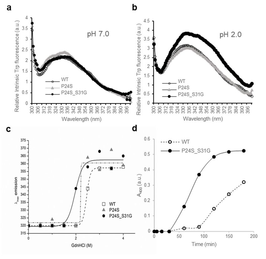
Int. J. Mol. Sci. 2020, 21, 6504                                                                     2 of 18

GD, and GS) interfere the folding of structurally conserved Greek-key motifs or change the surface
properties of proteins. Mutations reducing the solubility causing the aggregation of βγ-crystallins are
associated with congenital or juvenile cataract [11].
     βB2-crystallin, the least modified and the most soluble β-crystallin, is the predominant β-crystallin
in human eye lens [12]. It exists as homo- and hetero-dimers and oligomers with other β-crystallins [13].
There are two known cataract-causing mutants: A2V has a poor thermal stability due to its poor ability
to make tetramers at high concentrations [14], and Q155X has a structure, that makes it less stable [15].
γD-crystallin is highly stable and is not denatured by conventional denaturing agents and heat [16].
The N-terminal domain of γD-crystallin is less stable than the C-terminal domain [17]. The mutants
identified in the N-terminal domain of γD-crystallin are as follows: R15C mutation causes protein
aggregation as result of the formation of disulfide-linked oligomers [18]. P24S, P24T, R37S, and R59H
mutations decrease the protein solubility and crystallize easily [8,19–21]. W43R mutation increases the
proteolytic susceptibility and facilitates to aggregate [22].
     In this study, we found two new nuclear cataract-associated mutants in βB2- and γD-crystallins
by an analysis of the proteomics dataset of cataract patients employing bioinformatics tools followed
by a study of the effects of these mutations, S175G/H181Q on βB2-crystallin and P24S/S31G on
γD-crystallin. Both two double mutations are located in the largest loops connecting two distant
β-sheets in the Greek key motif. We examined the structural changes employing spectroscopic and
mass spectrometric analysis. We found significant structural changes of βB2-crystallin S175G/H181Q
mutant and demonstrated perturbation of the C-terminal Greek key 4 motifs by studying protein
structural dynamics via HDX-MS [23]. We also found structural changes of γD-crystallin P24S/S31G
mutant, but these are only in acidic condition, because of the high stability of γD-crystallin in normal
condition. We found that increase of cataract-related modifications including oxidation occur only in
this mutant. These studies reveal that two new mutations found in this study are located in the largest
loop between distant β-sheets in Greek key motif. We believe they promote cataract development due
to the structural changes of Greek key motif. They could serve as biomarkers of cataract development.

2. Results

2.1. Selection of Two New Nuclear Cataract-Specific Mutations by Bioinformatics Analysis
     Five normal human lenses (3-day and 2-, 18-, 35-, and 70-year old) and two nuclear cataract
lenses (70- and 93-year old) were obtained from previous studies, where age-related changes in human
crystallins were analyzed [24,25]. To define cataract-specific amino acid mutations in crystallin proteins,
we retained peptides that were identified with amino acid mutations from 14 crystallin proteins. The
mutations were consistently observed in both 70- and 93-year cataract lens samples but were not
observed in other normal lens samples. Mutations were accepted only when there were no possible
chemical modifications matching the delta masses. The resulting ten peptides are shown with the
amino acid mutations in Table 1.

                    Table 1. The list of novel mutations from nuclear cataract human lens samples.

                     Protein              From           To                       Peptide
           Alpha-crystallin B chain         57           69       APSW(F→I/L)DTGLSEMR
           Alpha-crystallin B chain        124          149       IPADVDPL(T→A)ITSSLSSDGVLTVNGPR
              Beta-crystallin A4           178          192       EWGSHA(P→Q)TFQVQSIR
              Beta-crystallin B1            93          110       SIIVS(A→T)GPWVAFEQSNFR
              Beta-crystallin B1           215          230       HWNEWGAFQPQMQ(S→G)LR
              Beta-crystallin B2           122          140       ME(I→V)IDDDVPSFHAHGYQEK
              Beta-crystallin B2           169          188       GDYKDS(S→G)DFGAP(H→Q)PQVQSVR
              Beta-crystallin B3            56           71       VGSIQVESGPWLAFE(S→R)
              Beta-crystallin B3           116          127       LHLFENPAF(S→G)GR
            Gamma-crystallin D              16           32       HYECSSDH(P→S)NLQPYL(S→G)R
Int. J. Mol. Sci. 2020, 21, 6504                                                                   3 of 18

     To determine the characteristics of each mutation, we examined the location of each mutation in
the structure (Figures S1–S3). Two of the mutants are in α-crystallin and eight are in β/γ-crystallin.
αB-Crystallin, one of α-crystallins having a HSP domain, has a crystal structure in which only some
structures have been identified (PDB ID code 2Y1Y) [26]. In αB-crystallin, the location of Phe61 is
not yet known, but that of Thr132 is in the helix between immediately adjacent β-sheets (Figure S1).
Unlike α-crystallin, β/γ-crystallins have four Greek key motifs (Figures S1b–g, S2 and S3). The Greek
key motif is known to be evolutionarily well conserved super-secondary protein structural fold that
affords structural compactness and high intrinsic stability against various stress including ROS and
aging [17,27]. The Pro185 of βA4-crystallin is located in a loop near the C-terminal of the Greek key
motif 4, where the structure is not clear (PDB ID code 3LWK) (Figure S1b). Ser227 of βB1 (PDB ID
code 1OKI) and Ile124 of βB2-crystallin (PDB ID code 1YTQ) are located at the end of β-sheet of Greek
key motif 3 and 4 (Figure S1c,d). In βB3-crystallin, Ser71 and 125 are at the end of β-sheet of 2 and 3
motif, and Serine 187 is in the loop at end of Greek key 4 motif (PDB ID code 3QK3) (Figure S2e–g).
Intriguingly, two double mutations found in βB2- and γD-crystallin are very close in sequence and
were identified in one tryptic digested peptide. Both Ser175 and His181 of βB2-crystallin and Pro24
and Ser31 of γD-crystallin are located in the largest loop connecting to distant β-sheets in the Greek
key motif (Figures S2 and S3). Although large loops do not form disulfide or coordinate with metals
in βB2 and γD-crystallins, these large loops are important to maintain stability of the Greek key
motif [28]. We selected and validated these two double mutations employing recombinant proteins of
these mutants.

2.2. Structural Characteristics of βB2-Crystallin Double Mutants
     In order to investigate the structural changes caused by these mutations of βB2-crystallin,
we examined the intrinsic fluorescence changes of Trp and Tyr of recombinant wild-type (WT) and
S175G/H181Q βB2-crystallin. The intrinsic fluorescence maximum wavelength and the intensity of
aromatic amino acids (Trp and Tyr) were measured, because intrinsic fluorescence of Trp and Tyr varies
depending on the structural environment of these residues [29]. As shown in Figure 1, the changes of
fluorescence signals were induced by the double mutation S175G/H181Q βB2-crystallin. The maximum
emission wavelengths of intrinsic Trp fluorescence (λmax), the strongest intrinsic fluorescence, were 334
nm for WT, an about 5 nm red-shift by the mutation could be observed for the fluorescence (Figure 1a).
The maximum emission wavelengths of intrinsic Trp & Tyr fluorescence were 340 nm for both WT
and mutant without red shift (Figure 1b). The intensities of intrinsic Trp and Trp & Tyr fluorescence
of the mutant were 1.1~1.3 times higher than those of WT. This change is meaningful because these
changes are sum of different 9 Tyr and 4 Trp residues. These spectroscopic results indicate that the
mutations affect the conformational state and the microenvironments of the Trp and Tyr fluorophores.
βB2-crystallins are known to form homodimers and heteromers with other β-crystallins [13,30] which
are required to increase the solubility. Since higher oligomeric fraction of βB2-crystallin increases the
solubility of the other β-crystallins [31,32], we examined the dimeric states of WT and S175G/H181Q
mutant using size exclusion chromatography (SEC). As shown in Figure 1c, dimeric fraction of WT
βB2-crystallin is higher than that of mutant form, although all β-crystallins migrate on SEC as dimers
in rapid equilibrium with monomer form. This shows the possibility that S175G/H181Q mutation
destabilizes dimer formation and makes the mutant protein less soluble than WT one.
Int. J. Mol. Sci. 2020, 21, 6504 4 of 18
Int. J. Mol. Sci. 2020, 21, x 4 of 18
Figure 1. Effect
Figure 1. Effect of
of the
the double
double mutations
mutations (S175G/H181Q)
(S175G/H181Q) on on the
the secondary, tertiary structures
secondary, tertiary structures and
and
oligomerization
oligomerization of βB2-crystallin.
βB2-crystallin. (a)
(a)Intrinsic
IntrinsicTrp
Trp fluorescence
fluorescence excited
excited at
at 295
295 nm. (b)
(b) Intrinsic
Intrinsic Trp
Trp
and Tyr fluorescence excited at 280 nm. The proteins were prepared in PBS buffer with concentration
of 0.5 mg/mL. The fluorescence intensity was recorded for every 1 nm for WT (opened circles) and
the S175G/H181Q mutant (closed circles). (c) Size exclusion
exclusion chromatograms
chromatograms of WT and S175G/H181Q
S175G/H181Q
mutant βB2-crystallin
βB2-crystallin using
using Superdex
SuperdexTM TM200
200HR
HR10/30
10/30 column.
column. 450
450µg
µg of
of WT
WT protein (black dotted
line) and the S175G/H181Q mutant
mutant (black
(black line)
line) were
were loaded
loaded on
on column
columnequilibrated
equilibratedwith
withPBS
PBSbuffer.
buffer.
Albumin (66.4 kDa) and α-lactalbumin (14.2 kDa) were were used
used as
as molecular
molecular weight
weight markers.
markers.
2.3. Effects
2.3. Effects of
of Mutations
Mutations on βB2-Crystallin Stability
on βB2-Crystallin Stability and
and Folding
Folding
To investigate
To investigate the
the stability
stability differences
differences between
between WT WT and
and S175G/H181Q
S175G/H181Q mutantmutant βB2-crystallins,
βB2-crystallins,
we assessed red shift of intrinsic fluorescence changes by chemical
we assessed red shift of intrinsic fluorescence changes by chemical denaturant GdnHCldenaturant GdnHCl in in aa dose
dose
dependent manner.
dependent manner. As As shown
shown in in Figure
Figure 2a,2a, mutant
mutant at at 0.5~0.6
0.5~0.6 MM GdnHCl
GdnHCl is is more
more readily
readily denatured
denatured
than WT
than WT atat 0.6~0.8
0.6~0.8 M.M. To
To validate
validate these
these results,
results, we
we performed
performed ANSANS binding
binding assay
assay inin response
response to to
various concentrations of GdnHCl, since significant increase of ANS fluorescence can
various concentrations of GdnHCl, since significant increase of ANS fluorescence can be observed by be observed by
increasing hydrophobic
increasing hydrophobic surface
surface in
in denatured
denatured and and aggregated
aggregated form
form of
of protein
protein [33].
[33]. WT
WT βB2-crystallin
βB2-crystallin
did not affect the hydrophobic exposure during GdnHCl-induced unfolding, while S175G/H181Q
did not affect the hydrophobic exposure during GdnHCl-induced unfolding, while S175G/H181Q
mutant significantly
mutant significantly increased
increased the
the hydrophobic
hydrophobic exposure
exposure atat low
low 0.6~0.8
0.6~0.8 MM GdnHCl
GdnHCl concentrations
concentrations
(Figure 2b). This increase of ANS fluorescence is caused by the accumulation
(Figure 2b). This increase of ANS fluorescence is caused by the accumulation of denatured of denatured form having
form
large hydrophobic exposure or the appearance of aggregates.
having large hydrophobic exposure or the appearance of aggregates.
To evaluate
To evaluate the
the long-term stability, the
long-term stability, the protein
protein with
with aa concentration
concentration of of 55 mg/mL
mg/mL were
were incubated
incubated
at around physiological temperature (37 ◦ C) for 36 h and measured turbidity (A ). As shown
at around physiological temperature (37 °C) for 36 h and measured turbidity (A400). As shown 400 in
in Figure 2c, WT protein maintained transparent after 18 h, while the turbidity
Figure 2c, WT protein maintained transparent after 18 h, while the turbidity of mutant increase of mutant increase
continuously till 36 h incubation. That is, the mutant protein is susceptible to aggregation inducedInt. J. Mol. Sci. 2020, 21, 6504 5 of 18
Int. J. Mol. Sci. 2020, 21, x 5 of 18
continuously till 36 h incubation. That is, the mutant protein is susceptible to aggregation induced
long-term
long-term incubation.
incubation. This
This result
result also
also confirmed
confirmed that,
that, unlike
unlike WT,
WT, when
when mutants
mutants were
were overexpressed
overexpressed
in
in mammalian cells, the mutants were aggregated in a small puncta form (Figure S5). These
mammalian cells, the mutants were aggregated in a small puncta form (Figure S5). These results
results
indicate that S175G/H181Q mutation of βB2-crystallins reduced protein stability by affecting
indicate that S175G/H181Q mutation of βB2-crystallins reduced protein stability by affecting the protein
the
folding
protein and increased
folding the tendency
and increased to be aggregated.
the tendency to be aggregated.
Figure 2.2.Denaturation
Figure Denaturation of βB2-crystallin protein
of βB2-crystallin with various
protein concentrations
with various of GdnHCl.
concentrations of (a) Transition
GdnHCl. (a)
curves of protein
Transition curvesequilibrium unfolding monitored
of protein equilibrium unfoldingby Emax of Trp
monitored intrinsic
by Emax of fluorescence
Trp intrinsic (Ex: 280 nm).
fluorescence
Data for nm).
(Ex: 280 each Data
proteinfor samples weresamples
each protein fitted with
werea fitted
Boltzmann
with acurve using curve
Boltzmann Originusing
8.5 (OriginLab
Origin 8.5
Corporation, Northampton, MA, USA). The Emax did not change at GdnHCl
(OriginLab Corporation, Northampton, MA, USA). The Emax did not change at GdnHCl concentrations above 1 M.
(b) Hydrophobic exposure during GdnHCl-induced unfolding was monitored
concentrations above 1 M. (b) Hydrophobic exposure during GdnHCl-induced unfolding was by ANS fluoresescence
intensity
monitored at by
470ANS
nm. fluoresescence
Data for each intensity
proteins were
at 470fitted
nm. with
Data afor
GaussAmp curvewere
each proteins using Origin
fitted 8.5.
with a
(c) Long-term stability revealed by incubating 5.0 mg/mL ◦
GaussAmp curve using Origin 8.5. (c) Long-term stabilityproteins
revealedatby
37 incubating
C continuously, and turbidity
5.0 mg/mL proteins
changes
at 37 °C at 400 nm wereand
continuously, measured.
turbidity changes at 400 nm were measured.
2.4. Identification of Structural Changes in Mutant βB2-Crystallin Employing Hydrogen Deuterium Exchange
2.4. Identification
Mass Spectrometryof(HDX-MS)
Structural Changes in Mutant βB2-Crystallin Employing Hydrogen Deuterium
Exchange Mass Spectrometry (HDX-MS)
To determine the structural changes induced by mutation, we predicted mutant structure
To determine
employing the structural
protein modeling analysis changes
using the induced
Phyre2 by mutation,
software we shown
[34]. As predicted mutant
in Figure structure
3a, mutation
employing
induced protein structural
significant modeling analysis
changes in usingthe the Phyre2 software
C-terminus, but not in [34].
theAs shown in Figure
N-terminus. Based on 3a,
mutation induced significant structural changes in the C-terminus,
these predicted results, we assumed that mutation caused the clumping of the C-terminal Greek but not in the N-terminus. Based
on these
key motif,predicted
which isresults,
importantwe assumed that mutation
for the stability caused the clumping
of βB2-crystallin [35], causes of the C-terminal
protein Greek
aggregation.
key
To motif, this
confirm which is important
possibility, for the stability
we employed of βB2-crystallin
hydrogen-deuterium [35], causesspectrometry
exchange-mass protein aggregation.
(HDX-MS), To
confirm this possibility, we employed hydrogen-deuterium exchange-mass
which measures the differential deuterium incorporation in proteins and peptides between WT and spectrometry (HDX-MS),
which measures
S175G/H181Q the differential
mutant deuterium
βB2-crystallins and incorporation
is a powerful in proteins
tool and peptides
to elucidate structuralbetween WT [36].
changes and
S175G/H181Q mutant βB2-crystallins and is a powerful tool to elucidate
Differential deuterium incorporations of identified peptides of WT and mutant in a time dependent structural changes [36].
Differential
manner deuterium
are listed in Tableincorporations
S1. Combined of identified
stitching peptides of WT andratios
the H/D exchange mutant in a time
of each pepticdependent
peptide
manner are listed in Table S1. Combined stitching the H/D exchange
showed the diagram of whole protein shown in Figure 3b. Peptide MS coverage was 98%. Significant ratios of each peptic peptide
showed the diagram of whole protein shown in Figure 3b. Peptide MS
conformational changes were observed in the Greek key motif 4 of βB2-crystallin (Figure 3c). More coverage was 98%. Significant
conformational
deuterium changes were
incorporation observed
in mutant in the
occurred in Greek key motif
the peptide 4 of βB2-crystallin
containing (Figure the
the portion between 3c).Greek
More
deuterium
key motifs 3incorporation
and 4 (144–151) in mutant
and the occurred
largest in theof
loop peptide
the Greek containing
key motifthe portion
4 (166–186).between
Thesetheresults
Greek
key motifs 3 and 4 (144–151) and the largest loop of the Greek key motif
suggest the Greek key motif 4 with apparent roles in βB2-crystallin structure and assembly [35] is 4 (166–186). These results
suggest the
exposed to theGreek
proteinkeysurface
motif 4by with apparent
mutation androles in βB2-crystallin
is more flexible than WT. structure and assembly
This destabilizes [35] is
the Greek
exposed
key motifto4 the proteinMeanwhile,
structure. surface by mutation and is more
less deuterium exchange flexible
was than WT. This
observed destabilizes
in peptide the in
located Greek
the
Greek key motif 2 (62–72). This indicates that the Greek key motif 2 is more shielded by mutation. in
key motif 4 structure. Meanwhile, less deuterium exchange was observed in peptide located the
Here,
Greek
by key motif
confirming the2change
(62–72). notThis
onlyindicates that the Greek
in the substituted site, but keyalso
motif 2 is more
in other sites, shielded by mutation.
it can be assumed that
the observed HDX change is the result of solvent exposure due to structural flexibility and itstability,
Here, by confirming the change not only in the substituted site, but also in other sites, can be
assumed
and not duethattothe
theobserved HDX change
effect of amino is the result
acid substitution onofintrinsic
solvent exchange
exposure rate.due to structural
These resultsflexibility
indicate
and stability, and not due to the effect of amino acid substitution
that S175G/H181Q mutations of βB2-crystallin cause the conformational changes in the Greek on intrinsic exchange rate. These
key
results indicate that S175G/H181Q mutations of βB2-crystallin
motif 4 which play a role in maintaining stable structure of βB2-crystallin. cause the conformational changes in
the Greek key motif 4 which play a role in maintaining stable structure of βB2-crystallin.Int. J. Mol. Sci. 2020, 21, 6504 6 of 18
Int. J. Mol. Sci. 2020, 21, x 6 of 18
FigureFigure 3. Structural
3. Structural analysisbetween
analysis betweenwild-type
wild-type and
andS175G/H181Q
S175G/H181Q mutant of βB2-crystallin.
mutant (a) The
of βB2-crystallin. (a) The
predicted tertiary structure of the S175G/H181Q mutant. Phyre2 structure predictions for
predicted tertiary structure of the S175G/H181Q mutant. Phyre2 structure predictions for the mutant isthe mutant
is shown in red ribbon. WT is shown in blue ribbon (PDB ID code: 1YTQ). (b,c) Structural changes in
shown in red ribbon. WT is shown in blue ribbon (PDB ID code: 1YTQ). (b,c) Structural changes in
WT and the mutant of βB2-crystallin analyzed by hydrogen/deuterium exchange mass spectrometry.
WT and the mutant of βB2-crystallin analyzed by hydrogen/deuterium exchange mass spectrometry.
(b) Deuterium exchange rate (%) of WT and S175G/H181Q mutant during incubation with D2O.
(b) Deuterium exchange rate (%) of WT and S175G/H181Q mutant during incubation with D2 O.
Significantly changed representative peptides were marked by colored arrows. The corresponding
Significantly
deuteriumchanged
exchangerepresentative
levels for eachpeptides
peptide were marked
in percent are by colored
given arrows.
on the TheChanges
right. (c) corresponding
in
deuterium exchange levels
hydrogen/deuterium for each spectrometry
exchange-mass peptide in percent are
data of the givenofon
mutant the right. compared
βB2-crystallin (c) Changes
to in
hydrogen/deuterium exchange-mass
the WT onto the structures of humanspectrometry data of the
βB2-crystallin structure mutant
(PDB of βB2-crystallin
ID: 1YTQ). compared to
Percentage difference
the WT onto the structures
of deuterium incorporationof human
in HDX-MS between WTstructure
βB2-crystallin (PDB ID: 1YTQ).
and S175G/H181Q mutant isPercentage difference of
colored according
to the key.
deuterium incorporation in HDX-MS between WT and S175G/H181Q mutant is colored according to
the key.
2.5. Effect of the Mutations on γD-Crystallin Structure Revealed by Biophysical Experiments
2.5. Effect of the Mutations on γD-Crystallin Structure Revealed by Biophysical Experiments
Another new γD-crystallin double mutant (P24S/S31G) was identified in cataract lenses (Table
1). Since single
Another new P24S mutant has
γD-crystallin alreadymutant
double been studied in the cataract
(P24S/S31G) aggregate-likely
was identified mutant
in cataract [20,21],
lenses (Table 1).
we compared the double mutant (P24S/S31G) with WT and single one (P24S). To determine
Since single P24S mutant has already been studied in the cataract aggregate-likely mutant [20,21],whether
single andthe
we compared double mutants
double mutanthave differentwith
(P24S/S31G) structural
WT and properties,
single one we(P24S).
measured the intrinsic
To determine whether
fluorescence of Trp of WT and single and double mutants. Since γD-crystallin is known to undergo
single and double mutants have different structural properties, we measured the intrinsic fluorescence
conformational changes at acidic pH (pH 2.0) [37], we measured intrinsic fluorescence of Trp at pH
of Trp of WT and single and double mutants. Since γD-crystallin is known to undergo conformational
changes at acidic pH (pH 2.0) [37], we measured intrinsic fluorescence of Trp at pH 7.0 and 2.0.
As shown in Figure 4a, no fluorescence difference between WT and two mutants was detected at pH 7.0,
which is consistent with previous result [37]. However, intrinsic fluorescence of Trp of double mutantconcentrations of GdnHCl, while γD-crystallin is more resistant to denaturation by GdnHCl [16]. To
investigate the stability differences between WT and the P24S and P24S/S31G mutants of γD-
crystallin, we assessed intrinsic fluorescence changes by chemical denaturant GdnHCl (pH 7.4) in a
dose dependent manner. As shown in Figure 4c, P24S/S31G mutant is more readily denatured at 2.0
M J.GdnHCl
Int. than21,
Mol. Sci. 2020, WT and P24S mutant at 2.5 M.
6504 7 of 18
To evaluate the long-term stability, the protein with a concentration of 5 mg/mL were incubated
at 37 °C for 36 h and turbidity changes were measured with absorbance at 400 nm. In the same way
was
withsignificantly higherIn
beta-crystallins. than those of
contrast to WT
the and P24S single
increase mutantdue
in turbidity at pHto 2.0 (Figure 4b).ofThe
aggregation maximum
βB2-crystallin
emission wavelengths of intrinsic Trp fluorescence (λmax) was 338 nm for WT, while
mutants, turbidity did not increase in γD-crystallin even after 36 h in both WT and mutant (Figure 4 nm red-shift for
double mutant
S6). Since gamma wascrystallin
observed.aggregates
The intensitywellof inintrinsic Trp fluorescence
acidic conditions, was also
the turbidity 1.3 times
of WT higher was
and mutant for
the double mutant than WT. These results indicate that P24S/S31G double mutations
measured at 37 °C in a buffer condition of pH 2.0. The turbidity of mutant began to increase after 30 of γD-crystallin
alter
min conformational
and increased until state150
andmin
microenvironments
to reach the maximum more than P24S
level, single turbidity
whereas mutation of at the
WTacidic pH.
gradually
increased as 90ismin
βB2-Crystallin readily denatured
passed. at lowthat
This indicates concentrations of GdnHCl,
the mutant protein is more while to aggregateisinmore
γD-crystallin
likely vitro.
resistant to denaturation by GdnHCl [16]. To investigate the stability differences
This result also observed that when mutants were overexpressed in mammalian cells, the mutants between WT and the
P24S
was and
small,P24S/S31G mutants in
but aggregation of the
γD-crystallin,
form of puncta we assessed intrinsic(Figure
was observed fluorescence changes
S5). These by chemical
results indicate
denaturant GdnHCl (pH
that S175G/H181Q 7.4) inofa dose
mutation dependent reduced
βB2-crystallins manner. As shown
protein in Figure
stability by4c, P24S/S31G
affecting the mutant
protein
isfolding
more readily denatured at 2.0 M GdnHCl
and increased the tendency to be aggregated. than WT and P24S mutant at 2.5 M.
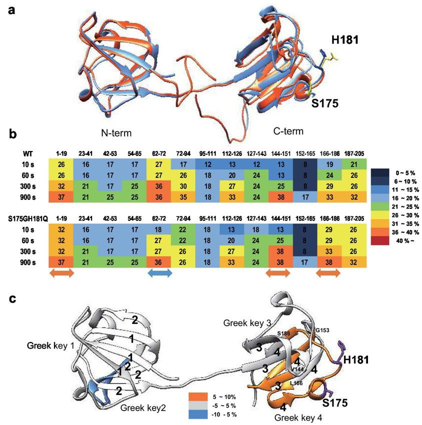
Figure 4. Effect of the double mutations (P24S/S31G) on the secondary and tertiary structures of
Figure 4. Effect
γD-crystallin byof the double mutations
spectroscopic methods. (P24S/S31G)
Intrinsic Trponfluorescence
the secondary and tertiary
excited structures
at 295 nm (a) at pHof 7.0,
γD-
crystallin
and (b) at by
pHspectroscopic methods.
2.0. The proteins wereIntrinsic
prepared Trp
in fluorescence excited
PBS buffer with at 295 nm (a)
concentration of at
0.5pH 7.0, and
mg/mL. (b)
The
fluorescence intensity was recorded for every 1 nm for WT (opened circles), the P24S mutant (gray
triangles) and the P24S/S31G mutant (closed circles). (c) Effect of P24S/S31G double mutations on
the folding and stability of γD-crystallin during denaturation induced by GdnHCl. WT and mutants
were incubated for 16 h with various concentrations of GdnHCl, and its intrinsic Trp fluorescence
intensities were monitored from 310 nm to 400 nm. Transition curves of protein equilibrium unfolding
monitored by Emax of Trp fluorescence. Data for each protein samples were fitted with a Boltzmann
curve using Origin 8.5. The final protein concentration was 0.2 mg/mL. (d) Long-term stability revealed
by incubating 2.5 mg/mL proteins at pH 2.0 and 37 ◦ C continuously, and turbidity data were measured
at given intervals.
To evaluate the long-term stability, the protein with a concentration of 5 mg/mL were incubated
at 37 ◦ C for 36 h and turbidity changes were measured with absorbance at 400 nm. In the same way
with beta-crystallins. In contrast to the increase in turbidity due to aggregation of βB2-crystallinmonitored by Emax of Trp fluorescence. Data for each protein samples were fitted with a Boltzmann
curve using Origin 8.5. The final protein concentration was 0.2 mg/mL. (d) Long-term stability
revealed by incubating 2.5 mg/mL proteins at pH 2.0 and 37 °C continuously, and turbidity data were
measured at given intervals.
Int. J. Mol. Sci. 2020, 21, 6504 8 of 18
2.6. Amyloid Fibril Formation of γD-Crystallin Mutants Measured by ThT Fluorescence Spectroscopy
A previous study showed that the fibrillogenesis of human γD-crystallin could be induced by
mutants,
acidic pHturbidity didP24S/S31G
[37]. Since not increasedouble
in γD-crystallin
mutant ofeven after 36 hisinunstable
γD-crystallin both WTat and mutant
acidic pH,(Figure S6).
we further
Since gamma crystallin aggregates well in acidic conditions, the turbidity
investigated whether this double mutant more readily aggregates at acidic pH. We examined of WT and mutant was
measured
amyloidalat 37 ◦ C inofaγD-crystallin
structure buffer condition of pH
mutants by2.0. The turbidity
employing of mutant
ThioflavinT (ThT),began to increase
a standard after
fluorescent
30 min and increased until 150 min to reach the maximum level, whereas turbidity of
dye that generates fluorescence by binding to amyloid structures. ThT is believed to bind to grooves WT gradually
increased
formed on asthe
90 min passed.
amyloid This surface
fibrous indicatesbythat the mutant
aligned protein
side chain is more
chains [38].likely to aggregate
As shown in Figurein vitro.
5a, at
This result
normal also observed
condition at 0 timethatpoint,
whenall mutants were overexpressed
γD-crystallins in mammalian
are similar fluorescence cells, the however,
intensities, mutants
was small,
during but aggregation
incubation in the
at pH 2.0, form ofinpuncta
increase wasofobserved
the rate (Figure S5).emission
ThT fluorescence These results indicate
intensity is inthat
the
S175G/H181Q mutation of βB2-crystallins reduced protein stability by affecting the
following order: P24S/S31G double mutant: P24S single mutant: and WT γD-crystallin. These results protein folding
and increasedthat
demonstrate the tendency
P24S/S31G todouble
be aggregated.
mutation promotes amyloid formation more readily than P24S
mutant and WT γD-crystallin at acidic condition.
2.6. Amyloid Fibril Formation of γD-Crystallin Mutants Measured by ThT Fluorescence Spectroscopy
A previous
2.7. Effect study pH
of Incubation showed
on thethat the fibrillogenesis
Tertiary of human γD-crystallin
Structure of γD-Crystallin could
Mutants Revealed by be
ANS induced by
acidic pH [37].
Fluorescence Since P24S/S31G double mutant of γD-crystallin is unstable at acidic pH, we further
Spectroscopy
investigated whether this double mutant more readily aggregates at acidic pH. We examined amyloidal
To investigate the effects of double mutation of γD-crystallin on the tertiary structure at low pH,
structure of γD-crystallin mutants by employing ThioflavinT (ThT), a standard fluorescent dye that
we examined the ANS spectra of WT and two mutants under acidic condition after various
generates fluorescence by binding to amyloid structures. ThT is believed to bind to grooves formed
incubation times. Destabilization of the protein structure was assessed using ANS binding technique
on the amyloid fibrous surface by aligned side chain chains [38]. As shown in Figure 5a, at normal
which measures the degree of exposure of hydrophobic regions. As shown in Figure 5b, ANS
condition at 0 time point, all γD-crystallins are similar fluorescence intensities, however, during
fluorescence intensities of all proteins increased during incubation time at pH 2.0. Increase in the rate
incubation at pH 2.0, increase in the rate of ThT fluorescence emission intensity is in the following order:
of ANS fluorescence intensity is the largest with P24S/S31G double mutant, followed by P24S and
P24S/S31G double mutant: P24S single mutant: and WT γD-crystallin. These results demonstrate that
WT. These results suggest that P24S/S31G double mutations induce more exposure of hydrophobic
P24S/S31G double mutation promotes amyloid formation more readily than P24S mutant and WT
moieties to the surface than WT as well as P24S mutant, making the structure unstable at acidic
γD-crystallin at acidic condition.
condition.
Figure 5.
Figure 5. Effect
Effect of
ofP24S/S31G
P24S/S31Gdouble
doublemutations
mutationsonon
thethe
fibril formation
fibril andand
formation tertiary structure
tertiary of γD-
structure of
crystallin in acidic pH. (a) ThioflavinT fluorescence emission intensity measurement (λ = 440
γD-crystallin in acidic pH. (a) ThioflavinT fluorescence emission intensity measurement (λEx = 440 nm,
Ex nm,
λEm = 485 nm) of WT, P24S, and P24S/S31G mutant γD-crystallin as a function of incubation time.
Samples were incubated at pH 2.0 for indicated time. (b) ANS fluorescence intensity measurement of
WT, P24S and P24S/S31G mutant γD-crystallin. ANS fluorescence emission intensity of γD-crystallin
sample as a function of incubation time are represented with WT (white bar), the P24S mutant (gray
bar) and the P24S/S31G mutant (black bar). The final concentration was 0.04 mg/mL. Data points are
presented as the means ± standard deviations (S.D.) of at least 3 independent measurements in each
Figure. * p < 0.05; ** p < 0.01 versus control.Int. J. Mol. Sci. 2020, 21, 6504 9 of 18
2.7. Effect of Incubation pH on the Tertiary Structure of γD-Crystallin Mutants Revealed by ANS Fluorescence
Spectroscopy
To investigate the effects of double mutation of γD-crystallin on the tertiary structure at low pH,
we examined the ANS spectra of WT and two mutants under acidic condition after various incubation
times. Destabilization of the protein structure was assessed using ANS binding technique which
measures the degree of exposure of hydrophobic regions. As shown in Figure 5b, ANS fluorescence
intensities of all proteins increased during incubation time at pH 2.0. Increase in the rate of ANS
fluorescence intensity is the largest with P24S/S31G double mutant, followed by P24S and WT. These
results suggest that P24S/S31G double mutations induce more exposure of hydrophobic moieties to the
surface than WT as well as P24S mutant, making the structure unstable at acidic condition.
2.8. Protein Modifications of WT and Mutant Recombinant γD-Crystallin Proteins
The structural changes of γD-crystallin in acidic condition cannot be examined with HDX-MS. Low
pH is essential for pepsin digestion of protein for HDX-MS procedure. However, since γD-crystallin
makes fibrils at low pH by aggregation [37], it is not possible to digest γD-crystallin aggregates at low
pH with pepsin to examine the structural changes with HDX-MS. Therefore, we tried to determine the
structural differences between WT and P24S/S31G mutant based on protein modifications, which vary
depending on the structural states of the protein. We isolated WT and P24S/S31G mutant γD-crystallin
recombinant proteins by SDS-PAGE and in gel-digestion with trypsin to obtain the peptides for
analysis. We comprehensively analyzed the protein modifications of WT and P24S/S31G mutant
γD-crystallin recombinant proteins employing peptide sequencing with nanoUPLC-ESI-q-TOF-MS/MS
with SEMSA [39] which is a sensitive method for identifying low abundant modifications. Precursor
ion chromatograms (XIC) of extracted precursor ions from both recombinant proteins are presented
in Figure 6. All intensities of precursor ions were normalized by total ion chromatograms (TIC) of
each sample run. Since protein modifications detected mainly in cataract lens are known as oxidation
and deamidation [40,41], we assessed the protein modifications of WT and mutant γD-crystallins as
shown in Table 2. Aging increases protein oxidation and cataract development is also promoted by
oxidation of the lens [42,43]. Oxidations of Cys residues have been identified in cataract samples [42],
and Met oxidation is known to be associated with cataractogenesis [44]. Of the six Cys residues of
γD-crystallin, four Cys residues (Cys19, 42, 109 and 111) are predicted to be exposed to the surface of
protein (Figure S7). Cys19 residue was not oxidized in WT, but was highly oxidized to sulfinic acid
(16HYECSSDHPNLQPYLSR32, ∆m = +32 Da) in P24S/S31G mutant (Figure 6a). Cys19 residue is
present in the largest loop of the Greek key motif 1 where the mutation occurred. Therefore, the oxidation
increase at Cys19 residue indicates the conformational change of Greek key motif 1 by mutation.
Cys42 residue in double mutant was mainly not oxidized, but small faction of Cys42 was oxidized
to thiosulfonic acid in double mutant (35SARVDSGCWMLYEQPNYSGLQYFLR59, ∆m = +64 Da)
(Figure 6b). Cys109 and Cys111 residues were detected in same tryptic peptide, and these are known
to function as antioxidant by forming disulfide crosslinking in WT [45]. Cys109 residue was oxidized
to dehydroalanine (∆m = −34 Da) and Cys111 was oxidized to sulfinic acid (∆m = +32 Da) in double
mutant (Figure 6c). This indicates that Cys109-Cys111 disulfide formation does not work in the double
mutant and lose their antioxidant function. In addition to Cys residue, Met residue is an oxidation
sensitive amino acid. Met147 of γD-crystallin is known to be an important residue for protein solubility
due to its proximity to the linker peptide and location at the interface between the N-terminal domain
(NTD) and C-terminal domain (CTD) [46]. Met147 was also found to be more oxidized to mono-
(143QYLLMPGDYR152, ∆m = +16 Da), or di-oxidation (143QYLLMPGDYR152, ∆m = +32 Da) in
the double mutant, in particular the intensity of the double oxidation was found to be significantly
increased compared to that of the unmodified peptide (Figure 6d–f). The results demonstrate that
P24S/S31G double mutant is more readily oxidized than WT γD-crystallin. The results show that
P24S/S31G mutant has different structure from WT γD-crystallin and is significantly aggregated in
response to various stimuli causing cataract.Int. J. Mol. Sci. 2020, 21, 6504 10 of 18
Int. J. Mol. Sci. 2020, 21, x 10 of 18
Figure 6.6.The
Figure Therelative
relative intensity
intensity of precursor
of precursor ion peptide
ion peptide containing
containing modifications
modifications in WT andinP24S/S31G
WT and
P24S/S31G
mutant mutant γD-crystallin
γD-crystallin (a–f).intensity
(a–f). The relative The relative intensity
of precursor ionofunmodified
precursor and
ion oxidized
unmodified and
peptide
oxidized peptide
including (a) Cys19,including (a) (c)
(b) Cys42, Cys19,
Cys109(b)and
Cys42,
111,(c) Cys109
(d–f) andwith
Met147 111,WT
(d–f) Met147
(white bar)with WT (white
and P24S/S31G
mutant
bar) and(black bar). NEM
P24S/S31G mutant means
(blackNEMbar).(N-ethylmaleimide) adducts of Cys residue
NEM means NEM (N-ethylmaleimide) that isofnot
adducts Cysoxidized.
residue
Total ionnot
that is chromatogram
oxidized. Total(TIC)
ionintensity of sample(TIC)
chromatogram as control normalize
intensity eachas
of sample intensity
controlofnormalize
modifications.
each
Each experiment was repeated twice.
intensity of modifications. Each experiment was repeated twice.
Table 2. Post-translational modifications detected in recombinant protein WT and double mutant
Table 2. Post-translational modifications detected in recombinant protein WT and double mutant
(P24S/S31G) of γD-crystallin. Peptides with the highest ion scores for each spot were tabulated.
(P24S/S31G) of γD-crystallin. Peptides with the highest ion scores for each spot were tabulated.
Modified residues are highlighted in bold. MS/MS spectra of modified peptides are presented in
Modified residues are highlighted in bold. MS/MS spectra of modified peptides are presented in
Figure S8.
Figure S8.
Mass (m/z) Mass Delta Mass
Residue Modification Start–End Delta (Da) Peptide Sequence
Experimental Theoretical
Start– Mass (m/z) Mass
Residue
C19 Modification
Nethylmaleimide 16–32 724.3238(3+) 2169.9496Mass −0.0042 Peptide Sequence
HYECSSDHPNLQPYLSR
Dioxidation
End Experimental
16–32
Theoretical
679.9601(3+) 2036.8585(Da) −0.0062 HYECSSDHSNLQPYLGR
C42 Nethylmaleimide
C19 Nethylmaleimide 16–32 38–59
724.3238(3+)932.0887(3+)
2169.9496 −0.0042 −0.0124
2793.2443 VDSGCWMLYEQPNYSGLQYFLR
HYECSSDHPNLQPYLSR
Trioxidation 35–59 758.5914(4+) 3030.3365 −0.0275 SARVDSGCWMLYEQPNYSGLQYFLR
Dioxidation
Thiosulfonic acid 16–32 679.9601(3+)
35–59 2036.8585
762.5911(4+) −0.0062
3046.3353 −0.044 HYECSSDHSNLQPYLGR
SARVDSGCWMLYEQPNYSGLQYFLR
C42
C109/ Nethylmaleimide 38–59 932.0887(3+) 2793.2443 −0.0124 VDSGCWMLYEQPNYSGLQYFLR
Nethylmaleimide/Nethylmaleimide 100–115 1062.9352(2+) 2123.8558 −0.004 GQMIEFTEDCSCLQDR
C111 Trioxidation 35–59 758.5914(4+) 3030.3365 −0.0275 SARVDSGCWMLYEQPNYSGLQYFLR
Cys->Dha/
Thiosulfonic acid 35–59 762.5911(4+)
100–115 3046.3353
983.4172(2+) −0.044 −0.0046
1964.8198 SARVDSGCWMLYEQPNYSGLQYFLR
GQMIEFTEDCSCLQDR
Nethylmaleimide
C109/ Nethylmaleimide/
Cys->Dha/
Dioxidation 100–115 1062.9352(2+)
100–115 2123.8558
936.8794(2+) −0.004 −0.0046
1871.7442 GQMIEFTEDCSCLQDR
GQMIEFTEDCSCLQDR
C111 Nethylmaleimide
M147 Cys->Dha/
Oxidation 143–152 636.307(2+) 1270.5994 −0.0022 QYLLMPGDYR
Dioxidation 100–115 983.4172(2+)
143–152 1964.8198
644.3046(2+) −0.0046 −0.0019
1286.5946 GQMIEFTEDCSCLQDR
QYLLMPGDYR
Nethylmaleimide
Cys->Dha/
100–115 936.8794(2+) 1871.7442 −0.0046 GQMIEFTEDCSCLQDR
3. Discussion
Dioxidation
M147 Oxidation 143–152 636.307(2+) 1270.5994 −0.0022 QYLLMPGDYR
In theDioxidation
present study, we identified
143–152 10 mutants
644.3046(2+) of crystallin
1286.5946 from proteomic
−0.0019 analysis dataset of
QYLLMPGDYR
human cataract patients, and by comparing human lens samples of normal and cataract patients.
Of these mutants, we focused on two cataract-specific double mutations, βB2-crystallin mutant,
S175G/H181Q and γD-crystallin mutant, P24S/S31G. We have shown that double mutation of
βB2-crystallin affects Greek key motif 4 structure in the C-terminus, which functions to maintain the
stability and solubility of βB2-crystallin protein, and that double mutation of γD-crystallin induces
structural changes and protein oxidation in Cys and Met residues that readily form amyloid fibril.Int. J. Mol. Sci. 2020, 21, 6504 11 of 18
These newly found mutations could serve as biomarkers of congenital cataract, as they appear to
damage lens transparency by inducing the aggregation of β/γ-crystallin, decreasing its solubility and
stability. These double mutations of crystallin occur adjacently in one tryptic peptide, which were in
the critical loop of the Greek key motif. This report is the first study to investigate dynamic structural
changes of crystallin mutants employing HDX-MS and to quantify protein oxidations of crystallin
mutants combining with spectroscopic and biophysical analysis.
β/γ-Crystallins have four Greek key motifs in two domains with twisted order [4]. The Greek key
motif is evolutionarily well conserved and crucial for the structure, function and stability of crystallin
proteins. Mutations occurred in Greek key motifs are known as typical causes of cataract [3,27].
Nonsense mutant Q155X in βB2-crystallin deteriorates protein stability by unfolding the Greek key
motif [15] and distorting only one Greek key motif of γD-crystalllin causes protein aggregation and
the development of nuclear cataracts [27]. Our new double mutation sites are present in the largest
loop connecting the distant β-sheets, which is an important factor in the stability of the Greek key
motif [28]. In S175G/H181Q βB2-crystallin, the mutated amino acid is located in the largest loop
between distant β-sheets in the Greek key motif 4. The sequence alignment indicated that S175 and
H181 residues conserved in βB2-crystallin across species except bovine (Figure S9). This indicates
that largest loop is important for preserving Greek key motifs. As shown in the Figure S2, Ser175 is
in the helix and has the potential to form hydrogen bonds with the surrounding glycine in a largest
loop. When it mutates to glycine, the helix structure can become unstable without forming hydrogen
bonds. In addition, His181 is likely to form hydrogen bonds with surrounding Gln183. When it is
substituted with glutamine, the hydrogen bond can be released and may not form the original loop
shape. In this study, the results show that double mutations affect the stability, oligomerization, and
folding of structures of βB2-crystallin. These changes were confirmed to analyze the dynamic structural
changes employing HDX-MS. S175G/H181Q mutation in βB2-crystallin made packing core of Greek
key motif 4 in C-terminus domain being loosened (Figure 3). Taken together, S175G/H181Q double
mutant is unable to retain the Greek key motif 4, which impedes the achievement of βB2-crystallin
folding, thereby causing cataracts. This is consistent with the results that misfolded βB2-crystallins
form aggregates in both the E. coli and HeLa cells [35].
γD-Crystallin mutant P24S/S31G, unlike the double mutant of βB2-crystallin, is mutated in
the Greek key 1 motif located in the N-terminus domain. The P24T and P24S mutants are known
as cataract-causing mutants by increasing surface hydrophobicity that promote destabilization and
aggregation [19,47]. New γD-crystallin mutant P24S/S31G we found is mutated not only in Pro24
but also in Ser31. Sequence alignment of γD-crystallins across species shows that Pro24 is about
60% conserved, while Ser31 is all conserved except bovine (Figure S9). Thus, it is possible that
Ser31 is important for γD-crystallin structure. Indeed, P24S and P24T was identified in peripheral
and nonnuclear cataract [27,47], while P24S/S31G mutation was found in nuclear cataract causing
severe cataract in this study. P24S mutant can increase the surface hydrophobicity and reduce protein
solubility [19]. Ser31 is presumed to form a hydrogen bond because the distance between nearby Arg15
and Arg32 is 4.44 and 6.60 Å, respectively in protein structure (Figure S3). Therefore, S31G mutant
can destabilize the Greek key motif without hydrogen bonding. This is confirmed by comparing the
structural properties of P24S/S31G double mutant with those of WT and P24S single mutant. P24S/S31G
double mutant is more readily denatured than WT and P24S at acidic pH. However, the dynamic
structural changes of γD-crystallin with HDX-MS could not be examined, because pepsin digestion in
HDX-MS experiment should be performed at low pH. Instead, we tried to determine the structural
difference between WT and P24S/S31G mutant based on the protein modifications. Protein oxidations
at Cys and Met residues, which protect proteins in response to oxidative stress, were measured and
compared between WT and P24S/S31G mutant. As shown in Figure 6, P24S/S31G mutant is more
readily oxidized than WT γD-crystallin. Since β/γ-crystallin is vulnerable to enzyme-independent
chemical modifications throughout lifespan [48,49], the results suggest that mutations make proteins
being more vulnerable to oxidative stress because of structural changes.Int. J. Mol. Sci. 2020, 21, 6504 12 of 18
In summary, this study identifies new two double mutations, S175G/H181Q in βB2- and P24S/S31G
in γD-crystallin, are sufficient to promote aggregation with distorting the largest loops connecting
the distant β sheets of Greek key motifs, possibly cause cataract. These new double mutants can be
used for early diagnosis of cataract patients. Further studies are required to understand whether these
mutants are involved in other crystallin-related diseases, i.e., γD-crystallin is known to be associated
with prostate cancer [50].
4. Materials and Methods
4.1. MS/MS Dataset and Bioinformatics Analysis
Five normal human lenses (3-day and 2-, 18-, 35-, and 70-year old) and two nuclear cataract lenses
(70-year old (Grade II, Pirie scale) and 93-year old (Grade III, Pirie scale)) were obtained from previous
studies, where age-related changes in human crystallins were analyzed [24,25]. The detailed description
of the data are provided in the previous studies [24]. In brief, the lenses were obtained from the Lions
Eye Bank of Oregon with IRB approval, dissected within 12 h of death, graded for cataract severity,
photographed, and frozen at −70 ◦ C until use. Individual lenses were homogenized on ice in a 20 mM
phosphate and 1.0 mM EDTA buffer (pH 7.0) using 1.0 mL of buffer per lens, and the homogenate was
centrifuged at 20,000× g for 30 min to pellet the water-insoluble proteins. The water-insoluble pellet
was re-suspended once in the same volume of homogenizing buffer, and again pelleted. The resulting
pellet was re-suspended by brief sonication (5 s × 2) on ice, and the protein content of both soluble
and insoluble fractions was determined in triplicate using a BCA assay (Pierce Biotechnology, Inc.,
Rockford, IL, USA). Portions (2.5 mg) of the samples were dried by vacuum centrifugation and stored
at −70 ◦ C until analysis.
Dried water-soluble or water-insoluble lens samples were dissolved in buffer containing 8.0
M deionized urea, 0.8 M Tris, 0.08 M methylamine, and 8.0 mM CaCl2 (pH 8.5). Cysteine residues
were reduced and alkylated by successive treatment with DTT and iodoacetamide, and sequencing
grade modified trypsin (Trypsin Gold from Promega, Madison, WI, USA) was added at a ratio of
1:25 protease/substrate, resulting in a dilution of the urea to a final 2.0 M concentration. Digestion
occurred during an 18 h incubation at 37 ◦ C with shaking. Following digestion, formic acid was
added to a final concentration of 5%, and peptides were solid-phase-extracted using Sep-Pak Light
cartridges (Millipore, Billerica, MA, USA). The peptides were separated by SCX chromatography and
were analyzed on a Thermo LCQ Classic instrument (Thermo Electron, San Jose, CA, USA). Their
MS/MS data sets were downloaded from public repository (MassIVE, MSV000078532) and the numbers
of MS/MS spectra for all lens datasets are shown in Table S2. The amount of water-insoluble material
from the 3-day old lens was negligible and not analyzed.
The MS/MS spectra were searched in an unrestrictive way using MODplus [51] because the human
lens tissue is often substantially modified post-translationally as it ages. The MODplus search allowed
946 chemical modifications and 360 amino acid substitutions (modifications in −150 and +350 Da
range listed in Unimod, July 2018) as variable modifications during search. The other parameters
were set as follows: precursor mass tolerance ±2.5 Da, fragment mass tolerance ±0.5 Da, trypsin
as enzyme, the number of enzymatic termini 1/2, any number of missed cleavages, any number of
modifications/peptide, Carbamidomethyl (Cys) as a fixed modification, SwissProt human database
(August 2019 release, 42,605 entries including common contaminant proteins). The protein database
was appended with pseudo-shuffled decoy sequences for FDR (False Discovery Rate) control using
target-decoy [52].
PSMs (peptide spectrum matches) with a minimum peptide length of 8 were identified at FDR
1% and the numbers of identified PSMs are shown in Table S2. The fractions of crystallin proteins in
each lens sample identified by MODplus are also shown in Figure S8. To conservatively call amino
acid mutations from identified PSMs, they were accepted only if there were no possible chemical
modifications matching the delta masses, the absolute size of delta masses were larger than 3 Da,Int. J. Mol. Sci. 2020, 21, 6504 13 of 18
and MODplus quality scores of their PSMs were higher than 0.1. As a result, total 112 mutations were
identified from all lens samples (Table S3). 85 mutations were observed in a single sample, 13 in both
normal and cataract samples, 2 in some normal samples but never in cataract samples, and 12 in all
cataract samples but never in other normal samples. The numbers of mutations observed for each
crystallin and sample are shown in Table S4a,b, respectively. More mutations were observed in cataract
samples and Table 1 shows mutated peptides containing 12 cataract-specific mutations (i.e., observed
in both 70- and 93-year cataract lens samples but never in other normal lenses). Such occurrence can
hardly happen by chance. For the identified 112 mutations across seven samples, the probability of
observing at least 12 mutations only in those two samples by chance is 9.264 × 10⁻¹⁰ .
4.2. Purification of Recombinant Proteins
For protein purification, the constructs encoding pGEX-4t-1 vector and a CRBB2, CRYGD cDNA
clone were obtained from human cDNA library as a template. All plasmid constructs were confirmed
by DNA sequencing. Mutant CRBB2 S175G/H181Q, and CRYGD P24S/S31G were generated using the
Quick-Change site-directed mutagenesis kit (Agilent Technologies, Santa Clara, CA, USA) according to
the manufacturer’s protocol using pGEX-4t-1 CRBB2 and CRYGD as a template. The primers used for
mutagenesis are: CRBB2 S175G_sense, 50 -ACTACAAGGACAGCGGCGACTTTGGGGCC-30 ; CRBB2
S175G_antisense, 50 -GGCCCCAAAGTCGCCGCTGTCCTTGTAGT-30 ; CRBB2 H181Q_sense, 50 -CTT
TGG GGC CCC TCA GCC CCA GGT-30 ; CRBB2 H181Q_antisense, 50 -ACC TGG GGC TGA GGG GCC
CCA AAG-30 ; CRYGD P24S_sense, 50 -CAG CAG CGA CCA CAG CAA CCT GCA GCC C-30 ; CRYGD
P24S_antisense, 50 -GGG CTG CAG GTT GCT GTG GTC GCT GCT G-30 ; CRYGD S31G_sense, 50 -TGC
AGC CCT ACT TGG GCC GCT GCA ACT C-30 ; CRYGD S31G_antisense, 50 -GAG TTG CAG CGG
CCC AAG TAG GGC TGC A-30 . Double mutants (CRBB2 S175G/H181Q, CRYGD P24S/S31G) were
sequentially generated using single point-mutated DNA as a template. All plasmid constructs were
confirmed by DNA sequencing. A pGEX-4t-1 plasmid carrying the human CRBB2, CRYGD gene was
expressed in BL21 (DE3) E. coli cells. The bacteria were cultured in LB medium at 37 ◦ C, and recombinant
fusion protein production was induced with 0.25 mM were isopropyl-β-d-thiogalactopyranoside (IPTG).
After 4 h of additional incubation at 37 ◦ C, the cells were harvested and lysed by vortexing with
lysozyme containing lysis buffer (lysozyme, protease inhibitor, triton X-100 in PBS (140 mM NaCl,
2.7 mM KCl, 10 mM Na2 HPO4 , pH 7.4)) on ice and sonicated. The soluble protein fraction was
recovered by centrifugation at 13,000× g for 30 min at 4 ◦ C. The GST-fused recombinant proteins
in the supernatant were purified by chromatography on a glutathione-agarose column followed by
washing, and equilibration by elution PBS buffer. To cleave crystallin from the GST-crystallin, the beads
were incubated overnight at room temperature with thrombin in crystallin elution buffer. After 16 h,
the purified crystallin was eluted and protein concentration was quantified with the BCA protein assay.
4.3. Measurement of Intrinsic Fluorescence of Tryptophan and Tyrosine
All spectroscopic experiments were carried out at 25 ◦ C using protein samples prepared in PBS
buffer (140 mM NaCl, 2.7 mM KCl, 10 mM Na2 HPO4 , pH 7.4). The fluorescence spectra were recorded
on SpectraMax i3x spectrophotometer (Molecular Devices, San Jose, CA, USA) with a slit width of 9 nm
for excitation and 14 nm for emission. The protein concentration was 0.2 mg/mL for the fluorescence
measurements. The excitation wavelength of the intrinsic Trp and Tyr fluorescence was 280 and 295 nm
respectively. The ANS (8-anilino-1-naphthalenesulfonic acid) fluorescence was determined at excitation
wavelength of 380 nm. For ANS fluorescence measurement at 470 nm, samples were prepared by
mixing the protein with ANS stock solution to final molar ratio of 75:1 (ANS:protein) and equilibrating
in the dark for 30 min at 25 ◦ C. Each experiment was triplicated with independent samples.
4.4. Protein Denaturation Induced by GdnHCl
Protein unfolding induced by guanidinium hydrochloride (GdnHCl) was performed by incubating
the WT and mutated crystallins in phosphate buffer containing various concentrations of GdnHCl (pHYou can also read