Hinge like domain motion facilitates human RBMS1 protein binding to proto-oncogene c-mycpromoter
←
→
Page content transcription
If your browser does not render page correctly, please read the page content below
Published online 17 May 2021                                                     Nucleic Acids Research, 2021, Vol. 49, No. 10 5943–5955
                                                                                                       https://doi.org/10.1093/nar/gkab363

Hinge like domain motion facilitates human RBMS1
protein binding to proto-oncogene c-myc promoter
                                   *                                                 *
Priyanka Aggarwal                      and Neel Sarovar Bhavesh

Transcription Regulation group, International Centre for Genetic Engineering and Biotechnology (ICGEB), Aruna Asaf
Ali Marg, New Delhi 110067, India

Received October 08, 2020; Revised April 02, 2021; Editorial Decision April 20, 2021; Accepted April 24, 2021

                                                                                                                                                              Downloaded from https://academic.oup.com/nar/article/49/10/5943/6276930 by guest on 15 October 2021
ABSTRACT                                                                        which master-regulates all the cellular processes. The net-
                                                                                work of protein–nucleic acid interactions inside a eukary-
DNA binding proteins recognize DNA specifically                                 otic cell is very complicated, far away from the oversimplifi-
or non-specifically using direct and indirect read-                             cation of specific and non-specific interactions. The atomic
out mechanisms like sliding, hopping, and diffu-                                resolution structural and mechanistic studies of protein-
sion. However, a common difficulty in explicitly elu-                           nucleic acid complexes are difficult to conduct without the
cidating any particular mechanism of site-specific                              prior knowledge of consensus sequences.
DNA-protein recognition is the lack of knowledge re-                               Human RNA Binding Motif Single Stranded Interact-
garding target sequences and inadequate account                                 ing Protein 1 (RBMS1) is one such protein that was first
of non-specific interactions, in general. Here, we                              isolated in 1994 as one of the family of myc gene single
decipher the structural basis of target search per-                             stranded binding proteins and has been shown to control
formed by the key regulator of expression of c-                                 the expression of proto-oncogene c-myc inside the human
                                                                                cell. c-myc protein is a transcription factor that binds both
myc proto-oncogene, the human RBMS1 protein. In
                                                                                specifically and non-specifically to activate the transcription
this study, we have shown the structural reorgani-                              of several downstream gene targets and therefore, even mi-
zation of this multi-domain protein required for rec-                           nor fluctuations in c-myc levels have profound effects on
ognizing the specific c-myc promoter sequence. The                              cellular growth and transformations (6). RBMS1 contains
results suggest that a synergy between structural                               two most abundantly present nucleic acid binding domains
re-organization and thermodynamics is necessary                                 in the eukaryotes, the RNA recognition motifs (RRMs
for the recognition of target sequences. The study                              also known as ribonucleoprotein (RNP) domain), with two
presents another perspective of looking at the DNA-                             highly conserved submotifs––octameric RNP-1 ((R/K)-
protein interactions.                                                           G-(F/Y)-(G/A)-(F/Y)-V-X-(F/Y)) and hexameric RNP-2
                                                                                ((L/I)-(F/Y)-(V/I)-X-(N/G)-L) within each of the domains
                                                                                (7,8). RBMS1 stimulates DNA replication, transcriptional
INTRODUCTION                                                                    regulation, and cell transformation by specifically binding
                                                                                to the 7 bp consensus sequence A/TCTA/TA/TT within
Protein-DNA interactions are the choreographers of cel-
                                                                                the 21 bp promoter sequence/autonomous origin of repli-
lular processes ranging from as basic as the chromosomal
                                                                                cation 2 kb upstream of c-myc gene (9–11).
organization to as complicated as translation (1). They are
                                                                                   The mechanism of c-myc promoter recognition by
responsible for maintaining the integrity of the genome as
                                                                                RBMS1 is not known at the molecular level. In this study,
well as controlling all major and minor cellular mecha-
                                                                                we report the structural and thermodynamics basis of
nisms (2,3). It is imperative that the DNA binding proteins
                                                                                the DNA binding mechanism of RBMS1. We have de-
locate their specific DNA targets in the highly dense nu-
                                                                                lineated the structural basis of specific recognition of c-
cleus of the cell (4). The functioning of DNA binding pro-
                                                                                myc promoter by RBMS1 protein with the help of the
teins relies on locating their precise DNA targets through
                                                                                three-dimensional structures of RBMS1 in free and c-myc
stochastic search processes. For achieving this, they bind a
                                                                                promoter DNA bound states determined in this study.
range of non-specific sequences, scan and recognize them
                                                                                This study provides a deeper understanding of the mech-
by combining 1D sliding, hopping, and 3D diffusion and
                                                                                anism that is followed by exclusive and stringent DNA pro-
ultimately reach their specific targets to perform their des-
                                                                                moter binding proteins during the stochastic DNA search
ignated functions (5). There is a symphony of specific/non-
                                                                                process.
specific DNA–protein interactions at the heart of the cell,

* To
   whom correspondence should be addressed. Tel: +91 11 2674 2360; Fax: +91 11 2674 2316; Email: neelsb@icgeb.res.in
Correspondence may also be addressed to Priyanka Aggarwal. Email: priyanka.pdm@gmail.com


C The Author(s) 2021. Published by Oxford University Press on behalf of Nucleic Acids Research.
This is an Open Access article distributed under the terms of the Creative Commons Attribution License (http://creativecommons.org/licenses/by/4.0/), which
permits unrestricted reuse, distribution, and reproduction in any medium, provided the original work is properly cited.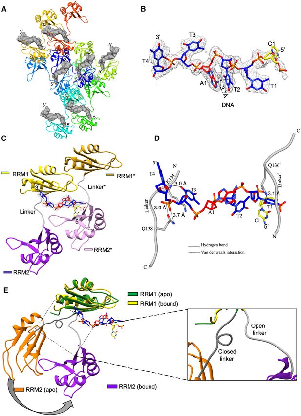
5944 Nucleic Acids Research, 2021, Vol. 49, No. 10

MATERIALS AND METHODS                                            about 100 mg of pure RBMS1 (58–224) from 1 liter of cul-
                                                                 ture. The cleavage of 6x-His tag was done using TEV pro-
Cloning, expression and purification of RBMS1 protein
                                                                 tease before performing size exclusion chromatography that
The coding sequence of RBMS1 (Uniprot Id P29558) was             left the tag related tetrapeptide GAMG at the N-terminal of
optimized for expression in Escherichia coli. The clone was      the protein. The NMR buffer used for final protein prepa-
synthesized from GeneArt (Life technologies). The bioin-         rations consisted of 20 mM sodium phosphate pH 7.1, 100
formatics software predicted the boundary of the second          mM NaCl and 5% D2 O (v/v).
RRM domain till 219 amino acid residues only and hence,
the construct initially was cloned from 58–219 amino acid        Cloning, expression and purification of isolated RRM1 and
residues in the pETM11 vector for protein expression and         RRM2 domains of RBMS1
the recombinant protein was purified. The 2D [15 N, 1 H]
HSQC spectrum, however, showed severe line broaden-              The two sub clones corresponding to RRM1 (amino acid
ing and overlap of resonance peaks. When the construct           residues 58–137) and RRM2 (amino acid residues 138–

                                                                                                                                 Downloaded from https://academic.oup.com/nar/article/49/10/5943/6276930 by guest on 15 October 2021
boundary was increased by the addition of five amino acid        224) domains were also made. Primers used for amplifica-
residues at the C-terminal end, the 2D [15 N,1 H] HSQC spec-     tion were 5 GCGCCATGGGAACCAATCTGTATATT
trum showed a very well folded and stable protein with           CGTGGTCTGCCT 3 forward primer and 5 GCG CT
much less line broadening and resonance overlap (Sup-            CGAG CTA ATC CTG TTC TTG CTG TTT TGC CAT
plementary Figure S1). The dramatic changes seen in the          3 reverse primer for RRM1, and 5 ‘GCG CCATGG GA
NMR spectra helped us in correctly determining the do-           ACAAACCTGTATATTAGCAATCTGCCG’ 3 forward
main boundaries. The subclone was then prepared corre-           primer and 5 GCGCTCGAGCTAATCCTGTTCTTGCT
sponding to the amino acid residues from 58–224. Primers         GTTTTGCCAT 3 reverse primer for RRM2. The cloning
used for amplification were 5 GCGCCATGGGAACC                    of both the constructs was done into the expression vec-
AATCTGTATATTCGTGGTCTGCCT 3 forward primer                       tor pETM11, downstream of 6X His tag, cleavable by to-
and 5 GCGCTCGAGCTAATCCTGTTCTTGCTGTTT                            bacco etch virus (TEV) protease. The vector plasmid was
TGCCAT 3 reverse primer. The cloning of the construct           transformed for protein expression into E. coli BL21(DE3)
was done in the expression vector pETM11, downstream             CodonPlus cells. The cloned genes were verified by sequenc-
of 6X Histidine-Tag cleavable by tobacco etch virus (TEV)        ing (Macrogen, Inc.). The purification protocol was the
protease. The vector plasmid was transformed for pro-            same as the one that was followed for the RBMS1 (58–224)
tein expression into Escherichia coli BL21(DE3) CodonPlus        construct.
cells. The cloned genes were verified by sequencing (Macro-
gen, Inc.).                                                      Oligonucleotides
   For expression of recombinant protein, E. coli bacterial      DNA oligonucleotides used as ligands were purchased from
cells were grown up to an OD600 of ∼0.8–1 in Luria–Bertani       Sigma-Aldrich in the desalted form. In order to understand
broth. The culture was induced using 0.5 mM IPTG at 25◦ C        the specificity of DNA, we designed 29 different DNA se-
for 16–20 h. Cells were collected by centrifugation at 3584xg    quences, in which either one of the bases of the seven nu-
rcf for 20 min, lysed by sonication by resuspension in a bind-   cleotide consensus binding DNA sequence from c-myc gene
ing buffer consisting of 20 mM sodium phosphate (pH 7.1),        promoter, i.e. TCTTATT was randomly changed to any of
300 mM NaCl, 5% (v/v) glycerol and 10 mM imidazole.              the other three bases. Some sequences were designed in a
The sample was added to Ni-NTA affinity chromatography           way to keep the TAT core sequence similar and change the
resin (Qiagen) and was washed with 20 column volumes of          other one or more bases to see the effect of nucleotide sub-
the same binding buffer but with 20 mM imidazole. Elution        stitution on protein binding. Other sequences were designed
was done in buffer with 20 mM sodium phosphate (pH 7.1),         in a way that the core sequence was not retained and other
300 mM NaCl, 5% (v/v) glycerol and 300 mM imidazole fol-         combinations from the 5’-3’ promoter or its complementary
lowed by cleavage with TEV protease for 16 h at 20 ◦ C. The      sequence were tested for their binding to the protein. Rest
TEV protease, 6X-His tag, and the uncleaved protein were         sequences were designed to check the specificity of bind-
removed by again performing Ni-NTA affinity chromatog-           ing for the length of the sequence, we took just the core se-
raphy, followed by size exclusion chromatography using S75       quence TAT and any random base at the start to see if the
16/60 GE column, in the buffer containing 20 mM sodium           core was still recognized by the protein.
phosphate (pH 7.1) and 100 mM NaCl. The centrifugal fil-
ters (Merck, Millipore) with 3000 Daltons molecular weight
cutoff was used for the concentration of fractions up to ∼1      Site directed mutagenesis
mM. Protease inhibitor cocktail (Roche) was added to the         Protein mutants (Y105S, F107L, Q135E and F185V) were
final protein preparation and storage of the protein aliquots    generated by site directed mutagenesis using a set of inter-
was done at −80 ◦ C.                                             nal PCR primers that contained the mutated sequence. The
   The preparation of isotopic labeled U-15 N or U-13 C,15 N-    mutant plasmids were verified by DNA sequencing. Mutant
labeled recombinant proteins was done using 2.5 g/L 13 C6 –      proteins were expressed and purified by using methods sim-
D-glucose and 1.0 g/l 15 NH4 Cl (Cambridge Isotope Lab-          ilar to those used for the wild type protein. The folding of
oratories), as sole carbon and nitrogen sources, respec-         protein mutants was confirmed using NMR spectroscopy.
tively in M9 minimal media, yielding the uniformly 13 C,15 N-    The primers used for each mutagenesis are shown in Sup-
labeled protein. Growth in M9 minimal medium yielded             plementary Table S4.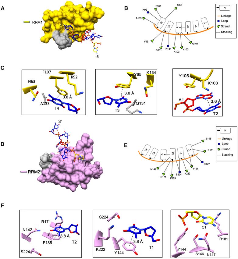
Nucleic Acids Research, 2021, Vol. 49, No. 10 5945
Solution-state NMR spectroscopy pulse sequence hsqcnoef3gpsi3d. The ratio of peak intensi-
ties with and without a 4 s proton saturation was used to ob-
Samples contained 1 mM RBMS1 protein in 20 mM
tain the steady-state 15 N–{1 H} nOe values. A recycle delay
sodium phosphate pH 7.1, 100 mM NaCl and 5% D2 O
(v/v). NMR experiments were conducted on a Bruker set to 2.5 s allowed the 15 N and 1 H spins to return to equilib-
Avance III equipped with a 5 mm cryogenic triple reso- rium. The spectra were processed using Topspin 3.1 (Bruker
nance TCI probe-head, operating at the field strength of AG) and all the calculations were done using the Dynam-
500.15 MHz at 303 K. Spectra were processed using Top- ics Center 2.5.4 (Bruker AG). The principal components of
spin 3.1 (Bruker AG) and analyzed using Computer Aided the anisotropic diffusion tensors were calculated using the
Resonance Assignment (CARA) software (12). The exper- ROTDIF 1.1 software (17). The residues whose 15 N–{1 H}
iments (13) used for protein resonance assignment were Het-nOe values were less than 0.65 were excluded from the
standard double and triple resonance spectra, namely, 2D diffusion tensor calculations.
[15 N,1 H]-HSQC, 2D [13 C,1 H]-HSQC [aliphatic (0 to 5 ppm
1
Hali ) and aromatic (4.7 to 10 ppm 1 Haro )], 3D CBCA- NMR spectroscopy of protein–DNA complexes
Downloaded from https://academic.oup.com/nar/article/49/10/5943/6276930 by guest on 15 October 2021
coNH, 3D HNCA, 3D HNCO, 3D HNCACB, 3D Hcc-
coNH, 3D hCccoNH,; all of them were acquired at 303 To map the interface of protein–DNA complexes, titration
K. For calculation of distance restraints, a set of NOESY of 0.5 mM U-15 N protein was done against the molar ra-
spectra (NOESY mixing time of 100 ms), namely, 3D 15 N- tios of DNA increasing in steps of 0.2 from 1:0 to 1:1.2. 2D
edited [1 H,1 H]-NOESY at 500.15 MHz spectrometer, 3D [15 N,1 H] HSQC spectrum was recorded at each step and was
13
Cali -edited [1 H,1 H]-NOESY in H2 O at 500.15 MHz spec- used for tracking changes in chemical shifts (chemical shift
trometer and in D2 O at 800.18 MHz spectrometer, and 3D perturbations, ␦) of the backbone amide protons at each
13
Caro -edited [1 H,1 H]-NOESY at 500.15 MHz spectrometer molar ratio. Calculation of CSPs was done using the equa-
were measured at 303 K. tion –
Manual assignment of backbone and side-chain reso-
δ 15NH 2
nances was done using Computer Aided Resonance As- δ 15NH ,HN = + (δHN )2
signment (CARA) software with 1 H shifts calibrated with 5
respect to 2,2-dimethyl-2-silapentane-5-sulfonate (DSS) at
where ␦HN and ␦15 NH are the changes in backbone
303 K (0.0 ppm). 13 C and 15 N chemical shifts were refer-
amide chemical shifts for 1 HN and 15 N resonances, respec-
enced indirectly to the DSS methyl proton resonance at 0
tively.
ppm in all spectra. TALOS-N was used for deriving back-
bone (, ) and side-chain ( 1) dihedral angle from the ob-
served chemical shifts (14). X-ray crystallography of protein–DNA complex
For co-crystallization, protein RBMS1 was mixed with
Solution structure calculation using NMR spectroscopy DNA sequence TCTTATT in an equimolar ratio of 1:1 and
was incubated at room temperature for 2 hours prior to set-
For calculation of the solution structure, the cross-peaks in ting up the crystal trays. The final protein concentration was
the NOESY spectra were used to derive the inter-proton re- 30 mg/ml in a buffer containing 20 mM HEPES pH 7.5, 50
straints up to a limit of 5 Å. NOE intensities were used for mM NaCl, 10 mM MgCl2 and 10 mM -Mercaptoethanol.
classification of distances as 1.8- 2.4 Å (strong), 1.8–3.5 Å The crystals were grown at 273 K by the hanging drop va-
(medium), 1.8–5.0 Å (weak). A total of 2185 distance con- por diffusion method and the reservoir contained 0.05 M
straints (around 17 per residue) were used for structure cal- Magnesium Sulfate Hydrate, 0.05 M HEPES sodium pH
culation using the program CYANA 3.98.13 (15), using dis- 7.0, 1.6 M lithium sulfate and 30% methanol. The thin
tance geometry and simulated annealing protocol of 20 000 plate-shaped crystals were soaked in cryoprotectant para-
steps. Further refinement of the top 20 cyana structures with tone oil and were directly mounted in a stream of cooled
the least residual target function and violations was done nitrogen gas at 100 K. Cu K␣ radiation ( = 1.54 Å) at
by simulated annealing and energy minimization in explicit 100 K was used for the collection of X-ray diffraction data
solvent using the SANDER module of the AMBER18. The using a Rigaku FR-E+ SuperBright microfocus rotating-
amber ff14SB force field (16) was used for the minimization. anode (dual-wavelength; Cu and Cr) X-ray generator that
The final ensemble comprised of 20 structures with the low- was equipped with an R-AXIS IV++ detector, operating at
est energy. 45 kV and 55 mA. Oscillation steps of 0.5◦ were used to col-
lect a total of 509 frames. The exposure time of each frame
was kept 240 s. The diffraction images set was processed and
Backbone 15 N relaxation experiments
scaled using the autoPROC package (18). The structure was
Backbone nuclear spin relaxation (s-ps dynamics) of solved using phaser-MR with HuD in complex with C-FOS
RBMS1 in free and DNA bound form were measured us- RNA as a template (38% sequence identity, PDB: 1FXL).
ing 15 N–{1 H}-heteronuclear nOe and 15 N T1, T2 relaxation The initial model was built using AutoBuild in PHENIX
experiments using Echo/Anti-echo-TPPI gradient selection (19) and was followed by multiple rounds of the manual
as pseudo 3D. Sixteen delays ranging from 20 to 1000 ms model building using Coot (20) in combination with run-
were used for T1, while 14 loop counters for the CPMG ning refinement cycles in PHENIX. UCSF Chimera (21)
pulse train were set to get T2 delays from 10 to 210 ms. and PyMol (http://www.pymol.org) softwares were used for
The 15 N–{1 H} heteronuclear nOe were measured using the all the structure visualizations and preparing images.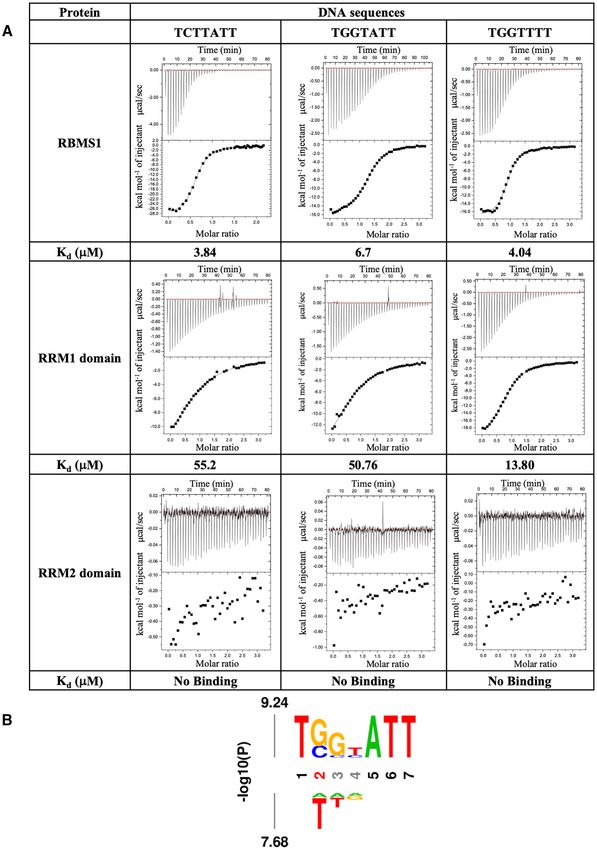
5946 Nucleic Acids Research, 2021, Vol. 49, No. 10
Isothermal titration calorimetry respect to the RRM1 domain (amino acid residues 58–132)
and when aligned with respect to the RRM2 domain (amino
ITC experiments were conducted at 303 K in the GE Mi-
acid residues 142–224) are shown in (Figure 1B and C).
croCal iTC200 calorimeter. ITC cell was filled with 0.1 mM
Both the domains had the canonical RRM fold of 1–␣1–
protein and 1 to 1.5 mM DNA was filled in the syringe. Both
2–3–␣2–4; with the two ␣-helices packed against an an-
protein and DNA were prepared in the buffer containing
tiparallel four stranded -sheet. The superimposition of the
20 mM sodium phosphate (pH 7.1) and 50 mM NaCl in
RRM1 and RRM2 domain has been shown separately in
filtered water. Protein and DNA concentrations were mea-
Figure 1d and e, respectively. The canonical RNA-binding
sured at 280 and 260 nm, respectively. Titrations consisted
ribonucleoprotein (RNP) sites, RNP1 and RNP2 were con-
of sequential injections of DNA with the first injection of
served (Supplementary Figure S2) and present on the 3
0.4 l followed by 39 injections of 1 l volume. A 120 s in-
and 1 strands of both the domains, respectively. A highly
terval was kept between the injections. The reaction mixture
flexible linker of 9 amino acid residues (amino acid residues
in the sample cell was constantly stirred at 750 rpm. To de-
133–141) connected the two domains and resulted in spa-
termine the change in enthalpy due to ligand dilution, titra-
Downloaded from https://academic.oup.com/nar/article/49/10/5943/6276930 by guest on 15 October 2021
tial heterogeneity of the domains about this region. Poor
tion of RBMS1 was performed with buffer alone. This was
convergence of structures in this region was attributed to a
then subtracted as background from the actual DNA bind-
small number of nOes observed for the residues in the linker
ing experiments. The results gave heats that were fitted to a
region due to conformational averaging. The flexibility in
one-site model using Origin 7 software.
the linker region was supported by the secondary chemi-
cal shifts and NMR relaxation parameters for the residues
MD simulations in the region (Supplementary Figures S2b and S3). RRM1
and RRM2 domains did not interact with each other in the
PDB files of RBMS1 and RBMS1-TCTTATT complex
free form of the protein and this was also evident from the
structures were prepared for Molecular Dynamics using
overlay of 2D [15 N,1 H] HSQC spectra of RRM1 domain
the Desmond 3.1 MD package (Schrödinger Inc.). The
(58–137) and the RBMS1 protein (58–224) as well as from
molecule was placed inside an orthorhombic box to impose
the overlay of 2D [15 N,1 H] HSQC spectra of RRM2 do-
periodic boundary conditions, ensuring a solvent shell of
main (138–224) and the RBMS1 protein (58–224) in which
at least 10 Å around the molecule, which was subsequently
no CSP were observed in the resonances of the amide pro-
filled with water molecules using the TIP3P solvent model
tons of the RRM1 domain and RRM2 domain, respectively
and was neutralized by the addition of Na+ /Cl– ion pairs
and all of them overlapped completely with those in the 2D
to reach a concentration of 150 mM. Prior to simulation,
[15 N,1 H] HSQC spectrum of the protein (58–224) (Supple-
the system was minimized for 100 ps. The simulation time
mentary Figure S4). The flexible linker resulting in the in-
was set to 1000 ns and the standard NPT ensemble sys-
dependent domain motion could be one of the possible rea-
tem (isobaric-isothermal condition) was used for simulation
sons that our attempts to obtain crystals of RBMS1 did not
wherein the constant temperature used was 300 K, and con-
succeed.
stant pressure 1.01325 bars under the force field OPLS3e
(22). The co-ordinate frames were saved at intervals of 4.8
ps for analysis. The time step used was 2 fs. Input and output
files were prepared, analyzed, and visualized using Maestro Both RRM domains of RBMS1 are required for DNA bind-
graphical user interface (GUI). ing
The truncated RBMS1 protein containing both RRM do-
RESULTS mains (58–224) was used for DNA binding studies. The
protein interacted with the full-length 21 bp promoter se-
RRM domains of RBMS1 form globular structure and do not
quence of c-myc with the affinity of 2.6 M and to the seven
interact with each other
nucleotides consensus sequence TCTTATT within the full-
The domain architecture of human RBMS1 protein com- length promoter sequence with an almost similar affinity
prised of two RRM domains separated by a stretch of only of 3.84 M in the ITC experiment (Supplementary Table
nine amino acid residues between them (Figure 1a). After S2). Hence, in order to further understand the role of indi-
the optimization of protein construct boundaries (see Ma- vidual RRMs in the promoter DNA binding, we made two
terials and Methods), protein (58–224) was found to exist as more constructs of the individual domains RRM1 (amino
a monomer by size exclusion chromatography (Supplemen- acid residues 58–137) and RRM2 (amino acid residues 138–
tary Figure S1). The purified homogeneous RBMS1 pro- 224), which were again purified to homogeneity (Supple-
tein was used to obtain complete sequence-specific NMR mentary Figure S1). The selected consensus sequence from
assignments and calculate solution structure. A superimpo- the upstream c-myc gene promoter sequence TCTTATT
sition of 20 lowest energy structures of RBMS1 (PDB id was titrated against both the shorter domain constructs of
7C36) is shown in Figure 1. The NMR structural param- RBMS1 protein, i.e. RRM1 domain, and RRM2 domain.
eter statistics for the energy-minimized 20 conformers of While the RRM2 domain showed no binding with the pro-
RBMS1 was calculated using cyana 3.98.13 and are given moter sequence, the binding affinity of the RRM1 domain
in Supplementary Table S1. The solution NMR structure decreased 18-fold from 3.84 M with RBMS1 (58–224) to
within the two domains was very well defined separately 55.2 M (Figure 2A). The presence of both the domains
and good convergence was seen within each of the domains. was deemed necessary for interaction with the promoter
The superimposition of the conformers when aligned with DNA sequence.Nucleic Acids Research, 2021, Vol. 49, No. 10 5947
Downloaded from https://academic.oup.com/nar/article/49/10/5943/6276930 by guest on 15 October 2021
Figure 1. (A) Schematic of the domain architecture of RBMS1 protein showing the construct boundaries. (B) Superposition of the backbone atoms of the
20 energy-minimized conformers of RBMS1 as aligned with respect to the RRM1 domain (residues 58–132). (C) Superposition of the backbone atoms of
the energy-minimized conformers of RBMS1 aligned with respect to the RRM2 domain (residues 142–224). (D) Superposition of the RRM1 domain of
RBMS1. (E) Superposition of the RRM2 domain of RBMS1. Secondary structural elements, N- and C-terminals are marked.5948 Nucleic Acids Research, 2021, Vol. 49, No. 10
Downloaded from https://academic.oup.com/nar/article/49/10/5943/6276930 by guest on 15 October 2021
Figure 2. (A) ITC thermograms and affinity (Kd ) values of the selected DNA sequences with the RBMS1 protein (58–224) and the two domains individually.
The RRM2 domain alone does not show binding with any of the DNA sequences. (B) Relative affinity of nucleotide substitutions within the cognate c-myc
promoter DNA sequence TCTTATT ligand calculated using ITC. The height of the A (adenine), C (cytosine), G (guanine) and T (thymine) is relative to
the determined values of the dissociation constant, Kd (Supplementary Table S2).Nucleic Acids Research, 2021, Vol. 49, No. 10 5949
Minimum length of six nucleotides of c-myc promoter se- Table 1. X-Ray structural parameters for the RBMS1-TCTTATT com-
quence having trinucleotide ATT at 3 end is required for bind- plex structure. The Ramachandran plot statistics were obtained using
ing with RBMS1 PSVS
In order to understand the specificity of DNA sequence in- PDB id 6M75
Data collection
teractions, we performed a series of ITC experiments with Wavelength (Å) 1.54
29 different DNA sequences, in which the bases of the 7 Detector type R-AXIS IV++
nucleotide consensus binding DNA sequence from c-myc Oscillation (◦ ) 0.5
gene promoter i.e. TCTTATT were randomly changed to Exposure (s) 240
No. of images 509
any of the other three bases. Also, in a few cases, more Software used for data processing autoPROC
than one nucleotide base in the sequence was changed or Space group P 21 21 2
where the length of the sequence was altered to have four Cell dimensions
or five nucleotides only. The entire list of DNA sequences, a, b, c (Å) 83.68, 114.95, 27.43
which were titrated for the binding studies and the ther- ␣, , ␥ (◦ ) 90, 90, 90
Downloaded from https://academic.oup.com/nar/article/49/10/5943/6276930 by guest on 15 October 2021
11.9 (2.4)
modynamic parameters obtained with them are given in Completeness (%) 99.7 (99.5)
Supplementary Table S2. The sequence logo that was de- CC1/2 0.995 (0.711)
rived from the sequences and the dissociation constant val- Resolution (Å) 67.65–2.57 (2.61–2.57)
ues obtained with them is shown in Figure 2B. The affinity Rmerge 0.166 (0.998)
Rmeas (%) 0.175 (1.052)
measurements using ITC showed that there is a sequence Rpim 0.055 (0.328)
preference for molecular recognition by the RBMS1 protein Redundancy 9.7 (10.0)
but confounded the idea that there was any simple code of No. of unique reflections 9032 (428)
recognition. When the length of the sequence was reduced Refinement
to four or five nucleotides only, the binding was abolished, Resolution (Å) 19.66–2.57 (2.65–2.57)
Rfree test set 900 reflections (10.00%)
showing that the protein preferred a minimum DNA length Rwork /Rfree (%) 0.211, 0.236
of six nucleotides for binding. Results also showed that the Total number of atoms 1466
binding preference of the protein for some DNA bases at Protein 1264
the given positions, for example, the substitution of C with DNA 120
Solvent 32
G at the second position of 7 nucleotide DNA sequence, Others 50
reduced the affinity of protein towards the resulting DNA Overall CC (real space correlation) 0.83
sequence TGTTATT from 3.84 to 15.2 M (Supplementary Average B, all atoms (Å2 ) 48.9
Table S2). Protein 48.1
Although we analyzed a lot of different DNA sequences DNA 78.7
R.m.s deviations
thermodynamically using ITC, a simple pattern of recogni- Bond lengths (Å) 0.006
tion was not seen for this protein. We, therefore, selected 3 Bond angles (º) 1.09
out of all of these DNA sequences for further analysis; this Ramachandran plot
included one specific consensus promoter sequence of c-myc Favoured/Allowed (%) 98.2/1.8
gene, TCTTATT, and two other sequences, TGGTATT and Rotamer outliers 0
C-beta outliers 0
TGGTTTT that showed good affinity. The rationale behind
selecting these two sequences amongst others was that these The values in the parenthesis are the statistics for the last resolution shell.
DNA sequences showed higher affinity than the other se-
quences for the RBMS1 protein (58–224) in the ITC experi-
ments. The three selected DNA sequences were then titrated Figure S5). Interestingly, the amino acid residues such as
against three constructs of RBMS1 protein, i.e. the RBMS1 K134 and Q135 in the linker region (amino acid residues
(58–224), the RRM1 domain (58–137), and the RRM2 do- 133–141) also showed significant perturbations indicating
main (138–224) (Figure 2a). There was no binding of any their possible role in binding to the DNA sequence. Another
of the three DNA sequences with the RRM2 domain. The interesting observation was that the binding of non-specific
binding affinity of the TGGTATT and TCTTATT sequence sequence TGGTTTT caused perturbations in the residues
decreased 10- and 18-fold, respectively with the RRM1 do- on RRM1 domain majorly, while the TGGTATT and TCT-
main as compared to the RBMS1 protein (58–224). TATT sequences caused perturbations in residues on both
In order to map specific residues on the protein sur- the RRM1 and RRM2 domains.
face, which were involved in binding to the DNA sequence,
we performed NMR titration of the DNA sequences with
RBMS1 (58–224) and calculated chemical shift perturba- RBMS1 protein binds with c-myc promoter DNA in a non-
tions (CSP). The common patches of amino acid residues canonical manner
involved in binding to the nucleic acid sequences fell mainly To delineate the atomic interactions between the protein
on the  strands of both the domains. In the RRM1 do- and DNA, we determined the crystal structure of RBMS1
main, amino acid residues on the 3 strand, such as T91, protein (58–224) with the c-myc promoter consensus se-
G104, Y105, while residues on 1 and 3 strands of the quence TCTTATT (PDB id 6M75). The data collection
RRM2 domain such as T141, L161, F185 showed sig- and refinement statistics are presented in Table 1. The crys-
nificant perturbation, indicating that these are the main tal asymmetric unit contained one molecule of protein
residues involved in nucleic acid binding (Supplementary and DNA each. RBMS1 adopted an open conformation5950 Nucleic Acids Research, 2021, Vol. 49, No. 10
wherein both domains were relatively far apart from each tional correlation time, c , obtained from the model-free
other. A comparison of the binding mode of the DNA to analysis of relaxation data, that increased to 11.0 ns from
RBMS1 with a similar type of structure of HuD protein 8.8 ns upon formation of the complex of RBMS1 with c-
(23) (PDB id 1FXL) wherein the two RRM domains form a myc promoter DNA. We calculated the average T1, T2, S2 ,
cleft in the shape of V to accommodate 11 nucleotide RNA R1/R2 and 15 N–{1 H} Het-nOe values for the RRM1 do-
sequence, suggested a deviation from the canonical bind- main, RRM2 domain, and the linker in both free and bound
ing. In the crystal structure, non-canonical binding mech- forms (Table 2). There was an overall decrease in the R1/R2
anism of DNA was observed wherein the DNA binding ratio (Table 2, Supplementary Figure S3a) and an overall in-
spanned from one domain in one asymmetric unit to the crease in the internal site-specific squared order parameter
other domain in the symmetry related molecule (Figure 3). S2 (S2 = Sf2 Ss2 ) of all the residues (Table 2, Supplemen-
Except for the 5 terminal nucleotide, for which electron tary Figure S3c). The values of c , R1 and R2 were within
density could not be observed, 5 nucleotides of the DNA the range of values expected for a protein of size 18.6 kDa
promoter consensus sequence TCTTATT made very spe- (24). The lower average of R1/R2 values of bound RBMS1
Downloaded from https://academic.oup.com/nar/article/49/10/5943/6276930 by guest on 15 October 2021
cific contacts with the aromatic amino acid residues on the (0.12) compared to free RBMS1 (0.17) suggested that the
protein in a 5 to 3 direction from RRM2 to RRM1 do- rotational diffusion of the RBMS1 became slower in the
mains. DNA promoter sequence’s nucleotide bases T3, T4, bound form (Table 2, Supplementary Figure S3a). Subtle
T6 and T7 were involved in parallel – stacking with the differences were seen as an increase in rigidity of the protein
amino acid residues Y144, F185, Y65 and F107, respec- by the increase in S2 values of some residues at sites where
tively (Figure 4), while A5 was involved in parallel displaced the DNA interacts with the protein to form the complex;
stacking with Y105. Y65 lies on the 1 strand of RRM1 S2 for F185 residue increased from 0.89 to 0.98 and that
and Y105 and F107 lie on the 3 strand of the RRM1 do- of Y65 increased from 0.87 to 0.97 upon complex forma-
main of RBMS1. The other two amino acids involved in tion. However, as nOes show sensitivity to the local flexibil-
stacking with DNA were positioned on the RRM2 domain ity; an increase in the average nOe values from 0.64 to 0.72
of the protein with Y144 and F185 lying on the 1 and for the linker suggested a decrease in its rapid local motion
3 strands of RRM2. These aromatic amino acid residues upon the complex formation (Table 2, Supplementary Fig-
showed high CSP values in the solution-state NMR spec- ure S3b). The local motions of the individual domains also
tra (Supplementary Figure S5) and were located on RNP decreased upon complex formation, however, the change in
sites of these RRM domains. The interactions observed in nOe values was not as pronounced as was seen for the linker.
the crystal structure were validated with the ITC data ob- The calculated R1, R2, 15 N–{1 H} Het-nOe values, and
tained from the mutations that were done in the aromatic the structural co-ordinates were used for calculating the
amino acid residues on the RRM1 domain (Y105S, F107L), overall rotational diffusion tensors using an anisotropic
which proved to be utmost crucial for binding affinity as diffusion tensor model for the free RBMS1, free individ-
well as specificity of RBMS1. The mutations done on the ual RRM1, and RRM2 domains and for the RBMS1,
RRM2 domain (F185V) showed they were important for RRM1 and RRM2 domains in the complex form, using
governing the specificity of the binding only. The mutation the program ROTDIF 1.117 (Table 3). The residues whose
of amino acid residue in the linker (Q138E) also affected the local motions were pre-dominant, that is, the ones having
15
affinity and specificity of the binding of RBMS1 protein to N–{1 H} nOe value less than 0.65 were excluded from the
the cognate DNA sequences (Supplementary Table S3). All calculations. We observed uniform increase in the diffusion
these findings supported the molecular mechanism of DNA tensors and this possibly could have implication in the bind-
binding that was delineated from the crystal structure. ing of DNA.
In order to further understand the role of dynamics in
DNA binding, we performed 1000 ns molecular dynam-
RRM domains undergo conformational and dynamic reorga-
ics (MD) simulations for free and TCTTATT DNA-bound
nization for DNA binding
states of RBMS1 protein. Both the free and DNA bound
The comparison of the free solution-state NMR struc- states of protein reached a state of equilibrium after 300 ns,
ture of RBMS1 (58–224) with the complex structure of as can be seen from the RMSD plots in Supplementary Fig-
RBMS1 in bound form with the DNA showed the confor- ure S6c and d. In the case of the free protein, the RMSD de-
mational change in RBMS1 upon binding to the DNA (Fig- viation within the protein was within 14.0–16 Å towards the
ure 3E). The opening of the 310 helix in the linker region end of MD, while in the bound form it deviated in the range
and movement of the RRM2 domain facilitated the binding of 12.0–13.5 Å, showing, it essentially became more stable
of RBMS1 to the DNA. The overall RMSD of individual upon complex formation. The RMSF plot (Supplementary
domain was calculated by separately overlaying individual Figure S6e and f), which shows residue-wise deviation, de-
domains in the X-ray structure of the complex and NMR picted major changes in the last 4 strand and the preceding
structure of the free protein (Supplementary Figure S6a and loop (amino acid residues 140–167) of RRM2 domain at the
b). As has been stated before, the mutagenesis studies of the C-terminal end of the protein that showed a deviation of 6
linker residue Q138 proved the importance of repositioning Å, coming down from 12 Å in case of the free protein struc-
of the linker to orient the domains in the correct pose. ture. Upon complex formation, the sheet of RRM2 domain
NMR relaxation dynamics analysis showed that the bind- came close to the 5 terminal end of the DNA sequence, i.e.
ing of RBMS1 (58–224) protein with the promoter DNA Y145 and F185 amino acid residues stacked parallel with
sequence led to minor changes in the flexibility of the pro- the T3 and T4 nucleotide bases of the DNA (supplemen-
tein. This could be seen from the effective (isotropic) rota- tary videos 1 and 2).Nucleic Acids Research, 2021, Vol. 49, No. 10 5951
Downloaded from https://academic.oup.com/nar/article/49/10/5943/6276930 by guest on 15 October 2021
Figure 3. (A) X-ray structure of the c-myc promoter bound to RBMS1 protein (58–224) and its symmetry related molecules. (B) The electron density
omit map of DNA at 1 in the bound form with the stacking distance of 3.7 Å between A1 and T2 nucleotides is shown. (C) Zoomed in view of DNA
binding between protein and one symmetry related molecule. (D) The interactions between the amino acid residues of the linker and the DNA nucleotides
of promoter DNA sequence are shown. (E) Superimposition of the solution-state NMR structure of apo RBMS1 with the crystal structure of RBMS1
bound with DNA showing the opening of the 310 helix in the linker region and movement of the RRM2 domain to bind DNA.5952 Nucleic Acids Research, 2021, Vol. 49, No. 10
Downloaded from https://academic.oup.com/nar/article/49/10/5943/6276930 by guest on 15 October 2021
Figure 4. (A) Surface view of the RRM1 domain (yellow) and the 3 terminal end of the DNA. (B) 2D representation of various types of molecular
interactions between the amino acid residues of the RRM1 domain and the 3 terminal end of the DNA with (C) Zoomed in views of the important
interactions. (D) Surface view of the RRM2 domain of symmetry related molecule and the 5 terminal end of the DNA with (E) 2D representation of
various types of molecular interactions between the amino acid residues of the RRM2 domain and the 5 terminal end of the DNA with (F) Zoomed in
views of the important interactions.
MD simulation videos clearly showed that the movement nucleotides of the DNA sequence. Therefore, the changes in
of RRM2 is crucial for the binding of RBMS1 to the DNA the orientation and positioning of the protein were required
and also explain why the two domains could not be fixed for protein to scan the bases and function at the specific pro-
with respect to each other in the solution-state NMR struc- moter DNA sequence site only.
ture. The stoichiometry calculated in solution from ITC,
NMR spectroscopy, and the MD simulation showed 1:1
DISCUSSION
binding wherein one protein molecule spanned across the
promoter DNA consensus sequence with the RRM1 do- DNA recognition by DNA binding proteins is a stochastic
main binding with the 3 terminal end nucleotides of DNA, process, with little affinity differences towards the specific
and the RRM2 domain binding with the 5 terminal end and non-specific DNA sequences (25). The thermodynam-Nucleic Acids Research, 2021, Vol. 49, No. 10 5953
Table 2. Comparison of average T1, T2, R1/R2, S2 and 15 N–{1 H} Het- binds the U2 nucleotide and U3 through U6 nucleotides
nOe values for the four constructs of RBMS1 protein in both free and are bound by the RRM2 domain (29). Our calorimetric
DNA bound forms ITC studies showed that the DNA sequences of less than
15 N–{1 H} 6 nucleotides in length did not bind to RBMS1 protein, in
Protein construct T1 (s) T2 (s) R1/R2 S2 Het-nOe vitro. Most RRM domain containing proteins, having two
or more than two RRMs do not have all RRM domains
RBMS1 (58–224) Free 0.58 0.10 0.17 0.87 0.67
Bound 0.63 0.08 0.12 0.93 0.71
participating in binding to the nucleic acid (23,28). This has
RRM1 (58–132) Free 0.57 0.10 0.17 0.87 0.67 been reported in the prp24 protein (30), wherein the first
Bound 0.64 0.07 0.12 0.94 0.71 two RRMs out of total 4 RRMs in the protein are involved
Linker (133–141) Free 0.56 0.10 0.17 0.87 0.64 in binding with the U6 RNA. In our case, when we sepa-
Bound 0.62 0.08 0.12 0.93 0.72 rated the two RRM domains of RBMS1, the RRM2 do-
RRM2 (142–224) Free 0.60 0.09 0.16 0.87 0.67
Bound 0.62 0.08 0.13 0.92 0.70 main alone did not bind to any of the nucleotide sequences
we titrated with it, and the binding of the RRM1 domain
Downloaded from https://academic.oup.com/nar/article/49/10/5943/6276930 by guest on 15 October 2021
alone to the nucleic acid sequences decreased ∼10–18 fold.
Table 3. The principal components of the anisotropic diffusion tensors of
This is because T3 and T4 nucleotides were involved in the
the different constructs of RBMS1 protein calculated using the program
ROTDIF 1.117
parallel – stacking with the Y144 and F185 amino acid
residues, which are present on the RRM2 domain. More-
over, the proper positioning of domain 1 with respect to
RBMS1– RRM1 RRM1 RRM2 RRM2 the DNA sequence was done with the help of the linker.
RBMS1 TCTTATT free bound free bound
Therefore, when the interaction of only the RRM1 domain
Dx × 107 s–1 0.96 1.40 0.94 1.41 0.91 1.33 was checked with the promoter DNA sequence, a 10–18
Dy × 107 s–1 1.02 1.49 1.02 1.47 1.01 1.46 fold reduction in affinity of interaction was deemed justi-
Dz × 107 s–1 1.07 1.77 1.08 1.70 1.12 1.92 fied. Hence, both the domains were necessary for RBMS1 to
perform its designated function. We argue that the versatil-
Dx , Dy and Dz denote the principal values of the anisotropic rotational
diffusion tensors in the x, y and z directions, respectively. ity shown by the RRM fold in binding to diverse sequences
comes from the cooperation of more than one RRM do-
main to carry out its function. The binding of RBMS1 pro-
ics and kinetics of DNA-protein interactions are the two tein to the c-myc promoter DNA sequence decreased 10
major determinants that govern the specific and the non- times if the amino acid residue in the linker of the RBMS1
specific binding (1–3). In this study, we report the DNA (58–224) was mutated (Supplementary Table S3). This re-
sequence recognition mechanism of a regulatory protein instilled the importance of the linker in correctly position-
RBMS1 that stringently regulates proto-oncogene c-myc ing the two domains to bind with the promoter DNA se-
levels and presents the future for developing efficient can- quence of c-myc.
cer targeted gene therapy against c-myc proto-oncogene. We explored the process of DNA scanning by RBMS1
The crystal packing revealed unique structural features protein by mutating the DNA sequence by changing one or
where the DNA binding spanned from one domain to the more bases. We wondered if the stacking interaction that
other domain of the symmetry related RBMS1 molecule. was seen between the bases of the promoter nucleotide se-
In the crystal structure, aromatic residues formed stack- quence was also a factor in deciding the orientation/pose
ing interaction with the DNA bases. The role of aromatic of RBMS1 and its specificity, or other factors were at play
residues has been implicated several times before in bind- too. Our results revealed that within all the sequences that
ing to the nucleic acid sequences inside the cell (26,27). The were used for the thermodynamics calculations, very little
DNA did not bind to the protein in an extended form and difference was seen in the affinity between the specific and
a stacking network was seen between the T4 and A5 nu- non-specific sequences. However, entropy is speculated to
cleotides of the DNA sequence (Figure 3B). The compari- play a major role in the modulation of the specificity of
son between the free NMR and complex X-ray structures interactions. It was observed that the change in entropy
(Figure 3E) revealed that the RRM2 domain underwent a of RBMS1’s interaction with TCTTATT was the lowest
major change in its orientation in order to bind the DNA amongst all the sequences that were analysed thermody-
molecule. We observed that the two domains did not in- namically using ITC.
teract with each other and tumbled independently in the The overall dynamics in the supplementary videos 1 and 2
solution. It is known that the presence of multiple RRM showed that the orientation of the RRM2 domain changed,
domains in a protein increases its affinity to a stretch of and it became ordered when it came close to the 5 terminal
nucleic acid as well as makes it possible for a nucleic acid of DNA. There were minor changes in the flexibility of the
binding protein to recognize a longer length of RNA/DNA RBMS1 protein upon binding but further experimental in-
nucleotides (28). For example, in the human HuD protein vestigations will help in establishing the role of dynamics in
(23), two RRMs form a complex with the c-fos AU rich 11 recognition of the c-myc promoter by the RBMS1 protein.
nucleotide sequence, wherein the RRM1 domain and the in- Site directed mutagenesis studies showed that the mutation
terdomain linker binds with the U5 through U10, while the of aromatic residues on the RRM1 domain had a larger
RRM2 accommodates two nucleotides U3 and U4; bind- impact on the binding of RBMS1 to the c-myc promoter,
ing a total of 7 nucleotides in the 11-nucleotide sequence. which complemented the structural finding that the RRM2
Similarly, in CUGBP1 protein, RRM1 and 2 together binds domain was mainly involved in correctly positioning the
to a 5 nucleotide long RNA sequence, where the RRM1 RRM1 domain onto the DNA. The complementarity in the5954 Nucleic Acids Research, 2021, Vol. 49, No. 10
conformation of the binding sequence’s nucleotides and the 3. Iwahara,J., Zweckstetter,M. and Clore,G.M. (2006) NMR structural
corresponding conformation of the RRM domain could be and kinetic characterization of a homeodomain diffusing and
hopping on nonspecific DNA. Proc. Natl. Acad. Sci. U.S.A., 103,
a crucial factor in governing the sequence specificity and the 15062–15067.
designated function. 4. Alberts,B., Johnson,A., Lewis,J., Raff,M., Roberts,K. and Walter,P.
To summarize, in this study we have determined the struc- (2014) In: Molecular Biology of the Cell 173–236. Garland Science.
tural and thermodynamics basis of c-myc promoter DNA 5. Wieczor,M. and Czub,J. (2017) How proteins bind to DNA: target
recognition by RBMS1 protein. Finally, more such struc- discrimination and dynamic sequence search by the telomeric protein
TRF1. Nucleic Acids Res., 45, 7643–7654.
tural and thermodynamics studies aimed at similar DNA- 6. Marcu,K.B., Patel,A.J. and Yang,Y. (1997) Differential regulation of
protein complexes need to be done to get more mechanistic the c-MYC P1 and P2 promoters in the absence of functional tumor
insights, and direct better designing of the future anti-gene suppressors: implications for mechanisms of deregulated MYC
therapies. transcription. Curr. Top. Microbiol. Immunol., 224, 47–56.
7. Nagai,K., Oubridge,C., Ito,N., Avis,J. and Evans,P. (1995) The RNP
domain: a sequence-specific RNA-binding domain involved in
DATA AVAILABILITY processing and transport of RNA. Trends Biochem. Sci., 20, 235–240.
Downloaded from https://academic.oup.com/nar/article/49/10/5943/6276930 by guest on 15 October 2021
8. Niki,T., Galli,I., Ariga,H. and Iguchi-Ariga,S.M. (2000) MSSP, a
Solution structures of free RBMS1 and crystal structure protein binding to an origin of replication in the c-myc gene, interacts
of TCTTATT DNA-bound form have been deposited in with a catalytic subunit of DNA polymerase alpha and stimulates its
polymerase activity. FEBS Lett., 475, 209–212.
the Protein Data Bank (PDB) under accession numbers 9. Negishi,Y., Nishita,Y., Saëgusa,Y., Kakizaki,I., Galli,I., Kihara,F.,
PDB id: 7C36 and PDB id: 6M75, respectively. The corre- Tamai,K., Miyajima,N., Iguchi-Ariga,S.M. and Ariga,H. (1994)
sponding deposition of NMR resonance assignments in the Identification and cDNA cloning of single-stranded DNA binding
BioMagResBank (BMRB) have been made under accession proteins that interact with the region upstream of the human c-myc
gene. Oncogene, 9, 1133–1143.
numbers BMRB id: 36354. All other data are available from 10. Takai,T., Nishita,Y., Iguchi-Ariga,S.M. and Ariga,H. (1994)
the corresponding author on a reasonable request. Molecular cloning of MSSP-2, a c-myc gene single-strand binding
protein: characterization of binding specificity and DNA replication
activity. Nucleic. Acids. Res., 22, 5576–5581.
SUPPLEMENTARY DATA 11. Niki,T., Izumi,S., Saëgusa,Y., Taira,T., Takai,T., Iguchi-Ariga,S.M.
and Ariga,H. (2000) MSSP promotes ras/myc cooperative cell
Supplementary Data are available at NAR Online. transforming activity by binding to c-Myc. Genes Cells, 5, 127–141.
12. Keller,R. (2004) The Computer Aided Resonance Assignment
Tutorial. CANTINA Verlag, Goldau.
ACKNOWLEDGEMENTS 13. Bax,A. and Grzesiek,S. (1993) Methodological advances in protein
We thank Dr. Bichitra Kumar Biswal and Ravi Kant Pal NMR. Acc. Chem. Res., 26, 131–138.
14. Shen,Y. and Bax,A. (2013) Protein backbone and sidechain torsion
for their help in X-ray data collection at the National In- angles predicted from NMR chemical shifts using artificial neural
stitute of Immunology (NII), New Delhi. We thank Prof. networks. J. Biomol. NMR, 56, 227–241.
Ranabir Das for the measurement of NOESY data on 800 15. Guntert,P. (2004) Automated NMR structure calculation with
MHz NMR spectrometer at TIFR-NCBS, Bengaluru. We CYANA. Methods Mol. Biol., 278, 353–378.
16. Maier,J.A., Martinez,C., Kasavajhala,K., Wickstrom,L.,
thank Dr. Arockiasamy Arulandu and Dr. Bichitra Kumar Hauser,K.E. and Simmerling,C. (2015) ff14SB: improving the
Biswal for their help and useful discussions. The authors accuracy of protein side chain and backbone parameters from
thank ICGEB New Delhi core funds and the Department ff99SB. J. Chem. Theory Comput., 11, 3696–3713.
of Biotechnology (DBT), Government of India for provid- 17. Berlin,K., Longhini,A., Dayie,T.K. and Fushman,D. (2013) Deriving
ing financial support for the high-field NMR spectrometer Quantitative Dynamics Information for Proteins and RNAs using
ROTDIF with a Graphical User Interface. J. Biomol. NMR, 57,
at ICGEB, New Delhi. 333–352.
Author contributions: P.A. and N.S.B. designed the experi- 18. Vonrhein,C., Flensburg,C., Keller,P., Sharff,A., Smart,O.,
ments. P.A. performed all the experiments. P.A. and N.S.B. Paciorek,W., Womack,T. and Bricogne,G. (2011) Data processing and
analyzed the data and wrote the manuscript. analysis with the autoPROC toolbox. Acta Crystallogr. D. Biol.
Crystallogr., 67, 293–302.
19. Adams,P.D., Afonine,P.V., Bunkóczi,G., Chen,V.B., Davis,I.W.,
FUNDING Echols,N., Headd,J.J., Hung,L.W., Kapral,G.J.,
Grosse-Kunstleve,R.W. et al. (2010) PHENIX: a comprehensive
ICGEB New Delhi core funds; P.A. is a recipient of Python-based system for macromolecular structure solution. Acta
the Department of Biotechnology (DBT) senior re- Crystallogr. D. Biol. Crystallogr., 66, 213–221.
20. Emsley,P. and Cowtan,K. (2004) Coot: model-building tools for
search fellowship; Department of Biotechnology, molecular graphics. Acta Crystallogr. D. Biol. Crystallogr., 60,
Government of India funded the ITC instrument 2126–2132.
[BT/PR13018/BRB/10/731/2009 to N.S.B.]. Funding 21. Pettersen,E.F., Goddard,T.D., Huang,C.C., Couch,G.S.,
for open access charge: ICGEB New Delhi publication Greenblatt,D.M., Meng,E.C. and Ferrin,T.E. (2004) UCSF Chimera
– a visualization system for exploratory research and analysis. J.
budget. Comput. Chem., 25, 1605–1612.
Conflict of interest statement. None declared. 22. Roos,K., Wu,C., Damm,W., Reboul,M., Stevenson,J.M., Lu,C.,
Dahlgren,M.K., Mondal,S., Chen,W., Wang,L. et al. (2019) OPLS3e:
extending force field coverage for drug-like small molecules. J. Chem.
REFERENCES Theory Comput., 15, 1863–1874.
1. Murphy,F.C.M. (2000) Nonsequence-specific DNA recognition: a 23. Wang,X. and Tanaka Hall,T.M. (2001) Structural basis for
structural perspective. Structure, 8, R83–R89. recognition of AU-rich element RNA by the HuD protein. Nat.
2. Iwaharaa,J., Zandarashvili,L., Kemmea,C.A. and Esadze,A. (2018) Struct. Biol., 8, 141–145.
NMR-based investigations into target DNA search processes of 24. Rossi,P., Swapna,G.V.T., Huang,Y.J., Aramini,J.M., Anklin,C.,
proteins. Methods, 148, 57–66. Conover,K., Hamilton,K., Xiao,R., Acton,T.B., Ertekin,A. et al.Nucleic Acids Research, 2021, Vol. 49, No. 10 5955
(2010) A microscale protein NMR sample screening pipeline. J. 28. Afroz,T., Cienikova,Z., Clery,A. and Allain,F.H.T. (2015) One, two,
Biomol. NMR, 46, 11–22. three, four! How multiple RRMs read the genome sequence. Methods
25. Siggers,T. and Gordan,R. (2014) Protein-DNA binding: complexities Enzymol., 558, 235–278.
and multi-protein codes. Nucleic. Acids. Res., 42, 2099–2111. 29. Teplova,M., Song,J., Gaw,H.Y., Teplov,A. and Patel,D.J. (2010)
26. Clery,A., Blatter,M. and Allain,F.H. (2008) RNA recognition motifs: Structural insights into RNA recognition by the alternate-splicing
boring? Not quite. Curr. Opin. Struct. Biol., 18, 290–298. regulator CUG-binding protein 1. Structure, 18, 1364–1377.
27. Daubner,G.M., Clery,A. and Allain,F.H. (2013) RRM-RNA 30. Bae,E., Reiter,N.J., Bingman,C.A., Kwan,S.S., Lee,D., Phillips,G.N.,
recognition: NMR or crystallography. . . and new findings. Curr. Opin. Butcher,S.E. and Brow,D.A. (2007) Structure and interactions of the
Struct. Biol., 23, 100–108. first three RNA recognition motifs of. splicing factor Prp24. J. Mol.
Biol., 367, 1447–1458.
Downloaded from https://academic.oup.com/nar/article/49/10/5943/6276930 by guest on 15 October 2021You can also read