Jumping Ahead with Sleeping Beauty: Mechanistic Insights into Cut-and-Paste Transposition - MDPI
←
→
Page content transcription
If your browser does not render page correctly, please read the page content below
viruses
Review
Jumping Ahead with Sleeping Beauty: Mechanistic Insights into
Cut-and-Paste Transposition
Matthias T. Ochmann                    and Zoltán Ivics *

                                           Division of Medical Biotechnology, Paul Ehrlich Institute, 63225 Langen, Germany; matthias.ochmann@pei.de
                                           * Correspondence: zoltan.ivics@pei.de

                                           Abstract: Sleeping Beauty (SB) is a transposon system that has been widely used as a genetic engineer-
                                           ing tool. Central to the development of any transposon as a research tool is the ability to integrate a
                                           foreign piece of DNA into the cellular genome. Driven by the need for efficient transposon-based
                                           gene vector systems, extensive studies have largely elucidated the molecular actors and actions
                                           taking place during SB transposition. Close transposon relatives and other recombination enzymes,
                                           including retroviral integrases, have served as useful models to infer functional information rele-
                                           vant to SB. Recently obtained structural data on the SB transposase enable a direct insight into the
                                           workings of this enzyme. These efforts cumulatively allowed the development of novel variants of
                                           SB that offer advanced possibilities for genetic engineering due to their hyperactivity, integration
                                           deficiency, or targeting capacity. However, many aspects of the process of transposition remain
                                           poorly understood and require further investigation. We anticipate that continued investigations
                                           into the structure–function relationships of SB transposition will enable the development of new
                                           generations of transposition-based vector systems, thereby facilitating the use of SB in preclinical
                                           studies and clinical trials.


Citation: Ochmann, M.T.; Ivics, Z.
                                           Keywords: transposon; strand transfer; excision; synaptic complex; DNA repair; integration; DNA
Jumping ahead with Sleeping Beauty:        binding; crystal structure; transposase; DNA recombination
Mechanistic Insights into
Cut-and-Paste Transposition. Viruses
2021, 13, 76. https://doi.org/
10.3390/v13010076                          1. Introduction
                                                The capacity of nucleic acids to move around and integrate into a new locus has
Academic Editors:
                                           evolved in manifold ways. Different enzymes have gained the capacity to process nucleic
Duane P. Grandgenett
                                           acids and integrate them—namely retroviruses [1,2], endogenous retroviruses [3], and
and Fabien Zoulim
                                           homologous recombination repair mechanisms [4]. Among them, the large family of
Received: 20 November 2020
                                           transposons first described by Barbara McClintock in maize [5] have the ability to move
Accepted: 28 December 2020
                                           their genetic information within the genome.
Published: 8 January 2021
                                                Transposable elements (TEs) can be classified into two groups according to their mech-
Publisher’s Note: MDPI stays neu-
                                           anism of movement. Class I TEs, also called retrotransposons, follow a copy-and-paste
tral with regard to jurisdictional clai-
                                           mechanism. After transcription of their DNA genome to RNA, a reverse transcription step
ms in published maps and institutio-       back into DNA is performed, and a reintegration into the genome occurs [6]. This process
nal affiliations.                          has certain similarities with retroviruses. Class I retrotransposons can be further subdi-
                                           vided into long terminal repeat (LTR) retrotransposons; retroviruses [1,2] and endogenous
                                           retroviruses (ERV) [3]; and non-LTR retrotransposons, including long interspersed nuclear
                                           elements (LINEs, such as the L1 element [7]) and short interspersed nuclear elements
Copyright: © 2021 by the authors. Li-      (SINEs, such as the Alu element [8]).
censee MDPI, Basel, Switzerland.                Class II transposons are DNA transposons solely relying on DNA intermediates
This article is an open access article
                                           in their transposition process. They can be subdivided into two subclasses. Subclass I
distributed under the terms and con-
                                           follows a cut-and-paste mechanism, during which the transposon is excised from one
ditions of the Creative Commons At-
                                           genomic location and reintegrates somewhere else [6]. In contrast, Subclass II transposons,
tribution (CC BY) license (https://
                                           such as members of the Helitron superfamily [9], follow a copy-and-paste mechanism,
creativecommons.org/licenses/by/
                                           during which the element generates copies of itself which integrate into the genome.
4.0/).

Viruses 2021, 13, 76. https://doi.org/10.3390/v13010076                                                      https://www.mdpi.com/journal/viruses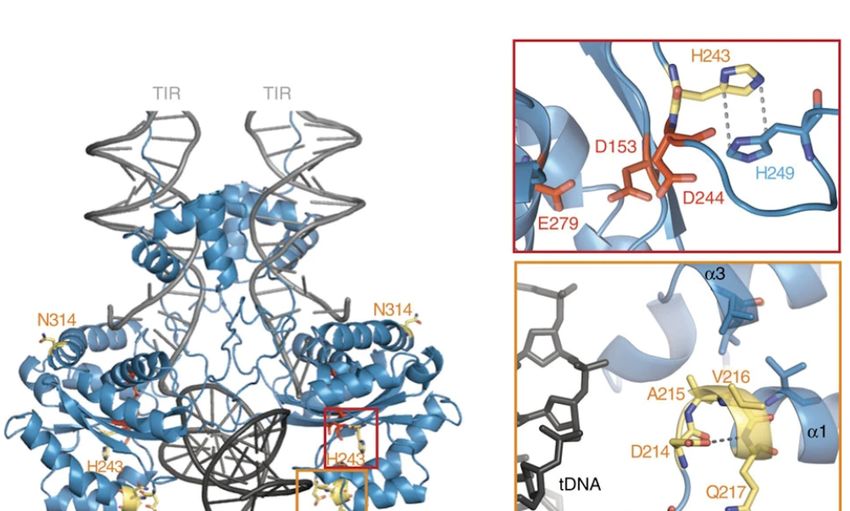
Viruses 2021, 13, 76                                                                                              2 of 17

                       However, unlike with retrotransposons, the copying mechanism does not involve an
                       RNA intermediate. Subclass I DNA transposons include the superfamilies Transib [10],
                       piggyBac [11], PIF/Harbinger [12], and Tc1/mariner [13].
                             All the members of the Tc1/mariner superfamily have in common that these elements
                       are flanked by terminal inverted repeats (TIRs), and contain a gene encoding a transposase,
                       an enzymatic factor catalyzing the transposition reaction [6]. The transposase binds to
                       the TIRs, excises the transposon from the donor locus, and reintegrates it adjacent to a TA
                       target sequence, leading to a TA target site duplication [6]. Members of the Tc1/mariner
                       family are ubiquitous in eukaryotes [6].
                             Because the TIRs and the transposase are considered to constitute the minimally
                       required components for the transposition reaction, a transposon that contains all these
                       elements is therefore considered autonomous [14]. However, many autonomous TEs
                       have given rise to non-autonomous derivatives by mutations, insertions, or deletions in
                       their transposase coding regions. These non-autonomous TEs can still be mobilized, but
                       need a functional transposase expressed by another element in the same cell [14]. It is
                       this trans-complementarity between two functional components (the transposase and the
                       specific TIRs that are recognized and mobilized by the transposase) that serves as the
                       basis of turning transposons into genetic vector systems suitable for moving any gene of
                       interest into the genome of a host cell. The Sleeping Beauty (SB) transposon system [14] is
                       widely used as a genetic engineering tool (recently reviewed in Amberger et al. [15]). The
                       structural features and mechanistic steps and processes taking place in the life cycle of SB
                       from DNA binding up to integration are described in the following sections.

                       2. Structural Features of the Sleeping Beauty Transposon System
                       2.1. The Sleeping Beauty Transposase
                             The SB transposase (Figure 1a) is composed of an N-terminal DNA binding domain
                       (DBD) (amino acids (aa) 1–110) and a C-terminal catalytic domain (DDE) (aa 114–340) con-
                       nected by a flexible linker region harboring a nuclear localization signal (NLS) (aa 97–123) [14].
                       The DBD consists of the two subdomains PAI and RED (PAIRED-like DBD) connected
                       by a linker [14,16]. Each subdomain is predicted to consist of three α-helices forming a
                       helix-turn-helix (HTH) motif which is found in many DNA binding proteins [17–20]. The
                       predicted HTH motif was confirmed by the NMR structure of the DBD subdomains [21,22]
                       (Figure 1b). The NMR structure shows that the three helices of the PAI subdomain are
                       located in the residues aa 12–22, aa 29–33, and aa 39–55, which are tightly packed. The
                       HTH motif is between the second and third helices [22]. Around 30% of the PAI subdomain
                       consists of positively charged amino acids, mainly arginines and lysines, leading to electro-
                       static repulsion and the destabilization of the structure in the presence of physiological salt
                       concentration and the absence of the TIRs [22]. The three helices of the RED subdomain
                       are located in the residues aa 67–77, aa 84–93, and aa 100–109 [21]. Helices 1 and 2 pack
                       against each other in an antiparallel arrangement, whereas helix 3 is located on top of
                       them [21]. The HTH motif is between helices 2 and 3; however, in contrast to the PAI
                       subdomain, it does not show a canonical β-turn connecting both helices, but a variation in
                       the β-turn with a longer turn-motif [21]. Additionally, helix 3 in the PAI subdomain is one
                       turn longer [21]. Similarly to the PAI subdomain, the RED subdomain is highly positively
                       charged, enhancing its DNA binding [21].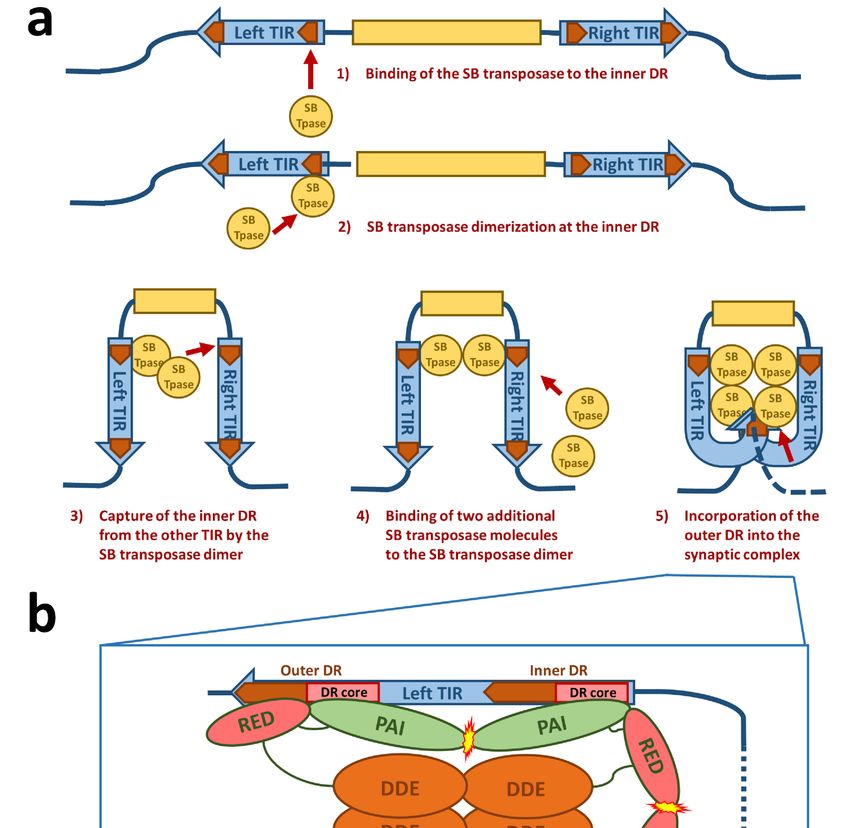
Viruses 2021, 13, 76                                                                                                            3 of 17

      Figure 1. Structural features of the Sleeping Beauty transposable element. (a) Schematic drawing of the domain structure of
      the SB transposase. The SB transposase has an N-terminal bipartite, paired-like DNA binding domain (green box) with the
      helix-turn-helix PAI subdomain (light green box) and RED subdomain (red box) and a GRRR AT-hook motif. It is followed
      by a bipartite nuclear localization signal (NLS, yellow boxes) and a C-terminal catalytic domain (orange box), with the
      DDE amino acid triad catalyzing the DNA cleavage and joining reactions. The clamp loop important for protein–protein
      interactions is overlapping with a glycine-rich box (light orange box). (b) NMR structure of the PAI and RED subdomains
      of the SB transposase. Reprinted from Protein Science [21] with permission from the publisher. (c) Crystal structure of
      the catalytic domain of the SB transposase with the catalytic triad (DDE) and the clamp loop. Reprinted from Nature
      Communications [23] with permission from the publisher. (d) Schematic drawing of the autonomous SB transposable element
      with the transposase coding region (yellow box) and the TIRs (blue arrows). An untranslated region (UTR, green box) is
      situated between the left TIR and the transposase coding region. The TIRs contain two binding sites for the transposase
      (orange arrows) represented by short directs repeats (DRs), one inner and one outer DR per TIR. In addition, the left TIR
      contains a “half-DR” sharing sequence similarities with the DRs. The DR core sequence, with which the PAI subdomain of
      the SB transposase interacts, is typed in red.

                                        The catalytic domain is predicted to have an RNaseH-like fold, similar to other DDE
                                  recombinases [24,25]. The catalytic triad of three acidic residues (DDE) [14], giving the
                                  domain its name, catalyze the DNA hydrolysis, required for excision, and transesterifica-
                                  tion, taking place in the integration reaction, in a two-metal-ion-dependent manner [26,27].
                                  Crystallographic structure analysis revealed the predicted RNaseH-like fold, consisting
                                  of a central five-stranded β-sheet surrounded by five α-helices [23] (Figure 1c). The three
                                  catalytic residues (D153, D244, and E279) are in close proximity, making up the active site
                                  of the enzyme [23]. The clamp loop (aa 159–190) between β1 and β2 includes a glycine-rich
                                  strip (aa 183–190) [14] which is curved and pivots on three consecutive glycines (aa 188–190)
                                  leading to an extended protein-protein surface [23]. The tip of the clamp loop has two short
                                  antiparallel β-strands (aa 169–174 and aa 174–176), forming a β-hairpin which is important
                                  for the protein–protein interaction with the inter-domain linker (aa 119–122) of a partner
                                  SB transposase molecule [23].
              1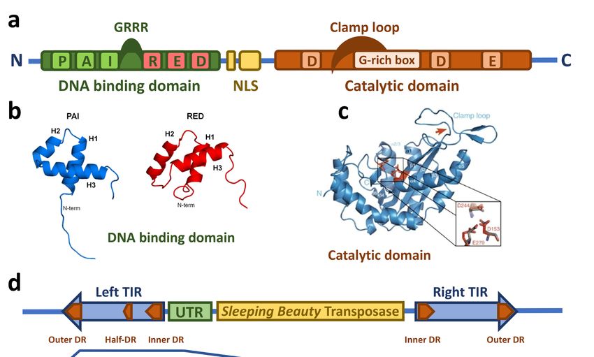
Viruses 2021, 13, 76                                                                                          4 of 17

                       2.2. The Sleeping Beauty Transposable Element
                             In addition to the transposase, the TIRs of the SB transposon flanking both ends
                       (Figure 1d) are also critically required for the transposition process. When SB is used as a
                       gene delivery tool, any genetic cargo can be placed between the TIRs and mobilized by
                       the transposase. The TIRs are ~220 bp in length and contain two direct repeats (DRs), one
                       outer and one inner, serving as binding sites for the SB transposase. This TIR arrangement
                       has been called the IR/DR structure [28]. Notably, the four DRs of SB are not identical: the
                       outer DRs are longer than the inner DRs by 2 bps (Figure 1d), and even slight variations
                       in the DR sequences can have a severe effect on the transposition efficiency [28–31]. The
                       left and right TIRs are not identical either; the left TIR has an extra “half-DR” element
                       showing sequence similarities to the transposase binding site (Figure 1d), which acts as
                       a transpositional enhancer [16]. Downstream of the left TIRs is an untranslated region
                       (Figure 1d) that contributes to the transcriptional regulation of the transposase [32,33].

                       3. The Mechanism of Sleeping Beauty Transposition
                       3.1. DNA Binding of the Sleeping Beauty Transposase
                            The transposition life cycle begins with binding of the transposase to the transposon
                       DNA (Figure 2a). The DNA binding domain of the transposase is mainly responsible for
                       the DNA recognition. Out of the two subdomains (PAI and RED), the PAI subdomain
                       has the dominant role in base-specific DNA binding [16]. The 30 -part of the transposase
                       binding site containing a core sequence conserved in all four DRs is recognized by the
                       PAI subdomain [16,22]. The DNA binding region of the PAI subdomain is located in
                       the residues aa 28, 29, 31, 33–36, 38–43, and 47, which are situated on the second and
                       third α-helices and on the loop connecting these helices of the HTH motif [22], which
                       is consistent with the role of HTH motifs in DNA binding [19]. The RED subdomain
                       interacts with the 50 -part of the DR adjacent to the core sequence [16]. This interaction of
                       the RED subdomain with DNA occurs only in the outer DRs and not the inner DRs [22].
                       Residues located at the third helix of the RED subdomain have been identified to be
                       primarily responsible for the DNA recognition of this subdomain, however helix 1 is also
                       highly positively charged and therefore potentially capable of binding DNA [21]. All
                       of the four transposase binding sites in the IR/DR structure in the TIRs are necessary
                       for SB transposition [34]. An important aspect for the next steps in the life cycle of SB
                       transposition is the formation of a transposase tetramer in a complex with the transposase
                       binding sites [16]. The inner DRs are bound by the transpose with a higher affinity than the
                       outer DRs [28,35], which was also confirmed by the NMR data on the PAI subdomain [22].
                       Additionally, the “half-DR” in the left TIR is bound by the PAI subdomain and mediates
                       protein–protein interactions with other transposase subunits [16]. The PAI subdomain
                       therefore fulfills three important functions: interaction with the DRs, interaction with the
                       “half-DR”, as well as transposase oligomerization. A GRRR amino acid motif contributes
                       as an AT-hook for specific substrate recognition [16]. In domain swapping experiments,
                       it was shown that primary DNA binding is not sufficient to determine the specificity
                       of the transposition reaction [16]. These experiments indicate that the RED subdomain
                       enforces specificity at a later step in transposition and therefore prevents the mobilization
                       of the SB transposon by transposases expressed by other, closely related subfamilies in the
                       same genome. It was also shown that the RED subdomain is involved in protein–protein
                       interactions and forms dimers upon DNA binding [36]. Helix 2 of the RED subdomain
                       has neutral or negative electrostatic potential and therefore could mediate protein–protein
                       interactions [21,36]. All these observations of the DNA-binding are consistent with the
                       crystal structures of protein-DNA complexes of closely related Tc1/mariner family members
                       such as Tc3 and Mos1 transposases [37,38]. Because the Tc3 and Mos1 transposons do
                       not have an IR/DR-like structure of their TIRs (instead, these transposons have a single
                       binding site for their transposases at each end of their short TIRs), the presence and strict
                       requirement for IR/DR in SB transposition suggests a regulatory role, which is discussed
                       in the next section.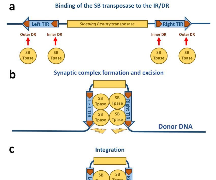
Viruses 2021, 13, 76                                                                                                            5 of 17

      Figure 2. Schematic drawing of Sleeping Beauty transposition. (a) The SB transposase (blue circle) binds to the DRs (orange
      arrows) within the TIRs. (b) The TIRs are brought together by SB transposase molecules in a synaptic complex. Excision of
      the SB transposon takes place from the donor DNA indicated by yellow flashes. (c) The excised transposon integrates into a
      TA site in the target DNA (green box) that is afterwards duplicated and flanks the new target site.

                                  3.2. Synaptic Complex Formation
                                       The next step required in the life cycle of SB transposition is the formation of a
                                  nucleoprotein complex called the synaptic complex (Figures 2b and 3). In this complex,
                                  both ends of the transposon are paired and held together by transposase subunits. For
                                  the formation of a synaptic complex, the complete TIRs with four transposase binding
                                  sites (DRs) and tetramerization-competent SB transposase are required. The “half-DR”
                                  motif in the left TIR is not essential for transposition, but functions as an enhancer of the
                                  transposition together with the PAI subdomain. It likely stabilizes the complexes formed
                                  by a transposase tetramer bound at the TIRs [16].

                                                                  2
Viruses 2021, 13, 76                                                                                                           6 of 17

      Figure 3. Schematic drawing of the synaptic complex formation. (a) At first, the SB transposase binds at the inner DR of
      the left TIR and forms dimers at this site. The SB dimer then captures the inner DR of the other TIR. Two additional SB
      transposase molecules are recruited to the nucleoprotein complex, leading to an incorporation of the outer DRs into the
      synaptic complex. (b) Protein–DNA and protein–protein interactions in the SB synaptic complex. The PAI subdomain of the
      N-terminal DNA-binding domain of the SB transposase interacts with the DR core sequence at both the inner and outer DRs.
      The RED subdomain contributes to DNA binding only at the outer DRs. At the inner DRs, the RED subdomain contributes
      to transposase dimerization. The relative positions of the four transposase monomers within the complex are arbitrarily
      drawn. Based on the structure of the Mos1 synaptic complex [37], it is likely that the catalytic DDE domains are acting in
      trans—that is, the DDE domain of an SB monomer bound at the left TIR executes cleavage at the right TIR and vice versa.

                                      For the formation of the synaptic complex, it has been proposed that a defined order
                                 of protein–DNA and protein–protein interactions is important [36] (Figure 3a). In this
                                 process, the assembly is mainly orchestrated by the interplay of the IR/DR structure and

                                                                 3
Viruses 2021, 13, 76                                                                                             7 of 17

                       the PAIRED-like DNA binding domain of the SB transposase. The specific primary DNA
                       recognition is performed by the PAI subdomain at an inner DR, which is bound at a higher
                       affinity than the outer DRs [22,28]. The contribution of the RED subdomain to the DNA
                       binding at the inner DR is limited, hence the transposase forms dimers through the protein–
                       protein interaction of the RED-RED interface located in helix 2 [36]. The SB transposase
                       could also bind to the inner DR as a preformed dimer. Once bound, this nucleoprotein
                       complex captures the inner DR from the other TIR (Figure 3a). The incorporation of an outer
                       DR into the synaptic complex by the transposase bound at the inner DR of the opposite TIR
                       does not result in productive transposition. In the next step, two additional SB transposase
                       molecules are recruited to the complex through the PAI-PAI protein interaction interface
                       (Figure 3a,b). This leads to the incorporation of the outer DRs in the synaptic complex [36]
                       (Figure 3a,b). In this step, the RED subdomain is required to complete the assembly process
                       by recognizing the outer DRs, thereby preparing the complex for strand cleavage executed
                       by the catalytic domain [36] (Figure 3b). This whole process is assisted by a host-encoded
                       cofactor called HMGB1, which is recruited by the SB transposase to the TIRs [35]. HMGB1
                       facilitates DNA bending at the inner DR, which could enhance the capture of the inner
                       DR on the other TIR [35]. However, the transposition reaction works also in the absence
                       of HMGB1 to a lower extent [35]. This ordered assembly is an important quality control
                       leading to functional transposition intermediates. It is important to note that if the ends of
                       the SB transposon are too close to each other (for example, in a circular DNA molecule), the
                       efficiency of transposition decreases [34]. Indeed, it has been established that efficient SB
                       transposition requires at least ~300 bp DNA bridging the TIRs [34]. A possible explanation
                       for this observation is that a certain length of DNA might be necessary to accommodate the
                       multimeric transposases and the host factor HMGB1 during the formation of the synaptic
                       complex. This orchestrated assembly of the synaptic complex shows that an alteration in
                       the DNA binding affinity of the SB transposase to the DRs does not necessarily enhance
                       the transposition reaction as a whole. Indeed, the replacement of the outer DR with
                       the sequence from the inner DR leads to insufficient SB transposition [28]. The ordered
                       assembly functions therefore as a “built-in” regulatory checkpoint mechanism, enforcing
                       synaptic complex formation before excision and ensuring that DNA cleavage occurs only
                       at the outer DRs, thereby leading to a higher level of accuracy and fidelity in contrast to
                       other transposons with simply structured TIRs [35,39,40].
                             It is notable that the mechanistic assembly of synaptic complexes is analogous between
                       SB transposition and V(D)J recombination. The sequences recognized by the RAG1/2
                       recombinase are related and binding is assisted by HMGB1 [41–43]. The regulation of an
                       ordered assembly of nucleoprotein complexes by somewhat dissimilar recombination sites
                       is also seen in V(D)J recombination [44], except that V(D)J recombination occurs between
                       heterologous partner sites (following the so-called 12/23 rule), whereas SB transposition
                       involves homologous sequences.

                       3.3. Excision of the Sleeping Beauty Transposon
                             Following the assembly of the synaptic complex, the excision of the SB transposon
                       from the donor locus occurs and DNA double-strand break (DSB) repair on the excision
                       site takes place (Figures 2b and 4). The excision step is crucial for the later integration step,
                       because it results in the exposure of a free 30 –OH group at the transposon ends required
                       for the strand transfer reactions taking place at the integration site [45] (Figure 4). The
                       first catalytic step in all transposition reactions is a Mg-cation-dependent hydrolysis of
                       the phosphodiester bond in the DNA backbone. This process is catalyzed by all DDE
                       recombinases in a similar way [46]—namely, first strand cleavage generates a single-strand
                       nick by a nucleophilic attack of a H2 O molecule, resulting in a free 30 –OH group [45]. The
                       nicking of the first strand is followed by the cleavage of the complementary DNA stand,
                       resulting in a double-strand break (DSB) that liberates the transposon from the donor
                       DNA. To catalyze second strand cleavage, DDE enzymes evolved versatile strategies [47].
                       Most DDE transposases, including piggyBac, Tn10, hAT, and the RAG1/2 recombinase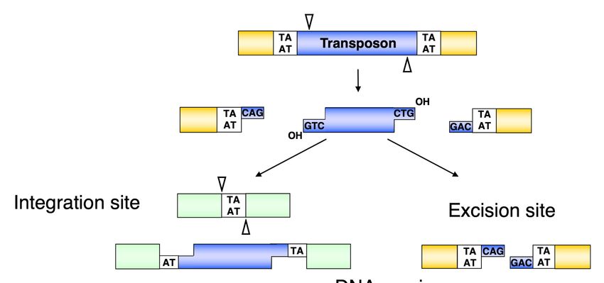
Viruses 2021, 13, 76                                                                                                           8 of 17

                                 catalyzing V(D)J recombination, use a single active site to cleave both DNA strands at
                                 one transposon end via a DNA hairpin intermediate either on the transposon end or
                                 on the flanking donor DNA [48–52]. However, members of the Tc1/mariner family do
                                 not transpose via a hairpin intermediate, indicating that double-strand cleavage is the
                                 result of two sequential hydrolysis reactions by the transposase [53,54]. Indeed, it has
                                 recently been shown that all the chemical steps of mariner transposition are executed
                                 by a single transposase dimer, in which one monomer performs two sequential strand
                                 cleavage and one strand transfer reactions at the same transposon end [55]. The Mos1
                                 mariner transposase cleaves the non-transferred strand first [56], and we infer that the first
                                 cleavage event during SB transposition also occurs at the non-transferred strand of the SB
                                 transposon (Figure 4). The first nick introduced by the SB and mariner transposases occurs
                                 three nucleotides inside the element [57,58] (Figure 4), which, following second strand
                                 cleavage at the exact tip of the transposon, generates three-nucleotide-long 30 –overhangs at
                                 the ends of both the excised transposon and those of the flanking donor DNA. The DSBs
                                 can be repaired by the non-homologous end joining (NHEJ) or homologous recombination
                                 (HR) DNA repair pathways [59,60]. The dominant way to repair transposon excision
                                 sites in somatic mammalian cells is NHEJ, which leads to transposon “footprints” being
                                 identical to the 30 –overhangs left at the donor site after SB excision [54,61] (Figure 4).
                                 Factors including Ku70 and DNA-PKcs of the NHEJ pathway have been shown to be
                                 required for SB transposition, because they are key contributors to the NHEJ repair of
                                 the excision site [54]. A physical interaction of Ku70 with the SB transposase has been
                                 observed [54], suggesting the active recruitment of repair factors to transposon excision
                                 sites by the transposase. NHEJ components have also been shown to be required for
                                 efficient retroelement integration and V(D)J recombination [62,63]. However, in contrast
                                 to V(D)J recombination, HR-dependent repair at the excision site can also occur in SB
                                 transposition [54]. The interaction of different repair factors at DNA DSBs generated by
                                 DNA transposition, retroviral integration, or V(D)J recombination probably defines how
                                 mechanistically very similar processes can lead to different products.

      Figure 4. Molecular events leading towards the formation of transposon footprints and target site duplications in Sleeping
      Beauty transposition. The SB transposase excises the transposon with staggered cuts and reintegrates it at a TA target
      dinucleotide. The single-stranded gaps at the integration site and the double-strand DNA breaks at the donor DNA are
      repaired by the host DNA repair machinery. After repair, the target TA is duplicated at the integration site, and a small
      footprint is left behind at the site of excision. Reprinted from CMLS [64] with permission from the publisher.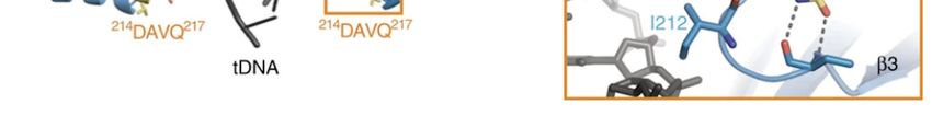
Viruses 2021, 13, 76                                                                                             9 of 17

                             CpG methylation of chromosomal DNA, leading to the formation of heterochromatin,
                       decreases the transposition activity of different transposons [65]. However, in the case of SB
                       transposition, CpG methylation in mouse embryonic stem (ES) cells leads to an enhanced
                       transposition activity [66]. This effect is not restricted to SB transposons but is a feature
                       that transposons with the characteristic IR/DR structure share [67]. A possible explanation
                       for the enhanced transposition activity upon CpG methylation could be that due to the
                       formation of a tight chromatin structure at the donor site, the SB transposase can more
                       efficiently bring the distant DR sites in the TIRs closely together.

                       3.4. Integration of the Sleeping Beauty Transposon
                             The free 30 –OH-groups exposed at the ends of the excised transposon are essential for
                       the integration step because they act as nucleophiles attacking the phosphodiester bond of
                       the target DNA (Figure 2c). This reaction can be chemically defined as a transesterification
                       reaction that results in a covalent coupling of the transposon ends to the target DNA [14].
                       In Tc1/mariner transposition, the transposon ends attack the double-stranded target DNA
                       in staggered positions, displaced from one another by 2 bp on the opposite strands. Thus,
                       integration of the two ends of the transposon with 30 -overhangs at staggered positions
                       in the target DNA results in single-stranded gaps which are filled up by the DNA repair
                       machinery [14] (Figure 4). This characteristic leads to a duplication of the target site flanking
                       the element called target side duplication (TSD), which is commonly observed with many
                       transposons. In the case of SB, the integration occurs at TA dinucleotides, leading to a
                       characteristic TA TSD [68–71], although SB integration can rarely occur at non-TA target
                       sites [68,72].
                             Additional molecular mechanisms involved in the integration of SB remain largely
                       unknown. However, studies on related transposases such as Mu [73] and the Tc1/mariner
                       superfamily member Mos1 [37] can be related to the integration mechanism of SB. In the
                       case of Mu transposition, the target DNA has to be bent by 140◦ [73]. This bend is promoted
                       by extended interactions along the DNA backbone and by a C-terminal coiled-coil domain,
                       reducing the electrostatic repulsion between the target DNA arms [73]. Additionally,
                       a sharp bend of 147◦ was observed in the Mos1 complex [74]. It is important to note
                       that the Mos1 post-excision complex [37] has an equivalent protein and transposon DNA
                       arrangement, such as the strand transfer complex occurring in the integration step [74].
                       This implies that target DNA binding and integration occurs without major changes
                       in the rest of the complex. Hence, the target DNA bending is important to bring the
                       phosphate group into the active site of the preassembled transposase. This allows then
                       the 30 -OH group of the transposon end to attack the phosphate group of the target DNA.
                       Another important aspect of the target DNA bending is that possibly after integration
                       at the active site the DNA snaps away, making this reaction irreversible. This product
                       escape has been observed in different strand-transfer complexes [73–77]. In addition,
                       the different spacing of the transposon ends with respect to the target DNA—which in
                       the case of Tc1/mariner transposases a TA dinucleotide pair—requires a different degree
                       of target DNA bending. It is therefore expected that the SB transposase, such as Mos1,
                       should be equipped with the ability to severely deform the DNA double helix at >140◦ .
                       Furthermore, it is likely that certain sequence-specific features at integration sites contribute
                       to target DNA bending. Alternating pyrimidine-purine bases, known to be associated with
                       bendable DNA structures, are often enriched in the insertion sites of most transposases
                       and integrases [74,78]. Biochemical studies have indeed shown that flexible, bent, or
                       mismatched sites are more suitable targets for integration [79–82]. The model of the SB
                       target capture complex also revealed that only bent target DNA can fulfill the requirement
                       for staggered integration [23] (Figure 5). Although the integration pattern of SB on the
                       genome level is close to random [71], a direct interaction with the conserved TA target
                       site has to occur. Additionally, the Mos1 strand transfer complex structure can serve here
                       as a model for SB transposition, because it revealed a direct interaction with the adenine
                       in the conserved TA target dinucleotide [74]. The structure shows that the adenine flips
Viruses 2021, 13, 76 10 of 17
out into the extra-helical space and forms base-specific contacts with a valine (V214) of
the transposase. The deformed DNA backbone is stabilized by salt bridges and hydrogen
bonds with the transposase.
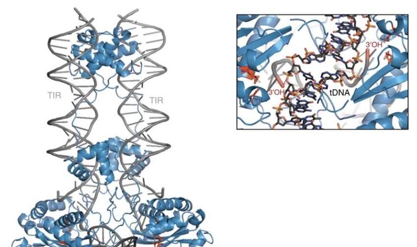
Figure 5. Model of the Sleeping Beauty strand transfer complex. Cartoon representation of the model: SB100X dimer (blue),
transposon ends (TIRs, grey), and bent target DNA substrate (tDNA, dark grey). Close up of the target site showing the
30 -OH group attacking the phosphate of the TA target DNA in a staggered way. Reprinted from Nature Communications [23]
with permission from the publisher.
Although 75% of SB transposon excision events are coupled to chromosomal integra-
tion, there is a loss of 25% of the events, which are not detectable as extrachromosomal
molecules [61]. A possible explanation for this is the suicidal autointegration of the trans-
poson into itself. This suicidal autointegration has been observed in the SB transposon [40]
but also in other transposons such as Tn10 [83] or Mu [84]. The efficacy of transposition
usually negatively correlates with the increasing size of the transposon [34,68,85–87]. One
possible explanation for this drop in efficacy is the increased numbers of target sites within
the transposon itself, which can lead to a higher frequency of autointegration [40]. A host
factor called barrier-to-autointegration factor (BAF or BANF1) that has been identified
to protect retroviruses [88–91] from autointegration was shown to interact with the SB
transposase in human cells and found to inhibit the autointegration of SB [40].
The molecular mechanisms involved in SB transposition also have a dramatic impact
on the distribution of integrations across the genome. Indeed, although SB integration is
close to random over the genome when transposition is launched out of extrachromosomal
plasmids [71], target site distribution is fundamentally different when the SB transposon is
mobilized out of a chromosomal site. When mobilized from a chromosome, an effect called
“local hopping” can be observed. Local hopping is a phenomenon where transposition out
of a chromosome leads to preferred integration into cis-linked sites in the close vicinity of the
donor locus. This feature seems to be shared by all transposons following the cut-and-paste
mechanism, but the extent of this effect varies between different transposons. In the case ofViruses 2021, 13, 76 11 of 17
the P-element transposon from Drosophila, the rate to insert within a window of 100 kb from
the donor site is ~50-fold higher than in regions outside this window [92]. Chromosomal
SB transposition results in 30–80% of re-integrations occurring locally [61,93–99], but in
a larger (up to 15 Mb) window around the donor site [98,100,101]. The extent of local
hopping is not only divergent between different transposons but is also dependent on the
host genome and the donor locus itself [102]. The underlying mechanism of this effect
remains unknown, but a potential explanation could be varying affinities of the transposase
for chromatin-associated factors in different hosts and locations within the chromosome
or the instability of the post-excision complex itself, which could limit the diffusion of the
complex away from the donor locus.
4. New Sleeping Beauty Variants Offering New Possibilities for Genetic Engineering
4.1. Hyperactive Sleeping Beauty Transposase Variants
SB was reconstructed from non-autonomous Tc1 family transposons in fish genomes [14],
and continued efforts to increase the transposition activity of the SB transposase have
identified several mutations that lead to an overall higher integration efficiency. These
mutations culminated in a hyperactive transposase variant of SB called SB100X [103]. This
variant has a 100-fold increased integration efficiency compared to the first-generation
SB transposase. SB100X was generated by molecular evolution and a combination of
different mutants. The mutations present in this hyperactive variant were rationalized by
the resolved crystal structure of the catalytic domain [23] (Figure 6). The T314N mutation
in SB100X may aid the proper folding of the transposase, which has been shown to be
a limiting factor in transposition [23,103]. M243H is located next to the catalytic residue
D244 and forms together with H249 a π-stack, which helps to position D244 in the active
site of the transposase [23]. The RKEN214—217DAVQ mutations form a part of the target
binding groove, so it is likely that these mutations lead to an ideal positioning of this β3-α1
linker for an interaction with the transposon DNA [23]. By understanding the structure
and mechanism of SB transposition, further hyperactive mutations could be rationally
designed in the future—for example, to address the need for the efficient chromosomal
integration of large SB transposons, which otherwise tend to transpose less efficiently than
shorter ones [34]. Having new hyperactive variants could facilitate the integration of even
larger DNA fragments over 100 kb [104] in gene therapy.
4.2. New Vector Platforms for Sleeping Beauty Transposition
The generation and use of new hyperactive SB transposase variants can not only
increase integration efficiency. The vector platform—i.e., the DNA molecules from which
transposition is initiated—can also have significant effects on the transposition efficiency.
Because, as described above in the context of excision, the TIRs of the SB transposon
need to be brought closely together for the catalytic steps to commence, it is likely that
derivatives of circular plasmid vectors with minimal DNA sequences connecting the
transposon TIRs could enhance this step. Indeed, the use of minicircles (circular genes
derived from plasmids lacking bacterial backbone sequences [105]) enabled a ~20-fold
increase in transposition efficiency as compared to conventional plasmids [106]. With SB
minicircles, the cellular toxicity triggered by the electroporation of naked DNA was reduced
up to 50% as compared to plasmid DNA in human CD34+ cells [106]. By delivering the
SB transposase in the form of mRNA instead of an expression plasmid, the transposition
efficiency was further increased and biosafety was improved due to the limited time of SB
transposase present in target cells [106]. The state of the art in clinical trials is to deliver the
components for SB transposition as minicircle DNA for the transposon and mRNA for the
transposase [107].Viruses 2021, 13, 76 12 of 17
Figure 6. Rationalizing the hyperactive mutations of SB100X. The DAVQ (aa 214–217) mutations (highlighted in yellow box)
target the binding groove of the target DNA (tDNA). The H243 mutation (highlighted in red box) together with H249 helps
to position D244 in the active site of the transposase. Reprinted from Nature Communications [23] with permission from the
publisher.
4.3. Integration-Deficient Sleeping Beauty Transposase Variant
An interesting variant of the SB transposase which has recently been described is the
K248T mutant [108]. This mutant is competent in transposon excision but has the feature
of deficiency in transposon integration [108]. After excision by K248T, extrachromosomal
transposon circles are formed which apparently cannot undergo integration. A structural
model indicates that K248 is involved in interaction with the target DNA [108]. It is thus
likely that the K248T mutant impairs the interaction with the target DNA, resulting in this
integration-deficient transposase variant. This variant has been used for the generation of
reprogramming factor-free induced pluripotent stem cells [108].
4.4. Targeted Sleeping Beauty Transposition
Targeting the integration of transposons into defined genomic regions has been chal-
lenging. However, several attempts have been made to fuse DNA binding domains to
transposases, thereby targeting integration in the vicinity of sites specified by the DNA
binding domains [109]. These attempts resulted in a low efficiency of targeted transpo-
sition [70] or worked only in an artificial in vitro environment [93] or in inter-plasmid
settings [110,111]. Targeted transposition was successful in a bacterial context [112,113],
but still remains challenging in eukaryotic cells. By using the CRISPR/Cas9 system with
its high targeting efficiency [114], SB100X transposase fused with catalytically inactive
Cas9 (dCas9), and single guide RNAs (sgRNAs) targeting the human Alu retrotransposon,
a slight bias towards integration with Alu elements could be accomplished in human
cells [115]. However, the efficiency of the targeted transposition events remained low and
further studies are required to bias SB integrations towards defined target regions.
6Viruses 2021, 13, 76 13 of 17
5. Conclusions
In this review, we described the structural features and mechanistic steps involved
in SB transposition. In the last few years, new structural information on the domains of
the SB transposase has provided new insight into important regions, motifs, and amino
acids required for transposition [21–23]. We have also discussed the key mechanistic steps
taking place in SB transposition, from DNA binding and synaptic complex formation to
SB transposon excision and up to integration. However, certain mechanistic steps—for
example, during transposon integration—are not well understood in SB transposition, and
need further investigation. Well-studied transposon relatives such as Mos1 offer models
from which we can infer information for SB transposition [74]. However, structural features
also revealed differences within the transposase structure. To gain further understanding
of the underlying structure-function relationships that are relevant in the context of a
transposition reaction, the full-length SB transposase as present in a synaptic complex
and/or strand transfer complex needs to be analyzed. Our state-of-the-art comprehension
of SB transposition yielded new variants, such as hyperactive transposase variants [103],
new vector systems based on minicircles [106], and integration-deficient SB transposase
variants [108]. The further and deeper understanding of SB transposition could facilitate
the generation of new variants, facilitating the development of an even richer SB transposon
toolbox.
Author Contributions: Conceptualization, M.T.O. and Z.I.; writing—original draft preparation,
M.T.O.; writing—review and editing, Z.I.; supervision, Z.I. All authors have read and agreed to the
published version of the manuscript.
Funding: The authors were supported by funding from the European Union0 s Horizon 2020 program
for research and innovation under Grant Agreement No. 754658.
Institutional Review Board Statement: Not applicable.
Informed Consent Statement: Not applicable.
Data Availability Statement: Readers are encouraged to consult the primary research articles cited
in this review article for accessing research data.
Conflicts of Interest: Z.I. is an inventor of patents relating to Sleeping Beauty transposon technology.
References
1. Seelamgari, A.; Maddukuri, A.; Berro, R.; de La Fuente, C.; Kehn, K.; Deng, L.; Dadgar, S.; Bottazzi, M.E.; Ghedin, E.; Pumfery, A.;
et al. Role of viral regulatory and accessory proteins in HIV-1 replication. Front. Biosci. 2004, 9, 2388–2413. [CrossRef] [PubMed]
2. Frankel, A.D.; Young, J.A. HIV-1: Fifteen proteins and an RNA. Annu. Rev. Biochem. 1998, 67, 1–25. [CrossRef] [PubMed]
3. Bannert, N.; Kurth, R. The evolutionary dynamics of human endogenous retroviral families. Annu. Rev. Genom. Hum. Genet. 2006,
7, 149–173. [CrossRef]
4. Jasin, M.; Rothstein, R. Repair of strand breaks by homologous recombination. Cold Spring Harb. Perspect. Biol. 2013, 5, a012740.
[CrossRef] [PubMed]
5. McClintock, B. Induction of Instability at Selected Loci in Maize. Genetics 1953, 38, 579–599. [PubMed]
6. Wicker, T.; Sabot, F.; Hua-Van, A.; Bennetzen, J.L.; Capy, P.; Chalhoub, B.; Flavell, A.; Leroy, P.; Morgante, M.; Panaud, O.; et al. A
unified classification system for eukaryotic transposable elements. Nat. Rev. Genet. 2007, 8, 973–982. [CrossRef] [PubMed]
7. Beck, C.R.; Garcia-Perez, J.L.; Badge, R.M.; Moran, J.V. LINE-1 elements in structural variation and disease. Annu. Rev. Genomics
Hum. Genet. 2011, 12, 187–215. [CrossRef]
8. Deininger, P. Alu elements: Know the SINEs. Genome Biol. 2011, 12, 236. [CrossRef]
9. Kapitonov, V.V.; Jurka, J. RAG1 core and V(D)J recombination signal sequences were derived from Transib transposons. PLoS Biol.
2005, 3, e181. [CrossRef]
10. Kapitonov, V.V.; Jurka, J. Rolling-circle transposons in eukaryotes. Proc. Natl. Acad. Sci. USA 2001, 98, 8714–8719. [CrossRef]
11. Sarkar, A.; Sim, C.; Hong, Y.S.; Hogan, J.R.; Fraser, M.J.; Robertson, H.M.; Collins, F.H. Molecular evolutionary analysis of
the widespread piggyBac transposon family and related “domesticated” sequences. Mol. Genet. Genom. 2003, 270, 173–180.
[CrossRef] [PubMed]
12. Jurka, J.; Kapitonov, V.V. PIFs meet Tourists and Harbingers: A superfamily reunion. Proc. Natl. Acad. Sci. USA 2001, 98,
12315–12316. [CrossRef] [PubMed]Viruses 2021, 13, 76 14 of 17
13. Shao, H.; Tu, Z. Expanding the diversity of the IS630-Tc1-mariner superfamily: Discovery of a unique DD37E transposon and
reclassification of the DD37D and DD39D transposons. Genetics 2001, 159, 1103–1115. [PubMed]
14. Ivics, Z.; Hackett, P.B.; Plasterk, R.H.; Izsvák, Z. Molecular Reconstruction of Sleeping Beauty, a Tc1-like Transposon from Fish,
and Its Transposition in Human Cells. Cell 1997, 91, 501–510. [CrossRef]
15. Amberger, M.; Ivics, Z. Latest Advances for the Sleeping Beauty Transposon System: 23 Years of Insomnia but Prettier than Ever:
Refinement and Recent Innovations of the Sleeping Beauty Transposon System Enabling Novel, Nonviral Genetic Engineering
Applications. Bioessays 2020, 42, e2000136. [CrossRef] [PubMed]
16. Izsvák, Z.; Khare, D.; Behlke, J.; Heinemann, U.; Plasterk, R.H.; Ivics, Z. Involvement of a bifunctional, paired-like DNA-binding
domain and a transpositional enhancer in Sleeping Beauty transposition. J. Biol. Chem. 2002, 277, 34581–34588. [CrossRef]
17. Pabo, C.O.; Sauer, R.T. Transcription factors: Structural families and principles of DNA recognition. Annu. Rev. Biochem. 1992, 61,
1053–1095. [CrossRef]
18. Czerny, T.; Schaffner, G.; Busslinger, M. DNA sequence recognition by Pax proteins: Bipartite structure of the paired domain and
its binding site. Genes Dev. 1993, 7, 2048–2061. [CrossRef]
19. Brennan, R.G.; Matthews, B.W. The helix-turn-helix DNA binding motif. J. Biol. Chem. 1989, 264, 1903–1906. [CrossRef]
20. Aravind, L.; Anantharaman, V.; Balaji, S.; Babu, M.M.; Iyer, L.M. The many faces of the helix-turn-helix domain: Transcription
regulation and beyond. FEMS Microbiol. Rev. 2005, 29, 231–262. [CrossRef]
21. Konnova, T.A.; Singer, C.M.; Nesmelova, I.V. NMR solution structure of the RED subdomain of the Sleeping Beauty transposase.
Protein Sci. 2017, 26, 1171–1181. [CrossRef] [PubMed]
22. Carpentier, C.E.; Schreifels, J.M.; Aronovich, E.L.; Carlson, D.F.; Hackett, P.B.; Nesmelova, I.V. NMR structural analysis of Sleeping
Beauty transposase binding to DNA. Protein Sci. 2014, 23, 23–33. [CrossRef] [PubMed]
23. Voigt, F.; Wiedemann, L.; Zuliani, C.; Querques, I.; Sebe, A.; Mátés, L.; Izsvák, Z.; Ivics, Z.; Barabas, O. Sleeping Beauty
transposase structure allows rational design of hyperactive variants for genetic engineering. Nat. Commun. 2016, 7, 11126.
[CrossRef] [PubMed]
24. Hickman, A.B.; Chandler, M.; Dyda, F. Integrating prokaryotes and eukaryotes: DNA transposases in light of structure. Crit. Rev.
Biochem. Mol. Biol. 2010, 45, 50–69. [CrossRef]
25. Rice, P.A.; Baker, T.A. Comparative architecture of transposase and integrase complexes. Nat. Struct Biol. 2001, 8, 302–307.
[CrossRef]
26. Montaño, S.P.; Rice, P.A. Moving DNA around: DNA transposition and retroviral integration. Curr. Opin. Struct. Biol. 2011, 21,
370–378. [CrossRef]
27. Yang, W.; Lee, J.Y.; Nowotny, M. Making and breaking nucleic acids: Two-Mg2+ -ion catalysis and substrate specificity. Mol. Cell
2006, 22, 5–13. [CrossRef]
28. Cui, Z.; Geurts, A.M.; Liu, G.; Kaufman, C.D.; Hackett, P.B. Structure–Function Analysis of the Inverted Terminal Repeats of the
Sleeping Beauty Transposon. J. Mol. Biol. 2002, 318, 1221–1235. [CrossRef]
29. Liu, G.; Aronovich, E.L.; Cui, Z.; Whitley, C.B.; Hackett, P.B. Excision of Sleeping Beauty transposons: Parameters and applications
to gene therapy. J. Gene Med. 2004, 6, 574–583. [CrossRef]
30. Zayed, H.; Izsvák, Z.; Walisko, O.; Ivics, Z. Development of hyperactive sleeping beauty transposon vectors by mutational
analysis. Mol. Ther. 2004, 9, 292–304. [CrossRef]
31. Geurts, A.M.; Yang, Y.; Clark, K.J.; Liu, G.; Cui, Z.; Dupuy, A.J.; Bell, J.B.; Largaespada, D.A.; Hackett, P.B. Gene transfer into
genomes of human cells by the sleeping beauty transposon system. Mol. Ther. 2003, 8, 108–117. [CrossRef]
32. Walisko, O.; Schorn, A.; Rolfs, F.; Devaraj, A.; Miskey, C.; Izsvák, Z.; Ivics, Z. Transcriptional activities of the Sleeping Beauty
transposon and shielding its genetic cargo with insulators. Mol. Ther. 2008, 16, 359–369. [CrossRef] [PubMed]
33. Moldt, B.; Yant, S.R.; Andersen, P.R.; Kay, M.A.; Mikkelsen, J.G. Cis-acting gene regulatory activities in the terminal regions of
sleeping beauty DNA transposon-based vectors. Hum. Gene Ther. 2007, 18, 1193–1204. [CrossRef] [PubMed]
34. Izsvák, Z.; Ivics, Z.; Plasterk, R.H. Sleeping Beauty, a wide host-range transposon vector for genetic transformation in vertebrates.
J. Mol. Biol. 2000, 302, 93–102. [CrossRef] [PubMed]
35. Zayed, H.; Izsvák, Z.; Khare, D.; Heinemann, U.; Ivics, Z. The DNA-bending protein HMGB1 is a cellular cofactor of Sleeping
Beauty transposition. Nucleic Acids Res. 2003, 31, 2313–2322. [CrossRef]
36. Wang, Y.; Pryputniewicz-Dobrinska, D.; Nagy, E.É.; Kaufman, C.D.; Singh, M.; Yant, S.; Wang, J.; Dalda, A.; Kay, M.A.; Ivics, Z.;
et al. Regulated complex assembly safeguards the fidelity of Sleeping Beauty transposition. Nucleic Acids Res. 2017, 45, 311–326.
[CrossRef]
37. Richardson, J.M.; Colloms, S.D.; Finnegan, D.J.; Walkinshaw, M.D. Molecular architecture of the Mos1 paired-end complex: The
structural basis of DNA transposition in a eukaryote. Cell 2009, 138, 1096–1108. [CrossRef]
38. Watkins, S.; van Pouderoyen, G.; Sixma, T.K. Structural analysis of the bipartite DNA-binding domain of Tc3 transposase bound
to transposon DNA. Nucleic Acids Res. 2004, 32, 4306–4312. [CrossRef]
39. Bouuaert, C.C.; Liu, D.; Chalmers, R. A simple topological filter in a eukaryotic transposon as a mechanism to suppress genome
instability. Mol. Cell. Biol. 2011, 31, 317–327. [CrossRef]
40. Wang, Y.; Wang, J.; Devaraj, A.; Singh, M.; Jimenez Orgaz, A.; Chen, J.-X.; Selbach, M.; Ivics, Z.; Izsvák, Z. Suicidal autointegration
of sleeping beauty and piggyBac transposons in eukaryotic cells. PLoS Genet. 2014, 10, e1004103. [CrossRef]Viruses 2021, 13, 76 15 of 17
41. West, R.B.; Lieber, M.R. The RAG-HMG1 complex enforces the 12/23 rule of V(D)J recombination specifically at the double-hairpin
formation step. Mol. Cell. Biol. 1998, 18, 6408–6415. [CrossRef]
42. Van Gent, D.C.; Hiom, K.; Paull, T.T.; Gellert, M. Stimulation of V(D)J cleavage by high mobility group proteins. EMBO J. 1997,
16, 2665–2670. [CrossRef]
43. Agrawal, A.; Eastman, Q.M.; Schatz, D.G. Transposition mediated by RAG1 and RAG2 and its implications for the evolution of
the immune system. Nature 1998, 394, 744–751. [CrossRef]
44. Jones, J.M.; Gellert, M. Ordered assembly of the V(D)J synaptic complex ensures accurate recombination. EMBO J. 2002, 21,
4162–4171. [CrossRef] [PubMed]
45. Mizuuchi, K. Polynucleotidyl transfer reactions in transpositional DNA recombination. J. Biol. Chem. 1992, 267, 21273–21276.
[CrossRef]
46. Craig, N.L. Unity in transposition reactions. Science 1995, 270, 253–254. [CrossRef] [PubMed]
47. Hickman, A.B.; Dyda, F. Mechanisms of DNA Transposition. Microbiol. Spectr. 2015, 3, 531–553. [CrossRef]
48. Hencken, C.G.; Li, X.; Craig, N.L. Functional characterization of an active Rag-like transposase. Nat. Struct. Mol. Biol. 2012, 19,
834–836. [CrossRef] [PubMed]
49. Zhou, L.; Mitra, R.; Atkinson, P.W.; Hickman, A.B.; Dyda, F.; Craig, N.L. Transposition of hAT elements links transposable
elements and V(D)J recombination. Nature 2004, 432, 995–1001. [CrossRef]
50. Mitra, R.; Fain-Thornton, J.; Craig, N.L. piggyBac can bypass DNA synthesis during cut and paste transposition. EMBO J. 2008,
27, 1097–1109. [CrossRef]
51. Bischerour, J.; Chalmers, R. Base flipping in tn10 transposition: An active flip and capture mechanism. PLoS ONE 2009, 4, e6201.
[CrossRef] [PubMed]
52. Bischerour, J.; Lu, C.; Roth, D.B.; Chalmers, R. Base flipping in V(D)J recombination: Insights into the mechanism of hairpin
formation, the 12/23 rule, and the coordination of double-strand breaks. Mol. Cell. Biol. 2009, 29, 5889–5899. [CrossRef] [PubMed]
53. Richardson, J.M.; Dawson, A.; O’Hagan, N.; Taylor, P.; Finnegan, D.J.; Walkinshaw, M.D. Mechanism of Mos1 transposition:
Insights from structural analysis. EMBO J. 2006, 25, 1324–1334. [CrossRef] [PubMed]
54. Izsvák, Z.; Stüwe, E.E.; Fiedler, D.; Katzer, A.; Jeggo, P.A.; Ivics, Z. Healing the Wounds Inflicted by Sleeping Beauty Transposition
by Double-Strand Break Repair in Mammalian Somatic Cells. Mol. Cell 2004, 13, 279–290. [CrossRef]
55. Claeys Bouuaert, C.; Chalmers, R. A single active site in the mariner transposase cleaves DNA strands of opposite polarity.
Nucleic Acids Res. 2017, 45, 11467–11478. [CrossRef]
56. Dawson, A.; Finnegan, D.J. Excision of the Drosophila Mariner Transposon Mos1. Mol. Cell 2003, 11, 225–235. [CrossRef]
57. Miskey, C.; Papp, B.; Mátés, L.; Sinzelle, L.; Keller, H.; Izsvák, Z.; Ivics, Z. The ancient mariner sails again: Transposition of the
human Hsmar1 element by a reconstructed transposase and activities of the SETMAR protein on transposon ends. Mol. Cell. Biol.
2007, 27, 4589–4600. [CrossRef]
58. Lampe, D.J.; Churchill, M.E.; Robertson, H.M. A purified mariner transposase is sufficient to mediate transposition in vitro.
EMBO J. 1996, 15, 5470–5479. [CrossRef]
59. Lohe, A.R.; Timmons, C.; Beerman, I.; Lozovskaya, E.R.; Hartl, D.L. Self-inflicted wounds, template-directed gap repair and a
recombination hotspot. Effects of the mariner transposase. Genetics 2000, 154, 647–656.
60. Engels, W.R.; Johnson-Schlitz, D.M.; Eggleston, W.B.; Sved, J. High-frequency P element loss in Drosophila is homolog dependent.
Cell 1990, 62, 515–525. [CrossRef]
61. Luo, G.; Ivics, Z.; Izsvák, Z.; Bradley, A. Chromosomal transposition of a Tc1/mariner-like element in mouse embryonic stem
cells. Proc. Natl. Acad. Sci. USA 1998, 95, 10769–10773. [CrossRef] [PubMed]
62. Daniel, R.; Katz, R.A.; Skalka, A.M. A role for DNA-PK in retroviral DNA integration. Science 1999, 284, 644–647. [CrossRef]
[PubMed]
63. Jackson, S.P.; Jeggo, P.A. DNA double-strand break repair and V(D)J recombination: Involvement of DNA-PK. Trends Biochem. Sci.
1995, 20, 412–415. [CrossRef]
64. Miskey, C.; Izsvák, Z.; Kawakami, K.; Ivics, Z. DNA transposons in vertebrate functional genomics. Cell. Mol. Life Sci. 2005, 62,
629–641. [CrossRef] [PubMed]
65. Yoder, J.A.; Walsh, C.P.; Bestor, T.H. Cytosine methylation and the ecology of intragenomic parasites. Trends Genet. 1997, 13,
335–340. [CrossRef]
66. Yusa, K.; Takeda, J.; Horie, K. Enhancement of Sleeping Beauty transposition by CpG methylation: Possible role of heterochromatin
formation. Mol. Cell. Biol. 2004, 24, 4004–4018. [CrossRef]
67. Jursch, T.; Miskey, C.; Izsvák, Z.; Ivics, Z. Regulation of DNA transposition by CpG methylation and chromatin structure in
human cells. Mob. DNA 2013, 4, 15. [CrossRef]
68. Li, X.; Ewis, H.; Hice, R.H.; Malani, N.; Parker, N.; Zhou, L.; Feschotte, C.; Bushman, F.D.; Atkinson, P.W.; Craig, N.L. A resurrected
mammalian hAT transposable element and a closely related insect element are highly active in human cell culture. Proc. Natl.
Acad. Sci. USA 2013, 110, E478–E487. [CrossRef]
69. Liu, G.; Geurts, A.M.; Yae, K.; Srinivasan, A.R.; Fahrenkrug, S.C.; Largaespada, D.A.; Takeda, J.; Horie, K.; Olson, W.K.; Hackett,
P.B. Target-site preferences of Sleeping Beauty transposons. J. Mol. Biol. 2005, 346, 161–173. [CrossRef]
70. Voigt, K.; Gogol-Döring, A.; Miskey, C.; Chen, W.; Cathomen, T.; Izsvák, Z.; Ivics, Z. Retargeting sleeping beauty transposon
insertions by engineered zinc finger DNA-binding domains. Mol. Ther. 2012, 20, 1852–1862. [CrossRef]Viruses 2021, 13, 76 16 of 17
71. Moldt, B.; Miskey, C.; Staunstrup, N.H.; Gogol-Döring, A.; Bak, R.O.; Sharma, N.; Mátés, L.; Izsvák, Z.; Chen, W.; Ivics, Z.; et al.
Comparative genomic integration profiling of Sleeping Beauty transposons mobilized with high efficacy from integrase-defective
lentiviral vectors in primary human cells. Mol. Ther. 2011, 19, 1499–1510. [CrossRef] [PubMed]
72. de Jong, J.; Akhtar, W.; Badhai, J.; Rust, A.G.; Rad, R.; Hilkens, J.; Berns, A.; van Lohuizen, M.; Wessels, L.F.A.; de Ridder, J.
Chromatin landscapes of retroviral and transposon integration profiles. PLoS Genet. 2014, 10, e1004250. [CrossRef] [PubMed]
73. Montaño, S.P.; Pigli, Y.Z.; Rice, P.A. The µ transpososome structure sheds light on DDE recombinase evolution. Nature 2012, 491,
413–417. [CrossRef] [PubMed]
74. Morris, E.R.; Grey, H.; McKenzie, G.; Jones, A.C.; Richardson, J.M. A bend, flip and trap mechanism for transposon integration.
Elife 2016, 5. [CrossRef] [PubMed]
75. Passos, D.O.; Li, M.; Yang, R.; Rebensburg, S.V.; Ghirlando, R.; Jeon, Y.; Shkriabai, N.; Kvaratskhelia, M.; Craigie, R.; Lyumkis, D.
Cryo-EM structures and atomic model of the HIV-1 strand transfer complex intasome. Science 2017, 355, 89–92. [CrossRef]
76. Yin, Z.; Shi, K.; Banerjee, S.; Pandey, K.K.; Bera, S.; Grandgenett, D.P.; Aihara, H. Crystal structure of the Rous sarcoma virus
intasome. Nature 2016, 530, 362–366. [CrossRef]
77. Maertens, G.N.; Hare, S.; Cherepanov, P. The mechanism of retroviral integration from X-ray structures of its key intermediates.
Nature 2010, 468, 326–329. [CrossRef]
78. Maskell, D.P.; Renault, L.; Serrao, E.; Lesbats, P.; Matadeen, R.; Hare, S.; Lindemann, D.; Engelman, A.N.; Costa, A.; Cherepanov,
P. Structural basis for retroviral integration into nucleosomes. Nature 2015, 523, 366–369. [CrossRef]
79. Yanagihara, K.; Mizuuchi, K. Mismatch-targeted transposition of Mu: A new strategy to map genetic polymorphism. Proc. Natl.
Acad. Sci. USA 2002, 99, 11317–11321. [CrossRef]
80. Fuller, J.R.; Rice, P.A. Target DNA bending by the Mu transpososome promotes careful transposition and prevents its reversal.
Elife 2017, 6. [CrossRef]
81. Kuduvalli, P.N.; Rao, J.E.; Craig, N.L. Target DNA structure plays a critical role in Tn7 transposition. EMBO J. 2001, 20, 924–932.
[CrossRef] [PubMed]
82. Pribil, P.A.; Haniford, D.B. Target DNA Bending is an Important Specificity Determinant in Target Site Selection in Tn10
Transposition. J. Mol. Biol. 2003, 330, 247–259. [CrossRef]
83. Benjamin, H.W.; Kleckner, N. Intramolecular transposition by Tn10. Cell 1989, 59, 373–383. [CrossRef]
84. Maxwell, A.; Craigie, R.; Mizuuchi, K. B protein of bacteriophage mu is an ATPase that preferentially stimulates intermolecular
DNA strand transfer. Proc. Natl. Acad. Sci. USA 1987, 84, 699–703. [CrossRef] [PubMed]
85. Karsi, A.; Moav, B.; Hackett, P.; Liu, Z. Effects of insert size on transposition efficiency of the sleeping beauty transposon in mouse
cells. Mar. Biotechnol. 2001, 3, 241–245. [CrossRef] [PubMed]
86. Lampe, D.J.; Grant, T.E.; Robertson, H.M. Factors affecting transposition of the Himar1 mariner transposon in vitro. Genetics 1998,
149, 179–187.
87. Fischer, S.E.; van Luenen, H.G.; Plasterk, R.H. Cis requirements for transposition of Tc1-like transposons in C. elegans. Mol. Genet.
Genom. 1999, 262, 268–274. [CrossRef]
88. Mansharamani, M.; Graham, D.R.M.; Monie, D.; Lee, K.K.; Hildreth, J.E.K.; Siliciano, R.F.; Wilson, K.L. Barrier-to-autointegration
factor BAF binds p55 Gag and matrix and is a host component of human immunodeficiency virus type 1 virions. J. Virol. 2003, 77,
13084–13092. [CrossRef]
89. Suzuki, Y.; Craigie, R. Regulatory mechanisms by which barrier-to-autointegration factor blocks autointegration and stimulates
intermolecular integration of Moloney murine leukemia virus preintegration complexes. J. Virol. 2002, 76, 12376–12380. [CrossRef]
90. Lee, M.S.; Craigie, R. A previously unidentified host protein protects retroviral DNA from autointegration. Proc. Natl. Acad. Sci.
USA 1998, 95, 1528–1533. [CrossRef]
91. Lee, M.S.; Craigie, R. Protection of retroviral DNA from autointegration: Involvement of a cellular factor. Proc. Natl. Acad. Sci.
USA 1994, 91, 9823–9827. [CrossRef] [PubMed]
92. Tower, J.; Karpen, G.H.; Craig, N.; Spradling, A.C. Preferential transposition of Drosophila P elements to nearby chromosomal
sites. Genetics 1993, 133, 347–359.
93. Ruf, S.; Symmons, O.; Uslu, V.V.; Dolle, D.; Hot, C.; Ettwiller, L.; Spitz, F. Large-scale analysis of the regulatory architecture of the
mouse genome with a transposon-associated sensor. Nat. Genet. 2011, 43, 379–386. [CrossRef]
94. Dupuy, A.J.; Fritz, S.; Largaespada, D.A. Transposition and gene disruption in the male germline of the mouse. Genesis 2001, 30,
82–88. [CrossRef] [PubMed]
95. Kokubu, C.; Horie, K.; Abe, K.; Ikeda, R.; Mizuno, S.; Uno, Y.; Ogiwara, S.; Ohtsuka, M.; Isotani, A.; Okabe, M.; et al. A
transposon-based chromosomal engineering method to survey a large cis-regulatory landscape in mice. Nat. Genet. 2009, 41,
946–952. [CrossRef] [PubMed]
96. Liang, Q.; Kong, J.; Stalker, J.; Bradley, A. Chromosomal mobilization and reintegration of Sleeping Beauty and PiggyBac
transposons. Genesis 2009, 47, 404–408. [CrossRef] [PubMed]
97. Horie, K.; Yusa, K.; Yae, K.; Odajima, J.; Fischer, S.E.J.; Keng, V.W.; Hayakawa, T.; Mizuno, S.; Kondoh, G.; Ijiri, T.; et al.
Characterization of Sleeping Beauty transposition and its application to genetic screening in mice. Mol. Cell. Biol. 2003, 23,
9189–9207. [CrossRef]
98. Carlson, C.M.; Dupuy, A.J.; Fritz, S.; Roberg-Perez, K.J.; Fletcher, C.F.; Largaespada, D.A. Transposon mutagenesis of the mouse
germline. Genetics 2003, 165, 243–256.You can also read