Na+, K+-ATPase Signaling and Bipolar Disorder - MDPI
←
→
Page content transcription
If your browser does not render page correctly, please read the page content below
International Journal of
           Molecular Sciences

Review
Na+, K+-ATPase Signaling and Bipolar Disorder
David Lichtstein *, Asher Ilani, Haim Rosen, Noa Horesh, Shiv Vardan Singh, Nahum Buzaglo
and Anastasia Hodes
 Department of Medical Neurobiology, Institute for Medical Research Israel-Canada, The Hebrew
 University-Hadassah Medical School, Jerusalem 91120, Israel; asheri@ekmd.huji.ac.il (A.I.);
 haimr@ekmd.huji.ac.il (H.R.); noa.rosenthal1@mail.huji.ac.il (N.H.); vardanshiva@gmail.com (S.V.S.);
 buzaglo@gmail.com (N.B.); anastasi.singalevich@mail.huji.ac.il (A.H.)
 * Correspondence: davidli@ekmd.huji.ac.il; Tel.: +972-2-675-8522; Fax: +972-2-643-9736

 Received: 1 July 2018; Accepted: 26 July 2018; Published: 7 August 2018

 Abstract: Bipolar disorder (BD) is a severe and common chronic mental illness characterized by
 recurrent mood swings between depression and mania. The biological basis of the disease is
 poorly understood and its treatment is unsatisfactory. Although in past decades the “monoamine
 hypothesis” has dominated our understanding of both the pathophysiology of depressive disorders
 and the action of pharmacological treatments, recent studies focus on the involvement of additional
 neurotransmitters/neuromodulators systems and cellular processes in BD. Here, evidence for the
 participation of Na+ , K+ -ATPase and its endogenous regulators, the endogenous cardiac steroids
 (ECS), in the etiology of BD is reviewed. Proof for the involvement of brain Na+ , K+ -ATPase
 and ECS in behavior is summarized and it is hypothesized that ECS-Na+ , K+ -ATPase-induced
 activation of intracellular signaling participates in the mechanisms underlying BD. We propose that
 the activation of ERK, AKT, and NFκB, resulting from ECS-Na+ , K+ -ATPase interaction, modifies
 neuronal activity and neurotransmission which, in turn, participate in the regulation of behavior and
 BD. These observations suggest Na+ , K+ -ATPase-mediated signaling is a potential target for drug
 development for the treatment of BD.

 Keywords: bipolar disorder; depression; mania; Na+ , K+ -ATPase; cardiac steroids; signaling;
 ERK; AKT

1. Depressive and Bipolar Disorder (BD)
     Major depressive disorder, dysthymia, and bipolar disorder (BD), commonly referred to as
depressive disorders, are a serious and devastating group of diseases. Affecting some 10% of the
population, they pose a significant public health issue. These disorders are manifested by a combination
of symptoms that interfere with the ability to work, study, sleep, eat, and enjoy once pleasurable
activities. BD is one of the most distinct syndromes in psychiatry and has been described in numerous
cultures over the course of history, in a manner suggesting considerable similarity of the syndrome in
time and place [1]. BD is characterized by episodes of extreme mood states, mania and depression,
interspersed with periods of euthymia. Symptoms of mania include elevated mood, hyperactivity,
racing thoughts, insomnia, irritability, and risky behavior. Depression is associated with symptoms
such as sad mood, poor self-esteem, lethargy, and anhedonia. The unique phase of the illness is mania.
However, depression can be the most prominent phase and the ratio of depression to mania over the
course of the disorder is highly variable [2,3]. BD is a frequent disease; depending upon the study, the
estimated lifetime prevalence of BD among adults worldwide is 1 to 3% [4]. Family, twin, and adoption
studies demonstrate that inherited factors are involved in the pathogenesis of BD [5]. Despite the
availability of a broad range of drugs, treatment remains inadequate. Some patients do not respond to

Int. J. Mol. Sci. 2018, 19, 2314; doi:10.3390/ijms19082314                         www.mdpi.com/journal/ijms
Int. J. Mol. Sci. 2018, 19, 2314                                                                   2 of 13

treatment and many suffer from frequent relapses [6]. A better understanding of the mechanisms of BD
could therefore contribute to the development of targeted therapies and is of the utmost importance.
     Despite the devastating impact of BD on millions worldwide, the underlying mechanisms
of the etiology and neurobiology of the disease is poorly understood. Historically, the brain
systems that receive the greatest attention in neurobiological studies of mood disorders are the
monoaminergic neurotransmission, which are distributed extensively throughout the network of
limbic, striatal, and prefrontal cortical neuronal circuits that are thought to support the behavioral
manifestations of mood disorders [7]. This notion began following the unexpected discovery that
reserpine, a drug used for the treatment of hypertension, caused depression in a few patients [8].
Further experimental analysis revealed that reserpine inhibited vesicular monoamine transporters
and depleted brain monoamine levels, implicating serotonin and norepinephrine in mood disorder
pathobiology [9]. Later, it was shown that administration of monoamine oxidase inhibitors and
tricyclic antidepressants altered monoamine neurotransmitter levels and relieved depressive symptoms.
These findings gave rise to the hypothesis that monoamine depletion contributes to mood disorder
pathology [10], a notion referred to as the monoamine hypothesis. Accordingly, monoamine neuronal
reuptake and degradation inhibitors were developed for the treatment of mood disorders. Although
this strategy has proved useful in alleviating symptoms, the inhibitors’ slow pace of action (3–5
weeks), extensive side-effects, and poor response in a significant proportion of patients (65–75%)
constitute significant limitations [11,12]. Moreover, the fact that monoamine depletion fails to produce
depressive symptoms in healthy individuals [13] suggests that additional mechanisms participate in
the pathophysiology of mood disorders and BD in particular. To this end, studies in recent years focus
on the involvement of additional neurotransmitter/neuromodulators systems and cellular processes
in BD. These include alterations in the metabolism and action of cholinergic [14], glutaminergic [15],
GABAergic [16], and opioid [17] neurotransmission as well as changes in the activity of proteins
located at the post-synaptic densities [18]. In addition, strong evidence showed that mitochondrial
function [19,20] and oxidative stress [21] and inflammation [22,23] participate in the etiology of BD:
Reduced antioxidant capacity was described in bipolar patients, manifested by decreased levels of
glutathione in post-mortem prefrontal cortex samples [24]. Downregulation of a number of antioxidant
genes, including Superoxide dismutase (SOD1), was found in BD [25]. Reduced antioxidant capacity
leads to the accumulation of Reactive oxygen species (ROS), which, in turn, causes oxidative damage
to macromolecules. Indeed, biomarkers indicating oxidative damage were reported in BD patients:
higher levels of protein carbonylation and lipid peroxidation [26]. In addition to the permanent
changes, several studies found a correlation between manic or depressive mood states and the levels
of oxidative damage biomarkers in bipolar individuals [26]. In addition, neurotrophic factors, mainly
brain-derived neurotrophic factor (BDNF), are important for neuroplasticity, a process that is impaired
in patients suffering from BD [27,28], and Wnt and GSK-3 signaling [29] participate in the etiology of
the disease. Despite these findings, none of these directions have led to the development of established
anti-depressive or anti-manic drugs and all the available drugs are compounds that modify the
monoamine system in the brain [7,12]. For the past 10 years, we and other laboratories presented
evidence that the Na+ , K+ -ATPase and endogenous cardiac steroids (ECS) are involved in the etiology
of BD. Although a complete description of Na+ , K+ -ATPase and ECS is beyond the scope of this article,
a cursory review of these entities will be presented before focusing on their possible involvement in
BD. The reader is referred to the excellent reviews on a more comprehensive presentation on Na+ ,
K+ -ATPase and CS-induced signaling included in this special issue of IJMS.

2. Na+ , K+ -ATPase
     Sodium, potassium-activated adenosine triphosphatase (Na+ , K+ -ATPase), an enzyme present in
the plasma membrane of most eukaryotic cells, hydrolyzes ATP and uses the free energy to drive the
transport of potassium into the cell and sodium out of the cell, against their electrochemical gradients.
This pump is the major determinant of the Na+ and K+ electrochemical gradient. As such, it has an
Int. J. Mol. Sci. 2018, 19, 2314 3 of 13
important role in regulating cell volume, plasma membrane electrical potential, as well as cytoplasmic
pH and Ca2+ levels through the Na+ /H+ and Na+ /Ca2+ exchangers, respectively and in driving a
variety of secondary transport processes [30]. Na+ , K+ -ATPase is a hetero-oligomer composed of
stoichiometric quantities of two major polypeptides: its α and β-subunits. The 100–112 kDa α-subunit
is a multi-spanning membrane protein that is responsible for the catalytic and transport properties
of the enzyme and contains the binding sites for the cations, ATP, cardiotonic steroids (CS) and a
group of regulatory proteins [31]. The β-subunit is a 45–55 kDa type II glycoprotein that transverses
the membrane once and is part of the functional core of the pump and is required for its trafficking
to the plasma membrane [32]. A third protein, FXYD, named after a shared PFxYD motif in the N
terminal extracellular part of the single transmembrane protein, is associated with Na+ , K+ -ATPase
and modulates ion transport [33]. There are four genes encoding the α-subunits α1, α2, α3, and α4,
four genes encoding the four β isoforms β1, β2, β3, and β4, and seven genes encoding the seven
FXYD isoforms. The α, β and FXYD-isoforms exhibit a species-, tissue-, and cell-specific pattern of
expression. Their distribution has been extensively studied and reviewed [30].
The α1 subunit is essentially omnipresent at the tissue and cellular levels. The α2 isoform is
predominantly expressed in muscle (heart and skeletal) and brain (in astrocytes and glia cells) [34].
The α3 isoform is mainly expressed in the brain, ovaries, and white blood cells [35]. In the brain this
isoform is mainly localized in neuronal projections [36] and to some extent in dendritic spines [37].
All three β subunits, which affect the kinetic properties of the pump, reducing the apparent potassium
affinity and raising the extracellular sodium affinity, are found in the brain. Of the seven FXYD proteins,
at least five (FXYD1 (phospholemman), FXYD2 (gamma-subunit of Na+ , K+ -ATPase), FXYD3 (Mat-8),
FXYD4 (CHIF), and FXYD7), are auxiliary subunits of Na+ , K+ -ATPase and regulate pump activity in a
tissue- and isoform-specific way [30,33,38].
3. Na+ , K+ -ATPase and Behavior
Numerous studies have shown that mutations in the Na+ , K+ -ATPase α isoform elicit behavioral
changes. Moseley and colleagues showed that α1 heterozygous mice exhibit an increased locomotor
response to AMPH, whereas α2 heterozygous mice show reduced locomotor activity and increased
anxiety-related behavior [39,40]. The α3 heterozygous mice displayed spatial learning and memory
deficits, increased locomotor activity, and an increased locomotor response to methamphetamine.
Schaefer and colleagues found that the α2-ouabain resistance mutation (α2R/R) caused decreased
locomotor activity, impaired learning, and increased responsiveness to methamphetamine [41].
The heterozygous mice for the loss-of-function disease-mutation G301R in the α2 isoform (α2 +/G301R )
shows hypo-locomotion in female mice and a stronger response to aversive acoustic stimuli of both
males and females, compared with WT mice [42]. Mice harboring a heterozygous hot spot disease
mutation, D801Y (α3+/D801Y ) in the α3 isoform exhibited hyper-locomotion relative to WT mice and
increased sensitivity to chemically-induced epileptic seizures [43]. And finally, Myshkin mice carrying an
inactivating mutation in the α3 subunit display deficits in social behavior [44], circadian disruptions [45]
as well as increased exploratory locomotion and sensitivity to AMPH [46]. Cumulatively, these studies
strongly support the notion that Na+ , K+ -ATPase activity is involved in determining behavior.
4. Cardiac Steroids (CS) and Endogenous CS (ECS)
Cardiac steroids, which include cardenolides (such as ouabain and digoxin), and bufadienolides
(such as bufalin and marinobufagenin), have been used for centuries, and are used today to
treat cardiac failure, arrhythmias, and other maladies in Western and Eastern medicine [47–50].
In the past few decades, compounds similar or identical to CS were identified in mammalian
tissues. These include ouabain [51], digoxin [52], and several bufadienolide-like compounds such as
19-norbufalin [53], 3β-hydroxy 14α 20:21-bufenolide [54], proscillaridin A [55], marinubufagenin [56],
and telocinobufagin [57]. The most studied ECS is the ouabain-like steroid. The presence of endogenous
ouabain was demonstrated in numerous studies showing the presence of a compound that interactsInt. J. Mol. Sci. 2018, 19, 2314 4 of 13
with specific and sensitive anti-ouabain antibodies and which was consequently purified and identified
according to mass spectrum analysis [58]. Although this steroid was found in human plasma and
urine more than 25 years ago, its exact structure is still under debate. Some claim that the endogenous
ouabain is indistinguishable from the plant steroid [51,59], others maintain that the mass spectrum data
relating to the endogenous steroid do not support this conclusion [60–62]. Clearly, additional analytical
studies are required to solve this dispute. The biosynthetic pathway for these steroids in mammalian
tissue has not been established. However, numerous studies support the notion that endogenous
ouabain is synthesized in and released from the adrenal gland and hypothalamus [63,64]. Furthermore,
results of experiments with a radioactive tracer chase support the notion that cholesterol is the substrate
for the synthesis of cardenolides and that cholesterol side-chain cleavage and 3β hydroxylation are
the first reactions in this process [65,66]. On the other hand, it was recently demonstrated in human
trophoblast and rat adrenocortical cells that the biosynthesis of marinobufagenin from cholesterol
occurs via a novel acidic bile acid pathway [56]. The lack of detailed information on the biosynthesis
of the ECS impedes the acceptance of these steroids as hormones. Clearly, studies based on substrate
utilization, inhibitors, and tracer methods, in combination with chromatographic and mass spectrum
analyses, are crucial. Despite this limitation, many consider the ECS a hormone family involved in
numerous physiological processes and pathological states, including salt homeostasis and regulation
of blood pressure, cell growth, and differentiation and behavior [59,67–72].
5. Na+ , K+ -ATPase-Induced Intracellular Signaling
It is now accepted that in addition to its main transport function, Na+ , K+ -ATPase also acts as
a signal transducer. The pioneering observation that the addition of low concentrations of ouabain
to cultured neonatal cardiac myocytes or A7r5 smooth muscle cells rapidly activates Src [73] set
the ground for intense and versatile research into the signaling processes of CS-Na+ , K+ -ATPase
interactions. For almost 20 years, research on the molecular basis of the CS-induced signaling,
unequivocally led by Dr. Zijian Xie and his colleagues, has been conducted in many laboratories.
These hundreds of studies have established that the interaction of CS with Na+ , K+ -ATPase is directly
responsible for the activation of signal transduction cascades in cardiac myocytes, renal epithelial
cells, neuronal, and several other cell types. The signaling activates Src, phopholipase C, MAPK, Akt,
and reactive oxygen species, slows Ca2+ oscillation, and consequent NFκB activation [74,75]. It is also
well recognized that Na+ , K+ -ATPase-mediated signaling is involved in many physiological processes,
including cell growth, differentiation, inflammation, muscle contractility, kidney function, and behavior
(as described in detail in IJMS in this Journal). In most, if not all, studies Na+ , K+ -ATPase-mediated
signaling is manifested following the addition of CS. Hence, the so-called Na+ , K+ -ATPase-mediated
signaling is actually CS-Na+ , K+ -ATPase-mediated signaling and strengthens the versatile roles of
the ECS. Importantly, the activation of the intracellular signaling reactions by CS-Na+ , K+ -ATPase
interactions occurs at cardenolide and bufadienolide concentrations (nM and sub-nM) similar to those
found in the human circulation [59,67–69,71,76].
6. Na+ , K+ -ATPase and ECS in BD
Genetic, molecular, behavioral, and pharmacological studies in the past decade provided strong
evidence for the involvement of the Na+ , K+ -ATPase/ECS system in BD:
1. An allelic association between BD and a Na+ , K+ -ATPase α subunit gene (ATP1A3) has been
reported [77]. The significant association with BD of six single SNPs in the three genes of the Na+ ,
K+ -ATPase α isoforms, suggests that this enzyme plays a role in the etiology of the disease [78].
It was also shown that a genetic dysfunction of the neuron-specific Na+ , K+ -ATPase α3 isoform
(Myshkin mice) induces manic-like behavior [79].
2. BD has been consistently associated with abnormalities in Na+ , K+ -ATPase activity in
erythrocytes [80,81]. Meta-analysis of erythrocyte Na+ , K+ -ATPase activity in bipolar illness
showed a significant mood-state-related decrease in the enzyme’s activity in both manic andInt. J. Mol. Sci. 2018, 19, 2314 5 of 13
BD patients [82]. Furthermore, Na+ , K+ -ATPase density was significantly lower in BD patients
than in major depressed and schizophrenic patients [83]. In addition, a reduction in brain Na+ ,
K+ -ATPase α1 isoform expression was found in mice treated with the mood stabilizer lithium [83].
3. The plasma levels of endogenous CS were significantly reduced in manic individuals, compared
with those in normal controls [84,85]. The levels of these compounds were increased in the
parietal cortex of post mortem samples from BD patients, vs schizophrenic, major depressed, and
normal individuals [86].
4. Numerous studies have demonstrated that intracerebroventricular (i.c.v.) injection of ouabain
induces hyperactive behavior in rats [87–89]. Actually, some studies refer to an ouabain-induced
increase in activity as an animal model for mania [89–91]. Indeed, CS-induced hyperlocomotion
is reduced following the administration of lithium or valporic acid, common mood stabilizers
used in the treatment of bipolar disorder [92].
5. The i.c.v. administration of highly specific and sensitive anti-ouabain antibodies, which lower
brain ECS, resulted in anti-depressive effects, as measured in the forced swimming test in
normal rats [86] as well as in the Flinder Sensitive Line (FSL) of genetically depressed rats [93].
In addition, administration of anti-ouabain antibodies also elicited anti-depressive effects
in lipopolysaccharide-treated rats, another animal model of depression [86]. Furthermore,
this treatment caused significant changes in catecholamine metabolism in the hippocampus and
ventral tegmentum, two areas know to be associated with mood disorders [93].
6. Administration of amphetamine (AMPH), a potent central nervous system stimulant, to BALB/c and
black Swiss mice, resulted in a marked increase in locomotor activity, accompanied by a threefold
increase in brain ECS [94]. The reduction in brain ECS by i.c.v. administration of anti-ouabain
antibodies prevented the AMPH-induced hyperactivity and the increase in brain ECS levels [94].
7. AMPH caused oxidative stress in the hippocampus and frontal cortex, manifested by an increase
in SOD and a decrease in CAT and GPx activity, and a reduction in NPSH and an increase in
TBARS levels. The reduced brain ECS activity following i.c.v. administration of anti-ouabain
antibodies protected against these AMPH-induced effects [95].
7. Na+ , K+ -ATPase Signaling and BD
As described above and in detail in this issue, by interacting with Na+ , K+ -ATPase, CS activate
several intracellular signaling pathways, including ERK and Akt phosphorylation. Administration
of ouabain in the lateral brain ventricle in rats resulted in mania-like hyperactivity, affording
this experimental perturbation an animal model for mania [87,92,96]. In addition, ouabain
administration induced a dose-dependent increase in Akt phosphorylation in the frontal cortex,
striatum, and hippocampus [97]. Phosphorylation of GSK-3β (Ser9), FOXO1 (Ser256), and eNOS
(Ser1177), all downstream molecules of Akt, was also increased in a dose-dependent manner within
the same brain regions [98]. It was also well documented that the in vivo ouabain treatment stimulated
dose-dependently the MEK1/2-ERK1/2-p90RSK pathway [99]. These findings suggested that the
activation of these signaling pathways may underline the behavioral effects induced by ouabain.
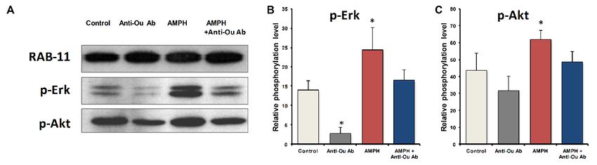
We recently examined the effect of the CNS stimulant amphetamine (AMPH) and the reduction in
brain ECS resulting from i.c.v. injection of specific anti-ouabain antibodies on behavior and ERK and
Akt phosphorylation in the mouse frontal cortex [94]. The results showed a reduction in AMPH-induced
hyperactivity [94], implicating the ECS in behavior. Furthermore, we have shown that anti-ouabain antibody
administration causes reduction in basal ERK phosphorylation in the mouse frontal cortex (Figure 1). In
agreement with previous studies [100,101], AMPH induced a 75% and 41% increase in p-ERK and p-Akt
levels, respectively, in the frontal cortex (Figure 1). The administration of anti-ouabain antibodies significantly
reduced the AMPH-induced increase in the phosphorylation levels of the two proteins (Figure 1). These
results suggest that the manic-like phase is characterized by activation of the ERK and Akt signaling
pathways in the frontal cortex, which is attenuated by a reduction in ECS levels. It is tempting to propose
that the alterations in ERK and Akt phosphorylation caused by changes in ECS are mediated by theirInt. J. Mol. Sci. 2018, 19, 2314 6 of 13
interactions with Na+, K+-ATPase. Such a sequence of events was proposed for the CS-induced effects on
Int. J. Mol. Sci.of
stimulation 2018,
cell19, x FOR PEER
viability REVIEW
[102], 6 of 13
increased heart contractility [103,104], and kidney development [105].
Int. J. Mol. Sci. 2018, 19, x FOR PEER REVIEW 6 of 13
Figure 1.
Figure 1. Effect
Effect of
of amphetamine
amphetamine and and anti-ouabain
anti-ouabain antibodies
antibodies on on ERK
ERK and
and Akt
Akt phosphorylation
phosphorylation levels
levels
Figure 1. Effect of amphetamine and anti-ouabain antibodies on ERK and Akt phosphorylation levels
in the
in thefrontal
frontalcortex.
cortex.Male
MaleBALB/c
BALB/c mice werewere administered
administeredsaline saline(10
(10mL/kg
mL/kg IP) and nonspecific
nonspecific IgG
IgG
in the frontal cortex. Male BALB/c mice were administered saline (10 mL/kg IP) and nonspecific IgG
(1 µg/kg ICV)
(1µg/kg (Control,nn==10),
ICV)(Control, 10),saline
salineand
andanti-ouabain
anti-ouabain antibodies
antibodies (1 µg/kg
(1 µg/kg i.c.v.) (Anti-Ou
i.c.v.) n = 10)
Ab, Ab,
(Anti-Ou n=
(1 µg/kg ICV) (Control, n = 10), saline and anti-ouabain antibodies (1 µg/kg i.c.v.) (Anti-Ou Ab, n =
or
10)AMPH
or AMPH (5 mg/kg
(5 mg/kgIP)IP)
and IgG
and (AMPH,
IgG (AMPH, n= n =10)
10)ororAMPH
AMPHand andanti-ouabain
anti-ouabainantibodies
antibodies(AMPH
(AMPH ++
10) or AMPH (5 mg/kg IP) and IgG (AMPH, n = 10) or AMPH and anti-ouabain antibodies (AMPH +
Anti-Ou
Anti-Ou Ab, ==10).10).The
The micewerewere sacrificed andthethe protein levels were determined by Western
Anti-OunAb, n = 10).mice sacrificed
The mice were and
sacrificed and protein
the proteinlevels were
levels weredetermined
determinedbybyWestern
Westernblot
blot
blot analysis.
analysis. The
The values
analysis.
values are presented
are presented
The values as the
are presented
as the
as mean
mean
the mean
± SE
± SE± (error (error
bars).
SE (error
bars). *
* p*Int. J. Mol. Sci. 2018, 19, 2314 7 of 13
8. Prospect and Future Directions
BD is a heterogeneous condition with a myriad symptoms varying in manifestation; dysregulation
of numerous biochemical pathways has been suggested to be involved in its pathogenesis. Research in
Na+ , K+ -ATPase-induced signaling is evolving. The goal of this overview was not to draw definitive
conclusions about Na+ , K+ -ATPase signaling in BD but to summarize the current knowledge, and to
discuss limitations and shortcomings in the existing research. The emerging literature provides
exciting initial evidence suggesting that alterations in Na+ , K+ -ATPase signaling is involved in BD.
However, additional work is necessary in order to establish a causal relationship between the two.
The uncovering of the metabolism and physiological role of ECS in the brain is the fundamental need.
Furthermore, pharmacological experiments evaluating the effects of ERK, AKT, and NFκB inhibitors
on behavior and examination of the consequence of alterations in ECS metabolism on Na+ , K+ -ATPase
signaling may provide important information on the issue. A deeper and clearer understanding of
the Na+ , K+ -ATPase-induced signaling cascades will establish a better understanding of the complex
mechanisms underlying the pathophysiology of BD and may lead to new venues for the development
of novel targets for the treatment of this disease.
9. Search Strategy
This review was based on search in the PUBMED data base for the key words “bipolar disorder”
or “depression” or “mania” with “Na+ , K+ -ATPase”, “ouabain”, “cardiac steroids”, “intracellular
signaling”, “ERK”, “AKT”, and “NFκB”. No language or time constraints were applied. The lists of
references were searched manually to find additional articles
Funding: This work was supported in part by Israel Science Foundation Grant No. 039-4964 to D.L.
Conflicts of Interest: The authors declare no conflict of interest.
References
1. Lewis-Fernandez, R.; Aggarwal, N.K. Culture and psychiatric diagnosis. Adv. Psychosom. Med. 2013, 33,
15–30. [CrossRef] [PubMed]
2. Hirschfeld, R.M. Differential diagnosis of bipolar disorder and major depressive disorder. J. Affect. Disord.
2014, 169 (Suppl. 1), S12–S16. [CrossRef]
3. Tondo, L.; Vazquez, G.H.; Baldessarini, R.J. Depression and Mania in Bipolar Disorder. Curr. Neuropharmacol.
2017, 15, 353–358. [CrossRef] [PubMed]
4. De la Vega, D.; Pina, A.; Peralta, F.J.; Kelly, S.A.; Giner, L. A Review on the General Stability of Mood Disorder
Diagnoses along the Lifetime. Curr. Psychiatry Rep. 2018, 20, 29. [CrossRef] [PubMed]
5. Neale, B.M.; Sklar, P. Genetic analysis of schizophrenia and bipolar disorder reveals polygenicity but also
suggests new directions for molecular interrogation. Curr. Opin. Neurobiol. 2015, 30, 131–138. [CrossRef]
[PubMed]
6. Nierenberg, A.A.; Kansky, C.; Brennan, B.P.; Shelton, R.C.; Perlis, R.; Iosifescu, D.V. Mitochondrial modulators
for bipolar disorder: A pathophysiologically informed paradigm for new drug development. Aust. N. Z.
J. Psychiatry 2013, 47, 26–42. [CrossRef] [PubMed]
7. Morsel, A.M.; Morrens, M.; Sabbe, B. An overview of pharmacotherapy for bipolar I disorder.
Expert Opin. Pharmacother. 2018, 19, 203–222. [CrossRef] [PubMed]
8. Muller, J.C.; Pryor, W.W.; Gibbons, J.E.; Orgain, E.S. Depression and anxiety occurring during Rauwolfia
therapy. J. Am. Med. Assoc. 1955, 159, 836–839. [CrossRef] [PubMed]
9. Shore, P.A.; Silver, S.L.; Brodie, B.B. Interaction of reserpine, serotonin, and lysergic acid diethylamide in
brain. Science 1955, 122, 284–285. [CrossRef] [PubMed]
10. Hirschfeld, R.M. History and evolution of the monoamine hypothesis of depression. J. Clin. Psychiatry 2000,
61, 4–6. [PubMed]
11. Dale, E.; Bang-Andersen, B.; Sanchez, C. Emerging mechanisms and treatments for depression beyond SSRIs
and SNRIs. Biochem. Pharmacol. 2015, 95, 81–97. [CrossRef] [PubMed]Int. J. Mol. Sci. 2018, 19, 2314 8 of 13
12. Iniesta, R.; Hodgson, K.; Stahl, D.; Malki, K.; Maier, W.; Rietschel, M.; Mors, O.; Hauser, J.; Henigsberg, N.;
Dernovsek, M.Z.; et al. Antidepressant drug-specific prediction of depression treatment outcomes from
genetic and clinical variables. Sci. Rep. 2018, 8, 5530. [CrossRef] [PubMed]
13. Salomon, R.M.; Miller, H.L.; Krystal, J.H.; Heninger, G.R.; Charney, D.S. Lack of behavioral effects of
monoamine depletion in healthy subjects. Biol. Psychiatry 1997, 41, 58–64. [CrossRef]
14. Jeon, W.J.; Dean, B.; Scarr, E.; Gibbons, A. The Role of Muscarinic Receptors in the Pathophysiology of Mood
Disorders: A Potential Novel Treatment? Curr. Neuropharmacol. 2015, 13, 739–749. [CrossRef] [PubMed]
15. Blacker, C.J.; Lewis, C.P.; Frye, M.A.; Veldic, M. Metabotropic glutamate receptors as emerging research
targets in bipolar disorder. Psychiatry Res. 2017, 257, 327–337. [CrossRef] [PubMed]
16. Lener, M.S.; Niciu, M.J.; Ballard, E.D.; Park, M.; Park, L.T.; Nugent, A.C.; Zarate, C.A., Jr. Glutamate and
Gamma-Aminobutyric Acid Systems in the Pathophysiology of Major Depression and Antidepressant
Response to Ketamine. Biol. Psychiatry 2017, 81, 886–897. [CrossRef] [PubMed]
17. Ehrich, E.; Turncliff, R.; Du, Y.; Leigh-Pemberton, R.; Fernandez, E.; Jones, R.; Fava, M. Evaluation of opioid
modulation in major depressive disorder. Neuropsychopharmacology 2015, 40, 1448–1455. [CrossRef] [PubMed]
18. Tomasetti, C.; Iasevoli, F.; Buonaguro, E.F.; De Berardis, D.; Fornaro, M.; Fiengo, A.L.; Martinotti, G.;
Orsolini, L.; Valchera, A.; Di Giannantonio, M.; et al. Treating the Synapse in Major Psychiatric Disorders:
The Role of Postsynaptic Density Network in Dopamine-Glutamate Interplay and Psychopharmacologic
Drugs Molecular Actions. Int. J. Mol. Sci. 2017, 18, 135. [CrossRef] [PubMed]
19. Morris, G.; Walder, K.; McGee, S.L.; Dean, O.M.; Tye, S.J.; Maes, M.; Berk, M. A model of the mitochondrial
basis of bipolar disorder. Neurosci. Biobehav. Rev. 2017, 74, 1–20. [CrossRef] [PubMed]
20. Cikankova, T.; Sigitova, E.; Zverova, M.; Fisar, Z.; Raboch, J.; Hroudova, J. Mitochondrial Dysfunctions in
Bipolar Disorder: Effect of the Disease and Pharmacotherapy. CNS Neurol. Disord. Drug Targets 2017, 16,
176–186. [CrossRef] [PubMed]
21. Erdem, M.A.S.; Pan, E.; Kurt, Y.G. Bipolar Disorder and Oxidative Stress. J. Mood Disord. 2014, 4, 70–79.
[CrossRef]
22. De Berardis, D.; Campanella, D.; Gambi, F.; la Rovere, R.; Carano, A.; Conti, C.M.; Sivestrini, C.;
Serroni, N.; Piersanti, D.; di Giuseppe, B.; et al. The role of C-reactive protein in mood disorders. Int.
J. Immunopathol. Pharmacol. 2006, 19, 721–725. [CrossRef] [PubMed]
23. Hamdani, N.; Doukhan, R.; Kurtlucan, O.; Tamouza, R.; Leboyer, M. Immunity, inflammation, and bipolar
disorder: Diagnostic and therapeutic implications. Curr. Psychiatry Rep. 2013, 15, 387. [CrossRef] [PubMed]
24. Gawryluk, J.W.; Wang, J.F.; Andreazza, A.C.; Shao, L.; Young, L.T. Decreased levels of glutathione,
the major brain antioxidant, in post-mortem prefrontal cortex from patients with psychiatric disorders.
Int. J. Neuropsychopharmacol. 2011, 14, 123–130. [CrossRef] [PubMed]
25. Benes, F.M.; Matzilevich, D.; Burke, R.E.; Walsh, J. The expression of proapoptosis genes is increased in
bipolar disorder, but not in schizophrenia. Mol. Psychiatry 2006, 11, 241–251. [CrossRef] [PubMed]
26. Siwek, M.; Sowa-Kucma, M.; Styczen, K.; Misztak, P.; Szewczyk, B.; Topor-Madry, R.; Nowak, G.; Dudek, D.;
Rybakowski, J.K. Thiobarbituric Acid-Reactive Substances: Markers of an Acute Episode and a Late Stage of
Bipolar Disorder. Neuropsychobiology 2016, 73, 116–122. [CrossRef] [PubMed]
27. Diniz, B.S. Decreased Brain-Derived Neurotrophic Factor (BDNF) in Older Adults with Bipolar Disorder:
Meaning and Utility? Am. J. Geriatr. Psychiatry 2016, 24, 602–603. [CrossRef] [PubMed]
28. Munkholm, K.; Vinberg, M.; Kessing, L.V. Peripheral blood brain-derived neurotrophic factor in bipolar
disorder: A comprehensive systematic review and meta-analysis. Mol. Psychiatry 2016, 21, 216–228.
[CrossRef] [PubMed]
29. Muneer, A. Wnt and GSK3 Signaling Pathways in Bipolar Disorder: Clinical and Therapeutic Implications.
Clin. Psychopharmacol. Neurosci. 2017, 15, 100–114. [CrossRef] [PubMed]
30. Clausen, M.V.; Hilbers, F.; Poulsen, H. The Structure and Function of the Na,K-ATPase Isoforms in Health
and Disease. Front. Physiol. 2017, 8, 371. [CrossRef] [PubMed]
31. Blanco, G.; Mercer, R.W. Isozymes of the Na-K-ATPase: Heterogeneity in structure, diversity in function.
Am. J. Physiol. 1998, 275, F633–F650. [CrossRef] [PubMed]
32. Hilbers, F.; Kopec, W.; Isaksen, T.J.; Holm, T.H.; Lykke-Hartmann, K.; Nissen, P.; Khandelia, H.; Poulsen, H.
Tuning of the Na,K-ATPase by the beta subunit. Sci. Rep. 2016, 6, 20442. [CrossRef] [PubMed]
33. Li, Z.; Langhans, S.A. Transcriptional regulators of Na,K-ATPase subunits. Front. Cell Dev. Biol. 2015, 3, 66.
[CrossRef] [PubMed]Int. J. Mol. Sci. 2018, 19, 2314 9 of 13
34. McGrail, K.M.; Phillips, J.M.; Sweadner, K.J. Immunofluorescent localization of three Na,K-ATPase isozymes
in the rat central nervous system: Both neurons and glia can express more than one Na,K-ATPase. J. Neurosci.
1991, 11, 381–391. [CrossRef] [PubMed]
35. Romanovsky, D.; Moseley, A.E.; Mrak, R.E.; Taylor, M.D.; Dobretsov, M. Phylogenetic preservation of α3
Na+ ,K+ -ATPase distribution in vertebrate peripheral nervous systems. J. Comp. Neurol. 2007, 500, 1106–1116.
[CrossRef] [PubMed]
36. Bøttger, P.; Tracz, Z.; Heuck, A.; Nissen, P.; Romero-Ramos, M.; Lykke-Hartmann, K. Distribution of
Na/K-ATPase alpha 3 isoform, a sodium-potassium P-type pump associated with rapid-onset of dystonia
parkinsonism (RDP) in the adult mouse brain. J. Comp. Neurol. 2010, 519, 376–404. [CrossRef] [PubMed]
37. Blom, H.; Ronnlund, D.; Scott, L.; Spicarova, Z.; Widengren, J.; Bondar, A.; Aperia, A.; Brismar, H. Spatial
distribution of Na+ -K+ -ATPase in dendritic spines dissected by nanoscale superresolution STED microscopy.
BMC Neurosci. 2011, 12, 16. [CrossRef] [PubMed]
38. Geering, K. FXYD proteins: New regulators of Na-K-ATPase. Am. J. Physiol. Renal Physiol. 2006, 290,
F241–F250. [CrossRef] [PubMed]
39. Moseley, A.E.; Williams, M.T.; Schaefer, T.L.; Bohanan, C.S.; Neumann, J.C.; Behbehani, M.M.; Vorhees, C.V.;
Lingrel, J.B. Deficiency in Na,K-ATPase alpha isoform genes alters spatial learning, motor activity, and
anxiety in mice. J. Neurosci. 2007, 27, 616–626. [CrossRef] [PubMed]
40. Lingrel, J.B.; Williams, M.T.; Vorhees, C.V.; Moseley, A.E. Na,K-ATPase and the role of alpha isoforms in
behavior. J. Bioenerg. Biomembr. 2007, 39, 385–389. [CrossRef] [PubMed]
41. Schaefer, T.L.; Lingrel, J.B.; Moseley, A.E.; Vorhees, C.V.; Williams, M.T. Targeted mutations in the
Na,K-ATPase alpha 2 isoform confer ouabain resistance and result in abnormal behavior in mice. Synapse
2010, 65, 520–531. [CrossRef] [PubMed]
42. Bottger, P.; Glerup, S.; Gesslein, B.; Illarionova, N.B.; Isaksen, T.J.; Heuck, A.; Clausen, B.H.; Fuchtbauer, E.M.;
Gramsbergen, J.B.; Gunnarson, E.; et al. Glutamate-system defects behind psychiatric manifestations in a
familial hemiplegic migraine type 2 disease-mutation mouse model. Sci. Rep. 2016, 6, 22047. [CrossRef]
[PubMed]
43. Holm, T.H.; Lykke-Hartmann, K. Insights into the Pathology of the alpha3 Na+ /K+ -ATPase Ion Pump in
Neurological Disorders; Lessons from Animal Models. Front. Physiol. 2016, 7, 209. [CrossRef] [PubMed]
44. Kirshenbaum, G.S.; Idris, N.F.; Dachtler, J.; Roder, J.C.; Clapcote, S.J. Deficits in social behavioral tests in a
mouse model of alternating hemiplegia of childhood. J. Neurogenet. 2016, 30, 42–49. [CrossRef] [PubMed]
45. Timothy, J.W.S.; Klas, N.; Sanghani, H.R.; Al-Mansouri, T.; Hughes, A.T.L.; Kirshenbaum, G.S.; Brienza, V.;
Belle, M.D.C.; Ralph, M.R.; Clapcote, S.J.; et al. Circadian Disruptions in the Myshkin Mouse Model of Mania
Are Independent of Deficits in Suprachiasmatic Molecular Clock Function. Biol. Psychiatry 2017. [CrossRef]
[PubMed]
46. Kirshenbaum, G.S.; Clapcote, S.J.; Duffy, S.; Burgess, C.R.; Petersen, J.; Jarowek, K.J.; Yucel, Y.H.; Cortez, M.A.;
Snead, O.C., 3rd; Vilsen, B.; et al. Mania-like behavior induced by genetic dysfunction of the neuron-specific
Na+ ,K+ -ATPase α3 sodium pump. Proc. Natl. Acad. Sci. USA 2011. [CrossRef] [PubMed]
47. Page, E. The Actions of Cardiac Glycosides on Heart Muscle Cells. Circulation 1964, 30, 237–251. [CrossRef]
[PubMed]
48. Roberts, D.M.; Gallapatthy, G.; Dunuwille, A.; Chan, B.S. Pharmacological treatment of cardiac glycoside
poisoning. Br. J. Clin. Pharmacol. 2016, 81, 488–495. [CrossRef] [PubMed]
49. Hong, Z.; Chan, K.; Yeung, H.W. Simultaneous determination of bufadienolides in the traditional Chinese
medicine preparation, liu-shen-wan, by liquid chromatography. J. Pharm. Pharmacol. 1992, 44, 1023–1026.
[CrossRef] [PubMed]
50. Krenn, L.; Kopp, B. Bufadienolides from animal and plant sources. Phytochemistry 1998, 48, 1–29. [CrossRef]
51. Hamlyn, J.M.; Blaustein, M.P. Endogenous Ouabain: Recent Advances and Controversies. Hypertension 2016,
68, 526–532. [CrossRef] [PubMed]
52. Goto, A.; Ishiguro, T.; Yamada, K.; Ishii, M.; Yoshioka, M.; Eguchi, C.; Shimora, M.; Sugimoto, T. Isolation
of a urinary digitalis-like factor indistinguishable from digoxin. Biochem. Biophys. Res. Commun. 1990, 173,
1093–1101. [CrossRef]
53. Lichtstein, D.; Gati, I.; Samuelov, S.; Berson, D.; Rozenman, Y.; Landau, L.; Deutsch, J. Identification of
digitalis-like compounds in human cataractous lenses. Eur. J. Biochem. 1993, 216, 261–268. [CrossRef]
[PubMed]Int. J. Mol. Sci. 2018, 19, 2314 10 of 13
54. Hilton, P.J.; White, R.W.; Lord, G.A.; Garner, G.V.; Gordon, D.B.; Hilton, M.J.; Forni, L.G.; McKinnon, W.;
Ismail, F.M.; Keenan, M.; et al. An inhibitor of the sodium pump obtained from human placenta. Lancet 1996,
348, 303–305. [CrossRef]
55. Schneider, R.; Antolovic, R.; Kost, H.; Sich, B.; Kirch, U.; Tepel, M.; Zidek, W.; Schoner, W. Proscillaridin A
immunoreactivity: Its purification, transport in blood by a specific binding protein and its correlation with
blood pressure. Clin. Exp. Hypertens. 1998, 20, 593–599. [CrossRef] [PubMed]
56. Fedorova, O.V.; Zernetkina, V.I.; Shilova, V.Y.; Grigorova, Y.N.; Juhasz, O.; Wei, W.; Marshall, C.A.;
Lakatta, E.G.; Bagrov, A.Y. Synthesis of an Endogenous Steroidal Na Pump Inhibitor Marinobufagenin,
Implicated in Human Cardiovascular Diseases, Is Initiated by CYP27A1 via Bile Acid Pathway.
Circ. Cardiovasc. Genet. 2015, 8, 736–745. [CrossRef] [PubMed]
57. Komiyama, Y.; Dong, X.H.; Nishimura, N.; Masaki, H.; Yoshika, M.; Masuda, M.; Takahashi, H. A novel
endogenous digitalis, telocinobufagin, exhibits elevated plasma levels in patients with terminal renal failure.
Clin. Biochem. 2005, 38, 36–45. [CrossRef] [PubMed]
58. Hamlyn, J.M.; Blaustein, M.P.; Bova, S.; DuCharme, D.W.; Harris, D.W.; Mandel, F.; Mathews, W.R.;
Ludens, J.H. Identification and characterization of a ouabain-like compound from human plasma. Proc. Natl.
Acad. Sci. USA 1991, 88, 6259–6263. [CrossRef] [PubMed]
59. Blaustein, M.P. The pump, the exchanger, and the holy spirit: Origins and 40-year evolution of ideas about
the ouabain-Na+ pump endocrine system. Am. J. Physiol. Cell Physiol. 2018, 314, C3–C26. [CrossRef]
[PubMed]
60. Lewis, L.K.; Yandle, T.G.; Hilton, P.J.; Jensen, B.P.; Begg, E.J.; Nicholls, M.G. Endogenous ouabain is not
ouabain. Hypertension 2014, 64, 680–683. [CrossRef] [PubMed]
61. Baecher, S.; Kroiss, M.; Fassnacht, M.; Vogeser, M. No endogenous ouabain is detectable in human plasma by
ultra-sensitive UPLC-MS/MS. Clin. Chim. Acta 2014, 431, 87–92. [CrossRef] [PubMed]
62. Vogeser, M. Letter to the editor: Comments on Blaustein (2018): “The pump, the exchanger, and the holy
spirit: Origins and 40-year evolution of ideas about the ouabain-Na+ pump endocrine system”. Am. J.
Physiol. Cell Physiol. 2018, 314, C640. [CrossRef] [PubMed]
63. Hamlyn, J.M. Biosynthesis of endogenous cardiac glycosides by mammalian adrenocortical cells: Three
steps forward. Clin. Chem. 2004, 50, 469–470. [CrossRef] [PubMed]
64. Laredo, J.; Hamilton, B.P.; Hamlyn, J.M. Ouabain is secreted by bovine adrenocortical cells. Endocrinology
1994, 135, 794–797. [CrossRef] [PubMed]
65. Perrin, A.; Brasmes, B.; Chambaz, E.M.; Defaye, G. Bovine adrenocortical cells in culture synthesize an
ouabain-like compound. Mol. Cell. Endocrinol. 1997, 126, 7–15. [CrossRef]
66. Lichtstein, D.; Steinitz, M.; Gati, I.; Samuelov, S.; Deutsch, J.; Orly, J. Biosynthesis of digitalis-like compounds
in rat adrenal cells: Hydroxycholesterol as possible precursor. Life Sci. 1998, 62, 2109–2126. [CrossRef]
67. Nesher, M.; Shpolansky, U.; Rosen, H.; Lichtstein, D. The digitalis-like steroid hormones: New mechanisms
of action and biological significance. Life Sci. 2007, 80, 2093–2107. [CrossRef] [PubMed]
68. Buckalew, V.M. Endogenous digitalis-like factors: An overview of the history. Front. Endocrinol. (Lausanne)
2015, 6, 49. [CrossRef] [PubMed]
69. Hamlyn, J.M.; Manunta, P. Endogenous cardiotonic steroids in kidney failure: A review and an hypothesis.
Adv. Chronic Kidney Dis. 2015, 22, 232–244. [CrossRef] [PubMed]
70. Bagrov, A.Y.; Shapiro, J.I.; Fedorova, O.V. Endogenous cardiotonic steroids: Physiology, pharmacology, and
novel therapeutic targets. Pharmacol. Rev. 2009, 61, 9–38. [CrossRef] [PubMed]
71. Hodes, A.; Lichtstein, D. Natriuretic hormones in brain function. Front. Endocrinol. (Lausanne) 2014, 5, 201.
[CrossRef] [PubMed]
72. Buckalew, V.M. Role of endogenous digitalis-like factors in the clinical manifestations of severe preeclampsia:
A sytematic review. Clin. Sci. (Lond.) 2018, 132, 1215–1242. [CrossRef] [PubMed]
73. Haas, M.; Askari, A.; Xie, Z. Involvement of Src and epidermal growth factor receptor in the
signal-=transducing function of Na,K-ATPase. J. Biol. Chem. 2000, 275, 27832–27837. [CrossRef] [PubMed]
74. Liu, J.; Xie, Z.J. The sodium pump and cardiotonic steroids-induced signal transduction protein kinases and
calcium-signaling microdomain in regulation of transporter trafficking. Biochim. Biophys. Acta 2010, 1802,
1237–1245. [CrossRef] [PubMed]
75. Cui, X.; Xie, Z. Protein Interaction and Na/K-ATPase-Mediated Signal Transduction. Molecules 2017, 22, 990.Int. J. Mol. Sci. 2018, 19, 2314 11 of 13
76. Buckalew, V. Is endogenous ouabain a physiological regulator of cardiovascular and renal function? Am. J.
Physiol. Heart Circ. Physiol. 2009, 297, H1972–H1973. [CrossRef] [PubMed]
77. Mynett-Johnson, L.; Murphy, V.; McCormack, J.; Shields, D.C.; Claffey, E.; Manley, P.; McKeon, P. Evidence
for an allelic association between bipolar disorder and a Na+ , K+ adenosine triphosphatase alpha subunit
gene (ATP1A3). Biol. Psychiatry 1998, 44, 47–51. [CrossRef]
78. Goldstein, I.; Lerer, E.; Laiba, E.; Mallet, J.; Mujaheed, M.; Laurent, C.; Rosen, H.; Ebstein, R.P.; Lichtstein, D.
Association between sodium- and potassium-activated adenosine triphosphatase alpha isoforms and bipolar
disorders. Biol. Psychiatry 2009, 65, 985–991. [CrossRef] [PubMed]
79. Kirshenbaum, G.S.; Burgess, C.R.; Dery, N.; Fahnestock, M.; Peever, J.H.; Roder, J.C. Attenuation of mania-like
behavior in Na+ ,K+ -ATPase α3 mutant mice by prospective therapies for bipolar disorder: Melatonin and
exercise. Neuroscience 2014, 260, 195–204. [CrossRef] [PubMed]
80. Nurnberger, J., Jr.; Jimerson, D.C.; Allen, J.R.; Simmons, S.; Gershon, E. Red cell ouabain-sensitive
Na+ -K+ -adenosine triphosphatase: A state marker in affective disorder inversely related to plasma cortisol.
Biol. Psychiatry 1982, 17, 981–992. [PubMed]
81. Naylor, G.J.; McNamee, H.B.; Moody, J.P. Changes in erythrocyte sodium and potassium on recovery from a
depressive illness. Br. J. Psychiatry 1971, 118, 219–223. [CrossRef] [PubMed]
82. Looney, S.W.; El-Mallakh, R.S. Meta-analysis of erythrocyte Na,K-ATPase activity in bipolar illness.
Depress. Anxiety 1997, 5, 53–65. [CrossRef]
83. Chetcuti, A.; Adams, L.J.; Mitchell, P.B.; Schofield, P.R. Microarray gene expression profiling of mouse brain
mRNA in a model of lithium treatment. Psychiatr. Genet. 2008, 18, 64–72. [CrossRef] [PubMed]
84. Grider, G.; El-Mallakh, R.S.; Huff, M.O.; Buss, T.J.; Miller, J.; Valdes, R.J. Endogenous digoxin-like
immunoreactive factor (DLIF) serum concentrations are decreased in manic bipolar patients compared
to normal controls. J. Affect. Disord. 1999, 54, 261–270. [CrossRef]
85. El-Mallakh, R.S.; Stoddard, M.; Jortani, S.A.; El-Masri, M.A.; Sephton, S.; Valdes, R.J. regulation of
endogenous ouabain-like factor in bipolar subjects. Psychiatry Res. 2010, 178, 116–120. [CrossRef] [PubMed]
86. Goldstein, I.; Levy, T.; Galili, D.; Ovadia, H.; Yirmiya, R.; Rosen, H.; Lichtstein, D. Involvement of
Na+ ,K+ -ATPase and endogenous digitalis-like compounds in depressive disorders. Biol. Psychiatry 2006, 60,
491–499. [CrossRef] [PubMed]
87. El-Mallakh, R.S.; El-Masri, M.A.; Huff, M.O.; Li, X.P.; Decker, S.; Levy, R.S. Intracerebroventricular
administration of ouabain as a model of mania in rats. Bipolar Disord. 2003, 5, 362–365. [CrossRef] [PubMed]
88. Riegel, R.E.; Valvassori, S.S.; Elias, G.; Réus, G.Z.; Steckert, A.V.; de Souza, B.; Petronilho, F.; Gavioli, E.C.;
Dal-Pizzol, F.; Quevedo, J. Animal model of mania induced by ouabain: Evidence of oxidative stress in
submitochondrial particles of the rat brain. Neurochem. Int. 2009, 55, 491–495. [CrossRef] [PubMed]
89. Varela, R.B.; Valvassori, S.S.; Lopes-Borges, J.; Mariot, E.; Dal-Pont, G.C.; Amboni, R.T.; Bianchini, G.;
Quevedo, J. Sodium butyrate and mood stabilizers block ouabain-induced hyperlocomotion and increase
BDNF, NGF and GDNF levels in brain of Wistar rats. J. Psychiatr. Res. 2015, 61, 114–121. [CrossRef] [PubMed]
90. Valvassori, S.S.; Resende, W.R.; Lopes-Borges, J.; Mariot, E.; Dal-Pont, G.C.; Vitto, M.F.; Luz, G.; de Souza, C.T.;
Quevedo, J. Effects of mood stabilizers on oxidative stress-induced cell death signaling pathways in the
brains of rats subjected to the ouabain-induced animal model of mania: Mood stabilizers exert protective
effects against ouabain-induced activation of the cell death pathway. J. Psychiatr. Res. 2015, 65, 63–70.
[PubMed]
91. Logan, R.W.; McClung, C.A. Animal models of bipolar mania: The past, present and future. Neuroscience
2016, 321, 163–188. [CrossRef] [PubMed]
92. Brocardo, P.S.; Budni, J.; Pavesi, E.; Franco, J.L.; Uliano-Silva, M.; Trevisan, R.; Terenzi, M.G.; Dafre, A.L.;
Rodrigues, A.L. Folic acid administration prevents ouabain-induced hyperlocomotion and alterations in
oxidative stress markers in the rat brain. Bipolar Disord. 2010, 12, 414–424. [CrossRef] [PubMed]
93. Goldstein, I.; Lax, E.; Gispan-Herman, I.; Ovadia, H.; Rosen, H.; Yadid, G.; Lichtstein, D. Neutralization
of endogenous digitalis-like compounds alters catecholamines metabolism in the brain and elicits
anti-depressive behavior. Eur. Neuropsychopharmacol. 2012, 22, 72–79. [CrossRef] [PubMed]
94. Hodes, A.; Rosen, H.; Deutsch, J.; Lifschytz, T.; Einat, H.; Lichtstein, D. Endogenous cardiac steroids in
animal models of mania. Bipolar Disord. 2016, 18, 451–459. [CrossRef] [PubMed]Int. J. Mol. Sci. 2018, 19, 2314 12 of 13
95. Hodes, A.; Lifschytz, T.; Rosen, H.; Cohen, B.A.H.; Lichtstein, D. Reduction in endogenous cardiac steroids
protects the brain from oxidative stress in a mouse model of mania induced by amphetamine. Brain Res. Bull.
2018, 137, 356–362. [CrossRef] [PubMed]
96. Valvassori, S.S.; Dal-Pont, G.C.; Resende, W.R.; Varela, R.B.; Peterle, B.R.; Gava, F.F.; Mina, F.G.; Cararo, J.H.;
Carvalho, A.F.; Quevedo, J. Lithium and Tamoxifen Modulate Behavior and Protein Kinase C Activity in the
Animal Model of Mania Induced by Ouabain. Int. J. Neuropsychopharmacol. 2017, 20, 877–885. [CrossRef]
[PubMed]
97. Yu, H.S.; Kim, S.H.; Park, H.G.; Kim, Y.S.; Ahn, Y.M. Activation of Akt signaling in rat brain by
intracerebroventricular injection of ouabain: A rat model for mania. Prog. Neuro-Psychopharmacol. Biol.
Psychiatry 2010, 34, 888–894. [CrossRef] [PubMed]
98. Kim, S.H.; Yu, H.S.; Park, H.G.; Ha, K.; Kim, Y.S.; Shin, S.Y.; Ahn, Y.M. Intracerebroventricular administration
of ouabain, a Na/K-ATPase inhibitor, activates mTOR signal pathways and protein translation in the rat
frontal cortex. Prog. Neuro-Psychopharmacol. Biol. Psychiatry 2013, 45, 73–82. [CrossRef] [PubMed]
99. Kim, S.H.; Yu, H.S.; Park, H.G.; Jeon, W.J.; Song, J.Y.; Kang, U.G.; Ahn, Y.M.; Lee, Y.H.; Kim, Y.S.
Dose-dependent effect of intracerebroventricular injection of ouabain on the phosphorylation of the
MEK1/2-ERK1/2-p90RSK pathway in the rat brain related to locomotor activity. Prog. Neuro-Psychopharmacol.
Biol. Psychiatry 2008, 32, 1637–1642. [CrossRef] [PubMed]
100. Zheng, W.; Zeng, Z.; Bhardwaj, S.K.; Jamali, S.; Srivastava, L.K. Lithium normalizes amphetamine-induced
changes in striatal FoxO1 phosphorylation and behaviors in rats. Neuroreport 2013, 24, 560–565. [CrossRef]
[PubMed]
101. Gonzalez, B.; Raineri, M.; Cadet, J.L.; Garcia-Rill, E.; Urbano, F.J.; Bisagno, V. Modafinil improves
methamphetamine-induced object recognition deficits and restores prefrontal cortex ERK signaling in
mice. Neuropharmacology 2014, 87, 188–197. [CrossRef] [PubMed]
102. Dvela, M.; Rosen, H.; Ben-Ami, H.C.; Lichtstein, D. Endogenous ouabain regulates cell viability. Am. J.
Physiol. Cell Physiol. 2012, 302, C442–C452. [CrossRef] [PubMed]
103. Tian, J.; Gong, X.; Xie, Z. Signal-transducing function of Na+ -K+ -ATPase is essential for ouabain’s effect
on [Ca2+ ]i in rat cardiac myocytes. Am. J. Physiol. Heart Circ. Physiol. 2001, 281, H1899–H1907. [CrossRef]
[PubMed]
104. Buzaglo, N.; Rosen, H.; Ben Ami, H.C.; Inbal, A.; Lichtstein, D. Essential Opposite Roles of ERK and Akt
Signaling in Cardiac Steroid-Induced Increase in Heart Contractility. J. Pharmacol. Exp. Ther. 2016, 357,
345–356. [CrossRef] [PubMed]
105. Dvela-Levitt, M.; Cohen-Ben Ami, H.; Rosen, H.; Ornoy, A.; Hochner-Celnikier, D.; Granat, M.; Lichtstein, D.
Reduction in maternal circulating ouabain impairs offspring growth and kidney development. J. Am.
Soc. Nephrol. 2015, 26, 1103–1114. [CrossRef] [PubMed]
106. Jefferys, J.G. Nonsynaptic modulation of neuronal activity in the brain: Electric currents and extracellular
ions. Physiol. Rev. 1995, 75, 689–723. [CrossRef] [PubMed]
107. Vizi, E.S.; Oberfrank, F. Na+ /K+ -ATPase, its endogenous ligands and neurotransmitter release.
Neurochem. Int. 1992, 20, 11–17. [CrossRef]
108. Noda, M.; Hiyama, T.Y. Sodium sensing in the brain. Pflugers Arch. 2015, 467, 465–474. [CrossRef] [PubMed]
109. Shimizu, S.; Akiyama, T.; Kawada, T.; Sata, Y.; Turner, M.J.; Fukumitsu, M.; Yamamoto, H.; Kamiya, A.;
Shishido, T.; Sugimachi, M. Sodium ion transport participates in non-neuronal acetylcholine release in the
renal cortex of anesthetized rabbits. J. Physiol. Sci. 2017, 67, 587–593. [CrossRef] [PubMed]
110. Cavalier, M.; Crouzin, N.; Ben Sedrine, A.; de Jesus Ferreira, M.C.; Guiramand, J.; Cohen-Solal, C.;
Fehrentz, J.A.; Martinez, J.; Barbanel, G.; Vignes, M. Involvement of PKA and ERK pathways in
ghrelin-induced long-lasting potentiation of excitatory synaptic transmission in the CA1 area of rat
hippocampus. Eur. J. Neurosci. 2015, 42, 2568–2576. [CrossRef] [PubMed]
111. Mao, L.M.; Wang, H.H.; Wang, J.Q. Antagonism of Muscarinic Acetylcholine Receptors Alters Synaptic ERK
Phosphorylation in the Rat Forebrain. Neurochem. Res. 2017, 42, 1202–1210. [CrossRef] [PubMed]
112. Yang, J.H.; Mao, L.M.; Choe, E.S.; Wang, J.Q. Synaptic ERK2 Phosphorylates and Regulates Metabotropic
Glutamate Receptor 1 In Vitro and in Neurons. Mol. Neurobiol. 2017, 54, 7156–7170. [CrossRef] [PubMed]
113. Wang, Q.; Liu, L.; Pei, L.; Ju, W.; Ahmadian, G.; Lu, J.; Wang, Y.; Liu, F.; Wang, Y.T. Control of synaptic
strength, a novel function of Akt. Neuron 2003, 38, 915–928. [CrossRef]Int. J. Mol. Sci. 2018, 19, 2314 13 of 13
114. Liu, G.; Feng, D.; Wang, J.; Zhang, H.; Peng, Z.; Cai, M.; Yang, J.; Zhang, R.; Wang, H.; Wu, S.; et al. rTMS
Ameliorates PTSD Symptoms in Rats by Enhancing Glutamate Transmission and Synaptic Plasticity in the
ACC via the PTEN/Akt Signalling Pathway. Mol. Neurobiol. 2018, 55, 3946–3958. [CrossRef] [PubMed]
115. Pen, Y.; Borovok, N.; Reichenstein, M.; Sheinin, A.; Michaelevski, I. Membrane-tethered AKT kinase regulates
basal synaptic transmission and early phase LTP expression by modulation of post-synaptic AMPA receptor
level. Hippocampus 2016, 26, 1149–1167. [CrossRef] [PubMed]
116. Caviedes, A.; Lafourcade, C.; Soto, C.; Wyneken, U. BDNF/NF-κB Signaling in the Neurobiology of
Depression. Curr. Pharm. Des. 2017, 23, 3154–3163. [CrossRef] [PubMed]
117. Dresselhaus, E.C.; Boersma, M.C.H.; Meffert, M.K. Targeting of NF-κB to Dendritic Spines Is Required for
Synaptic Signaling and Spine Development. J. Neurosci. 2018, 38, 4093–4103. [CrossRef] [PubMed]
118. Engelmann, C.; Haenold, R. Transcriptional Control of Synaptic Plasticity by Transcription Factor NF-κB.
Neural Plast. 2016, 2016, 7027949. [CrossRef] [PubMed]
© 2018 by the authors. Licensee MDPI, Basel, Switzerland. This article is an open access
article distributed under the terms and conditions of the Creative Commons Attribution
(CC BY) license (http://creativecommons.org/licenses/by/4.0/).You can also read