Low-grade developmental and epilepsy associated brain tumors: a critical update 2020
←
→
Page content transcription
If your browser does not render page correctly, please read the page content below
Slegers and Blumcke Acta Neuropathologica Communications                          (2020) 8:27
https://doi.org/10.1186/s40478-020-00904-x

 REVIEW                                                                                                                                                Open Access

Low-grade developmental and epilepsy
associated brain tumors: a critical update
2020
Rutger Juriaan Slegers1 and Ingmar Blumcke2*

  Abstract
  Brain tumors represent the second most frequent etiology in patients with focal seizure onset before 18 years of age
  and submitted to epilepsy surgery. Hence, this category of brain tumors, herein defined as low-grade, developmental,
  epilepsy-associated brain tumors (LEAT) is different from those frequently encountered in adults as (A): 77% of LEAT
  occur in the temporal lobe; (B): the vast majority of LEAT are of low malignancy and classified as WHO I°; (C): LEAT are
  often composed of mixed glial and neuronal cell components and present with variable growth patterns including
  small cysts or nodules; (D): LEAT do not share common gene driving mutations, such as IDH1 or 1p/19q co-deletions.
  Characteristic entities comprise the ganglioglioma (GG), the dysembryoplastic neuroepithelial tumor (DNT), the
  angiocentric glioma (AG), the isomorphic diffuse glioma (IDG) and the papillary glio-neuronal tumor (PGNT),
  representing 73.2% of 1680 tumors collected in a large German series of 6747 patients submitted to epilepsy surgery.
  In the realm of exciting discoveries of genetic drivers of brain tumors new genes have been also reported for LEAT.
  BRAF V600E mutations were linked to GG with CD34 expression, FGFR1 mutations to DNT, MYB alterations to AG and
  also IDG and PRKCA fusions to PGNT, suggesting the possibility to also develop a genetically driven tumor classification
  scheme for LEAT. Rare availability of LEAT in a single center is a challenging obstacle, however, to systematically
  unravel the neurobiological nature and clinical behavior of LEAT. Other challenges in need of clarification include
  malignant tumor progression of LEAT entities, seizure relapse in patients following bulk tumor resection and the
  controversial issue of associated focal cortical dysplasia as additional pathomechanism. In order to advance our
  understanding and promote reliable diagnostic work-up of LEAT, we recommend, therefore, international collaboration
  to achieve our goals.
  Keywords: Pathology, Seizure, Ganglioglioma, Astrocytoma, Oligodendroglioma

Introduction                                                                              most prevalent ones within the LEAT spectrum. The
Literally every brain tumor compromising the neocortex                                    ganglioglioma with its biphasic composition of glial and
or neuronal circuits thereof can cause a seizure and pro-                                 neuronal cell elements was introduced in 1926 by
gress into chronic epilepsy [25]. This common clinical                                    Perkins, and further described by Cushing in his mono-
scenario was first published by Hughlings Jackson in                                      graph in 1927 and by Courville in 1930 [18, 55]. The
1882 [71]. Later the first epilepsy-associated tumor en-                                  second most prevalent tumor associated with early epi-
tities were described, which are nowadays seen as the                                     lepsy onset, the DNT, was first described by Daumas-
                                                                                          Duport in 1988 in a series of 20 patients submitted to
* Correspondence: bluemcke@uk-erlangen.de
                                                                                          epilepsy surgery [21]. GG and DNT belong to the group
2
 Department of Neuropathology, University Hospitals Erlangen,                             of glio-neuronal tumors, as defined by the WHO. The
Schwabachanalge 6, 91054 Erlangen, Germany                                                term “long-term epilepsy associated tumors (LEATs)”
Full list of author information is available at the end of the article

                                           © The Author(s). 2020 Open Access This article is licensed under a Creative Commons Attribution 4.0 International License,
                                           which permits use, sharing, adaptation, distribution and reproduction in any medium or format, as long as you give
                                           appropriate credit to the original author(s) and the source, provide a link to the Creative Commons licence, and indicate if
                                           changes were made. The images or other third party material in this article are included in the article's Creative Commons
                                           licence, unless indicated otherwise in a credit line to the material. If material is not included in the article's Creative Commons
                                           licence and your intended use is not permitted by statutory regulation or exceeds the permitted use, you will need to obtain
                                           permission directly from the copyright holder. To view a copy of this licence, visit http://creativecommons.org/licenses/by/4.0/.
                                           The Creative Commons Public Domain Dedication waiver (http://creativecommons.org/publicdomain/zero/1.0/) applies to the
                                           data made available in this article, unless otherwise stated in a credit line to the data.
Slegers and Blumcke Acta Neuropathologica Communications (2020) 8:27 Page 2 of 11 was introduced by Luyken and coworker from the Bonn rare in LEAT; B) LEAT occur predominately in the tem- epilepsy center in 2003 to also recognize rare tumor en- poral lobe (Table 2) of either hemisphere and have no tities in patients with drug-resistant long-term epilepsy sex preference. The neurobiological and/or neurodeve- that do not match with the WHO description and nos- lopmental basis for their preferred growth into the ology, but are likely more close to the GG and DNT temporal lobe remains to be clarified; C) The histomor- spectrum [44]. Further examples for this hitherto ongoing phological spectrum of established LEAT entities is discovery of LEAT entities are papillary glio-neuronal broad (Table 2) and has significantly increased over the tumor described in 1998 [42], angiocentric gliomas de- last three WHO classification updates [2, 3]. However, scribed in 2005 [73], isomorphic astrocytoma described in inter- and intra-rater agreement is poor for the differen- 2004 [6] and now referred to as isomorphic diffuse glioma tial diagnosis of LEAT, also affecting the WHO grading [75], multinodular and vacuolating neuronal tumors of [3]. Second opinion for a histopathology review should the cerebrum in 2014 [10], and polymorphous low-grade be requested whenever a malignant cortical brain tumor, neuroepithelial tumor of the young in 2017 [34]. i.e. IDH1 wildtype glioma, is diagnosed in a young pa- In a very large European multicenter cohort of almost tient with early seizure onset as principle clinical symp- 10,000 histopathologically defined surgical brain speci- tom [30]; D) the vast majority of LEAT are benign and mens obtained from patients submitted to epilepsy sur- assigned to WHO I° with very few documented cases of gery [7] the group of LEAT were second most frequent malignant progression. However, malignant progression to hippocampal sclerosis. A similar observation holds does occur as will be discuss below [7, 44]; E) Molecular true for the German Neuropathology Reference Center neuropathology has revolutionized our understanding of for Epilepsy surgery (Table 1). However, the histopatho- tumor classification strategies and its impact on clinical logical classification of LEAT remained ever challenging treatment. However, these studies have very much fo- due to variable microscopic features including cellular cused on malignant tumors, such as oligodendrogliomas, components difficult to differentiate from preexisting glioblastomas and medulloblastomas, rather than LEAT. In neurons, and multiple architectural growth patterns oc- addition, commonly described molecular genetic findings curring in many LEAT entities, i.e. diffuse infiltration, do not play a role in LEAT, such as IDH1R132H, 1p/19q small cysts and/or white matter rarefaction and tumor co-deletions, TERT promotor mutations or MGMT DNA cell nodules. Despite the large morphological variability methylation. Hence, BRAF V600E, FGFR1, FGFR2, MYB/ in LEAT, common hallmarks were reported as following: L1 and PRKCA gene alterations have been recognized in A) seizure onset at a young age (< 18 years). In contrast common LEAT entities and likely translate into specific to adult-onset seizures due to a diffusely infiltrating gli- subgroups. DNA methylation array analysis has sup- oma or meningioma compromising the neocortex, seiz- ported this notion but needs further corroboration, in ure onset in LEAT occurs usually by the age of 13 years particular by addressing large enough and prospect- (Table 2). Focal neurological deficits will be, however, ively collected patient cohorts with LEAT. Table 1 Brain lesion categories encountered in the German Neuropathology Reference Center for Epilepsy Surgery Category Numbers Age @ Onset Disease Duration Age @ Surgery HS 2144 (31.2%) 11,4 22,7 34,1 Dual 262 (3.9%) 8,6 14,6 23,3 Tumors 1680 (25.9%) 15,4 11,5 26,8 Malformations 1238 (18.3%) 6,0 12,1 18,3 No Lesion 542 (8%) 11,9 15,0 27,9 Vascular 369 (5.5%) 23,1 12,7 35,9 Scars 344 (5.1%) 9,7 14,9 25,3 Encephalitis 138 (2%) 13,2 7,7 20,7 Double 30 (0.4%) 7,0 14,8 21,4 Total 6747 11,8 16,1 27,9 HS-Hippocampal sclerosis; Dual-dual pathology includes HS with any other principle lesion such as tumors, malformations of cortical development (excluding associated FCD Type IIIA according to the ILAE classification of 2013); vascular malformations, glial scars (excluding postsurgical scars), or encephalitis; Tumors see Table 2; Malformations of cortical development include Focal Cortical Dysplasia (ILAE classification of 2011), polymicrogyria, hemimegalencephaly, hypothalamic hamartoma or cortical tubers; No lesion – microscopic inspection of surgical sample could not reveal any specific lesion entity, including no-HS and gliosis only; Vascular malformations include cavernoma and meningoangiomatosis in Sturge-Weber syndrome, but not ischemic stroke or hypertensive hemorrhages; glial or glio-mesodermal scars include traumatic brain injury and any pre−/peri- or postnatal stroke lesion, excluding postsurgical scaring; Encephalitis includes Rasmussen, limbic or other focal infection; Double pathology represents a combination of at least 2 principal lesions, excluding HS; Age at onset, duration of epilepsy and age at surgery in years
Slegers and Blumcke Acta Neuropathologica Communications                 (2020) 8:27                                                            Page 3 of 11

Table 2 Brain tumors encountered in the German Neuropathology Reference Center for Epilepsy surgery
Tumor entity                    Number (%)              Location temporal              Age @ Onset               Disease Duration             Age @ Surgery
GG                              886 (52.7)              81.6%                          12,8                      11,8                         25,0
DNT                             288 (17.1)              72.5%                          14,6                      10,8                         25,0
PA                              90 (5.4)                65%                            14,9                      11,9                         24,2
LGNET                           62 (3.7)                78.3%                          17,3                      12,8                         29,4
IDG                             40 (2.4)                42.5%                          14,9                      15,3                         24,9
AG                              13 (0.8)                53.8%                          5,0                       14,3                         15,8
MVNT                            6 (0.4)                 66.7%                          17,3                      20,7                         35,2
PGNT                            4 (0.2)                 75%                            12,0                      1,0                          23,3
Total of LEAT                   1389 (82.7)             76.8%                          13,6                      11,8                         25,1
PXA                             41 (2.4)                85.3%                          18,6                      12,2                         29,7
CYSTS                           34 (2)                  82.4%                          22,4                      11,7                         35,2
OLIGO                           99 (5.9)                53.4%                          24,2                      12,4                         38,1
ASTRO                           70 (4.2)                57.1%                          26,2                      6,5                          32,8
MEN                             24 (1.4)                45.5%                          40,7                      6,2                          46,1
OTHER                           23 (1.4)                52.9%                          17,1                      9,5                          26,8
Total of non-LEAT               291 (17.3)              61.6%                          24,2                      10,0                         35,1
Total                           1680                    74.0%                          15,5                      11,5                         26,9
GG ganglioglioma, DNT dysembryoplastic neuroepithelial tumor, PA pilocytic astrocytoma, LGNET low-grade neuroepithelial tumors (not otherwise specified), IDG
isomorphic diffuse glioma, AG angiocentric glioma, PGNT papillary glio-neuronal tumor, MVNT multinodular and vacuolated neuronal tumor of the cerebrum, PXA
pleomorphic xanthoastrocytoma, CYSTS dermoid, epidermoid or arachnoidal cysts (excluding LEAT with cystic components), OLIGO diffuse gliomas with
oligodendroglial phenotypes, i.e. oligodendrogliomas or mixed oligoastrocytomas diagnosed before discovery of IDH1 mutations and 1p/19q co-deletions, ASTRO
diffuse glioma with astroglial phenotypes, MEN meningioma, OTHER brain tumors of low frequency including desmoplastic infantile ganglioglioma, neurocytoma,
osteoma, subependymoma, or teratoma). Location: specifying the percentage of tumors located in the temporal lobe; Age@onset: age at onset in years. Disease
duration: duration of epilepsy in years. Age@surgery: age at surgery in years

The issue of heterogeneous and not yet genetically                                  We concluded from these studies, that a systematic
defined LEAT entities                                                            molecular-genetic approach will be mandatory to im-
Our today’s body of knowledge has considerably matured                           prove diagnostic reliability in LEAT and to scientifically
into an advanced brain tumor classification scheme inte-                         address the many clinical challenges related to LEAT,
grating histopathology and molecular-genetic data that                           i.e. the issue of early seizure onset, chronic epilepsy, as-
can be translated into disease specific treatment regimen                        sociated focal cortical dysplasia and malignant tumor
[43]. Unfortunately, the current WHO classification                              progression. Stone and coworkers confirmed that LEAT
scheme of 2016 did not recommend specific molecular-                             can be divided into distinct molecular subgroups using a
genetic signatures for the neuropathological diagnosis of                        class discovery approach [65]. One class was predomi-
LEAT entities. In fact, some genetic biomarkers have been                        nated by astrocytic differentiation patterns and BRAF
unraveled for LEAT (see below) but not yet been system-                          V600E mutations whereas another class was enriched in
atically reviewed in a large and consecutive cohort of                           FGFR1 alterations and oligodendroglial differentiation
LEAT. This dilemma further contributes to the long-                              patterns. The groups were only partially concordant with
lasting challenge in achieving a reliable differential diagno-                   histology diagnosis, however, as gangliogliomas and
sis of LEAT [67] and recently confirmed by poor interob-                         DNT were represented by both groups, although GG
server agreement of only 40% amongst 18 observers in a                           were enriched in class 1 and DNT in class 2. Similar re-
series of 25 LEAT cases using a web-based, digital micros-                       sults were found by Qaddoumi and coworkers [58]
copy platform [3]. Significant difficulties related to the dif-                  forming three molecular subgroups: a ganglioglioma-like
ferential diagnosis of ganglioglioma and DNT, when the                           group driven by BRAF alterations, secondly a FGFR1
neuronal component was difficult to differentiate from                           group predominated by oligodendrocyte-like cells and
preexisting neurons overrun by glial tumor cells, either of                      lastly a MYB group with astrocytic and angiocentric pat-
astrocytic or clear cell/oligodendroglia-like phenotypes, or                     terns. These approaches confirmed molecular subgroups
when applying the many published variants of DNT, such                           in LEAT and need corroboration in large enough co-
as simple or complex DNT [21, 69]. Notwithstanding,                              horts to establish a reliable classification scheme for clin-
none of these DNT variants have been recognized by the                           ical diagnosis and patient stratification in future research
WHO classification panel.                                                        and/or clinical trials.
Slegers and Blumcke Acta Neuropathologica Communications   (2020) 8:27                                            Page 4 of 11

BRAF p. Val600Glu (V600E) mutations in ganglioglioma             BRAF V600E integration into the glial cell lineage. These
Davies and coworkers first described a BRAF V600E                studies experimentally confirmed our long-term propos-
mutation in 2002 in several tumor entities, especially in        ition that tumorigenesis is related to the glial component
malignant melanomas [22]. Later BRAF alterations were            in ganglioglioma, whereas the epileptogenic phenotype as-
also found in low-grade (pilocytic) astrocytoma and              sociates with post-mitotic dysplastic neurons [9].
ganglioglioma. A study from 2009 included 11 ganglio-               Interestingly, and important for any future genetically
glioma and detected a BRAF V600E mutation in 3 of                driven classification scheme of LEAT, the BRAF V600E
them [64]. This was confirmed in a larger cohort of 18           mutation was correlated with a worse recurrence-free
ganglioglioma of which 9 showed the BRAF V600E                   survival in a cohort 47 GG tested, of which 18 (38%)
mutation [24]. Furthermore, the BRAF V600E mutation              were immunohistochemically positive [20]. In another
was screened in a cohort of 1320 nervous system tumors           series of 28 brainstem GG, the BRAF V600E mutation
and detected in 18% of histopathologically diagnosed             was correlated with a faster tumor regrowth compared
ganglioglioma (14/77), 21% of adults (11/53) and 13% of          to wild type (p = 0.001) and shorter progression free sur-
children (3/24) [62]. Notwithstanding, the BRAF V600E            vival (p = 0.012) [16]. The notion that BRAF V600E mu-
mutation is more common in pleomorphic xanthoastro-              tations are pharmacologically addressable by next-
cytoma (42/64–66%) and in 15/23 (65%) of pleomorphic             generation kinase inhibitors, such as Vemurafenib, open
xanthoastrocytoma with anaplasia, characterizing BRAF            an important new avenue for personalized medicine in
V600E as a valuable marker for gene panel diagnostics            gangliogliomas difficult to approach surgically, i.e. in the
in all CNS tumors. The first BRAF V600E specific anti-           dominant hemisphere and close to eloquent cortical re-
body was reported in 2011 (clone VE1 [15];) and is used          gions, or with histopathologically atypical or anaplastic
nowadays to histopathologically screen for BRAF V600E            features [27, 39, 76].
mutations in the diagnostic work-up of formalin-fixed               Notwithstanding, many other genetic alterations have
and paraffin-embedded tissue specimens. The VE1 anti-            been described in GG amongst which genetic alterations
body was scientifically explored in 71 ganglioglioma by          of the MAP kinase signaling pathway were most promin-
Koelsche and coworkers in 2013 [40], detecting the mu-           ent. In a study of 40 GG, RAF1 (3%), KRAS (5%), NF1
tation in 58% of these tumors (41/71). DNA sequencing            (3%), FGFR1 (5%), FGFR2 (8%), ABL2 (3%), CDKN2A
in 60 of 62 cases analyzed in this study confirmed a             (8%) and PTEN (3%) were detected [54]. Up to date,
proper detection of the BRAF mutation by VE1 anti-               these studies were driven by a histopathological stratifi-
bodies. Interestingly, a BRAF V600E mutation was asso-           cation of included tumor tissue. The difficulties in histo-
ciated with younger patient age (compared to their               pathological agreement [3] and the morphological
previous report [62]) and not with proliferation. At the         heterogeneity within many LEAT entities (see below)
cellular level, the BRAF V600E-mutated protein was pre-          make this approach difficult to confirm and to use for a
dominantly observed in neurons. In many cases mutant             consensus classification. In contrast, we favor a genetic-
BRAF was also expressed by glial cells, indicating that          ally driven classification scheme of LEAT in the near
cells carrying a BRAF mutation remain capable to differ-         future.
entiate into both, neuronal and glial cell lineages, and
both of which represent the major cellular composition           FGFR1 alterations in DNT
of the tumor [40]. In another study, the BRAF V600E              FGFR1 gene alterations were first reported by Jones
mutation was found to be significantly associated with           et al. [38] in one case of pilocytic astrocytoma and sim-
the expression of CD34 in 38/93 (40,8%) of ganglioglioma         ultaneously also by Zhang et al. [80] in several primary
[56], confirming the quality of CD34 immunoreactivity as         brain tumors including one DNT. A more comprehen-
surrogate marker for GG and PXA [5, 60]. These studies           sive study revealed FGFR1 alterations in 18 of 22 DNTs
of human tumor tissue were recently confirmed and fur-           studied (82%), including 9 tyrosine kinase domain dupli-
ther corroborated by Koh and coworkers in an animal              cations, eight missense single nucleotide variants and 8
model expressing the BRAF V600E mutation [41]. The               FGFR1-TACC fusions. The group also noted that similar
mutation was electroporated into developing mice brain           mutations were present in tumors with an oligodendro-
to study its cellular lineage distribution and functional        glial phenotype [58]. Rivera et al. [61] confirmed the
consequences during tumor growth. When the mutation              above findings and showed 12 FGFR1 tyrosine kinase
was successfully integrated into neuronal cell progenies         domain duplications, 10 point mutations and 3 break-
90% of the mice showed spontaneous epileptic seizures            points in a series of 25 of 43 DNTs (58,1%). Thus,
after 4 weeks postnatally, averaging five generalized, tonic-    FGFR1 alterations have an approximate prevalence in
clonic seizures per day, and which could be rescued with         DNT of 58.1–82%. In a recently published DNA methy-
the FDA-approved BRAF V600E inhibitor vemurafenib.               lation profiling study of a small group of LEAT, there
The tumorigenic properties were, however, mostly due to          were no FGFR1 alterations found in the 5 DNT tested
Slegers and Blumcke Acta Neuropathologica Communications (2020) 8:27 Page 5 of 11
[3]. DNA methylation revealed, however, that the CD34- The issue of morphologically heterogeneous LEAT entities
negative group of DNT were distinct from the group of Herein we will not attempt to histopathologically de-
CD34 positive GG, corroborating earlier findings of scribe all LEAT entities. This has been tried many times
Stone et al. [65] and Qaddoumi and coworkers [58]. by the WHO classification or neuropathology textbooks.
However, it is also because the room would not be
enough to recognize all variants encountered in our
MYB/MYBL1 alterations in AG and IDG
large clinical experience with more than 1300 LEAT
MYB fusions have been reported as rare events in
samples (Table 2). As mentioned above, we expect that
pediatric low-grade gliomas, and first described in a total
molecular-genetic testing will be integrated in the histo-
of 9 tumors of which two were angiocentric gliomas
pathology diagnosis of LEAT in the near future. Still for
[80]. This has been confirmed by Qaddomi et al. [58]
those in our neuropathology community not having ac-
who studied 15 angiocentric gliomas, and found a MYB
cess to the necessary gene panel or DNA methylation
fusion in 14/15, being predominately a MYB-QKI fusion
technology, we attempt to recognize and summarize
in 13/15 patients. More recently, Wefers and coworkers
common principles in LEAT cell populations and
studied 26 tumors histologically characterized as iso-
growth patterns. We will also refer to most recently de-
morphic astrocytoma [6] using the DNA methylation
scribed new histomorphological entities, such as the
array approach. They renamed these tumors as iso-
polymorphous low-grade neuroepithelial tumor of the
morphic diffuse glioma (IDG) as they formed a separate
young (PLNTY) and multinodular and vacuolating neur-
cluster distinct from all reference cases including diffuse
onal tumor (MVNT), in which the classification into
astrocytoma, IDH-mutant, DNT’s and ganglioglioma. The
molecular classes still awaits clarification.
closest relation was found with a cluster of angiocentric
The neuronal component is per WHO definition
glioma. Interestingly, 77% of IDGs also had alterations in
mandatory to histologically approve a GG or DNT but
the MYBL1- or MYB-loci, mostly representing copy num-
can vary from almost normal and mature neurons difficult
ber alterations or MYBL1- and MYB-fusions as shown by
to distinguish from pre-existing neurons in the cortical
RNA sequencing [75]. Although IDG tumors were first re-
ribbon, to clustered large phenotypes not anatomically or
ported in 2004 and not yet been recognized by the WHO
otherwise explicable, i.e. in the subarachnoidal space. Lack
panel, they likely represent a distinct (3rd) group of genet-
of specific marker proteins for these dysplastic neurons re-
ically defined LEAT and which association/familiarity with
mains a challenging issue hindering better agreement
AG is in need of further clarification (Fig. 1).
amongst the neuropathological community. A number of
valuable immunohistochemical marker proteins has been
PRKCA translocations in PGNT published to help solving this issue, see CD34, p16 or
The group of PRKCA altered PGNT build a 5th EMA in Fig. 1. We strongly recommend to apply these
group of genetically defined LEAT. A PRKCA (9; markers in cases where routine microscopy work-up may
17)(q31;q24) translocation was first identified in 2013 suggest an IDH1 wildtype glioma in a child with focal sei-
by Bridge and coworkers in two PGNT and the re- zures and a difficult-to-classify histomorphological lesion,
spective SLC44A1-PRKCA fusion detected by RT-PCR neither fitting into the group of hypercellular glio-
and FISH analysis [11]. This was confirmed within neuronal neoplasia nor focal cortical dysplasia, in particu-
four pediatric cases of PGNT through FISH analysis lar when presenting in the temporal lobe.
whilst 15 PGNT mimics showed no fusion [50]. More The glial component is believed to represent the neo-
recently, Hou et al. [31] looked at 28 PGNTs by plastically transformed portion of the tumor, which has
using the DNA methylation array approach and per- been elegantly confirmed in a recent molecular-biologically
formed a hierarchical cluster analysis comparing with driven experimental animal model [41]. However, some tu-
130 reference cases from 13 distinct methylation clas- mors predominantly carry an astroglial phenotype and
ses. 17/28 tumors clustered with classes of other should thus classify rather into the group of ganglioglioma,
established tumor entities and were probably falsely isomorphic or pilocytic glioma. When an oligodendroglia-
classified by a previous histopathology diagnosis. The like/clear-cell phenotype prevail, a diagnosis of DNT being
remainder of 11 tumors formed a novel and distinct conform with the WHO classification should refer to a
cluster, however. All of the latter group were histo- nodular tumor with a specific glio-neuronal element [66]. It
pathologically confirmed as PGNT, and 9/18 exam- has to be emphasized, however, that clear-cell morph-
ined by RNA sequencing or FISH revealed a fusion ologies can also occur in GG and PLNTY, in particu-
with the PRKCA gene. This discovery history integrat- lar those characterized by CD34 immunoreactivity.
ing advancing scientific methods may pave the way Astrocytic phenotypes with an angiocentric presenta-
towards a genetically rather than histopathologically tion or ependymal-like architectures should rather
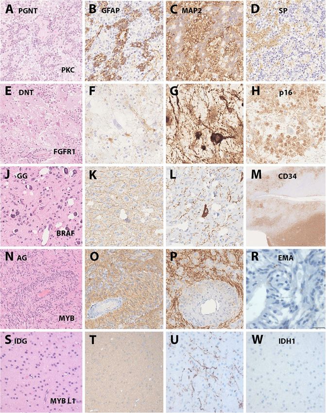
driven approach to classify LEAT in the near future. classify into the group of AG (Fig. 1).Slegers and Blumcke Acta Neuropathologica Communications (2020) 8:27 Page 6 of 11 Fig. 1 Histopathologically and genetically defined LEAT. Legend to Figure: Selected LEAT entities in which a common gene driving mutation has been discovered. a-d: a papillary glio-neuronal tumor (PGNT) with the characteristic presentation of papillary growth pattern (A – HE), glial (B – GFAP) and neuronal components (C – MAP2 and D – Synaptophysin). This tumor was included in the study by Hou et al. describing its distinct DNA methylation profile and SLC44A1-PRKCA fusion [31]. e-h: a dysembryoplastic neuroepithelial tumor (DNT) with the characteristic histological presentation of a specific glio-neuronal element (E – HE), lack of GFAP immunoreactivity in the clear-cell component (F - GFAP), floating neurons (G – MAP2) and a newly discovered p16 immunoreactivity shown in H, helpful to distinguish the DNT from other LEAT entities (unpublished observation, courtesy of Dr. Roland Coras, Erlangen, Germany). This tumor would typically present as FGFR1 altered CD34 negative DNT (not yet genetically confirmed in this tumor sample). j-m: a ganglioglioma (GG) with a characteristic glial-neuronal phenotype and small calcifications (J – HE), a predominant astroglial component (K-GFAP), dysplastic neurons (L-MAP2) and CD34 immunoreactivity in the tumor mass lesion shown in lower right corner as well as in diffusely infiltrated peritumoral grey and white matter (M-CD34). This tumor was included in the study of Blumcke et al. describing the distinct DNA methylation patterns of BRAF V600E mutated CD34 positive GG vs. CD34 negative DNT [3]. n-r: an angiocentric glioma (AG) with characteristic growth pattern around blood vessels (N-HE), a predominant astroglial phenotype (O-GFAP), enriched neuronal matrix (P-MAP2) and EMA-dots similar to ependymoma (R-EMA). This tumor showed a MYB fusion as previously described by Qaddomi et al. [58]. s-w: an isomorphic and diffusely infiltrating glioma (IDG) of low cellularity (S- HE), a predominant astroglial phenotype (T-GFAP), only few contained and pre-existing neurons (U-MAP2) and lack of IDH1R132H mutations (W-IDH mutation specific antibody). This tumor showed a MYBL1 fusion and was previously described by Wefers et al. [75] Growth patterns largely vary in most LEAT entities tumor mass, and nodular growth. In contrast to many including small cysts, white matter rarefaction, diffuse textbook edits and MRI interpretations, LEAT can show infiltration patterns or cell clusters remote from the diffuse infiltration. However, proliferation indices remain
Slegers and Blumcke Acta Neuropathologica Communications   (2020) 8:27                                          Page 7 of 11

usually very low in a given surgical sample. It is hypothe-      group of gangliocytoma and not yet assigned a grading
sized, therefore, although not yet scientifically con-           [43]. In a series of seven MVNTs no BRAF V600E muta-
firmed, that the early neurodevelopmental nature of              tions were found but one case showed a FGFR2 fusion
LEAT will allow tumor satellite clusters to dissolve into        [17]. In another series of eight MVNTs genetic alter-
cortical areas remote from the primary mass lesion. We           ations were found in BRAF other than V600E, MAP2K1
also believe that this infiltration pattern has the ability to   and FGFR2 (2/8, 5/8 and 1/8, respectively) [53]. As men-
promote seizure onset remote from the MRI visible                tioned above for PLNTY, the diverse molecular land-
tumor lesion, but this also needs scientific confirmation.       scape of findings reported in the literature make it
   All of the above-mentioned principles may expose              difficult to align this entity into a genetically driven
themselves in even yet unprecedented and unpublished             classification scheme and more studies are needed to
phenotypes, contributing to the growing list of published        clarify the etiology of this hitherto histomorphologi-
LEAT entities. We believe and hypothesize herein, that           cally defined entity. Lack of cell proliferation and lack
the early “developmental” origin of the tumor, most              of expansive or infiltrative growth reinforced also the
likely in prenatal periods, as well as continuous electric       debate whether MVNT should align with malforma-
bombardment of postnatally developing and maturing               tions of cortical development or cortical dysplasia ra-
neocortex contribute foremost to the broad histomor-             ther than with a neoplasm [68].
phological spectrum of LEAT rather than representing
distinct and clinically meaningful entities, i.e. with a         The issue of brain-tumor related epilepsy and associated
higher risk for seizure relapse or malignant tumor pro-          focal cortical dysplasia
gression. Our better understanding of genetic etiologies         Whereas our knowledge of molecular pathways driving
and epigenetic modifications thereof will eventually             neoplastic cell growth and malignant progression has
prove or disprove this hypothesis in times to come.              substantially matured, the issue of ictogenesis, i.e. why
                                                                 and how a seizure occurs in a patient with a brain
New histomorphological entities                                  tumor, and epileptogenesis, i.e. turning a normal into an
Polymorphous low-grade neuroepithelial tumor of the              epileptic brain prone to unprovoked recurrent seizures,
young (PLNTY) is an entity first described by Huse et al.        still awaits clarification. Brain tumors cause about 10 to
in 2017 [34]. The group presented ten patients with a            15% of all adult-onset and 0,2 to 6% of all child-onset
young age at diagnosis (between 4 and 32 years old) and          epilepsies [7, 12, 70]. Notwithstanding, many alterations
tumors with infiltrative growth patterns, a predominant          have been reported in human peritumoral brain tissue
oligodendoglioma-like glial cell component and intense           which have the potential to dramatically alter neuronal
CD34 immunoreactivity as most common features. Three             and glial homeostasis and the microenvironment in
out of eight tested tumors showed a BRAF V600E muta-             favor of a pro-epileptogenic state [19, 51, 70]. These
tion, one showed a FGFR3 fusion and three had a FGFR2            studies also proposed candidate therapeutic regimen for
fusion. Such FGFR2 fusions have not yet been detected in         treatment of patients with brain-tumor related epilepsy
any other LEAT category and might qualify as a distinctive       [32]. Two main hypotheses have been proposed; the
feature in the near future. The mixed molecular-genetic          tumorocentric and the epileptocentric approach [52].
background, however, including BRAF and FGFR genes               The tumorocentric approach states that the epileptic ac-
described above, makes PLNTY yet difficult to stratify into      tivity derives from the tumor itself which was recently
a classification scheme led predominately by gene driving        confirmed by experimental work of Koh et al. in neurons
mutations rather than histomorphological features. How-          transfected with the BRAF V600E mutation in vivo [41].
ever, DNA methylation profiling showed similarities with         The epileptocentric approach provides evidence that the
the methylation class of ganglioglioma but also suggested        infiltrated peritumoral neocortex is key for tumor-re-
that PLNTY may form a separate group [34]. Further               lated epileptic activity, due to glioma-related gluta-
studies will help to clarify this issue.                         matergic and γ-aminobutyric acid changes leading to
  In 2013 Huse et al. described multinodular and vacuo-          epileptogenicity [52]. The neurotransmitter glutamate
lating neuronal tumors (MVNT) in ten patients [33],              is used by glioma cells as a “tumor growth factor”.
which was confirmed 2014 by Bodi et al. in 2 additional          Altered expression of glutamate transporters by the
patients [10]. MVNT is defined nowadays by the WHO               tumor cells, including the cystine-glutamate trans-
as a benign tumor associated with seizures. These tu-            porter (xCT) system, also increases concentrations of
mors have a typical radiological pattern described as            extracellular glutamate contributing to epileptic dis-
FLAIR and T2-WI hyperintense lesions, clustered in               charges, tumor proliferation and neurotoxicity [32].
multiple small nodules, affecting subcortial white matter        However, such experimental studies have been per-
surrounded by normal-appearing parenchyma [13, 28].              formed so far only on histopathologically diagnosed
The WHO has subsumed this variant, therefore, into the           low-grade glioma and not LEAT.
Slegers and Blumcke Acta Neuropathologica Communications (2020) 8:27 Page 8 of 11
These considerations lead to the other important issue retrospective analysis of 55 pediatric ganglioglioma
which is how to achieve complete postsurgical seizure cases in a single center setting 53 had a ganglioglioma
control. Many studies have confirmed that active and and 2 an anaplastic ganglioglioma at time of diagnosis.
medically uncontrolled epilepsy significantly increases the After a mean follow-up time of 9,5 years 25 showed
risk of sudden death [23]. However, LEAT were amongst tumor progression and 6 transformation to a higher
the best candidates for complete postsurgical seizure con- grade after which a median survival of 9.1 months was
trol [7]. Planning for epilepsy surgery needs to take into reported [78]. Zanello et al. [79] analyzed 18 anaplastic
consideration, therefore, not only any MRI visible lesion ganglioglioma in a retrospective series forming 8% of
but to also resect the ictal onset zone. Scalp EEG may be their ganglioglioma cohort of 222 patients. They also
misleading when the ictal onset is buried deep in the cor- looked at molecular alterations and found BRAF V600E
tex, i.e. in temporo-mesial structures, and intracerebral in 39%, hTERT promotor mutations in 61%, p53 accu-
procedures were increasingly used to delineate the epi- mulation in 39%, ATRX loss in 17% and p.K27M
leptogenic as well as ictal onset zones. Correlations with H3F3A mutations in 17% of the cohort. A median
histopathology have not been performed, however, in a progression-free survival of 10 months and a median
systematic way to allow any conclusion about the struc- overall survival of 27 months was reported within this
tural and/or molecular correlate. One result of this di- cohort [79]. We suggest, however, re-examine these
lemma is the ongoing discussion about Focal Cortical tumor samples with a more objective measure, such as
Dysplasia associated with LEAT, hitherto classified as DNA methylation, as they may have been histopatho-
FCD IIIB by the ILAE classification scheme from 2011 [8]. logically assigned falsely into the group of GG (see the
Histopathology patterns of such FCD have never been sci- many pitfalls listed above) and belong rather into the
entifically defined. As a consequence, scientific reporting group of malignant glioma.
of the prevalence of dysplastic neocortex around LEAT,
including ganglioglioma and DNT, varies to a great extent Malignant dysembryoplastic neuroepithelial tumors
by 25 to 75% of the cases [1, 4, 26, 37, 48, 57]. The authors DNTs are considered as truly benign with a near to zero
expect that ongoing molecular-genetic studies will help to chance of malignant progression. In our own experience
clarify if these cases represent true FCD or pro- with more than 280 DNT, all of which can be classified as
epileptogenic molecular interactions of the tumor with classic variants with nodular growth, a specific glio-
surrounding peritumoral brain tissue. neuronal element and no tumor-cell related CD34-
immunoreactivity (Table 2), we have not observed a single
The issue of malignant progression in LEAT patient with tumor relapse due to malignant progression.
The proposal for surgical treatment in a young patient Heiland and coworkers [29] presented a case, however,
presenting with an MRI stable or only slowly growing which was characterized as DNT and which relapsed 5
LEAT, that is initially well controlled by antiepileptic years after tumor resection as a glioblastoma (GBM).
drugs, may raise considerable concern in patient man- DNA methylation analysis of the tissue samples showed a
agement. We strongly advocate, however, to counsel for methylation pattern distinct from typical GBM. Malignant
complete neurosurgical resection and a histopathological progression of DNTs were reported earlier by Ray and co-
diagnosis to confirm the benign nature of a neoplasia. workers in 2009 [59] or Thom and coworkers in 2011
As a matter of fact, malignant progression has been re- [69]. Notwithstanding, these reports were published before
ported in LEAT [46], although at very low frequency the era of DNA methylation classifier [14], which leaves us
and we do not have approved biomarker or molecular with the notion that malignant transformation can occur
signatures predictive for such malignant progression, in every tumor but need particular attention, re-review if
yet. Reported prevalence of malignant progression differ necessary, and current state-of-the-art molecular-genetic
among the LEAT entities. The most common tumor re-assessment when occurring in a DNT.
within this group, the ganglioglioma, has an estimated
chance of 3% for malignant progression whilst the sec- Pleomorphic xanthoastroctyoma (PXA)
ond most common tumor entity, the DNT (Table 2) has PXA are semi-malignant brain tumors sharing molecular
near to 0 % chance for dedifferentiation [36, 45, 49, 77]. and morphological commonalities with LEAT. We have
This highlights the importance of a reliable differential not addressed PXA as bona fide LEAT entity, however,
diagnosis as it will directly influence patient manage- due to their semi-benign nature with WHO tumor grad-
ment and also therapeutic regimens. ing of atypic II° and anaplastic III° subtypes. In addition,
disease onset had a mean age of 18,6 years in 41 PXA
Anaplastic ganglioglioma collected at the German Neuropathology Reference
Reported cases of LEAT with confirmed malignant pro- Center for Epilepsy Surgery (see Table 2). Histomorpho-
gression were mostly addressing ganglioglioma. In a logical similarities between PXA and LEAT include,Slegers and Blumcke Acta Neuropathologica Communications (2020) 8:27 Page 9 of 11
however, the broad spectrum of different tumor cell Authors’ contributions
components and growth patterns as described above. Rutger Slegers and Ingmar Blumcke both wrote equally on the manuscript.
The author(s) read and approved the final manuscript.
Reifenberger and coworkers reported an intense CD34
immunoreactivity in 73% of their PXA, which was more Funding
common in grade II (84%) than in grade III tumors IB was supported by grant BL 421/4–1 from the German Research council
(44%) [60]. Another similarity to GG is the BRAF V600E and the European Reference Network "EpiCare" under grant agreement #
870280 from the European Union.
hotspot mutation identified in 71.4% of 167 published
PXA [47], more often in grade II than in grade III tumors
Availability of data and materials
(75% and 47,4%, respectively [35, 63]. A homozygous dele- The datasets generated during and/or analyzed during the current study will
tion of CDKN2A/B, corresponding to loss of 9q21.3, is a be made available from the corresponding author on reasonable request.
rather distinctive molecular feature of PXA [74]. In a
study of 24 PXA and 14 anaplastic PXA, CDKN2A/B dele- Ethics approval and consent to participate
Not applicable.
tions were identified in 83 and 93%, respectively [72].
Knowledge of and access to the molecular-genetic land- Consent for publication
scape of these brain tumors will be most helpful, therefore, Not applicable.
to classify tumor entities into similar molecular classes
despite their variable histomorphological phenotypes. It Competing interests
The authors declare that they have no competing interests.
awaits further studies, however, to precisely define also
clinically meaningful entities. Author details
1
Faculty of Health, Medicine and Life Sciences, Maastricht University,
Universiteitssingel 40, NL – 6229ER Maastricht, The Netherlands. 2Department
Conclusion of Neuropathology, University Hospitals Erlangen, Schwabachanalge 6, 91054
The histopathological spectrum of LEAT is heterogeneous, Erlangen, Germany.
both morphologically and genetically! Even with currently
Received: 21 January 2020 Accepted: 29 February 2020
available molecular genetic markers such as BRAF V600E
and FGFR1 and immunohistochemical surrogate markers,
such as CD34 and p16, there is still a poor inter-rater References
agreement in the histopathological diagnosis. Any effort 1. Adachi Y, Yagishita A (2008) Gangliogliomas: characteristic imaging findings
and role in the temporal lobe epilepsy. Neuroradiology 50:829–834. https://
should be taken, therefore, to improve and standardize our doi.org/10.1007/s00234-008-0410-x
criteria and terminology, and to extent the use of molecular 2. Blumcke I, Aronica E, Becker A, Capper D, Coras R, Honavar M, Jacques TS,
genetic diagnostic tools over a histomorphology-based clas- Kobow K, Miyata H, Muhlebner A et al (2016) Low-grade epilepsy-associated
neuroepithelial tumours - the 2016 WHO classification. Nat Rev Neurol 12:
sification to specify clinically meaningful tumor entities 732–740. https://doi.org/10.1038/nrneurol.2016.173
within the LEAT spectrum. There is also a small risk for 3. Blumcke I, Coras R, Wefers AK, Capper D, Aronica E, Becker A, Honavar M,
malignant transformation of LEAT, but currently not pre- Stone TJ, Jacques TS, Miyata H et al (2019) Review: challenges in the
histopathological classification of ganglioglioma and DNT: microscopic
dictable with any available biomarker. A large enough and agreement studies and a preliminary genotype-phenotype analysis.
unselected, consecutive cohort of LEAT is a mandatory Neuropathol Appl Neurobiol 45:95–107. https://doi.org/10.1111/nan.12522
pre-requisite for our further progress in the field and will 4. Blumcke I, Cross JH, Spreafico R (2013) The international consensus classification for
hippocampal sclerosis: an important step towards accurate prognosis. Lancet
require international collaboration efforts. Neurol 12:844–846. https://doi.org/10.1016/s1474-4422(13)70175-3
5. Blumcke I, Giencke K, Wardelmann E, Beyenburg S, Kral T, Sarioglu N,
Abbreviations Pietsch T, Wolf HK, Schramm J, Elger CE et al (1999) The CD34 epitope is
AG: Angiocentric glioma WHO I°; ATRX: Alpha-thalassemia/mental-retardation expressed in neoplastic and malformative lesions associated with chronic,
syndrome-X-linked; BRAF: B-Raf Proto-oncogene, serine/threonine kinase; focal epilepsies. Acta Neuropathol 97:481–490. https://doi.org/10.1007/
CNS: Central nervous system; CNV: Copy number variants; s004010051017
DNT: Dysembryoplastic neuroepithelial tumor WHO I°; 6. Blumcke I, Luyken C, Urbach H, Schramm J, Wiestler OD (2004) An
EEG: Electroencephalography; FCD: Focal cortical dysplasia; FGFR1: Fibroblast isomorphic subtype of long-term epilepsy-associated astrocytomas
growth factor receptor 1; GBM: Glioblastoma multiforme WHO IV°; associated with benign prognosis. Acta Neuropathol 107:381–388.
GG: Ganglioglioma WHO I°; HS: Hippocampal sclerosis; IDG: Isomorphic https://doi.org/10.1007/s00401-004-0833-3
diffuse glioma; K27M H3F3A: Lysine 27 replacement with methionine in 7. Blumcke I, Spreafico R, Haaker G, Coras R, Kobow K, Bien CG, Pfäfflin M,
histone H3 variant H3.3; LEAT: Low-grade, developmental and epilepsy- Elger C, Widman G, Schramm J et al (2017) Histopathological findings in
associated tumor; LGNET: Low-grade neuroepithelial tumor (NOS); brain tissue obtained during epilepsy surgery. N Engl J Med 377:1648–1656.
MGMT: Methylguanine methyltransferase; MRI: Magnetic resonance imaging; https://doi.org/10.1056/NEJMoa1703784
MVNT: Multinodular and vacuolated neuronal tumor of the cerebrum; MYB/ 8. Blumcke I, Thom M, Aronica E, Armstrong DD, Vinters HV, Palmini A,
MYBL1: MYB proto-oncogene (like 1), transcription factor; PA: Pilocytic Jacques TS, Avanzini G, Barkovich AJ, Battaglia G et al (2011) The
astrocytoma WHO I°; PGNT: Papillary glio-neuronal tumor WHO I°; clinicopathologic spectrum of focal cortical dysplasias: a consensus
PRKCA: Protein kinase C alpha; PXA: Pleomorphic xanthoastrocytoma WHO classification proposed by an ad hoc task force of the ILAE diagnostic
II°; TERT: Telomerase reverse transciptase; xCT: Cystine-glutamate transporter methods commission. Epilepsia 52:158–174. https://doi.org/10.1111/j.1528-
1167.2010.02777.x
Acknowledgements 9. Blumcke I, Wiestler OD (2002) Gangliogliomas: an intriguing tumor entity
We want to thank Roland Coras for his help providing the images for the associated with focal epilepsies. J Neuropathol Exp Neurol 61:575–584.
figure used. https://doi.org/10.1093/jnen/61.7.575Slegers and Blumcke Acta Neuropathologica Communications (2020) 8:27 Page 10 of 11
10. Bodi I, Curran O, Selway R, Elwes R, Burrone J, Laxton R, Al-Sarraj S, Honavar transformation of a Dysembryoplastic Neuroepithelial tumor (DNET)
M (2014) Two cases of multinodular and vacuolating neuronal tumour. Acta characterized by genome-wide methylation analysis. J Neuropathol Exp
Neuropathol Commun 2:7. https://doi.org/10.1186/2051-5960-2-7 Neurol 75:358–365. https://doi.org/10.1093/jnen/nlw007
11. Bridge JA, Liu XQ, Sumegi J, Nelson M, Reyes C, Bruch LA, Rosenblum M, 30. Holthausen H, Blumcke I (2016) Epilepsy-associated tumours: what epileptologists
Puccioni MJ, Bowdino BS, McComb RD (2013) Identification of a novel, should know about neuropathology, terminology, and classification systems.
recurrent SLC44A1-PRKCA fusion in papillary glioneuronal tumor. Brain Epileptic Disord 18:240–251. https://doi.org/10.1684/epd.2016.0851
Pathol 23:121–128. https://doi.org/10.1111/j.1750-3639.2012.00612.x 31. Hou Y, Pinheiro J, Sahm F, Reuss DE, Schrimpf D, Stichel D, Casalini B, Koelsche
12. Brogna C, Gil Robles S, Duffau H (2008) Brain tumors and epilepsy. Expert C, Sievers P, Wefers AK et al (2019) Papillary glioneuronal tumor (PGNT) exhibits
Rev Neurother 8:941–955. https://doi.org/10.1586/14737175.8.6.941 a characteristic methylation profile and fusions involving PRKCA. Acta
13. Buffa GB, Chaves H, Serra MM, Stefanoff NI, Gagliardo AS, Yanez P (2019) Neuropathol 137:837–846. https://doi.org/10.1007/s00401-019-01969-2
Multinodular and Vacuolating neuronal tumor of the cerebrum (MVNT): a 32. Huberfeld G, Vecht CJ (2016) Seizures and gliomas--towards a single
case series and review of the literature. J Neuroradiol: Doi. https://doi.org/ therapeutic approach. Nat Rev Neurol 12:204–216. https://doi.org/10.1038/
10.1016/j.neurad.2019.05.010 nrneurol.2016.26
14. Capper D, Jones DTW, Sill M, Hovestadt V, Schrimpf D, Sturm D, Koelsche C, 33. Huse JT, Edgar M, Halliday J, Mikolaenko I, Lavi E, Rosenblum MK (2013)
Sahm F, Chavez L, Reuss DE et al (2018) DNA methylation-based Multinodular and vacuolating neuronal tumors of the cerebrum: 10 cases of
classification of central nervous system tumours. Nature 555:469–474. a distinctive seizure-associated lesion. Brain Pathol 23:515–524. https://doi.
https://doi.org/10.1038/nature26000 org/10.1111/bpa.12035
15. Capper D, Preusser M, Habel A, Sahm F, Ackermann U, Schindler G, Pusch S, 34. Huse JT, Snuderl M, Jones DT, Brathwaite CD, Altman N, Lavi E, Saffery R,
Mechtersheimer G, Zentgraf H, von Deimling A (2011) Assessment of BRAF Sexton-Oates A, Blumcke I, Capper D et al (2017) Polymorphous low-grade
V600E mutation status by immunohistochemistry with a mutation-specific neuroepithelial tumor of the young (PLNTY): an epileptogenic neoplasm
monoclonal antibody. Acta Neuropathol 122:11–19. https://doi.org/10.1007/ with oligodendroglioma-like components, aberrant CD34 expression, and
s00401-011-0841-z genetic alterations involving the MAP kinase pathway. Acta Neuropathol
16. Chen X, Pan C, Zhang P, Xu C, Sun Y, Yu H, Wu Y, Geng Y, Zuo P, Wu Z 133:417–429. https://doi.org/10.1007/s00401-016-1639-9
et al (2017) BRAF V600E mutation is a significant prognosticator of the 35. Ida CM, Rodriguez FJ, Burger PC, Caron AA, Jenkins SM, Spears GM,
tumour regrowth rate in brainstem gangliogliomas. J Clin Neurosci 46:50– Aranguren DL, Lachance DH, Giannini C (2015) Pleomorphic
57. https://doi.org/10.1016/j.jocn.2017.09.014 Xanthoastrocytoma: natural history and long-term follow-up. Brain Pathol
17. Choi E, Kim SI, Won JK, Chung CK, Kim SK, Choi SH, Choi S, Han B, Ahn B, 25:575–586. https://doi.org/10.1111/bpa.12217
Im SW et al (2019) Clinicopathological and molecular analysis of 36. Isler C, Erturk Cetin O, Ugurlar D, Ozkara C, Comunoglu N, Kizilkilic O, Oz B,
multinodular and vacuolating neuronal tumors of the cerebrum. Hum Kayadibi Y, Tanriverdi T, Uzan M (2018) Dysembryoplastic neuroepithelial
Pathol 86:203–212. https://doi.org/10.1016/j.humpath.2018.11.028 tumours: clinical, radiological, pathological features and outcome. Br J
18. Courville CB (1930) Ganglioglioma: tumor of the central nervous system; Neurosurg 32:436–441. https://doi.org/10.1080/02688697.2018.1476671
review of the literature and report of two cases. Arch Neurol Psychiatr 24: 37. Jehi LE, Luders HO, Naugle R, Ruggieri P, Morris H, Foldvary N, Wyllie E,
439–491. https://doi.org/10.1001/archneurpsyc.1930.02220150002001 Kotagal P, Bingaman B, Dinner D et al (2008) Temporal lobe neoplasm and
19. Cowie CJA, Cunningham MO (2014) Peritumoral epilepsy: relating form and seizures: how deep does the story go? Epileptic Disord 10:56–67. https://doi.
function for surgical success. Epilepsy Behav 38:53–61. https://doi.org/10. org/10.1684/epd.2008.0173
1016/j.yebeh.2014.05.009 38. Jones DT, Hutter B, Jager N, Korshunov A, Kool M, Warnatz HJ, Zichner T,
20. Dahiya S, Haydon DH, Alvarado D, Gurnett CA, Gutmann DH, Leonard JR Lambert SR, Ryzhova M, Quang DA et al (2013) Recurrent somatic
(2013) BRAF(V600E) mutation is a negative prognosticator in pediatric alterations of FGFR1 and NTRK2 in pilocytic astrocytoma. Nat Genet 45:927–
ganglioglioma. Acta Neuropathol 125:901–910. https://doi.org/10.1007/ 932. https://doi.org/10.1038/ng.2682
s00401-013-1120-y 39. Kaley T, Touat M, Subbiah V, Hollebecque A, Rodon J, Lockhart AC, Keedy V,
21. Daumas-Duport C, Scheithauer BW, Chodkiewicz JP, Laws ER Jr, Vedrenne C Bielle F, Hofheinz RD, Joly F et al (2018) BRAF inhibition in BRAF(V600)-
(1988) Dysembryoplastic neuroepithelial tumor: a surgically curable tumor mutant Gliomas: results from the VE-BASKET study. J Clin Oncol:
of young patients with intractable partial seizures. Report of thirty-nine Jco2018789990. https://doi.org/10.1200/jco.2018.78.9990
cases. Neurosurg 23:545–556 40. Koelsche C, Wohrer A, Jeibmann A, Schittenhelm J, Schindler G, Preusser M,
22. Davies H, Bignell GR, Cox C, Stephens P, Edkins S, Clegg S, Teague J, Woffendin Lasitschka F, von Deimling A, Capper D (2013) Mutant BRAF V600E protein
H, Garnett MJ, Bottomley W et al (2002) Mutations of the BRAF gene in human in ganglioglioma is predominantly expressed by neuronal tumor cells. Acta
cancer. Nature 417:949–954. https://doi.org/10.1038/nature00766 Neuropathol 125:891–900. https://doi.org/10.1007/s00401-013-1100-2
23. Devinsky O, Hesdorffer DC, Thurman DJ, Lhatoo S, Richerson G (2016) Sudden 41. Koh HY, Kim SH, Jang J, Kim H, Han S, Lim JS, Son G, Choi J, Park BO, Heo
unexpected death in epilepsy: epidemiology, mechanisms, and prevention. WD et al (2018) BRAF somatic mutation contributes to intrinsic
Lancet Neurol 15:1075–1088. https://doi.org/10.1016/s1474-4422(16)30158-2 epileptogenicity in pediatric brain tumors. Nat Med 24:1662–1668. https://
24. Dougherty MJ, Santi M, Brose MS, Ma C, Resnick AC, Sievert AJ, Storm PB, doi.org/10.1038/s41591-018-0172-x
Biegel JA (2010) Activating mutations in BRAF characterize a spectrum of 42. Komori T, Scheithauer BW, Anthony DC, Rosenblum MK, McLendon RE,
pediatric low-grade gliomas. Neuro-Oncology 12:621–630. https://doi.org/ Scott RM, Okazaki H, Kobayashi M (1998) Papillary glioneuronal tumor: a
10.1093/neuonc/noq007 new variant of mixed neuronal-glial neoplasm. Am J Surg Pathol 22:1171–
25. Fisher RS, Acevedo C, Arzimanoglou A, Bogacz A, Cross JH, Elger CE, Engel J Jr, 1183. https://doi.org/10.1097/00000478-199810000-00002
Forsgren L, French JA, Glynn M et al (2014) ILAE official report: a practical clinical 43. Louis DN, Perry A, Reifenberger G, von Deimling A, Figarella-Branger D,
definition of epilepsy. Epilepsia 55:475–482. https://doi.org/10.1111/epi.12550 Cavenee WK, Ohgaki H, Wiestler OD, Kleihues P, Ellison DW (2016) The 2016
26. Frater JL, Prayson RA, Morris IH, Bingaman WE (2000) Surgical pathologic World Health Organization classification of tumors of the central nervous
findings of extratemporal-based intractable epilepsy: a study of 133 system: a summary. Acta Neuropathol 131:803–820. https://doi.org/10.1007/
consecutive resections. Arch Pathol Lab Med 124:545–549. https://doi.org/ s00401-016-1545-1
10.1043/0003-9985(2000)1242.0.Co;2 44. Luyken C, Blumcke I, Fimmers R, Urbach H, Elger CE, Wiestler OD, Schramm
27. Garnier L, Ducray F, Verlut C, Mihai MI, Cattin F, Petit A, Curtit E (2019) J (2003) The spectrum of long-term epilepsy-associated tumors: long-term
Prolonged response induced by single agent Vemurafenib in a BRAF V600E seizure and tumor outcome and neurosurgical aspects. Epilepsia 44:822–
spinal Ganglioglioma: a case report and review of the literature. Front Oncol 830. https://doi.org/10.1046/j.1528-1157.2003.56102.x
9:177. https://doi.org/10.3389/fonc.2019.00177 45. Luyken C, Blumcke I, Fimmers R, Urbach H, Wiestler OD, Schramm J (2004)
28. Gonzalez-Quarante LH, Ruiz-Juretschke F, Sola Vendrell E, Gil de Sagredo Del Supratentorial gangliogliomas: histopathologic grading and tumor
Corral OL, Agarwal V, Garcia-Leal R (2018) Multinodular and vacuolating neuronal recurrence in 184 patients with a median follow-up of 8 years. Cancer 101:
tumor of the cerebrum. A rare entity New case and review of the literature. 146–155. https://doi.org/10.1002/cncr.20332
Neurocirugia (Astur) 29:44–55. https://doi.org/10.1016/j.neucir.2017.08.003 46. Majores M, von Lehe M, Fassunke J, Schramm J, Becker AJ, Simon M (2008)
29. Heiland DH, Staszewski O, Hirsch M, Masalha W, Franco P, Grauvogel J, Tumor recurrence and malignant progression of gangliogliomas. Cancer
Capper D, Schrimpf D, Urbach H, Weyerbrock A (2016) Malignant 113:3355–3363. https://doi.org/10.1002/cncr.23965Slegers and Blumcke Acta Neuropathologica Communications (2020) 8:27 Page 11 of 11
47. Mallick S, Benson R, Melgandi W, Giridhar P, Rath GK (2018) Grade II polymorphism-based genotype arrays results in a novel BRAF fusion gene.
pleomorphic Xanthoastrocytoma; a meta-analysis of data from previously Brain Pathol 19:449–458. https://doi.org/10.1111/j.1750-3639.2008.00225.x
reported 167 cases. J Clin Neurosci 54:57–62. https://doi.org/10.1016/j.jocn. 65. Stone TJ, Keeley A, Virasami A, Harkness W, Tisdall M, Izquierdo Delgado E,
2018.05.003 Gutteridge A, Brooks T, Kristiansen M, Chalker J et al (2018) Comprehensive
48. Morris HH, Estes ML, Prayson RA, et al. Frequency of different tumor types molecular characterisation of epilepsy-associated glioneuronal tumours.
encountered in the Cleveland Clinic epilepsy surgery program. Epilepsia. Acta Neuropathol 135:115–129. https://doi.org/10.1007/s00401-017-1773-z
1996;37(suppl.5):S96. 66. Stone TJ, Rowell R, Jayasekera BAP, Cunningham MO, Jacques TS (2018)
49. Nguyen HS, Doan N, Gelsomino M, Shabani S (2017) Dysembryoplastic Review: molecular characteristics of long-term epilepsy-associated tumours
Neuroectodermal tumor: An analysis from the surveillance, epidemiology, (LEATs) and mechanisms for tumour-related epilepsy (TRE). Neuropathol
and end results Program, 2004-2013. World Neurosurg 103:380–385. https:// Appl Neurobiol 44:56–69. https://doi.org/10.1111/nan.12459
doi.org/10.1016/j.wneu.2017.04.093 67. Thom M, Blumcke I, Aronica E (2012) Long-term epilepsy-associated tumors.
50. Pages M, Lacroix L, Tauziede-Espariat A, Castel D, Daudigeos-Dubus E, Ridola Brain Pathol 22:350–379. https://doi.org/10.1111/j.1750-3639.2012.00582.x
V, Gilles S, Fina F, Andreiuolo F, Polivka M et al (2015) Papillary glioneuronal 68. Thom M, Liu J, Bongaarts A, Reinten RJ, Paradiso B, Jager HR, Reeves C,
tumors: histological and molecular characteristics and diagnostic value of Somani A, An S, Marsdon D et al (2018) Multinodular and vacuolating
SLC44A1-PRKCA fusion. Acta Neuropathol Commun 3:85. https://doi.org/10. neuronal tumors in epilepsy: dysplasia or neoplasia? Brain Pathol 28:155–
1186/s40478-015-0264-5 171. https://doi.org/10.1111/bpa.12555
51. Pallud J, Audureau E, Blonski M, Sanai N, Bauchet L, Fontaine D, Mandonnet 69. Thom M, Toma A, An S, Martinian L, Hadjivassiliou G, Ratilal B, Dean A,
E, Dezamis E, Psimaras D, Guyotat J et al (2014) Epileptic seizures in diffuse McEvoy A, Sisodiya SM, Brandner S (2011) One hundred and one
low-grade gliomas in adults. Brain 137:449–462. https://doi.org/10.1093/ dysembryoplastic neuroepithelial tumors: an adult epilepsy series with
brain/awt345 immunohistochemical, molecular genetic, and clinical correlations and a
52. Pallud J, Capelle L, Huberfeld G (2013) Tumoral epileptogenicity: how does review of the literature. J Neuropathol Exp Neurol 70:859–878. https://doi.
it happen? Epilepsia 54(Suppl 9):30–34. https://doi.org/10.1111/epi.12440 org/10.1097/NEN.0b013e3182302475
53. Pekmezci M, Stevers M, Phillips JJ, Van Ziffle J, Bastian BC, Tsankova NM, 70. van Breemen MS, Wilms EB, Vecht CJ (2007) Epilepsy in patients with brain
Kleinschmidt-DeMasters BK, Rosenblum MK, Tihan T, Perry A et al (2018) tumours: epidemiology, mechanisms, and management. Lancet Neurol 6:
Multinodular and vacuolating neuronal tumor of the cerebrum is a clonal 421–430. https://doi.org/10.1016/s1474-4422(07)70103-5
neoplasm defined by genetic alterations that activate the MAP kinase 71. van Gijn J (2011) From the archives. Brain 134:2795–2797. https://doi.org/10.
signaling pathway. Acta Neuropathol 135:485–488. https://doi.org/10.1007/ 1093/brain/awr241
s00401-018-1820-4 72. Vaubel RA, Caron AA, Yamada S, Decker PA, Eckel Passow JE, Rodriguez FJ,
54. Pekmezci M, Villanueva-Meyer JE, Goode B, Van Ziffle J, Onodera C, Grenert Nageswara Rao AA, Lachance D, Parney I, Jenkins R et al (2018) Recurrent
JP, Bastian BC, Chamyan G, Maher OM, Khatib Z et al (2018) The genetic copy number alterations in low-grade and anaplastic pleomorphic
landscape of ganglioglioma. Acta Neuropathol Commun 6:47. https://doi. xanthoastrocytoma with and without BRAF V600E mutation. Brain Pathol 28:
org/10.1186/s40478-018-0551-z 172–182. https://doi.org/10.1111/bpa.12495
55. Perkins OC (1926) Ganglioglioma. Arch Pathol Lab Med 2:11 73. Wang M, Tihan T, Rojiani AM, Bodhireddy SR, Prayson RA, Iacuone JJ, Alles
56. Prabowo AS, Iyer AM, Veersema TJ, Anink JJ, Schouten-van Meeteren AYN, AJ, Donahue DJ, Hessler RB, Kim JH et al (2005) Monomorphous
Spliet WGM, van Rijen PC, Ferrier CH, Capper D, Thom M et al (2014) BRAF angiocentric glioma: a distinctive epileptogenic neoplasm with features of
V600E mutation is associated with mTOR signaling activation in Glioneuronal infiltrating astrocytoma and ependymoma. J Neuropathol Exp Neurol 64:
tumors. Brain Pathol 24:52–66. https://doi.org/10.1111/bpa.12081 875–881. https://doi.org/10.1097/01.jnen.0000182981.02355.10
57. Prayson RA, Estes ML, Morris HH (1993) Coexistence of neoplasia and 74. Weber RG, Hoischen A, Ehrler M, Zipper P, Kaulich K, Blaschke B, Becker AJ,
cortical dysplasia in patients presenting with seizures. Epilepsia 34:609–615. Weber-Mangal S, Jauch A, Radlwimmer B et al (2007) Frequent loss of
https://doi.org/10.1111/j.1528-1157.1993.tb00436.x chromosome 9, homozygous CDKN2A/p14(ARF)/CDKN2B deletion and low
58. Qaddoumi I, Orisme W, Wen J, Santiago T, Gupta K, Dalton JD, Tang B, TSC1 mRNA expression in pleomorphic xanthoastrocytomas. Oncogene 26:
Haupfear K, Punchihewa C, Easton J et al (2016) Genetic alterations in 1088–1097. https://doi.org/10.1038/sj.onc.1209851
uncommon low-grade neuroepithelial tumors: BRAF, FGFR1, and MYB 75. Wefers AK, Stichel D, Schrimpf D, Coras R, Pages M, Tauziede-Espariat A,
mutations occur at high frequency and align with morphology. Acta Varlet P, Schwarz D, Soylemezoglu F, Pohl U et al (2020) Isomorphic diffuse
Neuropathol 131:833–845. https://doi.org/10.1007/s00401-016-1539-z glioma is a morphologically and molecularly distinct tumour entity with
59. Ray WZ, Blackburn SL, Casavilca-Zambrano S, Barrionuevo C, Orrego JE, recurrent gene fusions of MYBL1 or MYB and a benign disease course. Acta
Heinicke H, Dowling JL, Perry A (2009) Clinicopathologic features of Neuropathol 139:193–209. https://doi.org/10.1007/s00401-019-02078-w
recurrent dysembryoplastic neuroepithelial tumor and rare malignant 76. Yau WH, Ameratunga M (2020) Combination of BRAF and MEK inhibition in
transformation: a report of 5 cases and review of the literature. J Neuro- BRAF V600E mutant low-grade ganglioglioma. J Clin Pharm Ther: Doi.
Oncol 94:283–292. https://doi.org/10.1007/s11060-009-9849-9 https://doi.org/10.1111/jcpt.13112
77. Yust-Katz S, Anderson MD, Liu D, Wu J, Yuan Y, Olar A, Fuller GN, Brown PD,
60. Reifenberger G, Kaulich K, Wiestler OD, Blumcke I (2003) Expression of the
de-Groot JF (2014) Clinical and prognostic features of adult patients with
CD34 antigen in pleomorphic xanthoastrocytomas. Acta Neuropathol 105:
gangliogliomas. Neuro-oncology 16: 409–413 Doi https://doi.org/10.1093/
358–364. https://doi.org/10.1007/s00401-002-0652-3
neuonc/not169
61. Rivera B, Gayden T, Carrot-Zhang J, Nadaf J, Boshari T, Faury D, Zeinieh M,
78. Zaky W, Patil SS, Park M, Liu D, Wang WL, Wani KM, Calle S, Ketonen L,
Blanc R, Burk DL, Fahiminiya S et al (2016) Germline and somatic FGFR1
Khatua S (2018) Ganglioglioma in children and young adults: single
abnormalities in dysembryoplastic neuroepithelial tumors. Acta Neuropathol
institution experience and review of the literature. J Neuro-Oncol 139:739–
131:847–863. https://doi.org/10.1007/s00401-016-1549-x
747. https://doi.org/10.1007/s11060-018-2921-6
62. Schindler G, Capper D, Meyer J, Janzarik W, Omran H, Herold-Mende C,
79. Zanello M, Pages M, Tauziede-Espariat A, Saffroy R, Puget S, Lacroix L,
Schmieder K, Wesseling P, Mawrin C, Hasselblatt M et al (2011) Analysis of
Dezamis E, Devaux B, Chretien F, Andreiuolo F et al (2016) Clinical, imaging,
BRAF V600E mutation in 1,320 nervous system tumors reveals high
Histopathological and molecular characterization of anaplastic
mutation frequencies in pleomorphic xanthoastrocytoma, ganglioglioma
Ganglioglioma. J Neuropathol Exp Neurol 75:971–980. https://doi.org/10.
and extra-cerebellar pilocytic astrocytoma. Acta Neuropathol 121:397–405.
1093/jnen/nlw074
https://doi.org/10.1007/s00401-011-0802-6
80. Zhang J, Wu G, Miller CP, Tatevossian RG, Dalton JD, Tang B, Orisme W,
63. Schmidt Y, Kleinschmidt-DeMasters BK, Aisner DL, Lillehei KO, Damek D
Punchihewa C, Parker M, Qaddoumi I et al (2013) Whole-genome
(2013) Anaplastic PXA in adults: case series with clinicopathologic and
sequencing identifies genetic alterations in pediatric low-grade gliomas. Nat
molecular features. J Neuro-Oncol 111:59–69. https://doi.org/10.1007/
Genet 45:602–612. https://doi.org/10.1038/ng.2611
s11060-012-0991-4
64. Sievert AJ, Jackson EM, Gai X, Hakonarson H, Judkins AR, Resnick AC, Sutton
LN, Storm PB, Shaikh TH, Biegel JA (2009) Duplication of 7q34 in pediatric Publisher’s Note
low-grade astrocytomas detected by high-density single-nucleotide Springer Nature remains neutral with regard to jurisdictional claims in
published maps and institutional affiliations.You can also read