Cholesterol as a modulator of cannabinoid receptor CB2 signaling - Nature
←
→
Page content transcription
If your browser does not render page correctly, please read the page content below
www.nature.com/scientificreports
OPEN Cholesterol as a modulator
of cannabinoid receptor CB2
signaling
Alexei Yeliseev1*, Malliga R. Iyer1*, Thomas T. Joseph2, Nathan J. Coffey1, Resat Cinar1,
Lioudmila Zoubak1, George Kunos1 & Klaus Gawrisch1
Signaling through integral membrane G protein-coupled receptors (GPCRs) is influenced by lipid
composition of cell membranes. By using novel high affinity ligands of human cannabinoid receptor
CB2, we demonstrate that cholesterol increases basal activation levels of the receptor and alters the
pharmacological categorization of these ligands. Our results revealed that (2-(6-chloro-2-((2,2,3,3-
tetramethylcyclopropane-1-carbonyl)imino)benzo[d]thiazol-3(2H)-yl)ethyl acetate ligand (MRI-2646)
acts as a partial agonist of CB2 in membranes devoid of cholesterol and as a neutral antagonist or
a partial inverse agonist in cholesterol-containing membranes. The differential effects of a specific
ligand on activation of CB2 in different types of membranes may have implications for screening of
drug candidates in a search of modulators of GPCR activity. MD simulation suggests that cholesterol
exerts an allosteric effect on the intracellular regions of the receptor that interact with the G-protein
complex thereby altering the recruitment of G protein.
Abbreviations
GPCR G protein-coupled receptor
ICL Intracellular loop (in GPCR)
TM Transmembrane domain (in GPCR)
HEK293 Human embryonic kidney
CHO Chinese hamster ovary
E. coli Escherichia coli
Sf9 cells A clonal isolate of Spodoptera frugiperda baculovirus infected insect cell
MD Molecular dynamics
hCB2 Human cannabinoid receptor C B2
hCB1 Human cannabinoid receptor C B1
MBP Maltose binding protein of E. coli
CP-55,940 Synthetic cannabinoid agonist 2-[(1R,2R,5R)-5-Hydroxy-2-(3-hydroxypropyl)cyclohexyl]-
5-(2-methyloctan-2-yl)phenol
[3H]-CP55,940 Radiolabeled synthetic ligand CP-55,940
SR-144,528 Synthetic cannabinoid inverse agonist 5-(4-Chloro-3-methylphenyl)-
1-[(4-methylphenyl)methyl]-N-[(1S,2S,4R)-1,3,3-trimethylbicyclo[2.2.1]
heptan-2-yl]-1H-pyrazole-3-carboxamide
[35S]-GTPγS Radiolabeled synthetic analog of guanosine triphosphate
MβCD Methyl-β-cyclodextrin
Gαi1 and Gβ1γ2 Subunits of heterotrimeric G protein
GEF assay Determines rates of nucleotide exchange on the G α subunit of G protein
POPC 1-Palmitoyl-2-oleoyl-glycero-3-phosphocholine
POPG 1-Palmitoyl-2-oleoyl-sn-glycero-3-phospho-(1′-rac-glycerol)
CHS Cholesteryl hemisuccinate
RMSD Root-mean-square deviation
RMSF Root mean square fluctuation
1
National Institute on Alcohol Abuse and Alcoholism, National Institutes of Health, Bethesda, MD 20852,
USA. 2Department of Anesthesiology and Critical Care, Perelman School of Medicine, University of Pennsylvania,
Philadelphia, PA 19104, USA. *email: yeliseeva@mail.nih.gov; malliga.iyer@nih.gov
Scientific Reports | (2021) 11:3706 | https://doi.org/10.1038/s41598-021-83245-6 1
Vol.:(0123456789)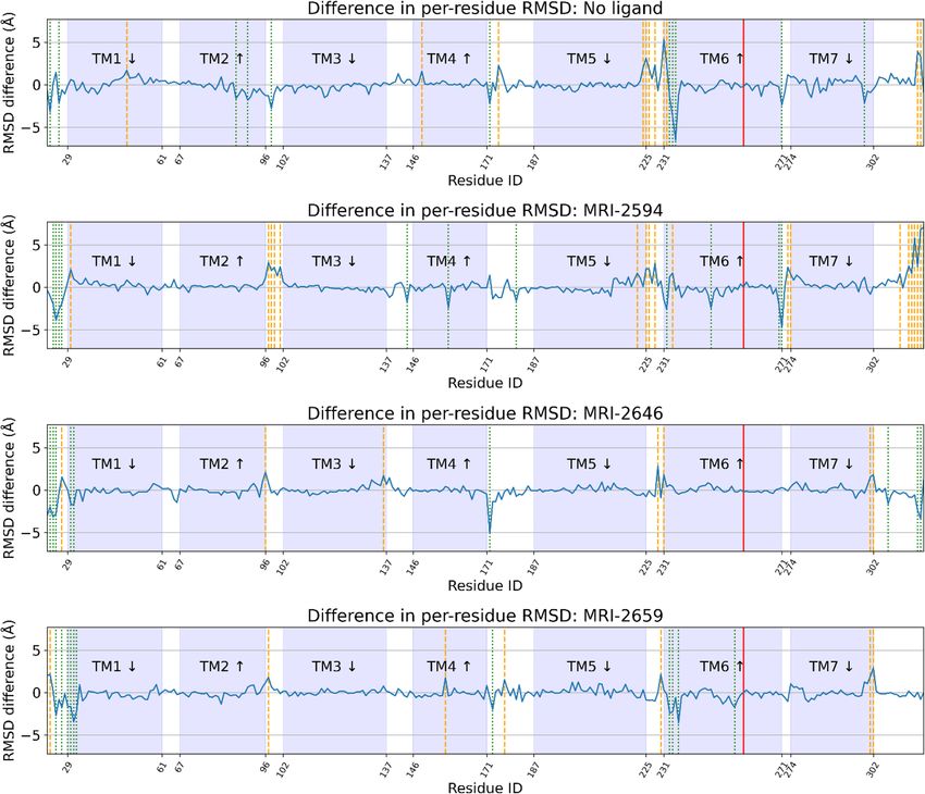
www.nature.com/scientificreports/
Lipid composition of membranes plays an important role in modulation of G protein-coupled receptors
(GPCR)1–4. In particular, cholesterol content varies significantly among different types of cell membranes, and
was shown to influence the ligand-induced signal transduction through GPCRs5–8. Early stage pharmacologi-
cal characterization of GPCR ligands typically involves screening of prospective drug candidates in cell- and
membrane-based assays9. Hence, it is important to understand how a physiological response of the target receptor
to small molecule ligands is modulated by the lipid composition of membranes.
Membranes of mammalian cells typically used for expression of recombinant GPCR for cell- and membrane-
based assays may contain significant amount of c holesterol10. Plasma membranes of human embryonic kidney
(HEK293) and Chinese hamster ovary (CHO) cells were reported to contain as much as 25–40 mol% of cho-
lesterol relative to phospholipid10–12. The lipid and cholesterol content of cultured mammalian cells fluctuates
depending on nutrient composition of growth media, temperature of cultivation, age of cell culture and other
growth parameters10. Other commonly used expression hosts such as Escherichia coli13,14, Spodoptera frugiperda
(Sf9), and Trichoplusia ni (Tn) synthesize only trace amounts of cholesterol or do not produce it at all11.
A large proportion of pharmacological drugs, currently on the market or under development, target mem-
brane proteins. Since pharmacological profiles of drug candidates could be influenced by the properties of cell
membranes harboring these receptors, variations in composition of the lipid bilayer may result in inconsistencies
of the receptor r esponse15,16. This is especially true for hydrophobic ligands targeting GPCR since interaction
between the drug and the lipid may influence the pharmacological characteristics of the compound such as its
binding affinity and selectivity17–19. While it was demonstrated that lipids and cholesterol can shift the response
of the receptor to ligand binding, it is not well known if the pharmacological categorization of ligands, i.e. ago-
nism, antagonism or inverse agonism, depends on composition of the lipid matrix. Here, we sought to elucidate
whether the functional response of the cannabinoid receptor C B2 to a series of novel specific ligands is affected
by the cholesterol content of membranes.
Cholesterol is critical for the formation of lateral domains (clusters)20, and it may induce negative curvature
ilayers21. The cholesterol-dependent increase in membrane stiffness increases the decay
elastic stress in lipid b
length of protein-induced perturbations in the lipid m atrix22. Furthermore, cholesterol may directly interact with
23–26
GPCR at sites identified in several r eceptors . Cholesterol was reported to negatively modulate the activity of
type 1 cannabinoid receptor ( CB1) in nerve c ells27. Furthermore, it was proposed that the C B1 receptor possesses
a specific cholesterol binding s ite28. However, the activity of the structurally close cannabinoid receptor C B2 was
reported to be not influenced by changes in membrane cholesterol c ontent29. The CB2 receptor is known for its
role as a regulator of inflammation and is commonly found in tissues containing variable levels of cholesterol30.
Therefore, it is important to understand if C B2 is sensitive to variation in cholesterol content.
Here, we examined the effects of cholesterol on C B2 by analyzing G protein activation by this receptor in
response to novel, rationally designed CB2 ligands (MRI-2646, MRI-2654, MRI-2687, MRI-2653, MRI-2659).
We will demonstrate that cholesterol increases the basal activation levels of C B2 receptor, thereby altering phar-
macological classification of these ligands from inverse agonists to partial agonists. We also use MD simulation
to probe structural effects of cholesterol and ligands.
Results
Based on the rational modification of the A-836339 thiazole scaffold, some of us previously reported that the syn-
thetic compounds MRI-2687 and MRI-2594 show contrasting activity on the CB2 receptor31. The two compounds
only differ in arm 1 featuring the extended central 6-methylbenzothiazole ring in MRI-2687 and 4,5-dimethylthi-
azole ring in MRI-2594, respectively. Unexpectedly, MRI-2687 behaved as an inverse agonist, whereas MRI-2594
acted as an agonist on CB2 (similar to A-836339)31,32. Detailed molecular docking analysis showed that these two
ligands adopt similar binding poses within C B2 wherein the different central rings reside in the same position
as arm 1 of the receptor bound AM10257 C B2 crystal s tructure31. The arm 1 of MRI-2687 along with its 6-Me
substitution forms π–π interactions with the side chain of Trp2586.48 and confines its conformation to a similar
rotamer as in the C B2-AM10257 structure. As opposed to MRI-2687, the lack of a large substituent on arm 1 of
MRI-2594 prevents it from extending sufficiently deep to constrain the conformation of Trp2586.48 and allows its
unrestrained movement and, thereby, leading to receptor activation. The proposed differential interactions with
the toggle switch residue T rp2586.48 prompted us to investigate the effect of these novel ligands on C B2 receptor
further in various cell-and membrane-based assays.
To assess the function of CB2 in vitro, we measured the activation of the receptor by quantifying the rates of
nucleotide exchange on the G α subunit of G protein that interacts with C B2. The full agonist CP-55,940 is not
well suited for studying modulatory effects of the lipid bilayer on receptor function since its strong activating
effect may mask moderate effects exerted by the lipid matrix. This is an important consideration since, unlike full
synthetic agonists, many endogenous agonists of GPCR only partially activate these receptors. We hypothesized
that our novel series of high-affinity ligands would allow detection of moderate modulating effects of the lipid
matrix on C B2 activation. Specifically, we sought to investigate the role of different substitutions at the 6-posi-
tion of the benzothiazole ring in this series of ligands. Elaborating on our earlier approach31, we synthesized
structurally-related compounds (Fig. 1) and evaluated their effects on CB2 receptor activity.
In this study, we used the recently developed cannabinoid ligands which were synthesized as shown in
Fig. 1A. The R group at the 6-position of the benzothiazole were varied to include Me, OMe, O CF3, Cl, and Br,
synthesized as shown in Fig. 1B.
The affinity of the MRI ligands for C B2 receptor was determined to be in 0.053–0.1 nM range in
hCB2-expressing-CHO cell membranes by a displacement binding experiment (Fig. 2a).
Then, we assessed the functional effect of the compounds on G-protein signaling in [35S]-GTPγS binding
assays using hCB2-expressing-CHO cell membranes obtained from PerkinElmer (see “Methods”). MRI-2646
Scientific Reports | (2021) 11:3706 | https://doi.org/10.1038/s41598-021-83245-6 2
Vol:.(1234567890)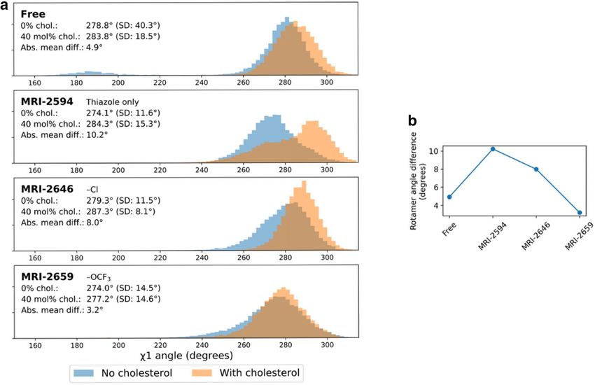
www.nature.com/scientificreports/
a
b
Figure 1. Benzothiazole-based CB2 ligands. (A) Structure of benzothiazole-based C
B2 ligands. (B) Synthesis
scheme for ligands, Reagents and conditions: a. 2-bromoethanol 90 °C; b. BOP, 2,2,3,3-tetramethylcyclopropane
carboxylic acid. c. Acetyl Chloride.
had no effect on basal signaling (Fig. 2b) whereas MRI-2654, MRI-2687, MRI-2653, and MRI-2659 reduced
basal signaling to 55% with high potency (Fig. 2a) as a function of inverse agonism. Unlike other structural
analogues, MRI-2594 demonstrated full agonism with high affinity ( hCB2R, Ki = 0.031 nM) and potency ( EC50:
0.09 nM) (Fig. 2a). We were intrigued by the seemingly neutral antagonist activity of MRI-2646 in CHO cells
even though its structural features were in line with the other benzothiazole ligands which behaved as inverse
agonist. Hence, in the present work, we used this novel, high affinity cannabinoid ligand to explore the role of
lipid environment in determining functional activity.
Based on our preliminary studies, we concluded that ligands such as MRI-2646 may act either as a weak
partial agonist or a neutral antagonist, depending on the cell type used in the assay. To better understand the
reason for these discrepancies, we assessed the activation of C B2 receptor by the in vitro G protein activation
assay that measures the rates of nucleotide exchange on the Gα subunit of G protein (GEF assay) as described
previously33,34. The assay reports the rates of formation of the complex of [ 35S]-GTPγS, a homolog of GTP, with
the Gα subunit of G protein. Typical assay conditions require small (nanogram) quantities of the receptor protein
either in cell membranes or reconstituted into lipid bilayers. The readout of the assay is the amount of the non-
hydrolysable complex of G α with [35S]-GTPγS which, under selected experimental conditions, is proportional
to the amount of activated receptor in the assay.
For convenience, the results of the GEF assay are normalized such that activation of C B2 in the presence of
CP-55,940 was set to 100%, and residual activity in the presence of saturating concentrations of the full inverse
agonist SR-144,528 was set to 0%.
We first compared two different types of membranes: E. coli BL21 (DE3) and a commercially available prepa-
ration of CHO cell membranes expressing C B2 (Millipore EMD, Cat. No HTS020M) in the GEF assay (Fig. 3).
Depending on the source of membranes, a significant difference in activation behavior of C B2 was observed. In
this assay, the tested ligands (with the exception of the previously described full agonist MRI-259431 and strong
inverse agonist MRI-2659) behaved as partial agonists of the C B2 receptor in E. coli membranes. However, these
same ligands acted as partial inverse agonists on CB2 expressed in CHO cells (Fig. 3). The activities of the ligands
relative to each other, which were roughly inversely correlated to the ligand substituent size, were not greatly
changed, and correlated roughly to the R-group size.
Intrigued by these observations, we compared several other available sources of membranes expressing C B 2.
MRI-2646 ligand was selected as a representative of a cohort of related benzothiazole ligands for all subsequent
measurements since it activated C B2 in E. coli cell membranes the most while being a partial inverse agonist of
CB2 in CHO membranes. Several preparations of membranes expressing C B2 were compared (Fig. 4). While
MRI-2646 behaved as a neutral antagonist of C B2 in membranes from CHO cells obtained from PerkinElmer,
it acted as a partial inverse agonist in two other commercial preparations of C B2 in CHO membranes procured
from EMD Millipore and Applied Cell Sciences (CHO-K1 membranes). Likewise, MRI-2646 was a partial inverse
agonist of CB2 expressed in membranes Expi293F and Expi293F GNTI- cells. On the other hand, this ligand
was a partial agonist of CB2 expressed in baculovirus infected insect Sf9 cells and in E. coli BL21 (DE3) cells.
Therefore, MRI-2646 exerts differential effects on CB2: in mammalian cell membranes this ligand acts as an
inverse agonist or neutral antagonist while in bacterial- and insect-cell membranes it behaves as a partial agonist
of CB2 receptor. Likely contributing factors to such discrepancies may include: (i) differences in lipid composition
Scientific Reports | (2021) 11:3706 | https://doi.org/10.1038/s41598-021-83245-6 3
Vol.:(0123456789)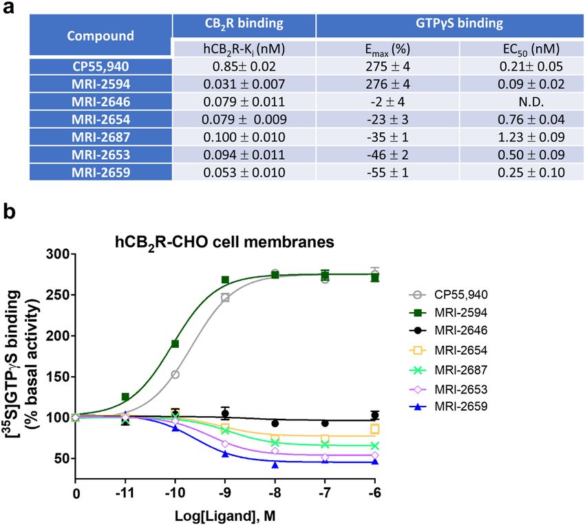
www.nature.com/scientificreports/
Figure 2. Affinity and functional effects of novel MRI ligands on CB2 receptor in CHO cell membranes. (a)
binding affinities (nM) and Emax (% and nM) of [35S]-GTPγS binding to CHO membranes (PerkinElmer,
Cat. No ES111-M400UA) expressing h CB2. (b) [35S]-GTPγS binding to membranes as a factor of ligand
concentration. Binding of [35S]-GTPγS was determined as described in “Methods”. Non-specific binding was
defined as 0% activity. The assay of GTPγS non-specific binding contains non-radioactive GTPγS.
Figure 3. GEF of CB2 expressed in: (a) E. coli membranes and (b) CHO membranes (Millipore EMD). 2 μg
of total protein per assay. Each point represents an average of four independent measurements (n = 4). Ligands
were added at a concentration of 2 μM to ensure saturation of the receptor, and G protein was added as
described in “Methods”. The dotted line indicates the rates of activation of G protein in the absence of a ligand.
The rates of activation with CP-55,9040 are set to 100%, and rates of activation in the presence of SR-144,528 to
0%.
Scientific Reports | (2021) 11:3706 | https://doi.org/10.1038/s41598-021-83245-6 4
Vol:.(1234567890)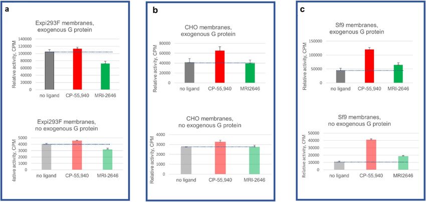
www.nature.com/scientificreports/
Figure 4. GEF on CB2 expressed in different cell lines. An amount of 2 μg of membrane protein per assay
was used. Each data points represents an average of four independent measurements (n = 4) with error rates
indicated by the bars. 100% represents full activation in the presence of 2 μM of CP-55,940, and 0%–with 2 μM
SR-144,528.
(in particular, cholesterol content) between mammalian, insect, and bacterial membranes; (ii) different pattern
of post-translational modifications (palmitoylation, glycosylation) of receptor molecules expressed in different
expression hosts; (iii) differences in expression levels of the receptor and densities of ligand-binding sites in
membrane preparations from different sources, and (iv) composition of endogenous G proteins in membrane
preparations. The endogenous membrane-associated G protein contained in preparations of CHO, HEK and
Sf9 cell membranes expressing CB2 contributes to the GEF signal. E. coli cells do not produce G protein. The
subtle role of the sterics and electronics of the 6-Cl substituent in this intricate modulatory mechanism cannot
be ruled out either.
Effects of lipid composition. We considered the effects of lipid composition of CB2-containing mem-
branes obtained from different expression hosts. Specifically, the CHO cell membranes are known to contain
high concentrations of cholesterol, unlike the E. coli cell membranes that are devoid of cholesterol12. We quanti-
fied the relative content of lipids and cholesterol in several preparations of membranes of mammalian, insect,
and E. coli cells (Supporting Fig. 1). Lipids were extracted from membranes, and their composition determined
by 1H-NMR as described in Legend to Supporting Fig. 1. Consistent with the previously published data, choles-
terol was not detected in membrane preparations from E. coli and from Sf9 cells11,35–37 while membrane preps
obtained from CHO cells and suspension culture of HEK Expi293F expressing C B2 contained 39% and 26% cho-
lesterol relative to phospholipids, respectively10,38. While the bacterial-, insect- and mammalian cell membranes
differ significantly not only in content of cholesterol but also in composition of phospholipids10,11,13, a variability
in cholesterol content between membranes from different expression cell lines correlates strongly with the sign-
aling pattern of CB2 activated by the novel ligand. Therefore, we hypothesized that the cholesterol content of
membranes affects the activation of the cannabinoid receptor CB2.
Endogenous vs. exogenous G protein. Besides lipids, the content of endogenous G proteins in cell
membrane preparations from different sources may also affect the readout of the [ 35S]-GTPγS binding and the
GEF G-protein activation assays used in this study (see “Methods”). The [ 35S]-GTPγS binding assay measures
the binding of the radiolabeled nucleotide analogue to the endogenous G protein that is already present in
membrane preparations while the GEF assay relies on the exogenous G protein subunits of G αi1 and Gβ1γ2 added
in large excess relative to receptor. Therefore, the GEF assay typically affords a good signal-to-noise ratio and
enables comparison of multiple samples at standardized conditions. The addition of G protein is necessary to
analyze the activation of CB2 in E. coli membranes since these membranes do not contain endogenous G protein.
On the other hand, the membranes obtained from mammalian and insect cell cultures contain endogenous G
proteins and, therefore in these membranes, the GEF assay reports on the rates of activation of a combined pool
of endogenous as well as exogenous G protein.
Scientific Reports | (2021) 11:3706 | https://doi.org/10.1038/s41598-021-83245-6 5
Vol.:(0123456789)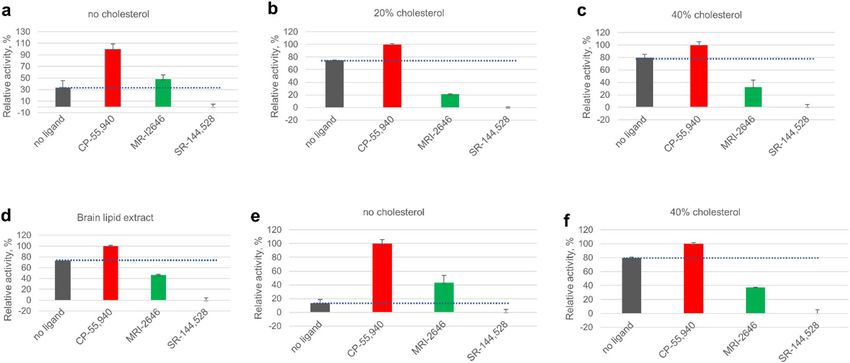
www.nature.com/scientificreports/
Figure 5. GEF analysis of three membrane preparations with or without supplementation with exogenous G
protein. (a) Expi293F membranes; (b) CHO membranes; (c) Sf9 membranes. Bars represent an average of four
independent measurements (n = 4). Dotted lines represent the rates of activation of G protein in the absence of a
ligand.
To assess the contribution of endogenous G protein to the total GEF signal, we performed the GEF assay on
membranes of Expi293F, CHO (Perkin Elmer) and Sf9, in the absence as well as in the presence of exogenous
G protein (Fig. 5).
As expected, in the absence of exogenous G protein the magnitude of the signal was significantly lower (three
to tenfold) compared to the standard GEF assay. Yet, the pattern of activation of CB2 by the MRI-2646 was simi-
lar in assays performed with- and without addition of exogenous G protein. While in membrane preps from
Expi293F cells, MRI-2646 acted as an inverse agonist of C B2 (Fig. 5a); in CHO cell membranes (PerkinElmer)
it behaved as a neutral antagonist (Fig. 5b); and in membranes of Sf9 cells expressing CB2, it acted as a partial
agonist (Fig. 5c).
Receptor density in membranes. We next examined the density of ligand binding sites (by satura-
tion 3H-CP-55,940 radioligand binding assay, Supporting Fig. 2). The density of the ligand binding sites varied
between ~ 22 and 50 pmol/ mg of membrane protein. However, there was no correlation between the density of
the ligand binding sites and behavior of the MRI-2646 ligand as an inverse agonist, neutral antagonist or partial
agonist of C
B2. Thus, the differential pattern of activation of C
B2 by the MRI-2646 cannot be explained by differ-
ences in expression levels and density of binding sites of receptor in membrane preparations.
The results above suggest that the functional effect of MRI-2646 on C B2 varies following the same pattern as
the content of cholesterol in membranes of cell lines expressing CB2. While in membranes devoid of cholesterol,
the MRI-2646 is a partial agonist, and, in cholesterol-containing membranes it acts either as an inverse agonist
or a neutral antagonist of CB2.
Treatment with methyl‑β‑cyclodextrin. The content of cholesterol in cell membrane preparations
can be altered by pre-treatment with methyl-β-cyclodextrin (MβCD)8,39 (Supporting Fig. 3). In membranes of
Expi293F cells expressing CB2, treated with 20 mM MβCD, the rates of activation of G protein on CB2 decreased
by about twofold although the levels of the C B2 receptor in these membranes were unchanged (Supporting
Fig. 3b). In the MβCD-treated membranes, the activity of CB2 in the presence of MRI-2646 was higher than
the basal signaling in the absence of ligands, indicating that MRI-2646 ligand acted as a partial agonist of C B2
receptor at these conditions.
MβCD can also be used as a carrier of cholesterol in order to enrich cell membranes with c holesterol40. To
test the effect of the exogenously added cholesterol on activation of C B2, we pre-treated the E. coli membranes
expressing CB2 with a solution of MβCD/cholesterol, and measured the rates of activation of G protein (Fig. 6a).
In the MβCD/cholesterol-treated membranes of E. coli, the MRI-2646 reproducibly acted as a neutral antagonist
of CB2, while in the untreated membranes it exhibited agonistic effects (Fig. 6b). There was no noticeable change
in the levels of CB2 in membranes, and the density of ligand binding sites did not change upon treatment with
MβCD/cholesterol (Supporting Fig. 4a,b) These results provide a more direct proof that the activation of C B2
bound to MRI-2646 is modulated by cholesterol content of membranes.
Scientific Reports | (2021) 11:3706 | https://doi.org/10.1038/s41598-021-83245-6 6
Vol:.(1234567890)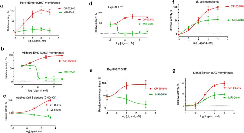
www.nature.com/scientificreports/
Figure 6. Effect of treatment with MβCD/Chol of E. coli membranes on activation of C B2 by synthetic ligands.
(a) Membranes expressing C B2 were treated with 20 mM MβCD/Chol for 1 h at 4 °C, washed with PBS, and
the activation of CB2 in the presence of ligands determined by GEF; (b) control, untreated E. coli membranes
expressing CB2. Each data point represents an average of four independent measurements (n = 4) with standard
deviation indicated by vertical bars.
Figure 7. Activation of G protein on C
B2 receptor reconstituted in liposomes. Liposome composition: 75%
POPC, 25% POPG and cholesterol content relative to total phospholipids as indicated, or total lipids extracted
from bovine brain, as indicated. Proteins were reconstituted at a protein-to-lipid ratio in the range 1:850 to
1:1100 mol/mol. (a–d) CB2 purified from E. coli cells. (e,f) CB2 purified from Expi293F GNTI- cells. Average
values of four independent measurements are plotted (n = 4). Dotted lines represent the rates of activation of G
protein in the absence of a ligand.
Liposome‑reconstituted CB2 and post‑translational modifications. To assess the possible role of
post-translational modifications of C B2 on its activation by the MRI ligands, the recombinant C B2 receptor
was isolated from two different expression cell lines, E. coli BL21 (DE3)41 and Expi293F GNTI-42. The protein
was purified and reconstituted into lipid bilayers containing 1-palmitoyl-2-oleoyl-glycero-3-phosphocholine
(POPC) and, 1-palmitoyl-2-oleoyl-sn-glycero-3-phospho-(1′-rac-glycerol) (POPG) at a molar ratio of 3/1,
either supplemented or not supplemented with cholesterol, as described in “Methods”10,13. We reported previ-
ously that the presence of phospholipids with a negatively charged headgroup stabilizes CB2 protein in lipid
bilayers43. Therefore, purified C
B2 was reconstituted into POPC/POPG (3/1, mol/mol) liposomes containing
0, 20 and 40 mol% cholesterol (reported as total content of lipids) as described in “Methods”. In one case, the
protein purified from E. coli cells was reconstituted into lipids extracted from brain tissue (Avanti Polar Lipids).
The ratio of protein-to-lipid in the resulting samples was in the range of 1:850 to1:1100 (mol/mol). The levels of
protein, and the density of ligand binding sites for receptor preparations reconstituted in liposomes with differ-
ent content of cholesterol varied only slightly (Supporting Fig. 5a,b).
The results of the GEF assay performed on these liposome-reconstituted CB2 samples demonstrate that
the MRI-2646 ligand acts as a partial agonist of bacterially expressed CB2 in liposomes devoid of cholesterol
(Fig. 7a). However, in the lipid matrix containing 20% or 40% of cholesterol or in liposomes composed of lipids
extracted from brain tissues (Fig. 7b–d), the basal (without ligand) activation of C B2 receptor was increased. In
Scientific Reports | (2021) 11:3706 | https://doi.org/10.1038/s41598-021-83245-6 7
Vol.:(0123456789)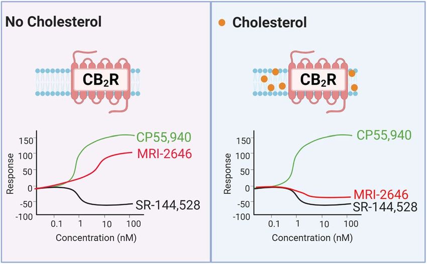
www.nature.com/scientificreports/
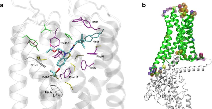
Figure 8. Depictions of ligand binding site and preferentially affected residues. (a) Representative snapshot
of MRI-2659 in orthosteric binding site. MRI-2646 and MRI-2659 share common backbone position; (b)
Cryo-EM structure (PDB: 6PT0)44. Those residues whose simulation average RMSD from the cryo-EM structure
changed by > 1 Å as a function of 0% vs 40 mol% cholesterol are highlighted by spheres. Such residues from free
and ligand-bound conditions (MRI-2594, MRI-2646, MRI-2659) are all shown. Note proximity to G α subunit
(grey). A number of these residues reside in the extracellular loops; the functional relevance is not known.
all cholesterol-containing liposomes, including those containing the brain lipid extract, the MRI-2646 ligand
acted as a partial inverse agonist of the receptor.
For comparison, the activation behavior of C B2 isolated from the Expi293F G NTI- cells42 and reconstituted
into liposomes was studied (Fig. 7e). In liposomes without cholesterol, the MRI-2646 acted as a partial agonist
of CB2, similar to its action on the bacterially expressed protein. At the same time, MRI-2646 acted as a partial
inverse agonist on HEK cell-expressed CB2 protein reconstituted into liposomes with 40% of cholesterol (Fig. 7f
and Supporting Fig. 5, 6). These results corroborate the above described inverse agonism of MRI-2646 on CB2 in
membrane preps containing cholesterol. Therefore, it can be concluded that cholesterol is involved in modulating
the activation behavior of C B2. Post-translational modifications of CB2 do not seem to play a significant role in
modulation the activation of the receptor by MRI-2646 in the presence of cholesterol.
Structural effects of cholesterol on CB2 in molecular dynamics simulation. Using molecular
dynamics (MD) simulations, we evaluated the effect of membrane cholesterol on three of the ligands’ interaction
with the known toggle switch residue T rp2586.48as well as the displacement and fluctuation of the ICL3-TM6
region of CB2, which would interact with the G-protein upon its recruitment. We hypothesized there would be a
significant effect of membrane cholesterol on these regions in free CB2.We anticipated three alternatives for the
effect of ligand type on the simulation results: (1) no obvious effect of ligand; (2) effects specific to the experi-
mentally observed ligand pharmacological category; or (3) effects correlating mainly to ligand size. MRI-2646.
MRI-2659, and MRI-2594 were simulated, chosen for their diversity.
We conducted equilibrium all-atom molecular MD simulations of the cryo-EM structure of C B2 (PDB:
6PT0)44 with and without 40% membrane cholesterol and a POPC/POPG, 3/1, mol/mol ratio. The cholesterol
molecules in the extracellular part of the TM5-6 region reported in the cryo-EM structure were retained, par-
ticularly since this is a known cholesterol binding site45,46. In both of these conditions, simulations were done
with no bound ligand as well as with each of the three chosen ligands. Each ligand was placed respectively in the
orthosteric site (Fig. 8) by analogy with the configuration of the structurally similar AM10257 ligand agonist
present in the X-ray structure of CB231. There were eight total simulations.
The R-groups of the ligands were in close proximity to the toggle switch residue Trp258 6.48. In each simula-
tion, the protein underwent an initial relaxation phase from the initial antagonist-bound cryo-EM conformation
within 10 ns, as shown by evolution of whole protein RMSD over time (Supporting Fig. 7). Each ligand remained
stable in its initial position throughout (Supporting Fig. 7). We discarded the first 50 ns of each production
simulation, to include the relaxation phase, for an aggregated grand total of 2 µs of simulation trajectory analyzed
across all conditions. Key binding site interactions are shown in Fig. 8a.
In GEF experiments, membrane cholesterol increased the constitutive activity of CB2, and the relative order of
MRI-2594, MRI-2646, and MRI-2659 by activity was preserved, even though these ligands would be categorized
differently relative to the benchmark of constitutive activity. Since G-protein recruitment is thought to depend
Scientific Reports | (2021) 11:3706 | https://doi.org/10.1038/s41598-021-83245-6 8
Vol:.(1234567890)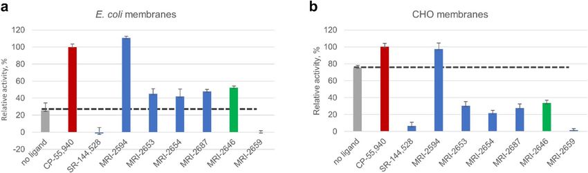
www.nature.com/scientificreports/
Figure 9. Difference in T
rp2586.48 rotamer angle related to presence of cholesterol. (a) Trp2586.48 χ1 rotamer
angle distributions with 0% and 40 mol% cholesterol. (b) Difference in mean Trp2586.48 χ1 rotamer angle
between no cholesterol vs 40 mol% cholesterol conditions; green (negative change) dashed line. TM domains are
annotated, with up and down arrows indicating direction of helix, with up from intracellular to extracellular.
on the toggle switch T rp2586.48, we hypothesized that the rotameric state of this residue would be differentially
changed by ligand type, particularly since the ligand substituent is in close proximity to it.
We found that the distribution of T rp2586.48 side-chain χ1 rotamer angles (from the N–Cα–Cβ–Cγ dihe-
dral) during the simulations varied by ligand arm 1 substituent size and presence of cholesterol. In ligand-free,
cholesterol-free CB2, the distribution of side-chain angles had a peak at ~ 280° (and very small peak at roughly
180o which will not be discussed further, since it is unlikely to relate to experimental results). In each case the
average magnitude was increased when cholesterol was present (Fig. 9a). The difference in mean χ1 angles
between cholesterol conditions for each bound ligand are shown in Fig. 9a and plotted in Fig. 9b. As the ligand
size grew, the effect of cholesterol on the rotamer angle decreased.
Some amount of conformational change from the reference cryo-EM structure is to be expected in simulation,
but we hypothesized that residues interfacing with G-protein, and therefore most likely to change the activation
rate, would be preferentially affected as a function of the presence of cholesterol. To test this idea, we compared
the average root-mean-square deviation (RMSD) of each Cα, with respect to the original cryo-EM structure at
each simulation frame, between 0% cholesterol and 40% cholesterol conditions by subtraction. This comparison
was made for free and each ligand-bound state. If a residue in the 40% cholesterol structure deviated more than
the same residue in the 0% cholesterol structure, the resulting quantity would be positive, and if less, negative.
Even though the reference cryo-EM structure is antagonist-bound, if there were no effect of cholesterol or ligand,
the RMSD would be expected to be similar across conditions, resulting in zero RMSD difference. We found that
residues that would interact with the G-protein complex, primarily in the ICL3 loop and the N-terminal side
of TM6 (residues 222–236), were preferentially displaced in the presence of cholesterol across all ligand condi-
tions (Fig. 10). The difference in average per-residue RMSD was relatively variable in this region compared to
the rest of the structure, where this quantity was mostly close to zero (Fig. 10). The number of residues in this
region with > 1 Å mean change in RMSD as a function of cholesterol presence was 9, 6, 2, and 3 in free, MRI-
2594, MRI-2646, and MRI-2659-bound simulations respectively. This number decreased with ligand substituent
size—though we cannot rule out other factors, particularly including the chemical composition of the substituent.
Given that RMSD change as a function of the presence of cholesterol was related to substituent size, we
hypothesized that the root-mean-square fluctuation (RMSF) of each residue with respect to the average structure
would also be affected in a systematic way (Supporting Fig. 8). A decrease in RMSF of specific residues would
suggest a localized decrease in entropy. The ICL3 and first part of TM6 (residue 221–236) exhibited a relatively
high RMSF (mean 2.58–3.80 Å with no cholesterol, 2.85–4.65 Å with 40% cholesterol). The magnitude of the
RMSF was inversely correlated to the size of the ligand substituent. In unbound C B2, the RMSF in this region in
the 40% cholesterol condition was substantially higher than in the 0% cholesterol condition (4.65 Å vs 2.58 Å).
While relatively small in magnitude, the mean of absolute per-residue difference in RMSF as a function of the
Scientific Reports | (2021) 11:3706 | https://doi.org/10.1038/s41598-021-83245-6 9
Vol.:(0123456789)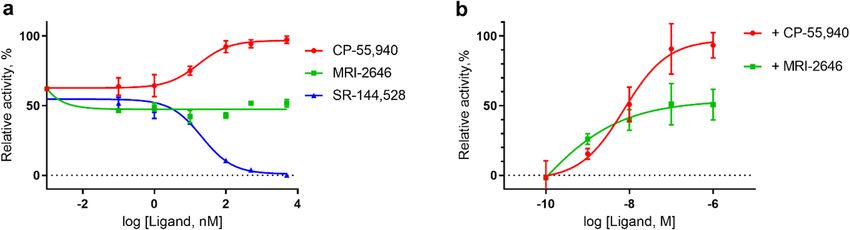
www.nature.com/scientificreports/
Figure 10. Difference in per-residue root-mean-square deviation (RMSD) between simulation and antagonist-
bound cryo-EM structure. Trp2586.48 highlighted in red, residues with > 1 Å difference highlighted in orange
(positive change) or green (negative change) dashed line. TM domains are annotated, with up and down arrows
indicating direction of helix, with up from intracellular to extracellular.
presence of cholesterol also decreased as ligand substituent size increased (1.01, 0.58, and 0.40 Å for MRI-2594,
MRI-2646, and MRI-2659 respectively, shown in Supporting Fig. 8).
Discussion
Here we demonstrated that cholesterol increases the basal activation levels of CB2 receptor, thereby altering the
pharmacological classification of novel synthetic cannabinoid ligands (Fig. 11). While in membranes devoid of
cholesterol the MRI ligands act as partial agonist of CB2, in cholesterol-enriched membranes that same ligands
became either inverse agonists or neutral antagonists of this receptor.
Some of us have previously reported the stabilizing effect of anionic lipids such as PG and PS on purified CB2
protein reconstituted in l iposomes43,47. It was also shown that the negatively charged cholesterol derivative, cho-
lesteryl hemisuccinate (CHS), stabilizes the recombinant CB2 protein in detergent micelles and l iposomes43. At
the same time, it was reported that cholesterol did not affect the activation of CB2 by the full agonist CP-55,94047.
Since these observations were made on a C B2 receptor activated by the high affinity full agonist CP-55,940, the
relatively moderate effects of the lipid matrix on receptor activation may have been masked.
Cholesterol is the major sterol found in higher e ukaryotes48. The rigid planar structure of cholesterol modu-
lates fluidity, thickness, curvature and permeability of m embranes49–51. The presence of cholesterol in membranes
increases ordering of lipid acyl chains. Physicochemical parameters of membranes have been implicated in the
regulation of function of integral membrane p roteins49,52,53. Lateral compression and hydrophobic matching
between the lipid bilayer and transmembrane domains of the protein are affected by cholesterol content. These
parameters are important for the structural stability of embedded membrane p roteins51.
Scientific Reports | (2021) 11:3706 | https://doi.org/10.1038/s41598-021-83245-6 10
Vol:.(1234567890)www.nature.com/scientificreports/
Figure 11. Membrane cholesterol dependent protean agonism.
It was previously reported that cholesterol rafts in human immune cell membranes modulate the activity of
CB1 receptor but do not affect the activation behavior of CB2 receptor29. That conclusion was reached by quan-
tifying several signaling pathways in cells treated with methyl-β-cyclodextrin (MβCD). However, the effects of
cholesterol/ MβCD were assessed in the presence of the full agonist of C B2, CP-55,940 that may have masked
moderate effects on activation of CB2 receptor by the lipid bilayer. Here, by using a novel synthetic ligand MRI-
2646 we demonstrated that the content of cholesterol in lipid bilayers modulates C B2 activation.
We have demonstrated that cholesterol increases the constitutive activity of the C B2 receptor. This effect was
first shown in membrane preparations of several types of cells expressing C B2. One can argue that the difference
between the membranes of mammalian cells (HEK, CHO) on the one hand, and insect and bacterial cells—on
the other hand is not only in the content of cholesterol but in many other parameters including composition
of phospholipids. Indeed, it has been reported that the content of phosphocholine (PC) lipids in HEK cell
membranes is about 33% of total lipids, about two-fold higher compared to Sf9 membranes which have higher
phosphoethanolamine (PE) (almost 40% of total lipids)10. The difference in lipid composition is even more
pronounced when E. coli membranes are considered: they consist predominantly (almost 75%) of zwitterionic
PE with the remainder consisting of anionic lipids (PG) and c ardiolipin54. To prove that the presence or absence
of cholesterol plays a major role in modulating the spontaneous signaling by C B2, we reconstituted the purified
receptor into lipid bilayers of defined composition, containing POPC/POPG, 3/1, mol/mol and cholesterol in
the range of 0–40% of total lipids. Indeed, the presence of cholesterol in these artificial bilayers resulted in an
increase of spontaneous signaling by CB2. This effect was observed for the recombinant receptor isolated from E.
coli cells as well as from the HEK Expi293F cells, providing additional evidence for an increase of basal activity
of the receptor by cholesterol.
What are the mechanisms by which cholesterol modulates function of GPCR? There are several examples of
GPCR that exhibit a certain affinity for cholesterol, and whose activities are regulated in response to the content of
cholesterol in membranes. This includes the β2-adrenergic receptor and the μ-opioid receptor for which specific
high affinity cholesterol binding sites have been reported near the transmembrane h elices7,45.
A putative cholesterol binding sequence (CRAC) was reported for transmembrane helix 7 of human C B1
receptor28. This sequence was proposed to be involved in directing the interaction of CB1 receptor with cho-
lesterol-rich microdomains of cell membranes. Moreover, the presence of a cholesterol molecule was recently
reported in a crystal s tructure55 and a cryo-EM s tructure56 of C B1 receptor. At the same time, there was no
evidence of a specific retention of cholesterol in a recently published C B2 crystal31 structure. There is choles-
44
terol included in PDB 6PT0, a cryo-EM structure of C B2 , although its origin is unclear since the protein was
expressed in Sf9 cells that produce very little if any c holesterol10. Also, any specific interaction of cholesterol
with certain sites on the receptor may not explain why modulation of receptor function occurs at relatively high
cholesterol concentrations in the lipid matrix surrounding the receptor.
Using MD simulations, we searched for structural and dynamical correlates of the experimental results.
In simulations of free C B2, the ICL3-TM6 region, known to interact with G α, deviated significantly in RMSD/
RMSF from the antagonist-bound conformation in the presence of cholesterol. Since the antagonist-bound
conformation would be less likely to recruit G protein by definition, deviating from it is consistent with the large
experimentally observed increase in constitutive activity. By contrast, the MRI-2659-bound structure showed
relatively little deviation in this region, consistent with the strong antagonist activity of MRI-2659 in both cho-
lesterol conditions.
The situation is less clear with the other two simulated ligands. We observed that the size of the ligand is
inversely correlated with the effect of cholesterol on both Trp258 and ICL3-TM6. Yet this correlation does not
map precisely to the corresponding changes in categorizing the ligands’ actions. When cholesterol is included
in the membrane, the strong agonist MRI-2594 remains a (less-strong) agonist, the weaker agonist MRI-2646
becomes an inverse agonist, and the strong inverse agonist MRI-2659 remains as such (Fig. 3). The ligands are
categorized relative to the baseline of ligand-free constitutive activity. The relative activity rank order of the MRI
ligand series ligands (Fig. 3, y-axes) is preserved in both cholesterol conditions. We consider that the simulation-
observed changes in the binding and ICL3-TM6 (i.e. G-protein binding) sites may be independent components
Scientific Reports | (2021) 11:3706 | https://doi.org/10.1038/s41598-021-83245-6 11
Vol.:(0123456789)www.nature.com/scientificreports/
contributing to the overall experimentally-observed effects, rather than ligand-specific effects that directly cor-
relate with the pharmacological categories of ligands, since the simulation results correlated with the ligand
sterics rather than their pharmacological categories. This is consistent with the hypothesis that the primary effect
of cholesterol is to modify the baseline constitutive activity that defines how the tested ligands are categorized.
Our simulation cannot elucidate the allosteric pathway from binding pocket to G protein that would be
responsible for the data—such an endeavor is well outside the scope of this work. While the chemical identity
of the ligand is presumably important, we have not specifically addressed the effects of specific ligands beyond
sterics. Future work might include simulating the C B2-G-protein complex and estimating the binding energy
difference as a function of cholesterol and ligand, but this is an exceedingly large task.
The data do not explain how the chloro- substituent at the 6-position of the benzothiazole arm may regulate
effects of cholesterol on CB2 activation. While the toggle switch Trp2586.48 functions as an important molecular
determinant in activation or deactivation of the receptor, it sheds limited light on differential interactions lead-
ing to neutral vs. inverse agonism. That the compound MRI-2654 with a bromo-substituent still behaves as an
inverse agonist attests to the subtle difference in size and electronegativity of the chloro-group in influencing the
molecular dynamics and signaling processes resulting in modulation of receptor function.
By using MRI-2646 ligand we demonstrate that cholesterol increases the constitutive activity of CB2 receptor.
The content of cholesterol in preparations of cell membranes expressing CB2 correlates with an increase in basal
signaling through CB2, which could be reversed by depletion of cholesterol using cyclodextrin. These results
suggest that the pharmacological properties of synthetic ligands can be influenced by the cholesterol composi-
tion of cell membranes harboring cannabinoid receptor. Such a regulatory mechanism may contribute for well
documented tissue- and cell-specific differences in the efficacy of partial CB2 agonists, such as the endocan-
nabinoid anandamide or the plant-derived cannabinoid Δ9-tetrahydrocannabinol57. For example, THC acted as
a full CB2 agonist in suppressing interferon-γ-induced activation of microglia58, whereas it had no CB2 agonist
activity and acted as a C B2 antagonist by blocking 2-AG-induced migration of natural killer c ells59, which have
high levels of membrane lipid, including c holesterol60.
Methods
Materials. Chromatographic resin Ni–NTA was purchased from Qiagen (Germantown, MD). Streptactin
XT was from IBA Life Sciences (Goettingen, Germany). The detergents CHAPS (3-[(3-Cholamidopropyl)-
Dimethylammonio]-1-Propane Sulfonate] • N,N-Dimethyl-3-Sulfo-N-[3-[[3α,5β,7α,12α)-3,7,12-Trihydroxy-
24-Oxocholan-24-yl]Amino]propyl]-1-Propanaminium Hydroxide, Inner Salt) , LMNG (Lauryl Maltose Neo-
pentyl Glycol) and DDM (Dodecyl-β-D-Maltoside) were from Anatrace (Maumwee, OH). CHS-Tris salt was
from Anatrace. The detergent Façade-TEG (3a,7a,12a-tri-((O-b-D-glucopyranosyl)ethyloxy)-cholane) and
lipids POPC, POPG, brain lipid extract and cholesterol were from Avanti Polar Lipids (Alabaster, AL).
The potent non-selective CB2 agonist CP-55,940 ((-)-cis-3[2-hydroxy-4-(1,1-dimethylheptyl)phenyl]-trans-
4-(3-hydroxtpropyl) cyclohexanol, the high affinity selective CB2 inverse agonist SR-144,528 5-(4-Chloro-
3-methylphenyl)-1-[(4-methylphenyl)methyl]-N-[(1S,2S,4R)-1,3,3-trimethylbicyclo[2.2.1]hept-2-yl]-1H-pyra-
zole-3-carboxamide were from Cayman Chemical (Ann Arbor, MI). 3H-labeled CP-55,940 was from Perkin
Elmer Life Sciences (Akron, OH). All other reagents were from Sigma-Aldrich (USA).
Preparations of CHO cell membranes expressing C B2 were obtained from the following sources: PerkinElmer
(Cat. No ES111-M400UA, Billerica, MA), Millipore ChemiScreen (Cat. No P34972, Burlington, MA) and Applied
Cell Sciences (Cat. No A318, Rockville, MD). Preparations of Sf9 cell membranes expressing CB2 were from
Signal Screen (Cat. No 6110130, Rockville, MD).
Expi293F HEK cells were obtained from ThermoFisher Scientific (Cat. No: A14527), and C B2 expressed and
membranes obtained in house as described e lsewhere42. The E. coli cells BL21 (DE3) were obtained from EMD
Millipore-Sigma (Cat. No. 69450), and CB2 expressed and membranes were obtained in house as described
elsewhere.34,61.
Chemistry. Commercially available regents were purchased and used as is. Proton (1H NMR) spectra were
recorded on a Varian 400 or Bruker 500 MHz spectrometer in solvents indicated with the values given in ppm
(TMS as internal standard) and J (Hz) assignments of proton resonance coupling. Mass spectra (HRMS) were
recorded on a JEOL SX102a mass spectrometer. Thin layer chromatography (TLC) analyses were carried out
on 5 cm × 10 cm silica gel GHLF 0.25 mm plates using various gradients of EtOAc:n-hexane with visualiza-
tion under UV light. Flash column chromatography was performed on Combiflash system. Product yields are
reported as un-optimized. Study compounds had ≥ 95% purity. Purity and structural characterization was done
by a combination of TLC, 1H-NMR, and LC/MS. LC–MS detection was carried out on Agilent 1200 using two
different methods/columns: Luna C18 3 um (3 × 75 mm) where the mobile phase was 4% to 100% acetonitrile
(0.05% TFA) standard gradient and EC18, 2.7 um (3 × 50 mm) where the method was 50% acetonitrile in water
(0.1% formic acid) for 3 min ramping up to 98% acetonitrile over 7.5 min. The LC–MS chromatogram showed
the correct molecular (MH+) ion as well as a single peak at UV (254 nm).
Synthesis and characterization of MRI‑268728 and MRI‑259431,32,62. Synthesis and characterization
of MRI‑2646, MRI‑2654, MRI‑2653 and MRI‑2659 were carried as outlined in Li et al.28. N-(6-methyl-3-(2-
hydroxyethyl)benzo[d]thiazol-2(3H)-ylidene)-2,2,3,3-tetramethylcyclopropane-1-carboxamide (4a)28.
N-(6-chloro-3-(2-hydroxyethyl)benzo[d]thiazol-2(3H)-ylidene)-2,2,3,3-tetramethylcyclopropane-
1-carboxamide (4b).
Scientific Reports | (2021) 11:3706 | https://doi.org/10.1038/s41598-021-83245-6 12
Vol:.(1234567890)www.nature.com/scientificreports/
2-amino-6-chlorobenzothiazole (1.84 g, 10.0 mmol) gave compound 4b (700 mg, 20%) as a white powder over
two steps. Mp 147–149 °C;1H-NMR (400 MHz, CDCl3): δ 7.56 (s, 1H), 7.36 (d, J = 8.7 Hz, 1H), 7.22 (d, J = 8.7 Hz,
1H), 4.49 (t, J = 4.6 Hz, 3H), 4.06 (t, J = 4.6 Hz, 3H), 1.58 (s, 2H), 1.32 (s, 8H), 1.22 (s, 8H). LCMS [M + H]+: 353.2.
N-(6-bromo-3-(2-hydroxyethyl)benzo[d]thiazol-2(3H)-ylidene)-2,2,3,3-tetramethylcyclopropane-
1-carboxamide (4c).
2-amino-6-bromobenothiazole (2.29 g, 10.0 mmol) gave compound 4c (950 mg, 24%) as a white powder
over two steps. Mp 173–175 °C;1H-NMR (400 MHz, C DCl3): δ 7.70 (s, 1H), 7.50 (d, J = 8.6 Hz, 1H), 7.17 (d,
J = 8.7 Hz, 1H), 4.49 (t, J = 4.5 Hz, 2H), 4.17 (s, 1H), 4.06 (s, 2H), 1.58 (s, 1H), 1.32 (s, 6H), 1.22 (s, 6H). LCMS
[M + H]+: 397.1.
N-(3-(2-hydroxyethyl)-6-methoxybenzo[d]thiazol-2(3H)-ylidene)-2,2,3,3-tetramethylcyclopropane-
1-carboxamide (4d).
2-amino-6-methoxybenothiazole (1.8 g, 10.0 mmol) gave compound 4d (920 mg, 26%) as a white powder
over two steps. Mp 164–166 °C;1H-NMR (400 MHz, CDCl3): δ 7.18 (d, J = 8.9 Hz, 1H), 7.11 (s, 1H), 6.99–6.96
(m, 1H), 4.82 (s, 1H), 4.48 (t, J = 4.5 Hz, 2H), 4.05 (d, J = 3.5 Hz, 2H), 3.83 (s, 3H), 1.55 (s, 4H), 1.32 (s, 6H), 1.21
(s, 6H). LCMS [M + H]+: 349.2.
N-(3-(2-hydroxyethyl)-6-(trifluoromethoxy)benzo[d]thiazol-2(3H)-ylidene)-2,2,3,3-tetramethylcyclopro-
pane-1-carboxamide (4e).
2-amino-6-trifluoromethoxybenothiazole (1.5 g, 6.4 mmol) gave compound 4 (610 mg, 24%) as a white
powder over two steps. Mp 149–151 °C; 1H-NMR (400 MHz, CDCl3): δ 7.46 (s, 1H), 7.30 (t, J = 7.6 Hz, 2H), 4.51
(s, 2H), 4.07 (s, 3H), 1.59 (s, 1H), 1.32 (s, 6H), 1.22 (s, 6H). LCMS [M + H]+: 403.2.
2-(6-methyl-2-((2,2,3,3-tetramethylcyclopropane-1-carbonyl)imino)benzo[d]thiazol-3(2H)-yl)ethyl acetate
(MRI-2687) (2a)28.
2-(6-Chloro-2-((2,2,3,3-tetramethylcyclopropane-1-carbonyl)imino)benzo[d]thiazol-3(2H)-yl)ethyl acetate
(MRI-2646) (2b).
Scientific Reports | (2021) 11:3706 | https://doi.org/10.1038/s41598-021-83245-6 13
Vol.:(0123456789)www.nature.com/scientificreports/
Compound 4b (300 mg, 0.85 mmol) was used as a starting material to give compound 2b (195 mg, 58%) as
a white powder. Mp 123–125 °C;1H-NMR (400 MHz, C DCl3): δ 7.54 (s, 1H), 7.34 (d, J = 8.8 Hz, 1H), 7.23 (s,
1H), 4.56 (t, J = 5.2 Hz, 2H), 4.46 (t, J = 5.3 Hz, 2H), 1.95 (s, 3H), 1.64 (s, 1H), 1.33 (s, 6H), 1.23 (s, 6H). LCMS
[M + H]+: 395.2.
2-(6-Bromo-2-((2,2,3,3-tetramethylcyclopropane-1-carbonyl)imino)benzo[d]thiazol-3(2H)-yl)ethyl acetate
(MRI-2654) (2c).
Compound 4c (300 mg, 0.75 mmol) was used as a starting material to give compound 2c (140 mg, 42%) as
a white powder. Mp 127–129 °C;1H-NMR (400 MHz, CDCl3): δ 7.68 (s, 1H), 7.48 (d, J = 8.3 Hz, 1H), 7.19 (d,
J = 8.6 Hz, 1H), 4.55 (d, J = 5.3 Hz, 2H), 4.47 (d, J = 5.2 Hz, 2H), 1.95 (s, 3H), 1.64 (s, 1H), 1.57–1.49 (m, 7H), 1.33
(s, 6H), 1.23 (s, 6H). LCMS [M + H]+: 439.2.
2-(6-Methoxy-2-((2,2,3,3-tetramethylcyclopropane-1-carbonyl)imino)benzo[d]thiazol-3(2H)-yl)ethyl acetate
(MRI-2653) (2d).
Compound 4d (400 mg, 1.1 mmol) was used as a starting material to give compound 2d (230 mg, 51%) as a
white powder. Mp 154–156 °C;1H-NMR (400 MHz, C DCl3): δ 7.21 (s, 1H), 7.09 (s, 1H), 6.96 (d, J = 9.3 Hz, 1H),
4.55 (d, J = 5.3 Hz, 2H), 4.47 (d, J = 5.2 Hz, 2H), 7.24–1.22 (m, 82H), 3.83 (s, 3H), 1.95 (s, 3H), 1.62 (s, 1H), 1.34
(s, 6H), 1.22 (s, 6H). LCMS [M + H]+: 391.2.
2-(2-((2,2,3,3-tetramethylcyclopropane-1-carbonyl)imino)-6-(trifluoromethoxy)benzo[d]thiazol-3(2H)-yl)
ethyl acetate (MRI-2659) (2e).
Compound 4e (300 mg, 0.75 mmol) was used as a starting material to give compound 2e (190 mg, 57%)
as a white powder. Mp 81–83 °C; 1H-NMR (400 MHz, C DCl3): δ 7.44 (s, 1H), 7.32 (d, J = 8.8 Hz, 1H), 7.26 (s,
2H), 4.57 (d, J = 5.4 Hz, 2H), 4.47 (t, J = 5.4 Hz, 2H), 1.95 (s, 3H), 1.64 (s, 1H), 1.33 (s, 6H), 1.23 (s, 6H). LCMS
[M + H]+: 445.3.
CB2 expression in E. coli and purification. CB2 was expressed as a fusion with the maltose binding pro-
tein (MBP) in BL21 (DE3) E. coli culture and purified on milligram-scale as previously described41,63. In brief, 10
Scientific Reports | (2021) 11:3706 | https://doi.org/10.1038/s41598-021-83245-6 14
Vol:.(1234567890)www.nature.com/scientificreports/
L of 2xYT media containing 0.2% glucose supplemented with ampicillin was inoculated with an overnight cul-
ture of E. coli. After reaching an optical density of 0.4, C
B2 expression was induced by addition of 0.5 mM IPTG
and 2.5 μM C B2 agonist CP-55,490. Expression was conducted for additional 42 h at 20 °C. After expression, cells
were harvested by centrifugation, washed with cold PBS, and lysed in a cell homogenizer (Avestin). Receptor was
solubilized for 1 h at 4 °C under continuous stirring by addition of concentrated detergent to final concentra-
tions of (0.1% CHS, 1.0% DDM, 0.5% CHAPS, all w/v). The insoluble material was removed by centrifugation at
170,000 × g for 1 h and the solubilized receptor was then purified by the affinity chromatography in 50 mM Tris
pH 7.5, 150 mM NaCl, 0.1% CHS, 0.1% DDM, 0.5% CHAPS, all w/v; 30% glycerol (v/v) and 10 μM CP-55,490
(buffer A) on Ni–NTA (Qiagen). MBP fusion partner was then removed upon incubation with the tobacco etch
virus (TEV) protease for 4 h at 4 °C, and the released receptor was further purified by chromatography on Strep-
Tactin XT (IBA Biosciences) and eluted in buffer A supplemented with 50 mM biotin as described p reviously41.
CB2 expression in HEK cells and purification. Biomass from 3 L of Expi293F GNTI- cells expressing CB2
containing N-terminal twin-Streptag and C-terminal His10 tag was obtained according to manufacturer’s proto-
col (ThermoFisher Scientific). Protein was solubilized in detergents and purified by the two successive rounds of
affinity chromatography on Ni–NTA resin and StrepTactin XT resin as described41.
Removal of the CHS and ligand from purified CB2. An amount of 2 mg of purified C B2 was bound
to 1200 μL of HisPur C o2+ (ThermoFisher) resin in buffer A and incubated under shaking for 2 h at 4 °C. The
protein sample was then transferred to a disposable gravity column and washed with 40 column volumes (CV)
of 0.5% CHAPS/0.1% DDM in 50 mM Tris–HCl, pH 7.5, 150 mM NaCl. Protein was then eluted with 6xCV
of the same buffer supplemented with 250 mM imidazole; combined eluates concentrated on 30 kDa MWCO
spin concentrator and washed 3 times to remove imidazole. Concentrated protein was supplemented with 15%
glycerol and aliquots stored at − 80 °C until further use. To confirm that both ligand and CHS have been removed
from the sample, 20 μL aliquot was mixed with 300 μL of chloroform–methanol mixture (1:1 v/v) and 1H-NMR
spectra acquired.
Reconstitution of CB2 into liposomes. Reconstitution of the purified CB2 into liposomes was performed
as described earlier43. Briefly, 200 μg of the purified protein was mixed with 2 mg of lipid mixture (POPC:POPG,
3:1, mol/mol without or with addition or 20 mol% or 40 mol% of cholesterol) solubilized in 1% CHAPS at a
concentration of 5 mg lipid/mL, and incubated on ice for 30 min. The detergents were then removed on 4 mL
Detergent Removal spin column (Pierce), according to manufacturer’s instructions. The combined filtrate con-
taining proteoliposomes was collected, and aliquots frozen in liquid nitrogen. Frozen liposomes were stored at
− 80 °C until further use. Content of protein in proteoliposomes was determined by BioRad DC assay.
Ligand‑binding assay in hCB2‑CHO‑K1 cell membranes. The assay was performed as described
reviously64. Briefly, binding affinity of the compounds to CB2R was determined by radioligand displacement
p
assays using 0.2 nM of [3H] CP-55,940 as the radioligand. Plasma membranes were from cultured CHO-K1
cells stably transfected with human CB2R (Perkin Elmer). Two microgram plasma membrane protein was used
in a 1 mL reaction mixture. Ki values were derived by computerized curve fitting and using the Cheng-Prusoff
equation to account for the affinity of the radioligand, using the GraphPad Prism 8 program (GraphPad Prism
Software Inc.).
[35S] GTPγS binding assay in hCB2‑CHO‑K1 cell membranes65. [35S] GTPγS binding was assayed as
described earlier65 with slight modifications. Briefly, hCB2-CHO-K1 cell membranes (4 µg) were incubated with
0.05 nM [35S] GTPγS, and the indicated concentrations of ligands in TEM buffer (50 mM Tris–HCl, 0.2 mM
EGTA, and 9 mM MgCl2, pH 7.4) containing 100 µM GDP, 150 mM NaCl, and 0.1% (w/v) bovine serum albu-
min in a total volume of 1 ml for 60 min at 30 °C.
[35S] GTP nucleotide exchange (GEF) assays. The subunits of G protein were expressed and purified as
described previously63. The nucleotide exchange assay was performed as previously described66.
Molecular dynamics simulations. The CHARMM36 force fi eld67 was used. Before ligand parameteriza-
tion, each ligand was geometry optimized using the B3LYP/6-31G** quantum mechanics level of theory and
basis set using Gaussian0968. Ligand parameters were derived from CGenFF69; these high affinity ligands were
not expected to explore the extremes of their conformational space.
The CB2 structure starting point was the cryo-EM structure previously described (Protein Data Bank: 6PT0).
This was oriented and placed in a lipid membrane using the Orientations of Proteins in Membranes (OPM)
database70 using the CHARMM-GUI71 input generator. In the with-cholesterol condition, the lipid membrane
consisted of 40% cholesterol, and POPC:POPG in a 3:1 ratio. Sodium and chloride atoms were added to 0.15 M
with excess for electroneutrality. The cholesterol molecules present in the cryo-EM structure were retained, par-
ticularly since the extracellular cholesterol conformations were analogous to those observed in the structurally
similar mu opioid receptor. The system was minimized and equilibrated with side chain and backbone restraints
which were subsequently released, and production simulations were run in the isothermic-isobaric ensemble at
303.15 K using NAMD 2.13 with GPU extensions. Particle Mesh Ewald summation of long-range interactions
was used, as were the Langevin barostat and thermostat.
Scientific Reports | (2021) 11:3706 | https://doi.org/10.1038/s41598-021-83245-6 15
Vol.:(0123456789)You can also read