Liquid-Liquid Phase Separation in Crowded Environments - MDPI
←
→
Page content transcription
If your browser does not render page correctly, please read the page content below
International Journal of
            Molecular Sciences

Review
Liquid–Liquid Phase Separation in
Crowded Environments
Alain A. M. André           and Evan Spruijt *
 Institute for Molecules and Materials, Radboud University Nijmegen, Heyendaalseweg 135,
 6525 AJ Nijmegen, The Netherlands; alain.andre@ru.nl
 * Correspondence: e.spruijt@science.ru.nl

 Received: 5 August 2020; Accepted: 13 August 2020; Published: 17 August 2020

 Abstract: Biomolecular condensates play a key role in organizing cellular fluids such as the cytoplasm
 and nucleoplasm. Most of these non-membranous organelles show liquid-like properties both in
 cells and when studied in vitro through liquid–liquid phase separation (LLPS) of purified proteins.
 In general, LLPS of proteins is known to be sensitive to variations in pH, temperature and ionic
 strength, but the role of crowding remains underappreciated. Several decades of research have
 shown that macromolecular crowding can have profound effects on protein interactions, folding and
 aggregation, and it must, by extension, also impact LLPS. However, the precise role of crowding
 in LLPS is far from trivial, as most condensate components have a disordered nature and exhibit
 multiple weak attractive interactions. Here, we discuss which factors determine the scope of LLPS in
 crowded environments, and we review the evidence for the impact of macromolecular crowding on
 phase boundaries, partitioning behavior and condensate properties. Based on a comparison of both
 in vivo and in vitro LLPS studies, we propose that phase separation in cells does not solely rely on
 attractive interactions, but shows important similarities to segregative phase separation.

 Keywords: liquid–liquid phase separation;                   intrinsically disordered proteins;   crowding;
 membraneless organelles

1. Introduction
      The cytosol is a complex mixture of macromolecules, including proteins, nucleic acids and
polysaccharides. Although the individual concentrations of these macromolecules varies between
cell types and organisms, their combined concentration can reach up to 400 mg·mL−1 [1,2]. This high
concentration means that the cytosol is densely packed with macromolecules. Under these conditions,
biochemical processes are strongly affected by limited diffusion and volume exclusion [3,4].
      Traditionally, biochemists have sought to understand biochemical processes by studying them in
isolation, using minimal models in the form of purified proteins. These in vitro (in this review used
to indicate cell-free) studies were commonly conducted in aqueous buffer solutions, where the effect
of crowding was omitted. To study the effect of crowding in vitro, scientists started to use synthetic
polymeric molecules such as polyethylene glycol (PEG), Ficoll and dextran [5]. In addition to synthetic
polymers, globular proteins such as bovine serum albumin (BSA), have been used a step towards
more biologically relevant crowders [6]. However, the cytosol also contains high levels of disordered
proteins and RNAs that are not specifically neutral [7]. Some of them form macroscopic puncta (or
bodies/foci/granules) in the cell, creating an additional level of complexity.
      Recent developments in cell biology aimed at understanding the cellular organization have
shed new light on the physical state of cellular fluids. Instead of a crowded, single-phase mixture
of macromolecules, the cytoplasm and nucleoplasm of many cells appear dotted with condensates
(granules, bodies, foci and puncta) that are separated from the surrounding fluid (Figure 1) [7]. These

Int. J. Mol. Sci. 2020, 21, 5908; doi:10.3390/ijms21165908                          www.mdpi.com/journal/ijms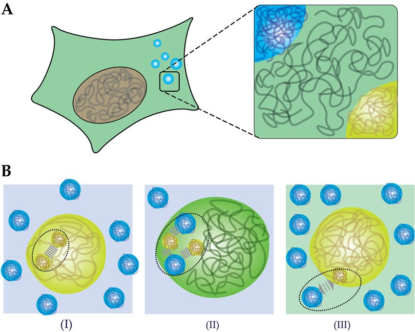
paraspeckles [9], and they have been collectively described as membraneless organelles (MLOs)
[10,11] or biomolecular condensates [9]. Many MLOs are likely formed through liquid–liquid phase
separation, resulting in very dense droplets [9,11].
        To elucidate the effect of crowding on MLOs, in vitro models using purified proteins combined
with
Int.     synthetic
     J. Mol. Sci. 2020,crowding
                        21, 5908 agents have been employed, similar to the first studies of the effect
                                                                                                     2 of of
                                                                                                          20
crowding on enzyme activity. In this review, we discuss the possible effects of crowded environments
on phase separation. To understand this, we first introduce the theoretical framework that has been
condensates seem to be dynamically formed and dissolved over time [8]. Many of these micro-niches in
used extensively to explain crowding effects on biochemical processes, and highlight the factors that
the cell have liquid-like properties, such as nucleoli, germ granules, stress granules and paraspeckles [9],
are particularly relevant to phase separating proteins. We then briefly describe the characteristics of
and they have been collectively described as membraneless organelles (MLOs) [10,11] or biomolecular
micro-compartments formed by phase separation. In the remainder of this review, we give an
condensates [9]. Many MLOs are likely formed through liquid–liquid phase separation, resulting in
overview of the use of crowders in in vitro MLO studies, and the observed effects of crowding on
very dense droplets [9,11].
phase separation, partitioning and the biophysical properties of condensates.

     Figure 1.1.Schematic
                 Schematic  overview
                          overview of a of a eukaryotic
                                        eukaryotic         cell containing
                                                   cell containing         membranous
                                                                   membranous         and membraneless
                                                                              and membraneless organelles.
     organelles.
      To elucidate the effect of crowding on MLOs, in vitro models using purified proteins combined
2. The
with    Effect of
      synthetic   Macromolecular
                crowding             Crowding
                           agents have            on Biochemical
                                        been employed,     similar toProcesses
                                                                      the first studies of the effect of crowding
on enzyme activity. In this review, we discuss the possible effects of crowded environments on
      Macromolecules occupy as much as 30% of the cellular volume [2], which makes the intracellular
phase separation. To understand this, we first introduce the theoretical framework that has been
space a crowded place. The abundance of macromolecules influences all aspects of biomolecular
used extensively to explain crowding effects on biochemical processes, and highlight the factors that
behavior, including protein folding, complexation, aggregation, and enzymatic activity. Since the
are particularly relevant to phase separating proteins. We then briefly describe the characteristics
importance of crowding was recognized in the 1960s, the effects of macromolecular crowding on
of micro-compartments formed by phase separation. In the remainder of this review, we give an
biomolecules have been extensively studied in a cell-free setting. For a comprehensive overview, we
overview of the use of crowders in in vitro MLO studies, and the observed effects of crowding on
refer to several excellent reviews by Ellis [3,12–14], Minton [4,15–17], and Rivas [5,18] on the physical
phase separation, partitioning and the biophysical properties of condensates.
aspects of crowding, and Zhou [19–21] on protein folding. To understand the role of crowding in
liquid–liquid
2. The Effect ofphase separation (LLPS),
                  Macromolecular           it is essential
                                     Crowding               to outlineProcesses
                                                  on Biochemical        some key concepts and approaches in
crowding research. Therefore, we will briefly describe the principles of excluded volume and
      Macromolecules
depletion    here, and occupy
                        discuss astheir
                                    much  as 30%onofbiomolecular
                                        effects      the cellular volume     [2], which
                                                                      structure           makes thefollowed
                                                                                   and assembly,      intracellular
                                                                                                                by
space  a  crowded   place.  The  abundance     of macromolecules       influences   all aspects of
additional effects of crowding on biomolecular dynamics and reactions, and finally, we discuss the  biomolecular
behavior,
most common including  protein approaches
                 experimental  folding, complexation,       aggregation,
                                             to mimic cellular    crowding. and enzymatic activity. Since the
importance of crowding was recognized in the 1960s, the effects of macromolecular crowding on
biomolecules    have been
2.1. Excluded Volume       extensively studied in a cell-free setting. For a comprehensive overview, we
                       Theory
refer to several excellent reviews by Ellis [3,12–14], Minton [4,15–17], and Rivas [5,18] on the physical
      Central to the effects of macromolecular crowding on biomolecular structure and dynamics is
aspects of crowding, and Zhou [19–21] on protein folding. To understand the role of crowding in
the notion that different species cannot occupy the same space: they exclude other species from a
liquid–liquid phase separation (LLPS), it is essential to outline some key concepts and approaches
certain volume [3,12,16,22]. The excluded volume is directly related to the size of molecules: large
in crowding research. Therefore, we will briefly describe the principles of excluded volume and
molecules (such as proteins) experience much larger excluded volumes than small molecules (Figure
depletion here, and discuss their effects on biomolecular structure and assembly, followed by additional
2A). More generally, the excluded volume depends on the size but also the shape of both the
effects of crowding on biomolecular dynamics and reactions, and finally, we discuss the most common
experimental approaches to mimic cellular crowding.

2.1. Excluded Volume Theory
     Central to the effects of macromolecular crowding on biomolecular structure and dynamics is the
notion that different species cannot occupy the same space: they exclude other species from a certain
volume [3,12,16,22]. The excluded volume is directly related to the size of molecules: large molecules
(such as proteins) experience much larger excluded volumes than small molecules (Figure 2A). More
processes (Figure 2B) [23].
       Rearranging of biomolecules can also result in overlapping of excluded volumes (Figure 2A)
 and, hence, a net increase in the volume that is available for crowders [23]. For inert crowders such
 an increase in available volume is entropically favorable, and manifests itself as an attraction between
 theJ.biomolecules,
Int.   Mol. Sci. 2020, 21,which
                          5908 is known in colloid–polymer mixtures as the depletion attraction [24,25].   It
                                                                                                     3 of 20
 is important to emphasize that the attraction depends strongly on the actual intermolecular
 interactions between the biomolecules of interest and the crowders (depletant): the simplified
generally, the excluded volume depends on the size but also the shape of both the biomolecules of
 Asakura–Oosawa–Verwey model that is often used to quantify depletion interactions only holds for
interest and the crowders [3]. An important consequence of this excluded volume is that the effective
 inert (hard-sphere) depletants. If the crowders are weakly attracted to the biomolecules, the depletion
concentration of these biomolecules can rise by several orders of magnitude compared to the global
 attraction diminishes rapidly and may disappear completely [26], a case we discuss in more detail
concentration, which can have a strong effect on the rates of biochemical processes (Figure 2B) [23].
 below in Section 2.4. Soft repulsion, on the other hand, may enhance the depletion attraction [27].

      Figure 2.2.Excluded
     Figure        Excludedvolume
                                volume     effect
                                        effect     of crowding.
                                               of crowding.     (A) A(A)  A particle
                                                                      small  small particle    (red) experiences
                                                                                      (red) experiences   a smaller aexcluded
                                                                                                                        smaller
      excluded    volume    than  a  large   particle  (black   sphere). The   blue  area  represents the
     volume than a large particle (black sphere). The blue area represents the free space for the particle; free  space  forforthe
                                                                                                                                 a
      particle;
     large       for athe
            particle     large  particle
                           effective  free the  effective
                                            volume          free volume
                                                      is limited.  (B) Manyis biochemical
                                                                               limited. (B) processes
                                                                                              Many biochemical        processes
                                                                                                       display a maximum
      display
     rate       a maximum
           at some   optimum rate       at some
                                 crowding,         optimum enhances
                                               as crowding       crowding,complexation
                                                                             as crowding(blueenhances
                                                                                                  curve),complexation      (blue
                                                                                                           but it also reduces
      curve), but(red
     diffusivity    it also reduces
                         curve)        diffusivity
                                 (C) The    effect of(red  curve) (C)
                                                       depletion       Thebringing
                                                                   forces, effect of molecules
                                                                                      depletion forces,
                                                                                                  togetherbringing
                                                                                                             reducesmolecules
                                                                                                                       the total
      together reduces
     excluded    volume. the    total2A
                            (Figure    excluded    volume.
                                          is adapted           (Figure 2001
                                                        from Minton     2A is[23],
                                                                              adapted
                                                                                   Figurefrom
                                                                                           2B isMinton
                                                                                                 adapted2001
                                                                                                           from[23],
                                                                                                                 EllisFigure
                                                                                                                       2001 [3],2B
      is adapted
     and   Figure from
                   2C is Ellis  2001from
                           adapted     [3], and  Figure
                                             Richter   20082C[24]).
                                                               is adapted from Richter 2008 [24]).

      Rearranging
 2.2. The             of biomolecules
           Effect of Crowding         can alsoStructure
                              on Biomolecular  result in overlapping
                                                         and Assemblyof excluded volumes (Figure 2A) and,
hence, a net increase in the volume that is available for crowders [23]. For inert crowders such an
       As illustrated in Figure 2C, the general consequence of crowding-induced depletion is that
increase in available volume is entropically favorable, and manifests itself as an attraction between
 compact states of biomolecules are favored: protein folding, oligomerization, complexation and
the biomolecules, which is known in colloid–polymer mixtures as the depletion attraction [24,25].
 aggregation are all more favored in crowded environments compared to the non-complexed state
It is important to emphasize that the attraction depends strongly on the actual intermolecular
 [24,25]. Since LLPS involves a condensation of proteins, it seems logical to assume that crowding
interactions between the biomolecules of interest and the crowders (depletant): the simplified
 favors phase separation for precisely the same reasons. However, the biomolecules involved in LLPS
Asakura–Oosawa–Verwey model that is often used to quantify depletion interactions only holds for
 are usually different from the globular proteins (commonly simplified as spherical objects) that are
inert (hard-sphere) depletants. If the crowders are weakly attracted to the biomolecules, the depletion
 typically considered in crowding studies. Proteins that play an important role in LLPS are often
attraction diminishes rapidly and may disappear completely [26], a case we discuss in more detail
 intrinsically disordered, and the effect of crowding on such disordered biomolecules is far from
below in Section 2.4. Soft repulsion, on the other hand, may enhance the depletion attraction [27].
 trivial. A detailed account of the role of crowding on disordered proteins was recently written by
 Fonin
2.2.  TheetEffect
            al. [28]. Here, weon
                  of Crowding  highlight two cases
                                 Biomolecular       thatand
                                              Structure  are Assembly
                                                             of particular relevance for LLPS.

     As illustrated in Figure 2C, the general consequence of crowding-induced depletion is that compact
states of biomolecules are favored: protein folding, oligomerization, complexation and aggregation are
all more favored in crowded environments compared to the non-complexed state [24,25]. Since LLPS
involves a condensation of proteins, it seems logical to assume that crowding favors phase separation
for precisely the same reasons. However, the biomolecules involved in LLPS are usually different
from the globular proteins (commonly simplified as spherical objects) that are typically considered in
crowding studies. Proteins that play an important role in LLPS are often intrinsically disordered, and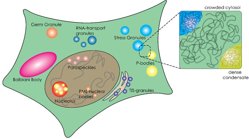
Int. J. Mol. Sci. 2020, 21, 5908                                                                       4 of 20

the effect of crowding on such disordered biomolecules is far from trivial. A detailed account of the
role of crowding on disordered proteins was recently written by Fonin et al. [28]. Here, we highlight
two cases that are of particular relevance for LLPS.
     First, intrinsically disordered proteins or regions of proteins (IDPs/IDRs) can become more
compact under crowded conditions, which could lead to secondary structure formation of so-called
foldons [19,28–31], for example, histone protein 1 (H1), a protein that is involved in chromatin regulation
and has recently gained attention as condensate regulator through the C-terminal IDR [32,33]. In the
case of H1, the C-terminal domain (CTD) was shown to gain secondary structure in the form of a
molten globular domain under polyethylene glycol (PEG) and Ficoll-70 conditions [34]. Furthermore,
secondary structure formation, including α-helix and β-turns, was also observed upon binding
of DNA [35], indicating the CTD of H1 acts as a semi-foldon. However, when studied in LLPS,
extensive NMR analysis by Stott and co-workers showed that the CTD remains unstructured even
upon complexation with DNA in a crowded condensed form [32].
     Second, crowding promotes intermolecular interactions of proteins, which can lead, for example,
to protein aggregation [36–38]. For example, synuclein shows enhanced aggregation under crowded
conditions [37,38]. This process is relevant to phase separation as many proteins such as fused in
sarcoma (FUS) and heterogenous nuclear ribonucleoprotein A1 (hnRNPA1) have shown a transition
from a dynamic fluid state to a solid aggregate state [39,40]. These solidification transitions have been
linked to neurodegenerative diseases such as Alzheimer’s disease. In Section 4.3 we will discuss some
examples where crowding changes the material properties of condensates in vitro.

2.3. From Assembly to Reactions
     Crowding not only affects the structure and assembly of biomolecules, it also influences the rates
of biochemical reactions [3,22]. It is commonly accepted that increasing crowdedness initially leads to
enhanced rates of biochemical processes that involve binding or complexation [4,22]. A combination
of higher effective concentrations (see Section 2.1) and a shift in chemical equilibria towards the
energetically favored compact, complex states (see Section 2.2) results in higher predicted rates of
many enzymatic processes. Indeed, crowding has been found to accelerate processes such as gene
expression [41], DNA replication [42] and protein phosphorylation [43]. Moreover, by favoring bound
states, crowding also affects processivity and stochasticity of processes [43,44]. The opposite effect, in
which crowding decreases the activity of certain enzymes, has also been reported, and is generally
attributed to a reduced conformational flexibility of enzymes that require significant changes in
conformation for their activity [45].
     A further increase in crowding typically results in decreasing rates of many biochemical processes,
which implies that the rate goes through a maximum at an intermediate crowder concentration
(Figure 2B) [3,22,23]. The decrease in rate is a result of strongly reduced diffusivities of the biomolecules
involved in a reaction at high crowding [3,46]. The corresponding maximum has been observed
experimentally for cell-free gene expression in solution [47]. It is interesting to note that an enhanced
enzyme activity, similar to what is commonly seen in intermediately crowded conditions, has also been
proposed to occur for “overcrowded” condensates [48–50]. Whether this is a consequence of crowding,
or has a different origin, is still an open question, which is beyond the scope of this review.

2.4. Mimicking Cellular Crowding
     To investigate the role of crowding in biochemical reactions, assembly processes, and LLPS,
in vitro experiments are generally performed using model crowding agents at varying concentrations.
General considerations for selecting an appropriate crowding agent are: (i) the crowding agent should
not interact specifically with the biological system [5,51], and (ii) the crowding agent should be highly
water soluble [5,16]. As we mentioned earlier, roughly 30% of the cellular volume is occupied by
macromolecules. Therefore, these crowding agents should reach concentrations up to 300 mg·mL−1
Int. J. Mol. Sci. 2020, 21, 5908 5 of 20
without precipitation [2,51]. The combination of these two criteria means that multiple crowding
agents should be tested to ensure that observed effects are not specific [5].
Polymers such as PEG and Ficoll are widely used as model crowding agents. These polymers
might be relatively inert in most systems, but they are very different from most cellular components.
As a step towards cellular crowding, highly water-soluble proteins have been introduced, such as
BSA and lysozyme [19,46,52]. Considering the first criterium of the crowder being an inert molecule,
proteins increase this complexity due to the large variety of amino acids that could introduce weak
non-covalent interactions. Furthermore, an advantage of polymeric crowders is that they are usually
available in a the large variety of sizes (e.g., PEG can range from 2.5 kDa up to 20 kDa), allowing
systematically varying the size of the crowders, while proteins have a fixed size (e.g., BSA is 68 kDa
and lysozyme 14 kDa) [5,52].
Although these synthetic crowders have given important insights into the thermodynamic effects
of ideal crowding, it is questionable how well Ficoll, dextran or PEG can mimic the cytosol. To make
this point, Elcock wrote in 2010: ‘I never once came across one [paper] that began: Ficoll and dextran
are incredibly important molecules and it is therefore vital that we understand their effects on protein
folding and protein-protein associations’ [53]. Therefore, bottom-up approaches have recently sought
to better understand the role of cellular crowding by using purified and concentrated lysates as natural
crowding solutions. For example, Huck and co-workers managed to condense Escherichia coli lysate,
and found enhanced transcription and translation rates compared to the dilute phase [41]. Furthermore,
at a single protein level, lysate showed non-specific interactions that inhibited folding of a FRET-based
crowding sensor, while the same sensor did show enhanced folding in Ficoll and PEG-rich media [26].
The usage of dense lysate thus narrows the gap between complicated in cell (in vivo) experiments (e.g.,
in cell NMR and super-resolution microscopy) and in vitro experiments using only polymers.
Lysate might sound like the best replacement of polymeric crowders, but it should not be thought
of as the perfect mimic of the real, complex cellular milieu, as Rivas and Minton have pointed
out [18]. First, because lysates have been cleared of lipid components, membrane–tracer interactions
are not present. Second, lysate contains a large amount of uncharacterized proteins which might
interact either specifically or non-specifically with probe molecules. Third, cells are known to contain
microcompartments, and the preparation of lysate could distort the stability or formation of these
compartments. In short, the ideal model crowded environment for systematic in vitro studies does not
exist yet, and to improve our understanding of the role of crowding on structure, dynamics and phase
behavior of biomolecules, a combination of approaches is crucial.
3. Organization of the Crowded Cytosol through Overcrowded Condensates
Cells are not only crowded, they are also compartmentalized. Biomolecular condensates, or
membraneless organelles, are an important class of microcompartments that function as cellular
regulators [51]. These microcompartments stand out from the traditional subcellular compartments,
such as mitochondria and lysosomes, because they lack a surrounding membrane [9]. Their separate
role in cellular organization was first recognized for P-granules [8], but the number of examples grew
rapidly and include stress granules [54,55], paraspeckles [56], P-bodies [57], nuage bodies [58] and
nucleoli [59,60]. These compartments form by condensation of proteins, nucleic acids and enzymes into
dense droplets, which have liquid properties [7,8,59]. To understand the effect of crowding on phase
separation, it is important to first highlight some of the typical structural features of the biomolecules
involved in phase separation.
3.1. Liquid–Liquid Phase Separation of Proteins and Nucleic Acids within Cells
The formation of biomolecular condensates has been linked to liquid–liquid phase separation
(LLPS), a process where macromolecules (e.g., DNA, RNA, and proteins) associate with each other
through weak non-covalent interactions to form dense liquid droplets, as illustrated in Figure 3A [9,11].
Proteins that are enriched in these droplets tend to have extensive intrinsically disordered regionsInt. J. Mol. Sci. 2020, 21, 5908 6 of 20
(IDRs) [61–63]. Harmon et al. describes phase separation of IDR-containing proteins using a model of
stickers and spacers [64]. This model includes some important features to describe the liquid behavior
of condensates, namely: weak non-covalent interactions (stickers), the valency (number of interactions)
and flexibility (IDRs that do not interact) [64,65]. Although Harmon et al. describe the stickers as single
Int. J. Mol. Sci. 2020, 21, x FOR PEER REVIEW 6 of 20
amino acids or short linear amino acids motifs [64], these motifs are not the only factors that can drive
phase
binding separation.
domains and Other examples of interaction
oligomerization domains, domains includeare
both of which structural binding in
widely present domains and
the phase-
oligomerization domains,
separating proteome [9,11]. both of which are widely present in the phase-separating proteome [9,11].
Figure
Figure 3.3. Liquid–liquid
Liquid–liquid phasephase separation
separation ofof proteins and polymers.
proteins and polymers. Illustration
Illustration of
of the
the difference
difference
between (A) associative phase separation which relies on attration between two macromolecules
between (A) associative phase separation which relies on attration between two macromolecules and
(B)
andsegregative phasephase
(B) segregative separation whichwhich
separation relies on repulsive
relies interactions.
on repulsive (C) The (C)
interactions. three model
The threesystems
model
from
systemsSection 4 which
from Section contains
4 which poly-U–spermine;
contains poly-U–spermine; FUS;
FUS;NPM1
NPM1condensates.
condensates. Poly-U–spermine
Poly-U–spermine
condensates
condensates relyrely solely
solely on
on charge
charge interactions
interactions between
between RNA
RNA (negative)
(negative) and
and spermine
spermine (positive).
(positive).
Increasing complexity, FUS has cation–π interactions between arginine (RGG
Increasing complexity, FUS has cation–π interactions between arginine (RGG motif, cation) motif, and
cation) and
tyrosine
tyrosine ([G/S]Y[G/S]
([G/S]Y[G/S] domain, domain, π) residues
π) residues within thewithin the disordered
disordered domains.domains. RNA play
RNA could coulda play a role
role but is but
not
is not included
included in the crowded
in the crowded studiesstudies discussed
discussed in Section
in Section 4. Finally,
4. Finally, NPM1 hasNPM1 has an oligomerization
an oligomerization domain
domain and a nuclear-binding
and a nuclear-binding domain in domain in addition
addition to chargetointeractions.
charge interactions.
low-complexity domains
A variety of motifs in low-complexity domains (LCDs)
(LCDs) have
have been
been identified
identified as
as weak
weak binders.
binders.
Such motifs
motifs include arginine-rich motifs (RGG) as found in FUS [66,67], Ddx4 [58,61], and[68,69],
include arginine-rich motifs (RGG) as found in FUS [66,67], Ddx4 [58,61], and LAF1 LAF1
prion-like domains (PrLD)
[68,69], prion-like domains containing
(PrLD) repeats [G/S]Y[G/N/A/S]AQ
containing as found in FUSas[39,67,70,71].
repeats [G/S]Y[G/N/A/S]AQ found in FUS The
RGG and PrLD domains can form cation–π interactions. These are the most important
[39,67,70,71]. The RGG and PrLD domains can form cation–π interactions. These are the most interactions of
FUS and will
important be furtherofdiscussed
interactions FUS and in Section
will 4 (Figure
be further 3C). in Section 4 (Figure 3C).
discussed
domains can be found in the form of RNA-recognition motifs (RRM), zinc
Structural binding domains
fingers (ZnF) and nucleic acid binding domains (NBD), which all interact with RNA and can increase
Structural domains
certain sequence specificity [67,72]. Structural domains areare not
not limited
limited to
to RNA
RNA binding
binding domains;
domains;
some also involve protein–protein interactions, for example, through PRM binding domains and SH2
domains [65,73].
[65,73].Finally,
Finally,oligomerization
oligomerization domains
domainsare important for increasing
are important the number
for increasing of binding
the number of
sites of a sites
binding protein.
of aG3BP1
protein.is known
G3BP1 tois form
known dimers, doubling
to form dimers,the number the
doubling of binding
numbersides [74]. A sides
of binding more
[74]. A more remarkable example is nucleophosmin (NPM1) which forms pentamers. This five-fold
increase is important for phase separation with rRNA as it relies on a single NBD (Figure 3C) [75].
3.2. The Role of RNA in Liquid–Liquid Phase SeparationInt. J. Mol. Sci. 2020, 21, 5908 7 of 20
remarkable example is nucleophosmin (NPM1) which forms pentamers. This five-fold increase is
important for phase separation with rRNA as it relies on a single NBD (Figure 3C) [75].
3.2. The Role of RNA in Liquid–Liquid Phase Separation
In addition to proteins, many biomolecular condensates are also enriched with RNAs. Although
most condensates that are enriched in RNA contain RNA binding proteins [63,76,77], there is strong
evidence that RNA–RNA interactions contribute to phase separation as well [78]. The lab of Gladfelter
has shown that RNA structure dictates the formation of distinct Whi3 condensates both in vivo and
in vitro [79,80]. The importance of RNA within biomolecular condensates has recently been reviewed
by Roden and Gladfelter [81].
3.3. Biophysical Properties of Membraneless Organelles
As a consequence of phase separation, the interior of these membraneless compartments is even
more crowded than the cytoplasm. It has been proposed that this overcrowding could play a role in
several suggested functions of MLOs, such as activation (e.g., nucleation of fibrils) and changing reaction
kinetics (e.g., bringing reactants in close proximity) [9,50,82,83]. Another consequence of overcrowding
is the reduced water content of condensates, which can affect other noncovalent interactions involved,
such as base pairing and host–guest binding [84]. A recent example was given in the case of the DEAD
box helicase Ddx4, which forms condensates through simple phase separation [58,61,85]. Due to the
relatively hydrophobic nature of the amino acids of Ddx4, the interior of Ddx4 condensates is more
similar to organic solvents, such as DMSO, than to water [85]. These condensates are able to “filter”
ssDNA from a pool of both dsDNA and ssDNA by preferential uptake [58,85]. Thus, biophysical
properties of condensates could affect sequestration of molecules and reaction selectivity [86].
To determine the molecular mechanisms underlying MLO formation and functioning, in vitro
studies are indispensable and have already provided tremendous insight into the characteristics
and potential roles of MLOs on biomolecular processes [9,11,50,76,82,83,86–94]. In these in vitro
experiments, the effects of pH, temperature and ionic strength on phase behavior are generally
explored. However, the effect of macromolecular crowding on LLPS is usually not discussed in great
detail, while crowding agents are commonly added to in vitro LLPS models. It is therefore pertinent to
look into the relation between macromolecular crowding and LLPS to understand the relevance of
these studies for LLPS in living cells.
4. How does Crowding Affect Liquid–Liquid Phase Separation?
Macromolecular crowding agents such as PEG, Ficoll and dextran are commonly used in cell-free
LLPS studies, as summarized in Table 1. Some of the earliest reported in vitro phase separated
condensates, such as the Nck/N-WASP [65], Ddx4 [58], and LAF-1 [69] were formed in non-crowded
buffers. However, as research into phase separation of cytoplasmic proteins progressed, the use
of macromolecular crowders became more ubiquitous. Currently, crowding agents are added in
almost all studies of in vitro model MLOs (Table 1). As an example, Tau [95–97], G3BP1 [97,98],
hnRNPA1/3 [40,68,99,100], EFhd2 [97], and TAF15 [99] are generally studied in the presence of 10%
(polymeric) crowders. These polymeric crowders vary in molecular weight: commonly used Ficoll-70
has an average molecular weight (Mw ) of 70 kDa; PEG is used in a range of 3.35 kDa up to 20 kDa. It is
important to consider the Mw of crowders as these can have different effects on phase separation, as
shown for lysozyme [101].
In studies of protein condensates from the nucleoplasm, crowding agents are also commonly
added. To study phase separation of the polymerase and mediator complex, Guo and co-workers used
16% Ficoll-400 [102], a much larger crowder than used in studies on cytosolic stress granule components,
where Ficoll-70 is typically used. Proteins involved in nucleolus formation, nucleophosmin (NPM1) and
fibrillarin (FBL/Fib1), have been exposed to three types of crowders as well in varying concentrations,
ranging from no crowding to 10–15% crowding [103–105]. Although the degree of crowding in theInt. J. Mol. Sci. 2020, 21, 5908 8 of 20
nucleoplasm is considered to be higher than in the cytoplasm, so far in vitro studies make no clear
distinction between the two with respect to the size or concentration of crowding agents added.
After analyzing the studies summarized in Table 1 for the use of crowding agents, we noticed that
systematic studies on the effect of crowding remain scarce. Nonetheless, we can distinguish at least
three main effects caused by crowding: (i) macromolecular crowding can induce LLPS (Section 4.1);
(ii) macromolecules may co-condense or partition into condensates (Section 4.2); (iii) macromolecular
crowders can change the biophysical properties of condensates (Section 4.3).
4.1. Crowding-Induced Phase Separation
Not many studies on biomolecular phase separation systematically analyze the influence of
crowding; even the simple presence or absence of crowding agents in buffers used is not always
explicitly stated in papers. However, there is ample evidence that crowding does affect LLPS. Two
examples that highlight the importance of crowders for the formation of condensates are: (i) for TIA-1,
a stress granule protein, there were no clear condensates detected in the absence of crowding. The
addition of 10% PEG was crucial to detect condensates by microscopy. (ii) FCA, a floral repressor
protein, showed very small dots without PEG, but the addition of 10% PEG increased the dots to tens
of microns, sizes that are similar to what is observed in other in vitro phase separation experiments.
In most cases, the addition of crowding agents leads to a decrease in the critical protein concentration
required for phase separation, or it is essential to observe phase separation at all (Table 1). Crowding thus
appears to induce phase separation in mixtures that would not show phase separation without crowders.
In the case of hnRNPA1 and FUS, for instance, crowding was found to alter the phase diagram towards
condensate formation at a lower protein concentration. Lin and co-workers showed that at 10% BSA, the
critical concentration of hnRNPA1 required for phase separation shifted from the micromolar regime to
nanomolar regime [100]. Similarly, Kaur observed a minimal FUS concentration of 0.5 µM when exposed
to 17.5% PEG-8k [106]. More recently, Yang and co-workers showed that doubling the Ficoll concentration
to 10%, caused a 4-fold reduction in the critical G3BP1 concentration [97]. These results show a strong
correlation between both polymeric and protein crowders and the critical protein concentration for phase
separation. While it has been suggested that the response of IDRs to crowding is very heterogeneous,
because IDRs could either (partially) fold or unfold depending on the amount type of crowding agent [28],
the examples reported in literature (Table 1) mostly suggest that crowding enhances phase separation. It is
possible that this correlation is biased, however, as the diversity of crowding agents used is limited to PEG,
Ficoll and dextran.
An open question remains what the mechanism behind crowding-induced phase separation is.
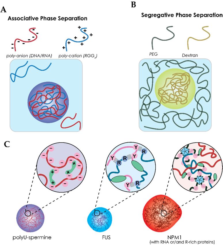
We propose that three effects could lead to enhanced phase separation by crowding agents (Figure 4B).
First, according to classical crowding theories, volume exclusion by the crowding agents could lead
to enhanced intermolecular attractions between proteins and between proteins and nucleic acids
(Figure 4B,I). As a result of stronger attraction, lower concentrations of protein are sufficient to nucleate
a condensed phase. Second, macromolecules could interact specifically with the proteins (Figure 4B,II).
When the interactions between the “crowding” agents and biomolecules are favorable, this could
enhance phase separation through co-condensation. Note that this is not a crowding effect, but
rather a form of associative phase separation. Evidence from several in vitro studies indicate that
even so-called inert crowding agents, like PEG and Ficoll, could be accumulated inside condensates
instead of being excluded, and we will discuss this phenomenon in Section 4.2. Third, the solubility of
proteins could change in the presence of crowders, which is typical of segregative phase separation
(Figure 4B,III). A classical example of segregative phase separation between synthetic polymers is the
combination of PEG and Ficoll [107]. Similarly, Julius et al. showed phase separation of lysozyme and
PEG [101]. It is not unlikely that polymeric or protein-based crowding agents have a similar effect
on the solubility of many biomolecules, in particular if they contain relatively hydrophobic sticker
motifs [64]. By extension, increasing the degree of crowding in an already phase separated mixture,
could lead to changes in density and fluidity of condensates, and ultimately result in aggregation. WeInt. J. Mol. Sci. 2020, 21, 5908 9 of 20
further discuss the effect of crowding on the material properties of condensates in Section 4.3, based on
several case studies.
Table 1. Crowding agents that have been used to induce or promote protein phase separation in vitro.
Membraneless Crowding
Protein Crowder [Crowding] 1 Ref.
Organelle Necessary?
FUS Stress Granules PEG 2–30% No [39,67,99,108]
Dextran 10% [39,67,99]
Ficoll-400 15% [109]
BSA 10% [100]
G3BP1 Stress Granules PEG (20k) 1–8% Yes [98]
Ficoll-400 5–20% [97]
TIA-1 Stress Granules PEG 10% Yes [110]
Tau (K18) Stress Granules PEG 7.5% No [95]
Tau-187 PEG 10% Yes [96]
TDP-43 Stress Granules Dextran 10% Yes [99,111]
hnRNPA1 Stress Granules PEG 10–20% No [40,100]
Ficoll-70 10% No [40,100]
Dextran 10% [67]
BSA 1.5–10% [63]
Yeast lysate 1% [63]
hnRNPA3 Stress Granules Dextran 10% No [67]
Efhd2 Stress Granules PEG 10% Yes [97]
EWSR1 Stress Granules Dextran 10% No [67]
TAF15 Stress Granules Dextran 10% No [67]
FMRP Neuronal Granules PEG 30% No [108]
FXR1 Neuronal Granules BSA 3% Yes [112]
SPOP/cDAXC Nuclear Speckles Ficoll 4–10% Yes [113]
FBL Nucleolus Dextran 10% No [105]
NPM1 Nucleolus PEG 5–15% Yes 2 [103,104]
Dextran 15% [103]
Ficoll 15% [103]
Ddx3x Stress Granules PEG 10% Yes
LAF1-(RGG Germ Granules
Dextran 1% No [68] 3
domain) (P-granules)
FCA (plant) nuclear bodies PEG 10% No [114]
Pol-II-CTD Transcriptional bodies PEG 10% Yes [102]
Ficoll-400 16% [102]
NusA Bacterial Bodies Dextran 10% Yes [115]
Brd4S Nuclear puncta PEG 2–4% Yes [116]
HeLa nuclear extract 0.3% [116]
BuGZ Spindle bodies PEG 10–40% Yes [117]
SH35 /PRM5 Synthetic Ficoll-70 2.5–40% No [118]
CBM-eADF3-CBM Synthetic Dextran 1–14% No [119]
Ficoll 1–14% [119]
1 2
Concentration crowding agent. Crowding is necessary for homotypic liquid-liquid phase separation (LLPS).
3 Dextran was not used as a crowder, but to study partitioning of different-sized dextran polymers.Int. J. Mol. Sci. 2020, 21, 5908 10 of 20
Int. J. Mol. Sci. 2020, 21, x FOR PEER REVIEW 10 of 20
Figure 4.
Figure 4. Cellular
Cellular condensates
condensates might arise from both associative associative and segregative phase separation.
(A) Phase
(A) Phase separation
separation results
results in
in aa crowded
crowded protein-dense
protein-densephase
phase that
that is
is situated
situated in
in the
the crowded
crowded cytosol
cytosol
(or nucleoplasm).
(or nucleoplasm). (B) (B) Three effects on phase separation caused by macromolecules:
on phase separation caused by macromolecules: (I) LLPS
LLPS is is
induced through
induced through depletion
depletion forces,
forces, increasing
increasing thethe attractive
attractive forces
forceswithin
withinthethecondensate.
condensate. (II)
(II) LLPS
LLPS isis
promoted through
promoted through co-condensation
co-condensation of of macromolecules
macromolecules through
through attractive
attractive interactions
interactions between
between the the
macromoleculesand
macromolecules and thethe proteins.
proteins. (III) LLPS
(III) LLPS is promoted
is promoted through segregation,
through segregation, as repulsiveasinteractions
repulsive
interactions
between between macromolecules
macromolecules and proteins and lowerproteins lower the
the solubility solubility of proteins.
of proteins.
4.2. Macromolecular
4.2.1. PartioningofMisleading
Attractive Interactions Mechanisms
Macromolecules of Phase Separation
can Regulate Regulators
Phase Separation
As indicated, many
Macromolecules thatmacromolecular
have been used crowders have been
to study crowding areshown to induce
normally phaseneutral
considered separation
inert
(Table 1). However, if they act as crowders to push other proteins together and facilitate
molecules [16]. Ghosh and co-workers showed that, depending on the strength of the interactions, phase
separation, they should
macromolecules can eitherbe promote
excludedor from the phase
inhibit condensed phase[118].
separation [16]. Using
While computational
most studies have not
models,
looked into the localization
they predicted of crowding agents,
that macromolecules there are
with strong indications
attractive that some could
interactions prototypical
promotecrowding
phase
agents do not always behave like ideal inert species that are excluded from condensed
separation [118] by co-condensation. As a model system, they used the complex phase separation phases. In this
of
section, we will
SH35 with PRM discuss how the spatial distribution of macromolecules can reveal more about the role
5 proteins designed by Rosen’s lab [66,120]. Heparin, a negatively charged
of macromolecules
macromolecule thatasattracts
regulators
the of phase separation.
positively charged PRM5, promoted phase separation and became
accumulated in the protein-dense phase [118]. This process is dependent on the concentration of
4.2.1. Attractive Interactions of Macromolecules can Regulate Phase Separation
heparin showing a reentrant behavior similar to what has been observed for RNA–protein
Macromolecules
condensates [118,121].that have been used to study crowding are normally considered neutral inert
molecules [16].example
A second Ghosh and co-workers
where showed
PEG promotes that,separation
phase dependingby onassociation
the strengthhasofbeen
the interactions,
reported by
macromolecules can either promote or inhibit phase separation [118]. Using
the lab of Keating, who pioneered RNA-based complex coacervates as MLO model systems [122– computational models, they
predicted that macromolecules
124]. Marianelli and co-workers with
usedstrong attractiveformed
coacervates interactions
from could promote
negatively phasepoly-U
charged separation
RNA[118]and
by co-condensation.
positively As a model
charged spermine system,
(Figure 3C),they
andused the complex
studied the effectphase separation of SH3
of macromolecular 5 with on
crowding PRM the
5
proteins designed by Rosen’s lab [66,120]. Heparin, a negatively charged
phase transition using PEG (8 kDa) and Ficoll-70 (70 kDa) [122]. While PEG decreased the criticalmacromolecule that attracts
the positively
charge charged poly-U/spermine
ratio between PRM5 , promoted phase separation
coacervates and to
(similar became accumulated
the examples in the4.1),
in Section protein-dense
this effect
phase [118]. This
was negligible in process is dependent
the presence on the
of Ficoll-70. concentration
Thus, the effect ofofPEG heparin
couldshowing a reentrant
not be explained bybehavior
classical
similar to what has been observed for RNA–protein condensates [118,121].
volume exclusion theory. Experiments with labelled Ficoll-70 and PEG revealed that only PEG was
A second
excluded fromexample where PEG
the coacervates, whilepromotes
Ficoll wasphase separation
enriched insidebytheassociation has been reported
dense coacervate phase [122]. by
the lab of Keating, who pioneered RNA-based complex coacervates as MLO
Therefore, the enhanced phase separation effect of Ficoll cannot be attributed to crowding. The model systems [122–124].
Marianelli and co-workers
authors proposed usedethylene
that PEG (and coacervates
glycol)formed
could fromdistortnegatively charged poly-U
the RNA secondary structure RNA and
through
positively
exclusion ofcharged
water spermine
[122]. PEG(Figure 3C), and
destabilizes the studied the effect of
poly-U structure, macromolecular
which crowding
results in a more on the
extended U
phase transition using PEG (8 kDa) and Ficoll-70 (70 kDa) [122]. While PEG
polymer, facilitating the interactions between poly-U and spermine and resulting in enhanced phase decreased the critical
charge ratio Conformational
separation. between poly-U/spermine
changes in coacervates
RNA structure (similarhaveto the
beenexamples
shown into Section 4.1), this
have large effect
effects on
was negligible
condensate in the presence
selectivity [79] and,oftherefore,
Ficoll-70. crowding
Thus, the effect
effectsofshould
PEG could not be explained
be carefully considered byasclassical
well.
A third type of regulator that is not directly linked to crowding is the weak interactor that
inhibits phase separation of two complex-forming biomolecules. Ghosh and co-workers proposed
that weak attractive interactions between the regulator and one of the phase separating moleculesInt. J. Mol. Sci. 2020, 21, 5908 11 of 20
volume exclusion theory. Experiments with labelled Ficoll-70 and PEG revealed that only PEG was
excluded from the coacervates, while Ficoll was enriched inside the dense coacervate phase [122].
Therefore, the enhanced phase separation effect of Ficoll cannot be attributed to crowding. The authors
proposed that PEG (and ethylene glycol) could distort the RNA secondary structure through exclusion
of water [122]. PEG destabilizes the poly-U structure, which results in a more extended U polymer,
facilitating the interactions between poly-U and spermine and resulting in enhanced phase separation.
Conformational changes in RNA structure have been shown to have large effects on condensate
selectivity [79] and, therefore, crowding effects should be carefully considered as well.
A third type of regulator that is not directly linked to crowding is the weak interactor that inhibits
phase separation of two complex-forming biomolecules. Ghosh and co-workers proposed that weak
attractive interactions between the regulator and one of the phase separating molecules would result in
competition between the regulator and the other phase separating molecule [118]. As an experimental
example, lysozyme was used. In a previous study by Protter and co-workers, proteins such as BSA
and lysozyme were shown to inhibit phase separation of hnRNPA1 [62]. This can be explained by a
comparison of the partition coefficients of lysozyme and heparin for the SH35 /RPM5 system: lysozyme
has a partitioning coefficient of 1, while heparin has a coefficient of 6.
4.2.2. Effect of Macromolecular Size in Partitioning
Besides the type of “crowding” agent, the size of macromolecules used to promote phase separation
could also impact the mechanism by which phase separation is facilitated. As an example, we want to
highlight the partitioning of dextran in a study by Schuster and co-workers [68]. Schuster used condensates
formed by the LAF1 RGG domain to study the partitioning of dextran of varying lengths [69]. While
low molecular weight dextran (4.4 kDa) was enriched in the dense phase, partitioning decreased with
increasing dextran size [68]. Dextran of 10 kDa was found to be the threshold for being equally distributed
between dense and diluted phase. Dextran of high molecular weight (70 kDa) was entirely excluded from
the condensates [68]. For this specific model, the effect of crowding could be studied by using dextran-70.
These examples show that macromolecules can regulate phase separation through partitioning,
and that not all effects of so-called crowding agents on phase separation can be attributed to crowding.
The strength of the interaction between “crowding” agents and proteins is important in these cases, just
like the size of the macromolecules. These insights also have implications for phase separation inside
living cells. Cells contain a large variety of small molecules, which have a weak or strong interaction
with several phase separating proteins, and their partitioning behavior could be affected by crowding.
As an example, Marianelli found that an oligo RNA exhibited a two-fold higher partitioning coefficient
under crowded conditions [122].
4.3. Crowding Affects Biophysical Properties of Condensates
For many biomolecular condensates, there exists a very fine phase boundary between the
liquid-to-gel (or solid) state [125]. When interactions are strengthened by the excluded volume effect
and depletion, it is possible that condensates are pushed across this boundary from liquid to solid.
Here, we will discuss three recently studied systems, in which the material properties of condensates
have been studied under crowded conditions.
4.3.1. The Effect of Crowding on FUS
Solidification of protein condensates (e.g., stress granules) has been linked to various neurodegenerative
diseases such as Alzheimer’s and Parkinson’s disease, where condensates lose their liquid properties
and start forming amyloid fibrils [40,100]. Molliex and Lin explained that this transition from liquid to
solid state is due to stabilization of the prion-like domains (PrLD) of RNA-binding proteins, also called
maturation [40,100]. As the proteins (generally FUS, but also other stress granule components) require
crowders to form condensates, Kaur and co-workers studied this specific effect for FUS.Int. J. Mol. Sci. 2020, 21, 5908 12 of 20
Fluorescence recovery after photobleaching (FRAP) is a technique commonly used to test whether
biomolecules in the dense condensate phase are mobile or immobile [126]. The faster the recovery of
the fluorescence signal after bleaching a spot, the faster the diffusion of fluorescently labelled molecules.
Kaur’s study showed that full-length FUS (FUSFL ) transits from a viscous fluid to a viscoelastic gel-like
state in a crowding dependent manner [106]. This gradual effect was also observed for the FUS RGG
domain, but not for the FUS prion-like domain (FUSPrLD ). The FUSPrLD showed a more switch-like
behavior: above 15% PEG, no recovery was observed for FUSPrLD . This effect was independent of the
molecular weight of PEG and dextran, and was attributed to the general increase in intermolecular
interactions caused by volume exclusion.
4.3.2. The Effect of Crowding on NPM1
Kriwacki’s lab observed a similar reduced mobility effect in nucleophosmin (NPM1)
condensates [104,105]. Under non-crowded conditions, NPM1 requires a second macromolecule
to form condensates, such as ribosomal RNA (rRNA) or proteins with an arginine-rich motif (e.g.,
surfeit locus protein 6 (SURF6)) [72]. When adding crowders such as Ficoll or PEG, Mitrea and
co-workers observed homotypic NPM1 droplets that arise from interactions between acidic and
basic tracks [105]. They then questioned how crowding influences the material properties of these
NPM1–NPM1-rich phases, and by extension, the nucleolus.
Ferrolino and co-workers discovered that the mobility of proteins in the dense phase of NPM1
homotypic droplets decreased rapidly under increasing crowding conditions [103]. Where the FRAP
recovery was reduced 2-fold at 5% PEG, there was almost no recovery at 15% PEG. As PEG was not
accumulated in the dense phase, this effect was attributed to the excluded volume effect that compacts
the disordered regions [103,127]. Maybe even more striking was the observation that NPM1 homotypic
droplets do age like stress granule condensates. After 3 h, the dense phase became immobile, although
this effect was reversible, in contrast to FUS ageing [40,103].
The situation was different for heterotypic droplets containing the arginine-rich protein SURF6.
SURF6 accumulates in the dense NPM1 droplets and under crowded conditions, and SURF6 itself
was mobile within the condensates at all tested ratios [103]. However, at low SURF6/NPM1 ratios,
NPM1 behaves more like homotypic condensates. When the SURF6-to-NPM1 ratio was increased to 4,
NPM1 showed a significantly higher FRAP recovery. Furthermore, Ferrolino discovered that at all
ratios, the homotypic NPM1 is predominantly present at the interface, suggesting a core–shell-like
structure. This could give more insight into how the fibrillar component of the nucleolus is structured.
4.3.3. Crowding Has No Effect on Protein Mobility of a Synthetic Silk-Like Protein
The previous two examples of both FUS and NPM1 showing reduced mobility under crowded
conditions indicate a transition from a viscous to a more gel-like state. However, within cells, many
of these condensates do show liquid-like behavior and they do recover after photobleaching [8,128].
Lemetti and coworkers engineered a silk-like protein CBM–eADF3–CBM (cellulose binding modulus,
CBM, linked by a silk repeat containing A- and Q-rich blocks, eADF3) that showed similar
unchanged biophysical properties upon crowding [119]. While crowding with dextran did decrease the
concentration required for phase separation, the FRAP recovery was not affected—almost full recovery
was observed within 30 seconds. Apparently, the effective density and viscosity of the condensed
phase of CBM–eADF3–CBM was not significantly affected by crowding.
5. Conclusions and Outlook
This review focused on the role of crowding in driving and shaping phase transitions of
biomolecular components. Although major progress has been made in understanding the phenomenon
of LLPS and the drivers of phase separation, the role of the dense crowded environment of the cell
remains underappreciated. Most of our current understanding of the role of crowding comes from
in vitro studies that aim to mimic the crowded cellular environment. The cases we have highlighted inInt. J. Mol. Sci. 2020, 21, 5908 13 of 20 this review show significant changes in the phase diagram, and suggest that crowding plays a very significant role in determining both the stability of mixtures of biomolecules and the extent of the region of phase separation. The addition of so-called crowding agents appears to induce or enhance LLPS in almost all in vitro studies we analyzed, but the mechanism underlying enhanced LLPS remains poorly characterized. We propose that at least three effects could lead to enhanced phase separation by crowding agents: (i) classical volume exclusion by crowding agents leads to enhanced intermolecular attractions, (ii) some macromolecules that are used as crowding agents could interact specifically with phase-separating biomolecules and enhance phase separation through co-condensation, and (iii) crowding agents could decrease the solubility of biomolecules, in a process of segregative phase separation. Moreover, these effects are likely to influence the biophysical properties of already formed condensates as well. In the complex, crowded environment of the cell, both the condensates and the crowding agents are much more diverse and heterogeneous than in any of the in vitro studies, and it is likely that all three effects play some role: weakly attractive proteins or nucleic acids could nucleate condensates, inert proteins exhibit a more idealized excluded volume effect, and weakly repulsive proteins are expected to segregate from each other. Additionally, many factors previously unaccounted for, such as chaperones, specific RNA-binding proteins, siRNAs, and the presence of lipid membranes, could also change the phase behavior of specific condensates. To fully understand each of these contributions, a more systematic approach will be needed. Funding: This research was funded by the Netherlands Organisation for Scientific Research (NWO-STARTUP). Conflicts of Interest: The authors declare no conflict of interest. Abbreviations Brd4S Bromodomain containing protein 4S BSA Bovine serum albumin BuGZ BUB3-interacting and GLEBS motif-containing protein zinc finger protein 207 CBM Cellulose binding domain Da Dalton DAXC Death domain-associated protein-6 Ddx4 DEAD box ATPase protein 4 DNA Deoxyribonucleic acid dsDNA Double stranded DNA EFhd2 EF-hand domain-containing protein D2 EWSR1 Ewing sarcoma breakpoint region 1 protein FBL/Fib-1 Fibulin/Fibrillarin FCA Flowering time control protein FCA FMRP Fragile X mental retardation protein FRAP Fluorescence recovery after photobleaching FRET Förster resonance energy transport FUS Fused in sarcoma protein FXR1 Fragile X mental retardation syndrome-related protein 1 G3BP1 Ras GTPase-activating protein-binging protein 1 H1 Histone protein 1 hnRNPA1 Heterogenous nuclear ribonucleoprotein A1 hnRNPA3 Heterogenous nuclear ribonucleoprotein A3 IDP Intrinsically disordered protein IDR Intrinsically disordered region LAF1 Transcription factor protein LAF1 LCD Low complexity domain LLPS Liquid-liquid phase separation MLO Membraneless organelle NBD Nucleic acid binding domain Nck Cytoplasmic protein Nck-1
You can also read