Roles of Long Non-coding RNAs in X-Chromosome Inactivation
←
→
Page content transcription
If your browser does not render page correctly, please read the page content below
Roles of Long Non-coding RNAs
in X-Chromosome Inactivation

J. Mauro Calabrese and Terry Magnuson

1 Introduction

Female mammals silence the majority of genes along one of their two X chro-
mosomes in a process termed X-chromosome inactivation (XCI). XCI likely
evolved in mammals as the X and Y chromosome, once homologous autosomal
pairs, diverged in sequence, largely through degeneration of the Y. This degen-
eration left males with only one functional copy of most X-linked genes, neces-
sitating the development of a compensation process that would equalize X-linked
gene dosage between the sexes (Livernois et al. 2012).
    XCI is critical for mammalian development. Severe defects in the process are
developmentally lethal, while abnormalities in X-chromosome dosage, which
occur in about 1 of 500 live births, can be pleiotropic disorders, associated with
forms of intellectual disabilities, infertility, and autoimmunity (Powell 2005). The
importance of regulating X-linked gene dosage is underscored by the chromo-
somal counting process inherent to XCI. Regardless of the total number of X
chromosomes an individual has, XCI ensures that one X per diploid genome
remains active, with the remainder subject to inactivation, in both males and
females. For example, XCI tends to silence two X’s in tetraploid female cells, and
only one in tetraploid male/female cell fusions (Monkhorst et al. 2008). In both
cases, the ratio of one active X per diploid genome is maintained. Similarly, in
humans, XCI shuts down two X’s in females with three (Triple X Syndrome), and
one X in males with two (Klinefelter’s Syndrome); the sole X in females with
Turner’s syndrome remains active. These chromosomal abnormalities are often
accompanied by chronic health issues (Powell 2005), indicating imperfect regu-
lation of X-linked dosage. However, the intrinsic capability of mammalian cells,
male or female, to sense and at least partially deal with abnormalities in

J. M. Calabrese (&)  T. Magnuson
Department of Genetics, Carolina Center for Genome Sciences, and Lineberger
Comprehensive Cancer Center, University of North Carolina, Chapel Hill, NC 27599, USA
e-mail: jmcalabr@email.unc.edu

A. M. Khalil and J. Coller (eds.), Molecular Biology of Long Non-coding RNAs,           69
DOI: 10.1007/978-1-4614-8621-3_3, Ó Springer Science+Business Media New York 2013
70 J. M. Calabrese and T. Magnuson X-chromosome dosage is remarkable and speaks to the physiological importance of XCI. In addition to its role in development and human health, XCI has emerged as a paradigm for epigenetic silencing mediated by noncoding RNA (ncRNA), given the critical role of Xist and other ncRNAs in the process. Advances in DNA sequencing technologies have led to the identification of thousands of ncRNAs expressed by the mammalian genome, many of which are developmentally reg- ulated and conserved (Dunham et al. 2012; Derrien et al. 2012; Cabili et al. 2011). Early studies have shown these RNAs have critical functions in a range of bio- logical processes, including stem cell maintenance, regulation of the DNA damage response, and developmental specification (Guttman and Rinn 2012). XCI was one of the first identified gene regulatory processes in mammals with a conserved role for ncRNAs (Brockdorff et al. 1992; Brown et al. 1992). Therefore, as the importance of ncRNA-mediated gene regulation has become broadly apparent, XCI has remained a flagship model for understanding ncRNA function. In the pages below, we describe the major features of XCI, with particular focus on the diverse roles that ncRNAs play in the process. 2 XCI Overview In the mouse, historically the field’s most utilized experimental model, XCI occurs in two waves during early development. The first is termed imprinted XCI, due to the exclusive inactivation of the paternally inherited X chromosome (Takagi and Sasaki 1975). Imprinted XCI occurs rapidly after formation of the zygote, initi- ating at the 4-cell stage of development, and nearing completion for some paternal loci at the formation of the early blastocyst, around the 32-cell stage (Kalantry et al. 2009; Okamoto et al. 2005; Patrat et al. 2009; Williams et al. 2011). This stark parent-of-origin bias appears to be independent of the meiotic sex chromo- some inactivation that occurs in the male germline (Okamoto et al. 2005), and instead is due to an imprint placed on the maternal X during oocyte maturation, which somehow blocks XCI from occurring on the chromosome (Tada et al. 2000). Cells of the extraembryonic lineage propagate a paternally derived inactive X (Xi) throughout their existence (Takagi and Sasaki 1975; West et al. 1977). In contrast, XCI is reversed in the inner cell mass (ICM) of the blastocyst, which gives rise to the embryo proper (Mak et al. 2004; Okamoto et al. 2004). Postim- plantation, XCI re-occurs in the epiblast, nearing completion around embryonic gestational day (E) 6.5 (Rastan 1982). In this second wave, termed random XCI, the choice to inactivate a given X is largely random and independent from its parent-of-origin (McMahon et al. 1983). Random XCI is maintained in all cells save the germline (Sugimoto and Abe 2007), resulting in adult females who are mosaics of paternally and maternally derived Xi’s. Not all mammals share the biphasic inactivation strategy of the mouse. While rats and cows show imprinted XCI in their extraembryonic tissue (Xue et al. 2002;
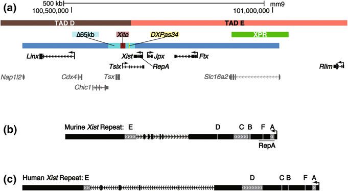
Roles of Long Non-coding RNAs in X-Chromosome Inactivation                                    71

Wake et al. 1976), suggesting a mouse-like biphasic inactivation strategy, other
eutherian mammals examined to date—humans, horses, and mules—appear to
undergo random XCI in all lineages (Moreira de Mello et al. 2010; Wang et al.
2012). In contrast, metatherians, such as the kangaroo and opossum, inactivate
their paternally inherited X in all tissues (Sharman 1971; Grant et al. 2012).

3 Control of XCI via the X-Inactivation Center

Studies of balanced chromosomal translocations in the mouse mapped the location
of a single X-linked region that invariably tracked with inactivation of adjoining
X-linked DNA, and often led to partial silencing of the fused autosome (Lyon, M.
F., Searle A. G., & International Committee on Standardized Genetic Nomen-
clature for Mice 1989). Because of the region’s ability to inactivate neighboring
DNA, it was proposed to contain the cis-mediated genetic signals required to

Fig. 1 Xist and the X-inactivation center. a The protein coding genes, noncoding RNAs, and
regulatory elements of the murine X-inactivation center, depicted to scale relative to UCSC
genome build mm9. Genes and regulatory regions in black text denote those discussed in the text
with documented or proposed roles in XCI. Genes in grey text have no known roles in XCI.
Exons and introns are depicted as solid bars and hashed lines, respectively. Regulatory regions
are depicted as colored bars above genes. Denoted TADs are those described in (Nora et al.
2012). The large blue bar spanning the majority of Fig. 1a denotes the genomic span of bacterial
and yeast artificial chromosomes that recapitulate aspects of XCI when integrated as multicopy
transgene arrays into mouse cell lines (Heard et al. 1999; Lee et al. 1996). b, c Mouse and human
Xist genomic loci. Exons and introns are depicted as in (a). Exonic regions in grey mark the
location of the six annotated Xist repeats, A through F, as described in (Brockdorff et al. 1992;
Brown et al. 1992; Nesterova et al. 2001). The location of the RepA transcript within the murine
Xist locus is underlined
72                                                       J. M. Calabrese and T. Magnuson

initiate and maintain XCI, and was termed the X-inactivation center (Xic) (Fig. 1a;
(Rastan and Brown 1990)). Subsequent analysis of structurally rearranged chro-
mosomes in humans identified a single homologous Xic, as well (Brown et al.
1991). Since then, a range of genetic and cell biological experiments have defined
several features contained within the Xic that are critical for proper execution of
XCI, including a surprising number of ncRNAs and regulatory elements that
produce ncRNA species. At the top of this regulatory cascade is Xist, which stands
for Xi-specific transcript. Xist is essential for XCI, coating the otherwise inactive
chromosome from which it was expressed. Several other ncRNAs have been
identified within the Xic, including Tsix, Jpx, Ftx, Linx, and RepA, most of which
have documented roles in XCI. Also, at least two critical regulatory regions within
the Xic, DXPas34, and Xite, have themselves been documented to produce RNA.
Most recently, it was discovered that a large ncRNA, termed Xact, is expressed
from the active X specifically in human pluripotent cells. Together with a complex
interplay of transacting factors, many of which remain undefined, the ncRNAs and
regulatory elements over the X establish a remarkably robust system of dosage
compensation that is capable of delivering a single active X (Xa) per diploid
genome, even in the presence of chromosomal abnormalities (Table 1).

4 Xist, A Long Noncoding RNA Required for XCI

One of the more striking cytological features of the Xi is the coating of the
chromosome by the Xist ncRNA, which can be visualized under a fluorescent
microscope via RNA fluorescence in situ hybridization (FISH). Xist was initially

Table 1 Proposed and validated functions of noncoding RNAs and regulatory elements asso-
ciated with XCI
Region     Classification Proposed/Validated   Seminal Reference(s)
                          Function
Xist      NcRNA        Master regulator of    (Brockdorff et al. 1992; Brown et al. 1992;
                          XCI                    Brown et al. 1991)
Jpx       NcRNA        Xist activator         (Tian et al. 2010)
Ftx       NcRNA        Xist activator         (Chureau et al. 2011)
Tsix      NcRNA        Xist repressor         (Lee et al. 1999)
DXPas34   Reg.         Tsix activator         (Courtier et al. 1995; Heard et al. 1993)
             Element
Xite      Reg.         Tsix activator         (Ogawa and Lee 2003)
             Element
Linx      NcRNA        Tsix regulator         (Nora et al. 2012)
RepA      NcRNA        Xist activator, PRC2   (Zhao et al. 2008)
                          recruitment
LINEs     DNA/RNA      Xist spreading, gene   (Chow et al. 2010)
                          silencing
XACT      NcRNA        Xa maintenance         (Vallot et al. 2013)
Roles of Long Non-coding RNAs in X-Chromosome Inactivation 73
identified as a candidate gene to control XCI because of its exclusive expression
from the Xi and its chromosomal localization within the region defined as the Xic
(Brown et al. 1991). Subsequent work defined the major characteristics of the gene
in both human and mouse: It is approximately 17 kb in length, can be detected as
spliced and polyadenylated, and is exclusively nuclear and untranslated (Brock-
dorff et al. 1992; Brown et al. 1992). Multiple spliceforms exist, some of which
appear to lack polyA tails (Brown et al 1991, 1992; Hong et al. 2000; Ma and
Strauss 2005; Memili et al. 2001). Consistent with its classification as a ncRNA,
Xist lacks conserved open reading frames, but does contain up to six regions of
tandemly arrayed repetitive sequence that may be responsible for aspects of its
function (Brockdorff et al. 1992; Brown et al. 1992; Nesterova et al. 2001). These
regions are on the order of 100 bp to 2 kb in length, and several are clearly
conserved between mouse and human (Fig. 1b, c; (Brockdorff et al. 1992; Brown
et al. 1992; Nesterova et al. 2001)).
Notably, recent work has identified an Xi-specific transcript in metatherian
mammals, termed Rsx (Grant et al. 2012). Rsx does not share sequence homology
with Xist, yet, similar to Xist, the RNA is expressed from the Xi, appears to coat
the chromosome in cis, lacks open reading frames, and is enriched for tandemly
repeated sequence at its 50 end (Grant et al. 2012). This apparent functional
conservation without sequence similarity suggests that ncRNA-mediated regula-
tion of dosage compensation arose at least twice during mammalian evolution,
highlighting the general utility of this regulatory strategy for the large-scale
management of gene expression programs.
Genetic ablation of Xist demonstrated its critical role in XCI. Mouse embryonic
stem cells (ESCs), which serve as a useful in vitro model because they have yet to
undergo XCI, show complete, nonrandom inactivation of a wild-type over a
mutant Xist allele during differentiation, which induces XCI in these cells (Penny
et al. 1996). Similarly, maternal inheritance of an Xist deletion results in non-
random inactivation of the wild-type, paternally inherited X in the mouse embryo.
Paternal inheritance of this same deletion results in lethality due to failure of XCI
in the extraembryonic lineages, where the wild-type, maternally inherited X is
resistant to silencing (Marahrens et al. 1997). These studies indicate that an X-
chromosome without Xist cannot undergo stable XCI.
While Xist coats the Xi in virtually every cell that contains one, the ncRNA is
only required during the initiation and early maintenance of the process, at least in
the mouse. Using an inducible Xist transgene integrated into an autosomal locus,
Wutz and Jaenisch were able to show that Xist is only capable of gene silencing in
ESCs up to 48 h postinduction of differentiation with retinoic acid. Before this
time point silencing was reversible and dependent on continued expression of Xist,
whereas afterwards XCI was irreversible even if Xist expression was extinguished
(Wutz and Jaenisch 2000). The in vivo correlate of this time frame is unclear, but it
is likely between E9.5 and 12.5, as deletion of Xist in mouse embryonic fibroblasts
(MEFs), which are frequently derived from these developmental time points, does
not result in X-reactivation (Csankovszki et al. 1999).74 J. M. Calabrese and T. Magnuson Other than gene silencing, coating of the Xi by Xist is the first documented cytological event during initiation of XCI in the mouse, and is seen as early as the four-cell stage of development (Okamoto et al. 2005). Xist stabilization and coating of the Xi is also observed at the onset of random XCI (Panning et al. 1997; Sheardown et al. 1997). The closely coupled timing of Xist coating and XCI’s initiation strongly suggest a role for Xist in the earliest stages of XCI, including the initiation of the process. Rigorous tests examining Xist’s role in initiating XCI in the mouse have thus far yielded conflicting results. To address the question, Kalantry and colleagues measured the kinetics of gene silencing during the earliest stages of imprinted XCI (Kalantry et al. 2009). They made the surprising observation that several X-linked genes exhibited indistinguishable patterns of silencing between wild-type mice and those carrying a paternally inherited Xist deletion at the 8- and 16-cell stage of development. At these early time points, silencing of certain genes was more affected by Xist loss than others, whereas all genes were affected at later time points. The results suggest imprinted XCI can initiate in the absence of Xist in the mouse. Moreover, they support an evolutionary model of XCI, which posits that inactivation evolved in a piece-meal fashion over the X chromosome (Lahn and Page 1999); Kalantry and colleagues found that genes whose silencing was most affected by Xist loss were those thought to be subject to dosage compensation for the longest amount of evolutionary time (Kalantry et al. 2009). In complete contrast, using a similar mutant allele and examining a similar set of X-linked genes, Namekawa and colleagues found that imprinted XCI did not initiate in the absence of Xist, suggesting the opposite conclusion reached by Kalantry and colleagues: Xist triggers the initiation of imprinted XCI (Namekawa et al. 2010). Methodological differences have been proposed to explain the discrepancy between these two studies (Namekawa et al. 2010; Brockdorff 2011). The two works also used different Xist mutant alleles. Whereas the mutant allele used by Kalantry and colleagues removed Xist exons 1 through 3, the mutant allele used by Namekawa and colleagues removed Xist exons 1 through 6 (Kalantry et al. 2009; Namekawa et al. 2010). Nonetheless, both alleles appear to be complete for loss of Xist function, making this difference unlikely to account for the discrepancy between the studies. A favored explanation is that differences between inbred mouse strains account for the differential detection of Xist-independent processes during the initiation of imprinted XCI. Genetic background differences often affect phenotypes of mutant mice, due to the presence of modifier alleles that associate with particular mouse strains; notable examples of this include mutational analyses of the Apc and Egfr genes (Montagutelli 2000). Whereas Kalantry and colleagues utilized F1 hybrids of M. m. musculus and M. m. molossinus mice (Kalantry et al. 2009), Namekawa and colleagues utilized F1 hybrids of M. m. musculus and M. m. castaneous mice (Namekawa et al. 2010). Therefore, differences in modifier alleles between the M. m. molossinus and M. m. castaneous subspecies could have been responsible for the differential detection of Xist sensitivity during the initiation of imprinted XCI. Under this assumption, the studies conducted by Kalantry and Namekawa indicate
Roles of Long Non-coding RNAs in X-Chromosome Inactivation                          75

that imprinted XCI can initiate in the absence of Xist over certain X-linked genes,
but that the strength of Xist-independent initiation varies with genetic background,
such that it is not detectable in M. m. castaneous/musculus hybrids (Kalantry et al.
2009; Namekawa et al. 2010).
   Whether similar Xist-independent processes are involved in the initiation of
random XCI is unclear. While many of the cytological features of the Xi are the
same in cells subject to imprinted and random XCI (coating in Xist and histone
H3-lysine27-tri-methylation (H3K27me3), late DNA replication, methylation of
CpG islands), a major difference exists in how the future Xi is chosen between the
two types of XCI. In imprinted XCI the identity of the Xi is pre-determined; in
random XCI it is not. Careful quantification of cell growth and death rates during
induction of random XCI via ESC differentiation showed that cells heterozygous
for a mutant Xist only ever chose the wild-type X for inactivation (Royce-Tolland
et al. 2010). This and other studies suggest Xist is required to trigger the initiation
of random XCI in the mouse (Royce-Tolland et al. 2010; Clerc and Avner 1998;
Gribnau et al. 2005; Lee and Lu 1999; Newall et al. 2001). Nevertheless, whether
random XCI can initiate in the complete absence of functional Xist is still an open
question. If it could, it would be predicted to be highly unstable in Xist’s absence,
given that cells heterozygous for Xist mutations never appear to inactivate the
mutant X (Penny et al. 1996; Marahrens et al. 1997; Royce-Tolland et al. 2010;
Gribnau et al. 2005).

5 Spread of Xist Over the Xi

Xist is an unusual RNA in that it appears to coat the gene-dense regions of the Xi
from which it is expressed (Chadwick and Willard 2004; Duthie et al. 1999; Mak
et al. 2002). Genetic tagging experiments performed in cell fusions have shown
Xist is retained on its chromosome of origin, suggesting the RNA spreads over the
Xi only in cis, and cannot dissociate to bind other X’s (Jonkers et al. 2008). This
banded pattern of association is stable during metaphase in mouse but not in
human (Duthie et al. 1999; Clemson et al. 1996). Curiously, in female MEFs
expressing transgenic Xist from an autosomal locus, endogenously produced RNA
diffuses away from its Xi of synthesis and accumulates over the integrated auto-
somal transgene (Jeon and Lee 2011). This phenomenon depends on a short
conserved region at Xist’s 50 end, Repeat F (Nesterova et al. 2001; Jeon and Lee
2011). Whether Xist ever leaves its chromosome of synthesis in more natural
settings is unclear, but these experiments indicate that diffusion is possible in
certain scenarios.
   Exactly how Xist manages to coat the gene-dense regions of the Xi is unclear.
The X chromosome is significantly, and specifically, enriched in LINE repetitive
elements relative to the autosomes. In mouse and human, 35 % of X-linked DNA
is LINE-derived, as compared to 20 % of autosomal DNA. Other repetitive ele-
ments do not display similar enrichment levels (Fujita et al. 2011). At a minimum,
76                                                     J. M. Calabrese and T. Magnuson

this enrichment indicates that the X chromosome provides a favorable genomic
environment for LINE insertions, and further suggests insertion of these elements
has been co-opted in some way to facilitate XCI. Toward the latter suggestion,
LINEs were initially proposed to serve as direct conduits, or booster elements, for
the spread of Xist over the Xi (Lyon 1998). Studies of Xist expression from various
autosomal loci have shown that high LINE-density positively correlates with the
ability of Xist to spread across autosomes, supporting a role for LINEs in Xist
coating (Chow et al. 2010; Popova et al. 2006; Tang et al. 2010). These elements
likely affect the propagation of Xist indirectly, however, as analysis of chromo-
some spreads indicates Xist is absent over the most LINE-dense regions of the Xi,
associating instead with the gene-dense regions of the chromosome (Chadwick and
Willard 2004; Duthie et al. 1999; Mak et al. 2002).
   In addition to the role that LINE-dense regions may play in the spread of Xist
over the Xi, mounting evidence supports an important role for the nuclear matrix
in the process. Disruption of chromatin structure via DNaseI and salt extraction
does not alter Xist localization in human cells, suggesting an indirect interaction
between the RNA and the Xi, potentially via the nuclear matrix (Clemson et al.
1996). Consistent with the nuclear matrix playing a role in Xist’s coating of the Xi,
a targeted siRNA screen identified the nuclear matrix protein Hnrnpu/SAF-A as
required for the process. Knockdown of Hnrnpu/SAF-A results in destabilization
of a long isoform of Xist, diffusion of a shorter isoform throughout the nucleus, and
defective induction of XCI (Hasegawa et al. 2010). Hnrnpu/SAF-A has both RNA
and DNA association domains, and it is possible that the protein serves as a direct
interface between Xist and regions of the Xi (Hasegawa et al. 2010). In support of
this model, this protein has been shown to coat the Xi in both mouse and human
cells (Pullirsch et al. 2010; Helbig and Fackelmayer 2003).
   A different screening approach led to the identification of SATB1 as a critical
factor in the initiation of Xist-mediated silencing (Agrelo et al. 2009). The protein
is known to be involved in the formation of chromatin loops, binding special AT-
rich DNA sequences at nuclear matrix attachment regions, again implicating the
nuclear matrix in Xist’s coating of the Xi (Alvarez et al. 2000; de Belle et al.
1998). SATB1 localizes to the area surrounding the Xi and Xist, rather than
directly over the chromosome (Agrelo et al. 2009). Based on these properties, it
has been proposed that SATB1 could anchor together the gene-poor, LINE-dense
regions of the Xi, which may, in turn, condense the Xi’s gene-dense regions, and
facilitate the spread of Xist RNA over the chromosome (Tattermusch and
Brockdorff 2011). Recent work has shown that the most LINE-dense regions of the
Xi are located adjacent to the Xist coat and gene-dense regions of the chromosome,
consistent with such a model (Calabrese et al. 2012).
   The transcription factor YY1 has been found to tether Xist to its site of synthesis
on the Xi (Jeon and Lee 2011). This tethering depends on YY1 binding sites in the
genomic DNA, located just upstream of Repeat F in the Xist locus (Jeon and Lee
2011). How this local tether relates to the nuclear matrix, or the spread of Xist over
the Xi, is unclear. Immunofluorescence analysis indicates YY1 does not form a
microscopically visible coat over the Xi, suggesting it is not directly involved in
Roles of Long Non-coding RNAs in X-Chromosome Inactivation                        77

the spread of Xist beyond the Repeat F locus (Jeon and Lee 2011). However,
siRNA knockdown of YY1 precludes Xist coating in MEFs, suggesting a critical
role for local docking of Xist in the spread of the RNA over the Xi (Jeon and Lee
2011).
   Multiple regions of the Xist RNA itself appear to mediate its ability to coat the
Xi. A landmark study, in which a series of inducible Xist transgenes harboring
various segmental deletions were inserted into the X-linked Hprt locus, found that
no single region of Xist was directly responsible for its spread over the Xi (Wutz
et al. 2002). In an endogenous setting, however, the spread of Xist is sensitive to
specific disruptions. Two groups, using different antisense technologies predicted
to disrupt RNA secondary structure, found that targeting of Xist’s Repeat C region
led to visible dissociation of the RNA from the Xi (Beletskii et al. 2001; Sarma
et al. 2010), indicating this region of the RNA likely plays a role in coating.
Sequence inversion of a region of Xist that encompasses the latter half of exon 1
(Repeat D), and exons 2 and 3, results reduced Xi localization and failure of XCI
in mutant carrier mice, suggesting this region may also be critical for Xist coating
(Senner et al. 2011).
   Finally, Xi coating by Xist is intimately linked to post-transcriptional pro-
cessing of the RNA. Only spliced Xist coats the Xi; the intron-containing RNA
does not (Sheardown et al. 1997; Panning and Jaenisch 1996). Furthermore, the
induction of XCI is accompanied by an increase in the post-transcriptional stability
of Xist and not necessarily increased rates of Xist transcription. Xist transcription
rates are similar between ESCs, which do not have an Xist-coated Xi, and female
fibroblasts, which do have one (Sheardown et al. 1997; Panning and Jaenisch
1996).

6 Post-Transcriptional Processing of Xist

A handful of factors have been identified as required for proper Xist processing,
and through that role, a functional XCI response. ASF/SF2, an important com-
ponent of the splicing machinery, binds Xist and is necessary for its processing and
the initiation of XCI (Royce-Tolland et al. 2010). A SAGE-based expression
screen for genes upregulated in female mouse embryos at the onset of XCI led to
the discovery of Upf1, Exosc10, and Eif1 as proteins required for Xist processing
and XCI (Bourdet et al. 2006; Ciaudo et al. 2006). How these latter three genes are
involved in Xist stabilization remains a mystery. Upf1 and Exosc10, components of
the nonsense mediated decay pathway and nuclear exosome, respectively, are
typically involved in the destruction of RNA, not its stabilization (Houseley and
Tollervey 2009). Similarly, Eif1 has a documented role in the selection of start
sites prior to translation initiation (Asano et al. 2000), but Xist is untranslated.
Establishing an ordered pathway for Xist processing and retention on the Xi will
likely yield critical insight into the mechanism of XCI.
78                                                     J. M. Calabrese and T. Magnuson

7 Xist and the Mechanism of XCI-Induced Gene Silencing

The microscopically visible exclusion of RNA Polymerase II (Pol II) and general
transcription factors from the nuclear domain occupied by Xist is one of the
earliest observable events after the initiation of XCI (Chaumeil et al. 2006).
Nevertheless, how the XCI machinery functions to inhibit Xi transcription remains
a mystery. Xist coating is required for the accumulation of several heterochromatic
marks over gene dense regions of the Xi, including H3K27me3, histone H2A
ubiquitylation, histone H4-lysine20-monomethylation (H4K20me1), and incor-
poration of the histone variant macroH2A (Mak et al. 2002; Costanzi and Pehrson
1998; Kohlmaier et al. 2004; Plath et al. 2003; Silva et al. 2003). Induction of this
heterochromatic state certainly is an important component of Xist-mediated gene
silencing. However, both the coating of the Xi by Xist and the silencing of many
X-linked genes are detected prior to Xi enrichment of these various heterochro-
matic marks, indicating they may be required to lock-in XCI-induced gene
silencing rather than initiate the process. Consistent with this idea, Eed, a core
component of the Polycomb Repressive Complex 2 (PRC2) that mediates depo-
sition of H3K27me3, is only required for maintenance of XCI in differentiated
extraembryonic derivatives, several cell division cycles after initiation of gene
silencing (Kalantry et al. 2006). Remarkably, trophoblast stem cells (TSCs)
lacking Eed lose Xi enrichment of all known heterochromatic marks, yet appear to
maintain silencing of at least one X-linked locus, and still exclude chromatin
modifications associated with active transcription from the genic Xi domain
(Kalantry et al. 2006). These results again indicate that XCI-induced transcrip-
tional repression can exist in the absence of enrichment for known, silencing-
associated epigenetic marks.
    Equally perplexing is the fact that coating of the Xi by Xist does not necessarily
indicate the presence of a silenced X-chromosome. In human blastocysts, Xist
coating and gene expression are co-detected at a high frequency over both X’s,
suggesting critical co-factors must co-localize with the RNA before gene silencing
can proceed (Okamoto et al. 2012). This observation raises the intriguing possi-
bility that some of the major players involved in the initiation of XCI during
embryogenesis remain undiscovered. Similar factors would be expected to exist in
mouse as well. Considering that imprinted XCI can initiate without Xist in certain
mouse strains, but silencing is rapidly lost in Xist’s absence (Kalantry et al. 2009),
such factors might be loaded onto the mouse X concurrently with, or prior to,
spread of Xist, but subsequently require the RNA for stabilization and immediate
maintenance of silencing. In random XCI, where Xi choice is not pre-determined,
loading of Xist onto the future Xi may be a prerequisite for recruitment of putative
silencing factors.
    Additional evidence indicating Xist coating is separable from X-linked gene
silencing comes from a study of X-reactivation in the mouse blastocyst (Williams
et al. 2011). As imprinted XCI nears completion during the early stages of mouse
development, cells of the epiblast reactivate their Xi before re-initiating the second
Roles of Long Non-coding RNAs in X-Chromosome Inactivation 79 round of XCI, which randomly targets the paternal or maternal X for silencing. Quantitative analysis of gene expression via RNA FISH showed that re-activation could be detected on the Xi prior to loss of the Xist coat (Williams et al. 2011). Moreover, re-activation kinetics were not altered by overexpression of Nanog, which results in precocious loss of the Xist coat specifically in epiblast cells (Williams et al. 2011). Together, similar to the situation described above for human embryos, these results indicate that the transcriptional repression mediated by XCI and Xist coating of the Xi can be regulated separately in vivo. A final piece of evidence indicating that Xist coating can be regulated sepa- rately from XCI-induced transcriptional repression comes from early transgenic studies of Xist itself. Systematic deletion of portions of the Xist cDNA in a transgenic mouse ESC model identified the Repeat A region as critical for the induction of gene silencing (Wutz et al. 2002). Although Repeat A mutant Xist was deficient in silencing, induced expression still led to Xist coating and accumulation of macroH2A, H3K27me3, and H4K20me1 over regions of the chromosome (Wutz et al. 2002; Kohlmaier et al. 2004; Plath et al. 2003). These data again support the notion that Xi coating by Xist and XCI-mediated transcriptional repression are separable events. Contrary to what would be expected from Repeat A deletion in Xist transgenes, where mutant Xist coats the X without efficiently silencing genes (Wutz et al. 2002; Chaumeil et al. 2006; Kohlmaier et al. 2004; Plath et al. 2003), deletion of the Repeat A region from the endogenous Xist locus in the context of mouse development or in ESCs results in XCI failure due to a complete absence of Xist coating, and lack of properly spliced Xist RNA (Royce-Tolland et al. 2010; Hoki et al. 2009). Transcription of Xist appears unaltered in mutant cells (Royce-Tol- land et al. 2010; Hoki et al. 2009). Together, these results indicate Repeat A is required for the post-transcriptional processing and stability of Xist RNA, in addition to its gene silencing properties. Inducible expression of wild-type or mutant Xist cDNAs from stably integrated transgenes appears to bypass XCI’s post-transcriptional processing requirements, thus facilitating the identification of Repeat A as critical for Xist-mediated gene silencing (Wutz et al. 2002; Kohlmaier et al. 2004; Plath et al. 2003). Beyond the requirement of Repeat A in Xist-mediated silencing, little is known about the mechanism by which XCI inhibits transcription. Early works showed that the nuclear domain occupied by Xist lacks nascent transcripts and is depleted of Pol II, general transcription factors, and splicing components (Clemson et al. 1996; Chaumeil et al. 2006; Clemson et al. 2006). Moreover, using DNA FISH to localize specific X-linked sequences relative to the mouse Xist domain, it was found that genes which escaped XCI were more frequently outside of the Xist domain than those that were subject to XCI (Chaumeil et al. 2006). Cot-1 DNA, which is primarily composed of LINE and SINE repetitive elements, also pro- duced signal that overlapped with Xist RNA in FISH assays, in both mouse and human cells (Chaumeil et al. 2006; Clemson et al. 2006). Based on these data, it was hypothesized that XCI induces the formation of a repeat dense nuclear compartment, marked by Xist, which physically excludes Pol II and associated
80                                                    J. M. Calabrese and T. Magnuson

transcription machinery from its occupied area (Namekawa et al. 2010; Chow
et al. 2010; Chaumeil et al. 2006; Clemson et al. 2006). In such a model, genes
subject to XCI enter the repeat-dense silent compartment coincident with inacti-
vation, whereas those that escape XCI remain exterior to it, allowing them access
to transcriptional machinery (Chaumeil et al. 2006).
    More recent work suggests revisions to this compartmentalized view of XCI
(Calabrese et al. 2012). Site-specific DNA FISH found that LINE-dense regions of
the Xi are most frequently located directly adjacent to the Xist coat, rather than at
its center, supporting previous observations that Xist associates with predomi-
nantly gene-dense rather than repeat-dense Xi regions (Mak et al. 2004; Chadwick
and Willard 2004; Duthie et al. 1999). Also, while genes escaping XCI were
frequently found outside of the Xist domain, so were the X-inactivated genes
situated adjacent to them. In this spatial conformation, escapers were frequently
expressed, but adjacent X-inactivated genes remained silent, as assessed via RNA
FISH and RNA-Seq (Calabrese et al. 2012).
    This latter observation is consistent with the recently described notion of
topologically associated chromatin domains (TADs). TADs are (roughly) meg-
abase-sized genomic regions that preferentially interact within themselves over
surrounding DNA (Dixon et al. 2012; Nora et al. 2012). TAD location is generally
consistent across cell types and differentiation states, and is often conserved
between species (Dixon et al. 2012; Nora et al. 2012). Although genes contained
within TADs are frequently co-regulated, differential expression within TADs also
occurs (Dixon et al. 2012; Nora et al. 2012). In regards to the Xi, the nuclear
position of individual TADs might largely be dictated by genes that escape XCI,
which would be expected to frequently interact with transcription factories located
outside of the Xi’s Xist-dense regions. Considering the existence of TADs, it
follows that X-inactivated and escaping genes present within the same or nearby
TAD would be located external to the Xist-dense Xi domain at similar frequencies.
    The observation that X-inactivated genes are not expressed, regardless of their
location relative to the microscopically detectable Xist cloud, supports a site-
specific model for XCI, where XCI-induced gene silencing is maintained inde-
pendently of a singular nuclear compartment dedicated to transcriptional silencing
(Calabrese et al. 2012). A collection of prior works supports this site-specific
model of XCI, showing that loci across the X differentially respond to the XCI
machinery in a manner that depends on both developmental and cellular context.
Examining the timing of X-inactivation for individual X-linked loci during the
initiation of imprinted XCI, Patrat and colleagues found that while some genes
were efficiently silenced at the 4-8 cell stage, during the onset of imprinted XCI,
others remained active and were not silenced until later in development, in some
cases well beyond the blastocyst stage (Patrat et al. 2009). Similarly, certain genes
appear more sensitive to Xist lost than others during the initiation of imprinted XCI
(Kalantry et al. 2009), and different subsets of X-linked genes escape XCI in
different cell types (Patrat et al. 2009; Calabrese et al. 2012; Carrel and Willard
2005; Cotton et al. 2011; Yang et al. 2010). Lastly, an allele-specific analysis of
Pol II distribution in human somatic cells found that while most X-inactivated
Roles of Long Non-coding RNAs in X-Chromosome Inactivation 81 genes lack Pol II association, a small number bind Pol II yet remain nontranscribed (Kucera et al. 2011). That XCI and escape can occur regardless of a gene’s nuclear position, and that both processes show variability between cell types and devel- opmental stages, suggests that the chromosome-level silencing capability of Xist requires some form of stably associated, developmentally regulated interface with specific regulatory sites to license the inactivation of individual loci. Further insight into the physical mechanism by which XCI inhibits transcription has come from a quantitative analysis of chromatin states surrounding Xi regu- latory elements. Recent work in F1 hybrid mouse TSCs found that X-inactivated promoters and intergenic regulatory elements maintained reduced levels of DNaseI hypersensitivity (DHS) despite excluding Pol II and other chromatin modifications associated with active transcription (Calabrese et al. 2012). This chromatin state appeared to be an epigenetic signature of XCI, as no single autosomal gene class— including autosomal Polycomb targets, lowly expressed, and nontranscribed genes—had a similar combination of DHS enrichment and Pol II exclusion. In autosomal contexts, DHS sites most frequently mark genomic locations bound by transcription factors engaged in the positive regulation of transcription (Song et al. 2011; Xi et al. 2007). The observation that X-inactivated regulatory elements still harbored detectable DHS in TSCs, albeit at reduced levels compared to the Xa, suggests they are still recognized and bound by cellular factors—these could be the transcription factors that bind cognate elements on the Xa, or unknown factors involved in XCI-induced silencing (Calabrese et al. 2012). Differentiating between these two possibilities, and determining whether cell types other than TSCs harbor similar Xi epigenetic signatures, will be important steps in understanding the mechanism of XCI. 8 Transcriptional Modulation of Xist as a Mechanism to Sense X-to-Autosome Ratios The more X-chromosomes a cell has, the more it inactivates. Remarkably, how- ever, the ratio between the number of Xa’s per diploid autosomal complement remains at one, regardless of overall ploidy (Brown et al. 1992; Webb et al. 1992; Rastan 1994). These data suggest a mechanism must exist for cells to sense X-to- autosome ratios. Quantification of XCI status in diploid and tetraploid fusion ESC lines supported the presence of one to several activators of XCI present on the X chromosome, whose abundance relative to undefined autosomal loci dictated the likelihood that individual X’s would undergo inactivation (Monkhorst et al. 2008). Subsequent BAC transgenic experiments identified the X-encoded ubiquitin ligase Rnf12 (now called Rlim) as one of the major X-linked XCI activators (Fig. 1a; (Jonkers et al. 2009)). Overexpression of Rlim in male and female ESCs led to ectopic induction of XCI on one or both X’s, respectively, and this induction depended on intact Rlim catalytic activity (Jonkers et al. 2009). Rlim therefore fit
82                                                     J. M. Calabrese and T. Magnuson

the proposed build of an XCI activator: the higher the ratio of Rlim-to-autosomes,
the higher the odds that any given X would be inactivated (Jonkers et al. 2009).
Genetic deletion of Rlim resulted in complete failure of XCI in some ESC lines
(Barakat et al. 2011), and no defect in others, suggesting additional XCI activators
may compensate for Rlim loss in a strain-specific manner (Shin et al. 2010).
Maternal loading of Rlim into oocytes is required for imprinted XCI in the mouse,
indicating the protein is the major XCI activator during this first wave of XCI
(Shin et al. 2010).
   Rlim activates XCI by indirectly inducing expression of Xist. A proteomic
screen found Rlim to interact with the autosomal transcription factor Rex1, and
target it for ubiquitylation and subsequent proteolytic degradation (Gontan et al.
2012). As a result, Rex1 protein levels inversely correlate with levels of Rlim.
Rex1 represses Xist transcription by binding to its promoter. Therefore, increasing
the ratio of Rlim (X-linked) to Rex1 (autosomal) is one way that cells increase
expression of Xist; high Rlim leads to Rex1 degradation, which in turn relieves
Xist repression (Gontan et al. 2012). Given the need for Xist in the establishment of
an Xi, it follows that regulated expression of the RNA is a major mechanism by
which cells sense X-to-autosome ratios.
   The ncRNA Jpx is another dose-dependent activator of Xist expression (Fig. 1a;
(Tian et al. 2010)). Deletion of a single copy of Jpx in female ESCs results in a
*10-fold loss of XCI induction, an effect that can be rescued by addition of
exogenous Jpx in trans. Jpx differs from Rlim in that it appears to activate Xist
expression directly, counteracting the repressive effects that Tsix has on the locus.
Jpx expression is induced *20-fold during ESC differentiation, suggesting a role
for the RNA in maintenance of Xist expression after XCI induction (Tian et al.
2010). How Jpx induces Xist expression is currently unknown.
   Lastly, another ncRNA, Ftx, may play a partially redundant role with Jpx in the
activation of Xist (Fig. 1a; (Chureau et al. 2011)). Like Jpx, Ftx is located adjacent
to Xist in the Xic, escapes XCI, and is upregulated upon ESC differentiation. The
RNA is also a miRNA precursor, an observation that may provide insight into its
mechanism of action. Deletion of Ftx in male ESCs reduces transcription at loci
across the Xic, most significantly of Xist, but also Tsix, Jpx, and intergenic tran-
scription between Jpx and Ftx. Whether Ftx exerts its transcriptional effects in a
cis- or trans-mediated manner is unclear. It is also currently unclear what role the
RNA plays in a functional XCI response. ESC deletion data would predict a role in
the broad regulation of ncRNA expression within the Xic (Chureau et al. 2011).

9 Transcriptional Silencing of Xist by Tsix

Just as the stabilization of Xist RNA on one X-chromosome is required to form an
Xi, the transcriptional silencing of Xist on the other is required to form an Xa. In
the mouse, this silencing is achieved primarily through the action of another long
ncRNA, Tsix. As its name implies, Tsix is transcribed antisense to Xist. Its
Roles of Long Non-coding RNAs in X-Chromosome Inactivation                        83

transcription extends over the entire murine Xist locus, initiating about 15 kb away
from Xist’s 30 end, and terminating about 2 kb after Xist’s 50 end (Fig. 1a; (Lee
et al. 1999)). Tsix has exons and the RNA can be spliced, but splicing is not
required for Xist silencing (Sado et al. 2006; Sado et al. 2001). Instead, tran-
scription over Xist’s promoter appears to be the mechanism by which Tsix exerts
its cis-mediated repressive effect (Luikenhuis et al. 2001; Ohhata et al. 2008). This
transcription results in the deposition of DNA methylation and other repressive
epigenetic modifications over Xist’s promoter that likely prevent its activation
during differentiation (Ohhata et al. 2008; Sado et al. 2005). Notably, Tsix
expression does not transcriptionally silence Xist in undifferentiated ESCs. Instead,
its expression deposits histone H3-lysine4-dimethylation over the Xist locus,
indicating Tsix’s repressive capacity is developmentally regulated (Navarro et al.
2005).
    Through repression of Xist expression, Tsix plays a central role in determining
which X-chromosome is chosen for silencing during random XCI. Deletion of a
65 kb region 30 to Xist that encompasses Tsix’s 50 end (D65 kb; Fig. 1a), or more
targeted deletions that prevent Tsix transcription, result in nonrandom inactivation
of the mutated allele in mice and ESCs (Clerc and Avner 1998; Lee and Lu 1999;
Sado et al. 2001). This bias is near-absolute: Tsix mutant mice inactivate their
mutant chromosome in 96 % of cells examined (Lee and Lu 1999; Sado et al.
2001). These studies indicate that transcription of Tsix plays a critical role in
repressing Xist expression on the future Xa. Similar to the situation observed for
Xist mutations in random XCI, Tsix mutants show evidence of a primary XCI
defect, meaning that the mutation appears to influence choice of Xi directly, and
not the maintenance of choice (Lee and Lu 1999). In the absence of Tsix, Xist
expression may be more easily maintained throughout the initiation process,
causing the severe inactivation bias.
    In addition to its role in choice, maintained Tsix expression is required to
prevent ectopic induction of XCI on the Xa during early mouse development. Male
and female embryos with a maternally inherited Tsix mutation are recovered at a
low frequency, between 1 and 15 % of what would be expected from normal
Mendelian inheritance (Sado et al. 2001; Lee 2000). This lethality results from
ectopic inactivation of the maternally inherited X in the extraembryonic lineages
(Ohhata et al. 2006). ESC lines deficient in Tsix expression also undergo low levels
of ectopic XCI upon differentiation (Luikenhuis et al. 2001; Sado et al. 2002;
Morey et al. 2001; Vigneau et al. 2006). These studies suggest that continued
expression of Tsix is required for normal Xa maintenance in both the embryonic
and extraembryonic lineages. The requirement for Tsix in Xa maintenance, in both
females and males, suggests Xist upregulation during the early stages of XCI is a
blanket mechanism that affects all X-chromosomes lacking Tsix expression.
    Tsix is not absolutely required for proper XCI. Surviving mouse embryos
carrying a maternally inherited Tsix mutation are runted, but display expected XCI
status and are fertile (Lee 2000). Similarly, in crosses between Tsix heterozygotes,
Tsix homozygous females are recovered at only 4 % of the expected frequency,
but are viable and display random XCI (Lee 2002). Female ESC populations
84 J. M. Calabrese and T. Magnuson
homozygous for this same Tsix mutation also are capable of proper XCI upon
differentiation, but display significantly elevated levels of cells carrying two Xi’s,
and have high levels of cell death upon differentiation (Lee 2005). The toxicity
associated with the inheritance of nonfunctional Tsix alleles speaks to the
importance of this ncRNA in the proper regulation of XCI. That certain cells are
able to establish a proper Xa-to-Xi ratio in the absence of functional Tsix indicates
a level of stochasticity associated with XCI that appears to confer robustness to the
dosage compensation process.
10 Regulation of Tsix Expression as a Mechanism Driving
Xi Choice
The transcriptional regulation of Tsix is a complex process that ultimately deter-
mines choice of Xi during random XCI. Beyond Tsix’s core promoter, several
separate regulatory regions appear to be important for expression of the RNA. The
most potent of these identified thus far is the DXPas34 enhancer, a 1.2 kb CG-rich
microsatellite repeat approximately 750 bp away from Tsix’s transcriptional start
site (Fig. 1a; (Courtier et al. 1995; Heard et al. 1993)). Deletion of DXPas34
results in reduction of Tsix transcription and nonrandom inactivation of the
mutated allele, similar to that observed for Tsix promoter deletions and truncations
(Vigneau et al. 2006; Cohen et al. 2007; Debrand et al. 1999). The region likely
serves as a loading site for positive regulators of Tsix transcription, as it has been
documented to recruit a host of transcriptional regulators, including CTCF, YY1,
Rex1, Klf4, and c-Myc (Donohoe et al. 2007; Navarro et al. 2008). Consistent with
an enhancer function for DXPas34, the element displays DHS, and increases basal
Luciferase activity in reporter assays (Stavropoulos et al. 2005). DXPas34 also
produces small RNA from both orientations in ESCs (Cohen et al. 2007), similar to
many known enhancer elements (Kim et al. 2010).
Another important player in the regulation of Tsix expression is Xite, which
stands for X-inactivation Intergenic Transcription Elements (Fig. 1a; (Ogawa and
Lee 2003)). Xite marks a cluster of intergenic transcription start sites that begins
upstream of Tsix’s basal promoter and extends to the Tsx gene (Ogawa and Lee
2003). Deletion of Xite reduces Tsix expression, albeit to a lesser extent than does
DXPas34 deletion, and as a consequence, Xite mutants show biased inactivation of
the targeted allele (Ogawa and Lee 2003). Truncation of Xite RNA via insertion of
a splice acceptor and polyadenylation sites does not bias XCI, suggesting that the
RNA per se does not modulate Tsix expression (Ogawa and Lee 2003). Rather,
Xite DNA itself appears to be an important regulator of XCI, as ESCs stably
transfected with extranumerary fragments of Xite fail to undergo XCI upon dif-
ferentiation (Lee 2005).
Most recently, a number of potential Tsix regulatory sites were identified in a
chromosome conformation capture screen examining the spatial organization of a
4.5 Mb region of the X-chromosome that surrounds the Xic (Nora et al. 2012).Roles of Long Non-coding RNAs in X-Chromosome Inactivation 85 This work found the Tsix locus and all of its previously known regulators to exist within a single TAD situated upstream of Xist’s 30 end (TAD D, Fig. 1a). Within this TAD, several previously unknown contact sites were identified that formed significant interactions with Tsix or Xite, and showed features reminiscent of regulatory regions. Strikingly, many fell within an 80 kb transcribed region, which was termed Linx, for large intervening transcript in the Xic (Fig. 1a). Linx has features typical of a ncRNA, including nuclear retention and high levels of intron- containing transcripts. Linx is co-expressed with Tsix in the epiblast from around the time of implantation onwards, and shows frequent mono-allelism, potentially indicative of a function in XCI (Nora et al. 2012). Future experiments targeting the Linx locus should shed light on the potentially important biological function of this RNA. Beyond the individual elements required for their transcription, the crucial factor driving Xi choice in random XCI is the establishment of asymmetrical expression patterns at Xist and Tsix. How this essential asymmetry is achieved is unknown. One potential clue comes from the analysis of DNA FISH patterns over the two X’s in ESCs (Mlynarczyk-Evans et al. 2006). DNA FISH signals for single loci on the same chromosome can often appear as doublets due to the spatial separation of replicated alleles. Mlynarczyk-Evans and colleagues showed that, in a given ESC, the X-chromosome destined to become the Xi shows a characteristic pattern of singlets and doublets in DNA FISH assays: the Xic to be inactivated appears as a singlet, while the genic loci across the chromosome appear as dou- blets (Mlynarczyk-Evans et al. 2006). Remarkably, the other X, destined to become the Xa, shows the reciprocal pattern, with a doublet at the Xic and singlets across the remainder of the chromosome. These patterns depend on functional copies of Xist and Tsix, can fluctuate within the same cell, and are not the result of asynchronous DNA replication (Mlynarczyk-Evans et al. 2006). Although their physiological relevance is unclear, these DNA FISH patterns stand alone as the earliest known markers of the future Xa/Xi, differentiating the two X’s prior to the induction of XCI. Extensive microscopic analyses have revealed another physiological event with potential importance in both the sensing of X-chromosome dosage and ultimate choice of Xi: the transient homologous pairing of X-chromosomes. Shortly after induction of XCI via differentiation of ESCs, the Xic’s of the two homologous X- chromosomes transiently co-localize in nuclear space (Xu et al. 2006; Bacher et al. 2006). This pairing is short-lived (about 45 min long), requires transcription and the trans-factors CTCF and Oct4, and can be driven by several regions within the Xic, including Tsix, Xite, and a region termed the X-paring region (Xpr, Fig. 1a; (Xu et al. 2006; Bacher et al. 2006; Donohoe et al. 2009; Xu et al. 2007; Masui et al. 2011; Augui et al. 2007)). The exact role of pairing in XCI remains ambiguous. Loss of pairing is seen in almost every scenario where random XCI is disrupted, including when XCI is completely inhibited, when it is nonrandom, and when it is induced on both X- chromosomes. For example, both pairing and XCI induction are disrupted by
86                                                   J. M. Calabrese and T. Magnuson

increasing dosage of Tsix and Xite sequences via stable transfection into ESCs
(Lee 2005; Xu et al. 2007). Conversely, Tsix/Xite deletions that result in non-
random XCI also disrupt pairing (Xu et al. 2006; Bacher et al. 2006). The add-back
of a 16 kb sequence that encompasses the Tsix promoter to these mutant cells can
restore pairing but not random XCI (Bacher et al. 2006). Lastly, RNAi-mediated
ablation of Oct4 results in loss of pairing with ectopic induction of Xist and
inactivation of both X’s—exactly the opposite effect of that seen in scenarios of
Tsix/Xite overdose, and different from the nonrandom XCI observed when a single
copy of Tsix is deleted (Donohoe et al. 2009). All together, these studies indicate
an intimate link between pairing and proper execution of random XCI. However,
pairing is not absolutely required for X-linked silencing, nor does the presence of
pairing ensure randomness of inactivation.
   In genetically normal cells, however, there is evidence to support a role for
pairing in choice of Xi. Using live-cell imaging followed by fixation and RNA
FISH, Masui and colleagues found that Tsix expression became monoallelic in
differentiating ESCs shortly after release of pairing (Masui et al. 2011). Pairing
may therefore play a role in the monoallelic assignment of Tsix transcription, and
through this, choice of Xi. Considering this, and the data showing loss of pairing
and XCI upon increased dosage of Tsix or Xite DNA (Lee 2005; Xu et al. 2006;
Xu et al. 2007), pairing may be linked to a chromosomal counting process that
requires the direct exchange of trans factors from one X to the other. The bio-
logical basis of pairing, and how it may impart monoallelic expression upon the
Tsix locus, remains to be determined.

11 Other ncRNAs Associated with XCI

Beyond Xist, Tsix, and the ncRNAs controlling their expression within the Xic, at
least three additional X-linked RNAs have potentially important roles in XCI.
RepA is a 1.6 kb RNA located within the larger Xist that contains the Repeat A
sequence (Figure S1A,B; (Zhao et al. 2008)). It was identified via immunopre-
cipitation of PRC2 complex components in ESCs and MEFs, followed by RT-PCR
detection of associated RNA. In PRC2 immunoprecipitates, RNA from the 50 end
of Xist, which overlapped the Repeat A sequence, was consistently detected, but
the remainder of Xist RNA was not. Northern blots probing with Repeat A
sequence subsequently identified a 1.6 kb RNA, which was termed RepA. RepA
associates with Ezh2, and induction of its expression from stably integrated
autosomal loci recruits the PRC2 complex. RepA is polyadenylated and may be
transcribed from its own promoter or processed from a larger Xist transcript.
shRNA knockdown of RepA is not possible without reduction of full-length Xist
transcripts, making it difficult to unambiguously ascribe function to the shorter
RNA. Nonetheless, initial results suggest RepA is a co-factor involved in Xist
activation and recruitment of PRC2 to the Xi (Zhao et al. 2008). It is important to
Roles of Long Non-coding RNAs in X-Chromosome Inactivation                        87

note that while RepA may play an important role in both processes, redundant
mechanisms are likely involved in PRC2 recruitment to the Xi; prior works have
shown that overexpression of Xist cDNAs lacking the Repeat A region still cause
H3K27me3 accumulation over the X, albeit at significantly reduced frequency
relative to wild-type Xist (Kohlmaier et al. 2004; Plath et al. 2003).
    RNA produced from full length LINE elements across the Xi may also be
involved in XCI (Chow et al. 2010). RNA FISH analysis in differentiating ESCs
showed a striking accumulation of LINE transcripts adjacent to, or directly
overlapping with, the Xist domain in the early and late stages of XCI, respectively.
These LINE transcripts were transcribed by Pol II and specific to the Tf- and Gf-
LINE subfamilies (Ostertag and Kazazian 2001). Other classes of repetitive ele-
ments, such as SINEs, showed no such accumulation within the Xist domain.
Furthermore, the induction of LINE transcripts was not specific to the Xi per se,
but rather occurred whenever Xist was induced; Xist expression from autosomal
stably integrated transgenes in male ESCs also led to localized accumulation of
Gf- and Tf-LINE RNA (Chow et al. 2010).
    The exact origin and function of these LINE-derived transcripts in XCI is
unknown. The highly repetitive nature of full-length LINEs makes it difficult to
pinpoint their expression to specific chromosomal loci. Furthermore, the induction
of LINE RNA appears to occur stochastically, being detected in about *25 % of
differentiated ESCs with an Xist domain (Chow et al. 2010). This apparent sto-
chasticity may be due to transient induction of LINE RNAs at a specific stage of
XCI, making them difficult to detect via RNA FISH in a heterogeneous population
of differentiating ESCs. LINE transcripts accumulate around the time that X-
linked genes become silenced, correlating LINE expression with transcriptional
silencing. Moreover, low abundance sense and antisense small RNAs were also
produced from at least one LINE-adjacent locus during XCI induction, potentially
linking LINE-derived transcripts to RNAi-mediated processes (Chow et al. 2010).
    Most recently, a long ncRNA expressed specifically from the Xa was discov-
ered in the analysis of RNA-seq data from human ESCs (Vallot et al. 2013). XACT
is a striking *252 kb in length, unspliced, polyadenylated and predominantly
nuclear. Similar to Xist, XACT accumulates in a cloud-like structure over its
chromosome of synthesis. Unlike Xist, however, XACT coats the Xa, and is
expressed in both male and female human ESCs. XACT expression is restricted to
pluripotent cells in humans. DNA FISH, RNA FISH, and RNA-seq failed to detect
XACT expression in the mouse, suggesting it is a human-specific ncRNA. The role
of XACT in dosage compensation is unknown. Given its expression pattern, it
likely functions as a regulator of the process specifically in undifferentiated cells
(Vallot et al. 2013). The recent identification of XACT serves as reminder of how
little is understood about XCI in humans, and the complex roles that X-linked
ncRNAs play in the process across mammals.
You can also read