THE YEAR IN CELL BIOLOGY 2020 - Rockefeller University Press
←
→
Page content transcription
If your browser does not render page correctly, please read the page content below
THE YEAR IN
CELL BIOLOGY
20 2 0

           THE YEAR IN CELL BIOLOGY: 2020
S E E W H AT Y O U ’ V E B E E N M I S S I N G

          H A M A M AT S U C A M E R A S . C O M

                                                   THE YEAR IN CELL BIOLOGY: 2020
W
                                                     e are pleased to present this collection of exceptional research published over the course of the last
                                                     year in the Journal of Cell Biology (JCB). As always, our goal with this collection is to feature papers
                                                     from across the breadth of cell biology and to highlight the articles that most captured the attention
                                                     and interest of our readers. We therefore considered the number of times full-text articles were
                                            downloaded in the first three months after their initial publication when making our selection.
                                            Although this collection cannot provide a full and comprehensive overview of all of the many exciting advances

THE YEAR                                    that have appeared in the pages of our journal, it does serve to offer a representative glimpse of the findings
                                            that our readers found most interesting this past year. From a study that demonstrates how cancer cells can
                                            “reprogram” natural killer cells to facilitate metastasis to work that reveals a role for optineurin and ATG9A

IN CELL                                     in modulating ubiquitin-induced mitophagy, these articles showcase a fraction of the diversity of the JCB
                                            community. Furthermore, with articles that examine the mechanisms by which microtubules prevent cell
                                            rupture during amoeboid migration and the role of proteasome distribution in regulating the GABAergic

BIOLOGY                                     response switch in neurons, this collection provides something for everyone in our diverse readership.
                                            Needless to say, this year has been unlike any other in recent memory, and the COVID-19 pandemic has greatly

  2020
                                            affected every aspect of our lives, including the scientific community’s ability to conduct research both safely
                                            and effectively. Despite this, the papers presented in this collection, as well as all of the work published over the
                                            past year, serve as a testament to the ingenuity, fortitude, and dedication of the cell biological community.
                                            We are eternally grateful to the many people who make it possible for us to publish some of the very best cell
                                            biological studies in the world: our reviewers, who volunteer their time and energy throughout the year, to you,
                                            our readers, and most especially to our authors. It is truly our privilege to publish all of the papers you see in
                                            JCB. We thank you for continuing to submit your very best work to us.
                                            We hope you enjoy reading this collection.

                                              6     A system to remove aberrant extracellular proteins
                                                    The chaperone Clusterin works in combination with heparan sulfate proteoglycans to bring misfolded
                                                    proteins into cells for degradation
                                                    Eisuke Itakura et al.

                                              7     ER contacts coordinate mitochondrial dynamics
                                                    Mitochondrial fission and fusion machineries colocalize at ER membrane contact sites, where they can
                                                    rapidly modulate mitochondrial morphology in response to metabolic cues
                                                    Robert G. Abrisch, Samantha C. Gumbin … Gia K. Voeltz

                                              8     VPS13 forms a lipid channel between membranes
                                                    Cryo-EM of VPS13 N-terminal fragment reveals a hydrophobic groove that may allow bulk lipid flow
                                                    between organelles
                                                    PeiQi Li … Karin M. Reinisch

                                              9     Autophagy targets nuclear pore complexes
                                                    Several different pathways selectively target NPCs and nucleoporins for autophagic degradation upon
                                                    inactivation of the TORC1 kinase complex
                                                    Yui Tomioka … Hitoshi Nakatogawa
                                             10     Proteasome distribution modulates GABA responses in developing neurons
                                                    The adaptor protein Ecm29 positions proteasomes at the axon initial segment to promote degradation of
                                                    the chloride transporter NKCC1 and limit neuronal excitability
                                                    Min Lee … Pei-Lin Cheng

                                             11     Neutral lipids control protein recruitment to lipid droplets
                                                    The neutral lipid core content is determinant to the affinity of amphipathic helices for artificial lipid droplets
                                                    Aymeric Chorlay and Abdou Rachid Thiam
                                             12     Unravelling the critical steps in mitophagy
                                                    Study reveals that mitochondrial ubiquitination and an interaction between OPTN and ATG9A are crucial
                                                    for inducing the selective degradation of damaged mitochondria
Brochure articles by Ben Short, PhD                 Koji Yamano … Noriyuki Matsuda
Design by Yuko Tonohira                             Breast cancer cells turn killer immune cells into allies
                                             13
On the cover: A confocal maximum                    Natural killer cells are reprogrammed by breast cancer cells to promote metastasis
intensity projection image of                       Isaac S. Chan … Andrew J. Ewald
HeLa cells. Core autophagy
protein ATG9A vesicles (orange)              14     ISG15 accelerates replication fork progression
are recruited to liquid droplets                    The ubiquitin-like molecule ISG15, which is induced by interferons and is often upregulated in cancer cells,
composed of Optineurin (green) and                  can increase genome instability and sensitize cells to genotoxic drugs
ubiquitin chain. Optineurin–ATG9A                   Maria Chiara Raso … Lorenza Penengo
interaction is critical for the selective
degradation of mitochondria (cyan).          15     Microtubules help migrating cells keep their shape
Nuclei are labeled in blue. Image                   Dendritic cells coordinate their movement through complex microenvironments by using the local
© 2020 Yamano et al. See the                        depolymerization of microtubules to trigger the actomyosin-mediated retraction of protrusions
associated article on page 12.                      Aglaja Kopf … Eva Kiermaier, Michael Sixt
WHY SUBMIT
      TO JCB?
AN EDITORIAL PROCESS GUIDED
BY YOUR COMMUNITY                                                                                                                 98% OF INVITED REVISIONS
                                                                                                                                       ARE ACCEPTED
At Journal of Cell Biology, all editorial decisions on research
manuscripts are made through collaborative consultation between
professional scientific editors and the academic editorial board.

                                                                                                                                  INITIAL DECISION IN 6 DAYS

                          Submission or transfer               Academic and scientific
                                                                   editors review
                                                                                                                              27% PAPERS ACCEPTED AFTER
                                                                                                                                 ONE ROUND OF REVIEW

         If appropriate, sent to          Academic and scientific                    Informed decisions
              Peer Review                editors review comments                       sent to authors

                                                                                                                               TIME IN PEER REVIEW: 31 DAYS

          Revised manuscript              Academic and scientific                   98% of invited
        submission and rereview               editors review                     revisions accepted*

                                                                                                                                      RAPID DECISIONS ON
                                                                                                                                    TRANSFER MANUSCRIPTS
                    Published online ~2 days after          Circulated via alerts and social
                       author proofs returned              media to readers around the world
                                                                                                                            *Median 2019

          Format Neutral                           Transfer Policy                                        Fair and Fast                       Open Access Options
                                                   We welcome submissions that include                    We limit rounds of revision,        Our options include
          You may submit your
                                                   reviewer comments from another journal.                and we strive to provide            Immediate Open
          papers in ANY format.
                                                   You may also request manuscript transfer               clear, detailed decisions that      Access (CC-BY) and open
                                                   between Rockefeller University Press                   illustrate what is expected in      access 6 months after
                                                   journals, and we can confidentially send               the revisions. Articles appear      publication (CC-BY-NC-SA).
                                                   reviewer reports and identities to another             online about two days after
                                                   journal beyond RUP.                                    author proofs are returned.

     CONNECT                       @JCellBiol                                            @rockefeller_university_press
                                                                                                                                           www.jcb.org
     WITH JCB                      Journal of Cell Biology                               jcb@rockefeller.edu

 4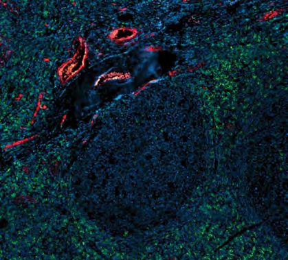
Editor-In-Chief                     Editorial Board
Jodi Nunnari                        John Aitchison                Rebecca Heald                                    Elior Peles
Executive Editor                    Johan Auwerx                  Martin Hetzer                                    Will Prinz
Tim Spencer                         Manuela Baccarini             Erika Holzbaur                                   Thomas Rando
email: tspencer@rockefeller.edu     Tamas Balla                   Martin Humphries                                 Samara Reck-Peterson
Editors                             Maureen Barr                  James Hurley                                     Daniel B. Rifkin
Arshad Desai                        Bill Bement                   Fumiyo Ikeda                                     Michael Rout
Pier Paolo Di Fiore                 Vann Bennett                  Luisa Iruela-Arispe                              Craig Roy
Elaine Fuchs                        Dominique Bergmann            Nancy Ip                                         Michael Rudnicki
Anna Huttenlocher                   Monica Bettencourt-Dias       Johanna Ivaska                                   Erik Sahai
Ian Macara                          Joerg Bewersdorf              Tarun Kapoor                                     Martin Schwartz
Ira Mellman                         Magdalena Bezanilla           Gerard Karsenty                                  Shu-ou Shan
Ana Pombo                           Cédric Blanpain               Scott Keeney                                     Andrey Shaw
Louis F. Reichardt                  Julius Brennecke              Alexey Khodjakov                                 Zu-Hang Sheng
Kenneth M. Yamada                   Tony Bretscher                Hiroshi Kimura                                   Agata Smogorzewska
Richard Youle                       Marianne Bronner              Jürgen Knoblich                                  Joan Steitz
                                    Valérie Castellani            Alberto R. Kornblihtt                            Harald Stenmark
Senior Scientific Editor
                                    Daniela Cimini                Thomas Langer                                    Aaron Straight
Melina Casadio
                                    Karlene Cimprich              Ana Maria Lennon-Dumenil                         Maria-Elena Torres-Padilla
email: mcasadio@rockefeller.edu
                                    Don W. Cleveland              Andres Leschziner                                Billy Tsai
Senior Scientific Editor, Reviews   Nika Danial                   Danny Lew                                        Bas van Steensel
Andrea Marat                        William Earnshaw              Jens Lykke-Andersen                              Patrik Verstreken
email: amarat@rockefeller.edu
                                    Jan Ellenberg                 Vivek Malhotra                                   Mark von Zastrow
Scientific Editors                  Scott Emr                     Brendan Manning                                  Erwin Wagner
Lucia Morgado Palacin               Anne Ephrussi                 Joan Massague                                    John Wallingford
email:lmorgado@rockefeller.edu
                                    Jeffrey Esko                  Satyajit Mayor                                   Tobias Walther
Dan Simon
                                    Sandrine Etienne-Manneville   Frauke Melchior                                  Xiaochen Wang
email:dsimon01@rockefeller.edu
                                    Marc Freeman                  Tobias Meyer                                     Lois Weisman
Managing Editor                     Judith Frydman                Liz Miller                                       Min Wu
Lindsey Hollander                   Hironori Funabiki             Alex Mogilner                                    Tim Yen
Phone: 212-327-8588
email: jcellbiol@rockefeller.edu    Melissa Gardner               Sean Munro                                       Tamotsu Yoshimori
                                    Larry Gerace                  Maxence Nachury                                  Li Yu
                                    David Gilbert                 Karla Neugebauer                                 Xiang Yu
                                    Bruce Goode                   Carien Niessen                                   Junying Yuan
                                    Yukiko Gotoh                  Eva Nogales                                      Marino Zerial
                                    Roger Greenberg               Karen Oegema                                     Hong Zhang
                                    Marcia Haigis                 Ewa Paluch                                       Yixian Zheng
                                    Ulrich Hartl                  Mark Peifer

Senior Preflight Editor             Senior Production Editor      Copyright to articles published in this journal is held by the authors. Articles are published
Laura Smith                         Samantha Wolner               by Rockefeller University Press under license from the authors. Conditions for reuse of the
                                                                  articles by third parties are listed at http://www.rupress.org/terms.
Preflight Editor                    Production Manager
Rochelle Ritacco                    Camille Clowery               Print ISSN: 0021-9525.
                                                                  Online ISSN: 1540-8140
Assistant Production Editor         Production Designer
Elissa Hunter                       Erinn A. Grady                Rockefeller University Press

5                                                                                                              THE YEAR IN CELL BIOLOGY: 2020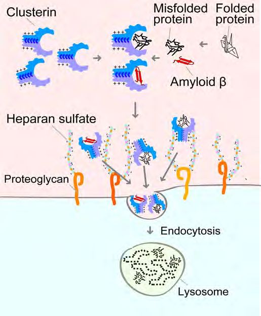
A SYSTEM TO REMOVE
ABERRANT EXTRACELLULAR
PROTEINS
The chaperone Clusterin works in combination with heparan sulfate
proteoglycans to bring misfolded proteins into cells for degradation

A number of diseases are believed to
be caused by the gradual buildup of
misfolded proteins that can aggregate
                                                 A model of the CRED
together and damage neurons and
                                                 pathway in which
other cells in the body. To help prevent         Clusterin and heparan
this damage, cells have developed                sulfate bring aberrant
numerous quality control systems that            extracellular proteins,
recognize misfolded proteins within the          including amyloid β,
cell and either fold them back into their        into cells where they
correct shape or degrade them before             can be degraded by
they start to aggregate.                         lysosomes. © 2020
                                                 Itakura et al.
“However, approximately 11% of
human proteins exist outside of the            CRISPR-based screen, the researchers              tissues and body fluids,” Itakura says.
cell, where they are subjected to even         found that, after binding to misfolded            The researchers named this system
more stresses that may cause them              proteins, Clusterin enters cells by               the chaperone- and receptor-mediated
to misfold,” says Eisuke Itakura, an           interacting with heparan sulfate                  extracellular protein degradation
assistant professor in the Department          proteoglycans, a large class of proteins          (CRED) pathway.
of Biology at Chiba University in Japan.       that are present on the surface of
“In addition, Alzheimer’s disease, the         almost all human cells. This interaction          Intriguingly, Itakura and colleagues
most prevalent cause of dementia               requires a group of conserved,                    found that the CRED pathway can
affecting 47.5 million people worldwide,       positively-charged residues on                    import amyloid β into cells for
is characterized by aggregates of              Clusterin that electrostatically interact         degradation. Mutations in the gene
amyloid β protein in the extracellular         with negatively-charged heparan                   encoding Clusterin have been linked
space. Despite this, how aberrant              sulfate.                                          to an increased risk of developing
extracellular proteins are degraded                                                              Alzheimer’s disease, and experiments
remains poorly understood.”                    Itakura and colleagues found that,                in rats have shown that injecting
                                               together, Clusterin and heparan                   Clusterin into the brain can prevent
A chaperone protein called Clusterin           sulfate proteoglycans allow many                  amyloid β–induced neurodegeneration.
can bind to misfolded extracellular            different cell types to internalize and           “Our results therefore suggest new
proteins and prevent them from                 degrade a wide variety of misfolded               avenues for the possible treatment
aggregating. Itakura and colleagues            extracellular proteins. “We therefore             or prevention of disorders such as
discovered that Clusterin can escort           think that this pathway is a general              Alzheimer’s disease that are associated
misfolded proteins into the cell and           extracellular protein quality control             with aberrant extracellular proteins,”
deliver them to the lysosomes for              system responsible for the clearance              Itakura says.
degradation. Using a genome-wide               of misfolded proteins from diverse

     RESEARCHER DETAILS                                                    ORIGINAL PAPER
                         Eisuke Itakura                                    Itakura, E., M. Chiba, T. Murata, and A. Matsuura. 2020.
                         Assistant Professor                               Heparan sulfate is a clearance receptor for aberrant
                         Chiba University                                  extracellular proteins. J. Cell Biol. 219: e201911126.
                         eitakura@chiba-u.jp                               https://doi.org/10.1083/jcb.201911126

 6                                                                                                        THE YEAR IN CELL BIOLOGY: 2020
ER CONTACTS COORDINATE
MITOCHONDRIAL DYNAMICS
Mitochondrial fission and fusion machineries colocalize at
ER membrane contact sites, where they can rapidly modulate
mitochondrial morphology in response to metabolic cues
The steady–state morphology of                  might also assemble at ER MCSs,”
the mitochondrial network within                explains Gia Voeltz, a Howard Hughes            Mfn1 (green) and Drp1 (magenta)
cells is maintained by a balance of             Investigator and Professor at the               colocalize at ER MCSs, where they
mitochondrial fission and fusion.               University of Colorado, Boulder.                mediate, respectively, the fusion
Disrupting this balance causes the                                                              (white arrows) and fission (magenta
                                                Using a variety of sophisticated imaging        arrows) of mitochondria (grey).
mitochondrial network to become either
                                                techniques, Voeltz and colleagues,              © 2020 Abrisch et al.
fragmented or elongated, potentially
                                                including first author Robert Abrisch,
resulting in altered cell metabolism and
                                                found that mitofusins do, in fact,
disease.
                                                accumulate at ER MCSs and that these          mitochondria can restore their
Mitochondrial fission preferentially            contact sites mark the location of            membrane potential by fusing with
occurs at sites where the mitochondria          ~90% of mitochondrial fusion events.          healthy neighbors at ER MCSs.
contacts the ER. The dynamin-related            “That’s strikingly similar to the 88% of
                                                                                              “Depolarization of individual
GTPase Drp1 accumulates at these                mitochondrial fission events scored to
                                                                                              mitochondria pushes the reaction at
membrane contact sites (MCSs), and              occur at ER MCSs,” Voeltz says.
                                                                                              ER MCSs toward fusion, resulting in
drives mitochondrial constriction
                                                Further experiments revealed that             the recovery of membrane potential,”
and division. Mitochondrial fusion is
                                                Drp1 and Mfn1 colocalize at ER MCSs           Voeltz says. “But other signals may
also regulated by dynamin-related
                                                and that a fusion event at one site is        push the reaction toward fission.
GTPases—the mitofusins Mfn1 and
                                                often followed by fission at the exact        “Future studies will investigate whether
Mfn2 promote outer membrane fusion
                                                same place. Voeltz and colleagues             fission and fusion rates are determined
while Opa1 controls fusion of the inner
                                                suspected that these hotspots of              by the relative recruitment and/or
mitochondrial membrane—but how
                                                mitochondrial dynamics might be               posttranslational modifications of the
these GTPase are positioned to define
                                                able to rapidly respond to metabolic          different machineries at ER MCSs, by
the site of fusion is unknown. However,
                                                cues and could, for example, allow            small signaling molecules like Ca2+,
fission and fusion may be spatially
                                                damaged mitochondria to quickly               or by the recruitment of activators or
coordinated because mitochondria can
                                                recover their function by transiently         inhibitors of these machineries.”
sometimes undergo “transient” fusions,
                                                fusing and exchanging components
in which two mitochondria fuse and
                                                with neighboring, healthy mitochondria.
share their contents, before separating
                                                Indeed, the researchers found that
again to resume their original
                                                ER MCSs often mark the boundaries
morphology.
                                                between polarized (healthy) and
“We hypothesized that if fission                depolarized (unhealthy) segments of
and fusion are coordinated, then                mitochondria, and that depolarized
the mitochondrial fusion machinery

     RESEARCHER DETAILS                                                                       ORIGINAL PAPER
                                                                                              Abrisch, R.G., S.C. Gumbin, B.T.
                                                                                              Wisniewski, L.L. Lackner, and G.K. Voeltz.
                                                                                              2020. Fission and fusion machineries
                                                                                              converge at ER contact sites to regulate
                                                                                              mitochondrial morphology. J. Cell Biol.
                                                                                              219: e201911122.
     Robert G. Abrisch           Samantha C. Gumbin                Gia K. Voeltz              https://doi.org/10.1083/jcb.201911122
     PhD student                 Undergraduate/Junior              HHMI Investigator and
     University of Colorado,     technician                        Professor, University of
     Boulder                     University of Colorado, Boulder   Colorado, Boulder
     (Currently a postdoctoral   (Currently a graduate student     gia.voeltz@colorado.edu
     fellow at UC San Diego)     at Stanford University)

 7                                                                                                    THE YEAR IN CELL BIOLOGY: 2020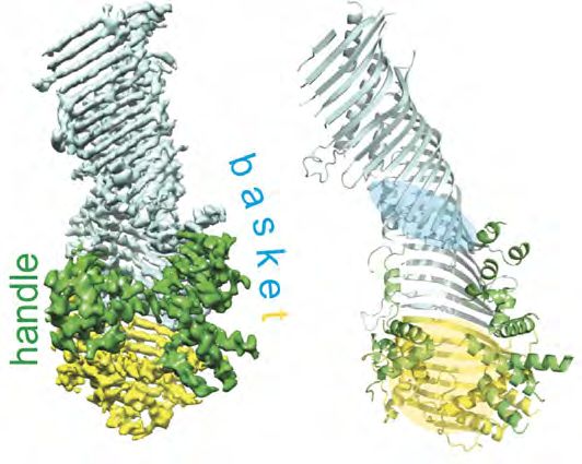
VPS13 FORMS A LIPID CHANNEL
BETWEEN MEMBRANES
Cryo-EM of VPS13 N-terminal                    The VPS13 family of lipid transport       organelles together via their N- and
fragment reveals a hydrophobic                 proteins may work differently, however.   C-termini and allowing lipids to flow
                                               These very large proteins are thought     from one membrane to the other
groove that may allow bulk lipid               to tether organelles together at          through their hydrophobic channels.
flow between organelles                        membrane contact sites and can bind       As an initial test of this idea, Li et al.
                                               tens of lipid molecules at a time.        created mutant versions of budding
Lipids constantly need to be exchanged         Budding yeast Vps13p is required          yeast Vps13p in which sections of the
between the various different                  for prospore membrane formation,          channel were switched to hydrophilic
membranes of eukaryotic cells. For             and, in humans, there are four family     residues instead of hydrophobic ones.
example, most membrane lipids are              members, some of which are linked to      These vps13 mutants could still bind
synthesized in the ER and must then            severe neurodegenerative disease.         lipids but they were unable to support
be delivered to other organelles. This                                                   sporulation, indicating that they are
is carried out, in part, by lipid transfer     Reinisch and colleagues, including
                                                                                         incapable of transferring lipids during
proteins that localize to the membrane         first author PeiQi Li, carried out a
                                                                                         prospore formation.
contact sites between organelles               single-particle cryo-EM reconstruction
and act as shuttles, extracting lipid          of a ~160-kD N-terminal fragment of       “The size of the VPS13 lipid-binding
molecules from one organelle and then          the VPS13 protein of the filamentous      cavity and, hence, the ability of these
depositing it in the membrane of the           fungus Chaetomium thermophilum. The       proteins to accommodate many lipids
other.                                         researchers found that the fragment       simultaneously suggests a role in
                                               adopts a basket-like structure whose      bulk lipid transfer,” says Reinisch,
“Typically, these lipid transport proteins     interior is lined with hydrophobic        who notes that the VPS13-like protein
comprise domains resembling lidded             residues. Thus, the N-terminal region     ATG2 may act similarly to promote
teacups, each with a hydrophobic cavity        of VPS13 forms an extended channel,       autophagosome assembly during
that accommodates one or two lipid             ~160 Å in length, that would be well-     autophagy.
molecules,” explains Karin Reinisch, a         suited to solubilizing
professor at Yale School of Medicine.          multiple lipid
This limited capacity is unlikely to           molecules at a time.
be sufficient when large amounts of
lipid need to be transferred rapidly,          Instead of shuttling
such as during the assembly of the             lipids between
autophagosome during autophagy,                membranes, VPS13
or the formation of the prospore               proteins might
membrane during yeast meiosis.                 therefore act like
                                               bridges, tethering

  The density map (left) and secondary structure (right) of
  VPS131-1390 show the fragment’s basket-like structure and the
  extended, hydrophobic channel that may facilitate lipid flow
  between membranes. © 2020 Li et al.

     RESEARCHER DETAILS                                                                  ORIGINAL PAPER
                                                                                         Li, P., J.A. Lees, C.P. Lusk, and K.M.
                                                                                         Reinisch. 2020. Cryo-EM reconstruction of
                                                                                         a VPS13 fragment reveals a long groove
                                                                                         to channel lipids between membranes.
                                                                                         J. Cell Biol. 219: e202001161.
                                                                                         https://doi.org/10.1083/jcb.202001161
     PeiQi Li                    Karin M. Reinisch
     Graduate student            Professor
     Yale School of Medicine     Yale School of Medicine
                                 karin.reinisch@yale.edu

 8                                                                                               THE YEAR IN CELL BIOLOGY: 2020
AUTOPHAGY TARGETS NUCLEAR                                                                  interact with either Atg11, which then
                                                                                           initiates autophagosome formation,
                                                                                           or Atg8, a protein located on forming
PORE COMPLEXES                                                                             autophagosomal membranes. Tomioka
                                                                                           et al. found that yeast lacking Atg11,
Several different pathways selectively target NPCs and                                     or yeast expressing an Atg8 mutant
nucleoporins for autophagic degradation upon inactivation of the                           unable to bind autophagy receptors,
TORC1 kinase complex                                                                       failed to degrade nucleoporins upon
                                                                                           TORC1 inactivation, indicating that
Composed of ~30 different nucleoporin         Nakatogawa and colleagues, including         NPCs/nucleoporins are selectively
proteins, nuclear pore complexes              first author Yui Tomioka, investigated       targeted to autophagosomes.
(NPCs) span the inner and outer               whether NPCs are broken down
                                                                                           Nakatogawa and colleagues determined
membranes of the nuclear envelope             by autophagy, a process in which
                                                                                           that some NPCs are targeted by a
and mediate transport between the             cellular components are engulfed
                                                                                           previously identified pathway called
nucleus and cytoplasm, thereby playing        by autophagosomes and delivered
                                                                                           nucleophagy, in which nuclear
critical roles in gene expression, cell       to lysosomes for degradation. The
                                                                                           envelope–derived vesicles are recruited
growth and division. Defects in NPC           researchers found that inducing the
                                                                                           to autophagosomes via an autophagy
function have been linked to aging            autophagy pathway in budding yeast by
                                                                                           receptor in the outer nuclear membrane
and a variety of illnesses including          inactivating the TORC1 kinase complex
                                                                                           called Atg39. But NPCs can also be
Alzheimer’s disease and cancer. Cells         resulted in the degradation of multiple
                                                                                           targeted via a different pathway, that
are therefore likely to employ a variety      nucleoporins. Blocking degradation
                                                                                           the researchers named NPC-phagy,
of quality control mechanisms to              caused these NPC components to
                                                                                           involving an as yet unknown autophagy
ensure they maintain an appropriate           accumulate in vacuoles—the yeast
                                                                                           receptor.
number of functional NPCs. “However,          equivalent of lysosomes—where they
the cellular systems that degrade the         appeared to be embedded within               In addition, Tomioka et al. found
NPC or nucleoporins are still largely         nuclear envelope–derived double-             that one nucleoporin, Nup159, can
unknown,” says Hitoshi Nakatogawa             membrane vesicles. This suggests             bind directly to Atg8. Disrupting this
from the Tokyo Institute of Technology.       that nucleoporins can be engulfed by         interaction impaired the turnover
                                                                     autophagosomes        of Nup159 but had no effect on the
                                                                     and delivered to      degradation of other nucleoporins,
                                                                     lysosomes while       suggesting that nucleoporins not
                                                                     they are still        assembled into NPCs can also be
                                                                     assembled into        targeted to autophagosomes via a
                                                                     NPCs.                 process the researchers dubbed
                                                                                           nucleoporinophagy.
                                                                       Though
                                                                       autophagy can       Together, these different selective
                                                                       be a nonselective   autophagy pathways are likely to
                                                                       process,            help cells maintain the structural and
                                                                       many cellular       functional integrity of their NPCs.
                                                                       components          “Our study provides a foundation
  In yeast cells lacking vacuolar proteases, TORC1 inactivation        are targeted to     for understanding the molecular
  leads to the accumulation of Nup159-containing structures            autophagosomes      mechanisms and physiological/
  (arrowhead) embedded in nuclear envelope-derived double-             by specific         pathological significance of these
  membrane vesicles located inside autophagosomes that have            autophagy           autophagy pathways in various
  been delivered to the vacuole. © 2020 Tomioka et al.                 receptors that      organisms,” Nakatogawa says.

     RESEARCHER DETAILS                                                                    ORIGINAL PAPER
                                           Hitoshi Nakatogawa (Left)                       Tomioka, T., T. Kotani, H. Kirisako, Y.
                                           Associate Professor                             Oikawa, Y. Kimura, H. Hirano, Y. Ohsumi,
                                           Tokyo Institute of Technology                   and H. Nakatogawa. 2020. TORC1
                                           hnakatogawa@bio.titech.ac.jp                    inactivation stimulates autophagy
                                                                                           of nucleoporin and nuclear pore
                                           Yui Tomioka (Right)                             complexes. J. Cell Biol. 219: e201910063.
                                           PhD Student
                                                                                           https://doi.org/10.1083/jcb.201910063
                                           Tokyo Institute of Technology

 9                                                                                                 THE YEAR IN CELL BIOLOGY: 2020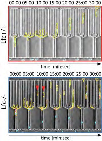
PROTEASOME DISTRIBUTION MODULATES GABA
RESPONSES IN DEVELOPING NEURONS
                                              somatodendritic
The adaptor protein Ecm29
                                              compartments of
positions proteasomes at                      neurons and controls
the axon initial segment to                   action potential
promote degradation of the                    firing. Cheng and
chloride transporter NKCC1                    colleagues found that,
                                              as the AIS assembles
and limit neuronal excitability
                                              in developing
                                              hippocampal and
The response of mouse neurons to the
                                              cortical neurons,
neurotransmitter GABA changes from
                                              an adaptor protein
excitatory to inhibitory during the first
                                              called Ecm29 tethers
few weeks after birth, a developmental
                                              proteasomes to the
switch that helps to establish the
                                              AIS by binding to
lifelong activity of local neuronal
                                              ankyrin G.
circuits. The change in GABA response
is due to a reduction in the intracellular    Neurons lacking Ecm29 showed                 NKCC1 levels in the AIS region of
chloride concentration in neurons that                                                     neurons are increased in the absence
                                              reduced protein turnover at the AIS,
                                                                                           of the proteasome adaptor Ecm29
is thought to be driven by increased          and Ecm29-deficient mouse brains             (bottom). A linear pseudocolored
expression of the chloride exporter           showed higher levels of NKCC1 that           scale shows the intensity of NKCC1
KCC2 and decreased levels of the              declined more slowly during the              staining within the AIS (marked by
chloride importer NKCC1. “However,            postnatal period. NKCC1 levels were          ankyrin G). © 2020 Lee et al.
how NKCC1 levels are down-regulated           particularly elevated in the AIS region
during the excitatory-to-inhibitory           of neurons. As a result, the switch from
GABA transition remains unclear,” says        excitatory to inhibitory GABA responses
Pei-Lin Cheng, an Associate Research                                                     “Our findings provide a developmental
                                              was delayed in Ecm29-knockout mice,
Fellow at the Institute of Molecular                                                     mechanism linking timing of AIS
                                              causing the animals’ neurons to be
Biology in Taiwan.                                                                       formation, proteasome transport, and
                                              hyperexcitable during this critical
                                                                                         subcellular proteostasis to the critical
                                              period in neuronal development.
Cheng and colleagues, including                                                          window of the GABAergic switch,
first author Min Lee, discovered that         “We hypothesized that prolonged            which governs excitability of maturing
developing neurons regulate NKCC1             NKCC1 expression/excitatory GABA           hippocampal/cortical neurons and is
levels and the GABA response by               responsiveness in Ecm29-deficient          mediated by Ecm29,” says Cheng, who
localizing the protein-degrading              immature neurons puts mice at higher       notes that changes in AIS-associated
proteasome to the axon initial segment        risk of seizures,” Cheng says. Indeed,     proteins have been linked to various
(AIS), a specialized membrane domain          Lee et al. found that Ecm29-null mice      neurodevelopmental and psychiatric
that forms around the same time as the        were more susceptible to chemical-         disorders, including epilepsy.
GABAergic switch. Composed of various         induced seizures, but were protected if
scaffold proteins such as ankyrin G,          treated with a potent NKCC1 inhibitor
the AIS segregates the axonal and             during the first few weeks of life.

   RESEARCHER DETAILS                                                                    ORIGINAL PAPER
                                                                                         Lee, M., Y.-C. Liu, C. Chen, C.-H. Lu,
                                                                                         S.-T. Lu, T.-N. Huang, M.-T. Hsu, Y.-P.
                                                                                         Hsueh, and P.-L. Cheng. 2020. Ecm29-
                                                                                         mediated proteasomal distribution
                                                                                         modulates excitatory GABA responses
                                                                                         in the developing brain. J. Cell Biol. 219:
                                                                                         e201903033.
   Min Lee                      Pei-Lin Cheng
   Research Assistant           Associate Research Fellow                                https://doi.org/10.1083/jcb.201903033
   Institute of Molecular       Institute of Molecular Biology,
   Biology, Academia Sinica     Academia Sinica
                                plcheng@imb.sinica.edu.tw

10                                                                                               THE YEAR IN CELL BIOLOGY: 2020
NEUTRAL LIPIDS CONTROL PROTEIN
RECRUITMENT TO LIPID DROPLETS
The neutral lipid core content is determinant to the affinity of
amphipathic helices for artificial lipid droplets
Lipid droplets (LDs) store neutral
lipids and control their hydrolysis to The AHs of the LD proteins Cav1 and
meet the cell’s demands for energy or Arfgap1 are recruited to artificial
LDs budding from the bilayer of giant
membrane synthesis. Formed at the
vesicles in vitro, preferentially binding
ER—the major site of lipid synthesis in LDs composed of the neutral lipid
eukaryotic cells—LDs are composed of triolein, compared with LDs made from
a central neutral lipid core, surrounded squalene. © 2020 Chorlay and Thiam
by a phospholipid monolayer containing
numerous regulatory proteins.
found that AH peptides derived from LD
Many of these proteins bind to LDs via proteins differentially bound to artificial
amphipathic helices (AHs), but how LDs, depending on the neutral lipid
these helices selectively target LDs—or making up the droplet’s core.
even specific subsets of LDs—remains
unclear. After all, AHs can also target “We found that neutral lipid
proteins to the cell’s many different composition determines the binding
membranes by inserting themselves level of AHs, and the ability of AHs to
into the hydrophobic core of lipid discriminate between neutral lipids “Overall, our data highlight an
bilayers. “In the case of LDs, AHs have relies on their amphipathic amino undervalued contribution of neutral
to expose their hydrophobic moieties to acid sequence,” Thiam explains. For lipids in controlling the binding of AHs
the neutral lipid core. Thus, interactions example, mutations in the AH of the to the surface of LDs,” Thiam says.
between the hydrophobic amino acids LD protein Cav1 either enhanced or “The phospholipid packing density
of an AH with neutral lipids could be a abolished its preference for artificial simply regulates the amount of
major driving force for LD targeting,” LDs composed of the neutral lipid exposed neutral lipids, which dictates
says Abdou Rachid Thiam, a professor triolein. the binding level of AHs. Clearly, the
at the École Normale Supérieure in Chorlay and Thiam also determined full picture of how AHs selectively
Paris. that the phospholipids surrounding LDs bind to LDs in a cellular context is not
influence AH binding by regulating the completely resolved yet, but our data
To test this idea, Thiam and graduate
access of AHs to the neutral lipid core. bring us a major step closer to it.”
student Aymeric Chorlay measured
the binding of various AHs to artificial However, while reducing the density
LDs embedded within the bilayer of of phospholipids on artificial LDs
giant vesicles in vitro (mimicking the increased AH binding, it didn’t change
emergence of cellular LDs from within the AHs’s relative preference for LDs
the ER bilayer). Chorlay and Thiam composed of particular neutral lipids.
RESEARCHER DETAILS ORIGINAL PAPER
Chorlay, A. and A.R. Thiam. 2020. Neutral
lipids regulate amphipathic helix affinity
for model lipid droplets. J. Cell Biol. 219:
e201907099.
https://doi.org/10.1083/jcb.201907099
Aymeric Chorlay Abdou Rachid Thiam
PhD student Professor
École Normale Supérieure École Normale Supérieure de
de Paris, PSL, Université de Paris, PSL, Université de Paris,
Paris, Sorbonne Université, Sorbonne Université, CNRS
CNRS thiam@ens.fr
11 THE YEAR IN CELL BIOLOGY: 2020UNRAVELLING THE CRITICAL
STEPS IN MITOPHAGY
ability of OPTN to support mitophagy
Study reveals that mitochondrial ubiquitination and an interaction even more than mutations in OPTN’s
between OPTN and ATG9A are crucial for inducing the selective ATG8-binding domain.
degradation of damaged mitochondria Other recent studies have shown
Damaged mitochondria are quickly demonstrates that the Parkin–PINK1 that the other crucial mitophagy
eliminated by a process called system is essential for ubiquitination adaptor, NDP52, can recruit the
mitophagy, a selective type of of damaged mitochondria but is not autophagy-activating ULK complex
autophagy in which the organelles required for autophagy activation per to damaged mitochondria. “Our
are engulfed by autophagosomes se,” Yamano says. study, in conjugation with these
and delivered to lysosomes for previous reports, indicates that the
Ubiquitinated mitochondria association of OPTN (and NDP52)
degradation. Two proteins linked
are thought to be linked to with ubiquitinated mitochondria
to familial Parkinson’s disease—the
autophagosomes by five different promotes the formation of an initial
ubiquitin kinase PINK1 and the
autophagy adaptors that, in platform that triggers the assembly
ubiquitin ligase Parkin—recognize
addition to binding ubiquitin, can of different autophagy core units
depolarized mitochondria and rapidly
also bind to ATG8 proteins on the through multivalent interactions,”
coat the outer membrane with
autophagosomal membrane. Two of Yamano says. “These functionalities
ubiquitin chains that act as a signal
these adaptors—OPTN and NDP52— thus fulfill a critical role for de novo
for the assembly of autophagosomal
appear to be particularly crucial for autophagosomal membrane formation
membranes around the damaged
mitophagy. Surprisingly, however, close to the ubiquitinated cargo.”
organelles.
Yamano and colleagues
“However, it was unclear whether determined that this
Parkin and PINK1 function solely in isn’t because OPTN and
the ubiquitination of dysfunctional NDP52 have a higher
mitochondria, or if they also affinity for ATG8 proteins.
physically communicate with the
Instead, the researchers
core autophagy machinery,” says Koji
discovered, OPTN plays a
Yamano from the Tokyo Metropolitan
critical role in mitophagy
Institute of Medical Science.
by recruiting ATG9A
To address this question, Yamano vesicles to ubiquitinated
and colleagues, including co-senior mitochondria (these
author Noriyuki Matsuda, devised vesicles are thought
ways to tag mitochondria with to supply components
ubiquitin chains independently of that aid autophagosome
PINK1, Parkin, or mitochondrial assembly). Yamano
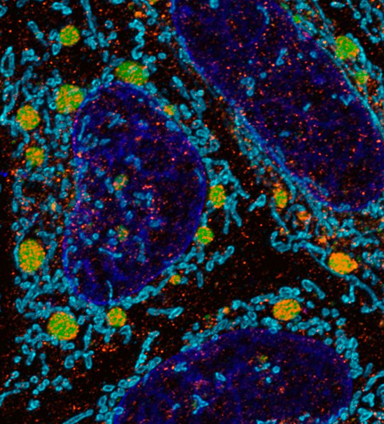
depolarization. The researchers found et al. found that the Following mitochondrial depolarization, ATG9A (red)
is recruited to mitochondria (blue) in cells expressing
that the addition of short, linear leucine zipper domain
wild-type OPTN (green, top row) but not in cells
ubiquitin chains to mitochondria was of OPTN binds to expressing OPTN with a mutated leucine zipper domain
sufficient to induce mitophagy and ATG9A. Mutations in (green, bottom row). © 2020 Yamano et al.
mitochondrial degradation. “This this domain reduced the
RESEARCHER DETAILS Noriyuki Matsuda (Left) ORIGINAL PAPER
Project Leader
Tokyo Metropolitan Institute of Yamano, K., R. Kikuchi, W. Kojima, R.
Medical Science Hayashida, F. Koyano, J. Kawawaki, T.
Shoda, Y. Demizu, M. Naito, K. Tanaka,
matsuda-nr@igakuken.or.jp
and N. Matsuda. 2020. Critical role of
Koji Yamano (right) mitochondrial ubiquitination and the
Senior Researcher OPTN–ATG9A axis in mitophagy. J. Cell
Tokyo Metropolitan Institute of Biol. 219: e201912144.
Medical Science https://doi.org/10.1083/jcb.201912144
yamano-kj@igakuken.or.jp
12 THE YEAR IN CELL BIOLOGY: 2020BREAST CANCER CELLS
TURN KILLER IMMUNE
CELLS INTO ALLIES
Natural killer cells are reprogrammed by breast
cancer cells to promote metastasis
Breast cancer cells spread to other reprogramming
parts of the body by invading the them so that they
surrounding, healthy breast tissue actually promote
until they reach the circulation, which the later stages
can carry them to other tissues of metastasis. Using Initially, the presence of NK cells (bottom row)
where they can form new metastatic several new assays to restricts the growth of tumor organoids in 3D
tumors, a major cause of death in model metastasis in collagen matrices. But after 36 h, the cancer cells
breast cancer patients. Natural killer the laboratory as well reprogram the NK cells, and the tumor starts to
(NK) cells are key components of as experiments in mice, invade its surroundings. © 2020 Chan et al.
the innate immune system that can the researchers found
recognize and kill cancer cells as they that, after they encounter tumor cells, treatment with anti-TIGIT or anti-
attempt to spread through the body. human and mouse NK cells lose the KLRG1 antibodies was particularly
ability to restrict tumor invasion and effective at preventing NK cells from
“Breast cancer cells must overcome
instead help cancer cells form new enhancing the metastatic potential of
NK cell surveillance in order to form
tumors. breast cancer cells.
distant metastases,” says Andrew
Ewald, a professor of cell biology NK cells exposed to tumors undergo “The synergistic effects of DNA
at Johns Hopkins University School dramatic changes, turning thousands methyltransferase inhibitors with
of Medicine and co-director of the of genes on and off and expressing receptor-blocking antibodies
Cancer Invasion and Metastasis different receptor proteins on their suggests a viable clinical strategy to
Program in the Sidney Kimmel surface. Ewald and colleagues found reactivate tumor-exposed NK cells
Comprehensive Cancer Center. that antibodies targeting two key to target and eliminate breast cancer
“However, we do not fully understand receptor proteins on the surface of metastases,” says Chan.
how breast cancer cells escape NK NK cells, called TIGIT and KLRG1,
Ewald notes, “Combined with
cell–mediated immunosurveillance prevented NK cells from helping
during their transit through the breast cancer cells seed new tumors. our observation that NK cells are
circulation and the initial seeding of The FDA-approved drugs decitabine abundant early responders to
distant organs.” and azacitidine had similar effects, disseminated breast cancer cells, our
likely because they prevent large- data provide a preclinical rationale
Ewald and colleagues, including first for the concept of NK cell–directed
scale changes in gene activity by
author Isaac Chan, discovered that, immunotherapies in the adjuvant
inhibiting enzymes known as DNA
although metastasizing breast cancer setting for breast cancer patients with
methyltransferases.
cells are initially vulnerable to NK high risk of metastatic recurrence.”
cells, they are quickly able to alter The researchers found that
the behavior of their would-be killers, combining decitabine or azacitidine
RESEARCHER DETAILS ORIGINAL PAPER
Isaac S. Chan Chan, I.S., H. Knútsdóttir, G.
Medical Oncology Fellow Ramakrishnan, V. Padmanaban, M.
Sidney Kimmel Comprehensive Cancer Center, Warrier, J.C. Ramirez, M. Dunworth, H.
Johns Hopkins University School of Medicine Zhang, E.M. Jaffee, J.S. Bader, and A.J.
isaac.chan@utsouthwestern.edu Ewald. 2020. Cancer cells educate natural
killer cells to a metastasis-promoting cell
Andrew J. Ewald state. J. Cell Biol. 219: e202001134.
Professor of Cell Biology
https://doi.org/10.1083/jcb.202001134
Sidney Kimmel Comprehensive Cancer Center,
Johns Hopkins University School of Medicine
andrew.ewald@jhmi.edu
13 THE YEAR IN CELL BIOLOGY: 2020ISG15 ACCELERATES REPLICATION activity,” Penengo says. Indeed,
increased ISG15 levels promoted
fork restart in a RECQ1-dependent
FORK PROGRESSION manner.
Elevated ISG15 might therefore be
The ubiquitin-like molecule accelerated fork progression, whereas detrimental to cancer cells by causing
ISG15, which is induced deleting the gene reduced the speed DNA replication to continue in the
of DNA replication in several cancer presence of genotoxic drugs that
by interferons and is often cell lines that usually overexpress would normally slow replication fork
upregulated in cancer cells, can ISG15. progression, resulting in genomic
increase genome instability and Raso et al. determined that ISG15
instability. Raso et al. found that
sensitize cells to genotoxic drugs cancer cells with high ISG15 levels
can regulate DNA replication fork were more sensitive to low doses
progression through non-covalent of the chemotherapeutic agents
ISG15 is strongly induced by type I mechanisms: overexpression of a
and type III interferons in response to camptothecin and cisplatin, because
mutant version incapable of being their replication forks continued
bacterial or viral infection. Though its conjugated to other proteins still
amino acid sequence is very different, unabated, leading to chromosome
accelerated fork progression. breakages and cell death.
ISG15’s 3D structure is similar to
ubiquitin and, like ubiquitin, it can The researchers found that ISG15 “The increased activity of RECQ1
be conjugated to other proteins by associates with several proteins at induced by high ISG15 levels
E3 ligases. But increasing evidence replication forks, including a DNA may thus represent an important
suggests that ISG15 can modulate helicase, RECQ1, that helps to restart vulnerability that can be exploited
the host immune response by non- stalled forks. “Depletion of RECQ1 for genotoxic anticancer treatments,”
covalently binding to other proteins or completely abolished the accelerated Penengo says. “Furthermore, the
even by acting as a cytokine secreted replication fork progression induced evaluation of ISG15 levels in tumor
from cells. by high levels of ISG15, suggesting samples may represent a predictive
that ISG15 may regulate RECQ1 parameter to stratify patients in
ISG15 is also induced by DNA function by unleashing its restart
damage and is frequently personalized cancer therapy.”
overexpressed in cancer cells.
“Elevated ISG15 levels occur in many
types of cancer and, in some cases, Proximity ligation
the robust expression of ISG15 has assays (magenta)
been reported to support tumor show that ISG15
growth,” explains Lorenza Penengo (red) associates
from the Institute of Molecular Cancer with the DNA
Research at the University of Zurich. helicase RECQ1
“However, its role in tumorigenesis is (green) in cells,
still controversial, and its mechanism even when the
of action is far from being clarified.” cells express a
mutant version
Penengo and colleagues, including of ISG15 (ΔGG)
first author Chiara Raso, discovered that cannot
that ISG15 localizes to DNA be covalently
replication forks, suggesting that conjugated to
it might modulate DNA replication. other proteins.
Inducing ISG15 expression © 2020 Raso et al.
RESEARCHER DETAILS ORIGINAL PAPER
Lorenza Penengo (Left) Raso, M.C., N. Djoric, F. Walser, S.
Professor Hess, F.M. Schmid, S. Burger, K.-P.
Institute of Molecular Cancer Research Knobeloch, and L. Penengo. 2020.
University of Zurich Interferon-stimulated gene 15 accelerates
penengo@imcr.uzh.ch replication fork progression inducing
chromosomal breakage. J. Cell Biol. 219:
Maria Chiara Raso (right) e202002175.
PhD student
Institute of Molecular Cancer Research https://doi.org/10.1083/jcb.202002175
University of Zurich
14 THE YEAR IN CELL BIOLOGY: 2020Kiermaier and first author Aglaja
MICROTUBULES HELP MIGRATING Kopf, therefore wondered whether
microtubules nucleated from the
CELLS KEEP THEIR SHAPE MTOC might control the shape of
migrating DCs. The researchers found
that, as the nucleus and associated
Dendritic cells coordinate their movement through complex MTOC move into one protrusion and
microenvironments by using the local depolymerization of microtubules guide the cell forward, protrusions
to trigger the actomyosin-mediated retraction of protrusions pointing in other directions lose their
microtubules and retract. “Upon
Cells navigate through complex tissues protrusion to another. passage of the MTOC, sheer geometry
by sending out multiple protrusions to may determine that all but the leading
How dendritic cells (DCs) control
explore different paths, but protrusions protrusion are cut off from microtubule
their shape as they surveil tissues
extending in the “wrong” direction supply because microtubules are too
for foreign antigens and deliver them
must then be retracted so that the cell inflexible to find their way into curved,
to lymph nodes is unclear, however.
can move forward without becoming narrow, and ramified spaces,” Kopf
DCs use an amoeboid mode of
entangled with its surroundings. says.
migration that is largely independent
Migrating fibroblasts are thought to
of adhesions and stress fibers, and Kopf et al. determined that the local
coordinate their protrusions using
they are too large and ramified for depolymerization of microtubules
mechanical signals transmitted via
membrane tension to coordinate their triggers the retraction of protrusions.
actomyosin-based stress fibers
protrusions. “We recently found that The Rho GTPase exchange factor GEF-
associated with focal adhesions,
when DCs migrate through complex H1/Lfc associates with microtubules
whereas neutrophils and other small
geometries, their nucleus acts as a and is released when they
leukocytes may use plasma membrane
mechanical gauge to lead them along depolymerize, leading to the activation
tension to relay signals from one
the path of least resistance and the of actomyosin contractility and
microtubule organizing center (MTOC), protrusion retraction. Depleting Lfc
which is closely associated with the impaired DC migration, often causing
nucleus, is critical for this navigational the cells to fragment as they failed to
task,” says Michael Sixt from the retract their protrusions and became
Institute of Science and Technology in entangled in their surroundings.
Austria.
“We propose that microtubules serve
Sixt and colleagues, including as an internal explorative system of the
co-corresponding author Eva cell that informs actomyosin whenever
a peripheral protrusion locates too
distant from the centroid and thereby
A wild-type DC (top) successfully initiates its retraction,” Kiermaier says.
navigates through a branched
microfluidic channel after sending “We also found that DCs lacking
out multiple protrusions to explore both Lfc and microtubules have even
the various pathways. A DC lacking more severe cell shape defects than
Lfc (bottom) fails to properly cells lacking Lfc only, indicating that
retract its exploratory protrusions
microtubule depolymerization induces
and undergoes fragmentation (red
arrowheads). © 2020 Kopf et al.
cell retraction via additional modes
that remain to be identified,” Sixt adds.
RESEARCHER DETAILS ORIGINAL PAPER
Kopf, A., J. Renkawitz, R. Hauschild,
I. Girkontaite, K. Tedford, J. Merrin, O.
Thorn-Seshold, D. Trauner, H. Häcker,
K.-D. Fischer, E. Kiermaier, and M. Sixt.
2020. Microtubules control cellular shape
and coherence in amoeboid migrating
Aglaja Kopf Eva Kiermaier Michael Sixt cells. J. Cell Biol. 219: e201907154.
Postdoctoral Researcher Professor Professor
CeMM Research Center Life and Medical Institute of Science and https://doi.org/10.1083/jcb.201907154
for Molecular Medicine Sciences Institute Technology Austria
of the Austrian Academy University of Bonn sixt@ist.ac.at
of Sciences ekiermai@uni-bonn.de
15 THE YEAR IN CELL BIOLOGY: 2020TOOLS FOR DISCOVERY
Journal of Cell Biology (JCB) publishes advances inany
area of basic cell biology as well as applied cellular
advances in fields such as immunology, neurobiology,
metabolism, microbiology, developmental biology,
and plant biology. All editorial decisions on research
Executive Director
manuscripts are made through collaborative
consultation between professional editors with Susan King
scientific training and academic editors who are Associate Finance Director
activein the field. Established in 1955, JCB publishes Laura Bisberg
12 issues per year. www.jcb.org
Director of Editorial Development
Teodoro Pulvirenti
Director of Publishing Technologies
Robert J. O'Donnell
Journal of Experimental Medicine (JEM) publishes Director of Communications and Marketing
papers providing novel conceptual insight into
Rory Williams
immunology, neuroscience, cancer biology, vascular
biology, microbial pathogenesis, and stem cell Institutional Sales Manager
biology. All editorial decisions are made by active Miguel Peralta
scientists in conjunction with professional editors.
Established in1896, JEM publishes 12 issues per year. Executive Assistant to Executive Director;
www.jem.org Office Administrator
Demantie (Sati) Motieram
Financial Analyst
Sarah S. Kraft
Senior Science Writer
Journal of General Physiology (JGP) publishes Ben Short
mechanistic and quantitative cellular and molecular
physiology of the highest quality; provides a best Marketing and Design Coordinator
in class author experience; and nurtures future Yuko Tonohira
generations of researchers. All editorial decisions
on research manuscripts are made through a
collaborative consultation between the Editor-in-
Chief and AssociateEditors, all of whom are active
scientists. Established in 1 918, JGP publishes 12
issues per year. www.jgp.org
Life Science Alliance (LSA) is a global, open-access,
Journal of Cell Biology (ISSN 0021-9525) is published monthly by
editorially independent, and peer-reviewed journal
Rockefeller University Press, 950 Third Avenue, New York, NY 10022.
launched in 2018 by an alliance of EMBO Press, Periodical postage paid at New York, NY and additional mailing offices.
Rockefeller University Press, and Cold Spring Harbor
2021 Subscription Rates
Laboratory Press. Life Science Alliance is committed Institutional Rates
to rapid, fair, and transparent publication of valuable Tier 1 Tier 2 Tier 3 Tier 4
research from across all areas in the life sciences. Online $2,670 $3,390 $4,160 $5,320
www.lsajournal.org Print + Online $5,930 $6,610 $7,180 $9,170
For more information, please contact our subscription office.
Phone: +1 212-327-8590
email: subs@rockefeller.edu
Advertising Requests
Phone: 201-767-4170
email: rupads@rockefeller.edu
Permission Requests
email: permissions@rockefeller.edu
Media Requests
email: news@rockefeller.edu
Postmaster
Send address changes to Journal of Cell Biology Subscriptions, Rockefeller
University Press, 950 Third Ave, 2nd Floor, New York, NY 10022.
LEARN MORE AT RUPRESS.ORGMAKE CONNECTIONS
Sign up for email alerts from JCB to stay informed of
the latest discoveries and make connections
that can impact your research.
CLICK HERE TO SIGN UP
17 THE YEAR IN CELL BIOLOGY: 2020Illuminate Microscopy
With Fluorophore
Antibody Conjugates
BioLegend offers one of the largest collections of antibodies, Vivid Antibody Staining for Multicolor Microscopy
with thousands of targets for immunology, neuroscience,
oncology, and stem cell research areas. These antibodies can
be combined with several fluorophore options, providing
directly conjugated primary antibodies for assays with reduced
background. Alternatively, we provide secondary fluorophore-
labeled reagents to amplify the signal of primary antibodies.
Quality-tested for IHC and ICC applications, our fluorophore
options include:
• Spark YG™️ 570: a new equivalent to Alexa Fluor®️ 555, Cy3,
and TRITC.
• Alexa Fluor®️ 488, Alexa Fluor®️ 594, and Alexa Fluor®️ 647.
• Brilliant Violet 421™️ and Brilliant Violet 510™️.
Find thousands of antibody/fluorophore combinations at:
biolegend.com/en-us/microscopy Human paraffin-embedded tonsil tissue co-stained with anti-human PNAd
(clone MECA-79) Biotin, and anti-human CD8a (clone C8/144B) Alexa Fluor®️
647 (green) followed by Streptavidin Spark YG™️ 570 (red). Nuclei were
counterstained with DAPI (blue).
biolegend.comYou can also read