A proteinase-free DNA replication machinery for in vitroand invivoamplified MicroRNA imaging
←
→
Page content transcription
If your browser does not render page correctly, please read the page content below
Published online 29 April 2020                                                               Nucleic Acids Research, 2020, Vol. 48, No. 10 e60
                                                                                                                      doi: 10.1093/nar/gkaa250

A proteinase-free DNA replication machinery for in
vitro and in vivo amplified MicroRNA imaging
                                                                                                                                                             *
Jie Wei, Huimin Wang, Xue Gong, Qing Wang, Hong Wang, Yangjie Zhou and Fuan Wang

Key Laboratory of Analytical Chemistry for Biology and Medicine (Ministry of Education), College of Chemistry and
Molecular Sciences, Wuhan University, Wuhan 430000, P. R. China

Received December 09, 2019; Revised March 05, 2020; Editorial Decision March 30, 2020; Accepted April 01, 2020

                                                                                                                                                                  Downloaded from https://academic.oup.com/nar/article/48/10/e60/5826808 by guest on 08 September 2020
ABSTRACT                                                                          tiveness of these enzyme-catalyzed machineries usually re-
                                                                                  lies on certain protein enzymes that might be susceptible to
The construction of robust, modular and compact                                   the delicate interference of external environment. Thus, it
DNA machinery facilitates us to build more intelli-                               is highly desirable to develop more robust and convenient
gent and ingenious sensing strategies in complex bi-                              sensing strategies for the complicated biological environ-
ological systems. However, the performance of con-                                ment.
ventional DNA amplifiers is always impeded by their                                  Recently, a fascinating group of proteinase-free catalytic
limited in-depth amplifications and miscellaneously                               machineries was developed for in vitro and in vivo nucleic
enzymatic requirements. Here, a proteinase-free re-                               acid amplification. These versatile proteinase-free sens-
ciprocal DNA replication machinery is developed by                                ing strategies hold great promise in clinical diagnosis of
exploiting the synergistic cross-activation between                               low-expression biomarkers owing to their convenient de-
hybridization chain reaction (HCR) and DNAzyme.                                   sign, robust execution, and high signal gain. And these
                                                                                  included DNAzyme-catalyzed machineries (9) and cas-
The DNAzyme provides an efficient way to sim-
                                                                                  caded hybridization-based machineries (10). DNAzymes
plify the sophisticated design of HCR machinery                                   are catalytic nucleic acids that have been recognized as
and simultaneously to promote the amplification ca-                               attractive amplifying labels for various biosensing events
pacity. And the HCR-assembled tandem DNAzyme                                      (11–15). DNAzyme-catalyzed machineries were accom-
nanowires produce numerous new triggers for re-                                   plished by the activation of the isothermal DNAzyme-
versely stimulating HCR amplifier as systematically                               based catabolism or anabolism process in solution (14,16)
explored by experiments and computer-aided sim-                                   and on surfaces (17,18). However, the amplification per-
ulations. The reciprocal amplifier can be executed                                formance of these different DNAzyme platforms is con-
as a versatile and powerful sensing platform for                                  strained by their inherent low catalytic efficiency. As a
analyzing miRNA in living cells and even in mice,                                 characteristic hybridization cascading machinery, HCR has
originating from the inherent reaction accelerations                              been recently developed with low signal leakage (19–21).
                                                                                  HCR provides a facile amplification mean for analyzing
and multiple-guaranteed recognitions. The recipro-
                                                                                  various nucleic acid analytes on cell surfaces as well as in-
cal catalytic DNA machine holds great potential in                                side living cells (22–25). HCR involves an efficient analyte-
clinical diagnosis and assessment.                                                triggered successive cross-opening of hairpin reactants for
                                                                                  generating nicked dsDNA copolymers (19). However, the
INTRODUCTION                                                                      relatively low reaction velocity of HCR system results in
The development of isothermal autonomous nucleic acid                             an unsatisfying signal amplification efficiency for analyz-
amplification systems attracted substantial research inter-                       ing biomarkers of trace amount which is especially appeal-
ests (1–3) owing to their extensive applications in clini-                        ing in earlier clinical diagnosis. To address this issue, HCR
cal diagnosis, food security, environmental monitoring and                        was further integrated with other amplification procedures
forensic analysis (4,5). Based on the different catalytic re-                     through a traditional stacked configuration, and achieved
action mechanisms, these strategies could be mainly classi-                       an improved sensing performance (26–31). Yet these con-
fied as enzymatic and nonenzymatic proteinase-free ampli-                         catenated systems are still hindered by their low amplifica-
fication machineries. These different enzyme-catalyzed ma-                        tion depth and efficiency.
chineries have been initially employed to amplify nucleic                            The reciprocal catalytic DNA machinery is thus pro-
acid, including rolling circle amplification (RCA) (6), loop-                     posed as the theoretically most efficient multi-series am-
mediated isothermal amplification (LAMP) (7) and strand-                          plification system in which the reciprocal partners could
displacement amplification (SDA) (8). However, the effec-                         reinforce mutually each other through the exquisite cross-

* To   whom correspondence should be addressed. Tel: +86 27 68756307; Fax: +86 27 68756307; Email: fuanwang@whu.edu.cn

C The Author(s) 2020. Published by Oxford University Press on behalf of Nucleic Acids Research.
This is an Open Access article distributed under the terms of the Creative Commons Attribution Non-Commercial License
(http://creativecommons.org/licenses/by-nc/4.0/), which permits non-commercial re-use, distribution, and reproduction in any medium, provided the original work
is properly cited. For commercial re-use, please contact journals.permissions@oup.com
e60 Nucleic Acids Research, 2020, Vol. 48, No. 10                                                                PAGE 2 OF 14

catalytic pathways. The trigger could be autonomously sup-        cially appealing for explicating and facilitating the more ad-
plemented for accelerating the reciprocal catalytic reaction      vanced design of DNA and DNAzyme circuits.
processes. Through the increasing accumulation of triggers,          Herein, we develop a proteinase-free and nanomaterials-
the advanced reciprocal catalytic amplifiers could substan-       free replicating HCR (R-HCR) machinery based on the
tially promote the reaction accomplishment until exhaust-         cross-invasion of HCR and DNAzyme biocatalysis. The
ing all reactants. These enzymatic reciprocal catalytic DNA       DNAzyme and HCR were adapted for the elaborate assem-
amplification systems have been extensively developed for         bly of a reciprocal catalytic amplifier. The sophisticated de-
in vitro bioanalysis (32–34), while rare interests are spent      sign and limited amplification of traditional HCR could be
on the proteinase-free reciprocal catalytic amplifier machin-     substantially eliminated by using the DNAzyme amplify-
ery for in vivo analysis (35,36). This might be attributed        ing mediator. Target activates an autonomous and contin-
to their inherently and distinctly different transducing in-      uous cross-opening of metastable hairpins in HCR ampli-
terfaces that require an extremely sophisticated design for       fier, leading to the assembly of tandem DNAzyme concate-
signal translations and integrations. Moreover, the hetero-       mers. The as-achieved DNAzyme then catalyzes the con-

                                                                                                                                   Downloaded from https://academic.oup.com/nar/article/48/10/e60/5826808 by guest on 08 September 2020
geneous reciprocal cross-catalytic circuits can significantly     secutive cleavage of substrate that supplements new targets
contribute to the reliable bioanalysis due to their multiple-     for reversely feeding back the HCR transducer. Based on
guaranteed molecular recognitions and successively accel-         the facile DNAzyme cofactors supply of endogenous Mg2+
erated molecular reactions. The performance of DNAzyme            ions and the commercial transfection agent, our DNAzyme
machines was promoted by anchoring DNAzyme walkers                amplification method is simple and easy-to-operate. Based
on surfaces, where the localized DNAzyme biocatalytic re-         on the DNAzyme-sustained accumulation of HCR trigger
action could substantially promote the successive RNA-            and the reverse HCR-facilitated activation of DNAzyme,
cleaving reaction (17,18). The DNAzyme amplifiers have            this reciprocal catalytic procedure leads to a remarkably
been also extensively developed by its integration with other     amplified transduction of the molecular recognition process
amplification means, including HCR and CHA amplifiers             as systematically explored by experiments and computer-
(37–39). Yet the depth of these integration is limited for        aided simulations. The versatile R-HCR amplifier was fur-
not considering the reverse DNAzyme, HCR and CHA                  ther examined as a general and robust sensing module for
feedback approaches. It is expected that the amplification        in vitro detection and in vivo imaging of different microR-
capacity of DNAzyme could be remarkably enhanced by               NAs with the aid of an auxiliary sensing module. The
the HCR-mediated cross-catalysis, thus significantly accel-       present nanomaterial-free DNAzyme amplifier represents
erating the entire reciprocal DNAzyme amplification pro-          a biocompatible system by preventing the inevitable expo-
cess. Especially, the rational assembly of DNAzyme and            sure and the unexpected adverse retention of transfecting
HCR modules could contribute to the ingenious construc-           nanomaterials. Our unprecedented R-HCR imaging system
tion of a reciprocal DNA amplifier by integrating the flex-       achieves a reliable signal localization of miRNA in living
ibility and reliability of each machinery constitute. Be-         cells and mice, thus holding great promise for in vivo detect-
sides, the proteinase-free reciprocal DNAzyme amplifica-          ing trace amount of biomarkers in cell biology and clinical
tion machinery is rarely explored for intracellular or in vivo    diagnosis.
miRNA imaging. We have recently reported an autocat-
alytic DNAzyme biocircuit for intracellular miR-21 imag-          MATERIALS AND METHODS
ing where an additional MnO2 nanoparticle is introduced
as DNAzyme cofactor suppliers and DNA probes carri-               Materials
ers (40) since the accessible Mn2+ ions are inadequate to         4-(2-Hydroxyethyl)piperazine-1      ethanesulfonic    acid
realize an efficient DNAzyme biocatalysis in living cells.        sodium salt (HEPES), magnesium chloride and sodium
The as-introduced MnO2 also revealed a comparably lower           chloride were of analytical grade and were purchased from
biocompatibility and essentially lower DNA loading ca-            Sigma-Aldrich (MO, USA) unless otherwise indicated.
pacity, which required more experimental optimization to          DNA marker and GelRed were purchased from Invitrogen
guarantee a delicate balance between the supply of ade-           (Carlsbad, CA, USA). All oligonucleotides (Supple-
quate DNAyzme cofactors and sufficient DNA delivering             mentary Tables S1–S3, Supporting Information) were
carriers. Noted that the unexpected adverse retention of          custom-designed and then synthesized by Sangon Biotech.
transfecting inorganic nanomaterials in vivo might bring          Co., Ltd (Shanghai, China). They were HPLC-purified by
unexpected biotoxicity, which remains the critical limita-        the company and further PAGE-purified prior to experi-
tions for their extensive biological applications. Due to their   ment in lab. Fetal bovine serum (FBS), Lipofectamine 3000
intrinsic good biocompatibility and essential high DNA            transfection Reagent, Dulbecco’s modified Eagle’s medium
loading capacity, the commercial facile transfection agents       (DMEM) was purchased from HyClone (Logan, UT,
have emerged as an attractive candidate for delivering var-       USA). Trypsin and Penicillin-streptomycin were purchased
ious DNA probes into living cells/tissues (24,41). More-          from Genview (USA). 3-(4,5-Dimethylthiazol-2-yl)-2,5
over, the DNAzyme amplifier is not a versatile and gener-         diphenyltetrazolium bromide (MTT) was purchased from
alized biosensing platform since all DNA probes need to           Beyotime Institute of Biotechnology (Shanghai, China).
be completely redesigned to detect another analyte. And           MCF-7, A549, MRC-5 and HeLa cells were obtained from
the MnO2 -based DNAzyme system is mainly based on ex-             Shanghai Institutes for Biological Sciences (SIBS). Human
perimental research without in-depth exploration into their       serum samples were kindly donated by healthy volunteers.
reaction procedure at single-molecular level, which is espe-      All solutions were prepared using ultrapure water, which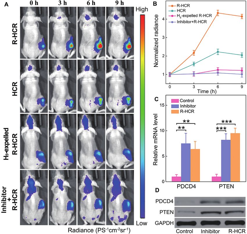
PAGE 3 OF 14 Nucleic Acids Research, 2020, Vol. 48, No. 10 e60
was obtained through a Milli-Q water purification system Intracellular R-HCR-imaging experiment
with an electric resistance >18.2 M·cm. Atomic force
MCF-7, A549, HeLa and MRC-5 cells were cultured
microscope (AFM) cantilever (SCANASYST-AIR) was
in DMEM medium containing 10% FBS and 1%
purchased from Bruker (Camarilla, CA, USA).
penicillin/streptomycin in 5% CO2 atmosphere at 37◦ C.
All cells above were plated in glass bottom culture dishes
Fluorescence assay and grown to 70% confluency for 12 h for the following
transfection. Briefly, the miR-21-targeting R-HCR mixture
Unless specifically presented, all of control experiments
containing H5 (0.05 nmol) and H1 +H2 +H3 +H4 +S+L
were carried out without changing the concentrations of
(0.1 nmol each) in Opti-MEM (200 l) was mixed with
specific DNA reactants except subtracting or replacing
lipofectamine 3000 (5 l) in Opti-MEM (200 l) for 5
the undesired oligonucleotides from their respective reac-
min. Then the above Opti-MEM transfection mixture and
tants. For amplified analysis of DNA by using R-HCR and
a supplementary 80 l of FBS were introduced into the
HCR amplifiers, the target was incubated with their re-
plated cells for 4 h at 37◦ C. Subsequently, the cultured cells
Downloaded from https://academic.oup.com/nar/article/48/10/e60/5826808 by guest on 08 September 2020
spective DNA mixtures (H1 +H2 +H3 +H4 +SM +L for HCR,
were washed three times with phosphate buffered saline
and H1 +H2 +H3 +H4 +S+L for R-HCR amplifier, 200 nM
(PBS) and cultured in DMEM medium (1.0 ml) containing
each) for 3 h at 25◦ C unless specified. For sensitive miR-
10% FBS for confocal imaging. When implementing
21 or miR-155 assay by the updated R-HCR circuit, the
the anti-miRNA antisense inhibitor oligonucleotide ex-
target (miR-21 or miR-155) was added into the mixture of
periment, MCF-7 cells were transfected with inhibitor
‘helper’ hairpin (H5 or H6 ) (100 nM) and R-HCR reactants
oligonucleotide (0.1 nmol) using lipofectamine 3000 (3 l)
(H1 +H2 +H3 +H4 +S+L, 200 nM each) at 25◦ C for 3 h un-
for 2 h, and were subsequently transfected and incubated
less specified. The real-time fluorescence monitoring of R-
with the R-HCR imaging system for 4 h. The detection of
HCR machine and its controls were implemented by using
miR-155 is the same as that of miR-21.
a Cary Eclipse spectrometer (Varian Inc) at 25◦ C. The flu-
orescence spectra were collected from 505 to 650 nm upon
exciting these samples at 490 nm. The time-dependent flu- Confocal laser scanning microscopy (CLSM) characteriza-
orescence change is recorded at the emission wavelength tion
of 520 nm and F0 represents the original fluorescence All cellular fluorescence images were obtained by Leica
intensity. TCS-SP8 laser scanning confocal microscopy system. A se-
ries of FRET, donor and acceptor filters were utilized to col-
Native polyacrylamide gel electrophoresis lect the following fluorescence readout: donor fluorescence
(FAM), acceptor fluorescence (TAMRA) from FRET and
Gel electrophoresis was performed to verify the external excitation. Upon exciting the system with 488 nm,
working principle of R-HCR system, 50 nM of the fluorescence emission spectra were collected from 500 to
target T was mixed with 200 nM of their DNA 560 nm for green channel of fluorophore donor (FAM), and
reactants (H1 +H2 +H3 +H4 +SM +L for HCR, and the FRET-generated fluorescence emission spectra was col-
H1 +H2 +H3 +H4 +S+L for R-HCR amplifier) in reac- lected from 575 to 635 nm for yellow channel of fluorophore
tion buffer (10 mM HEPES, 1 M NaCl, 50 mM MgCl2 , pH acceptor (TAMRA). Upon exciting the system with 561 nm,
7.2) for 3 h at 25◦ C. Then all samples were added into the the external excited fluorescence emission spectra were col-
freshly prepared native polyacrylamide (12%) gel matrix. lected from 575 to 635 nm for red channel of fluorophore
Electrophoresis was implemented at 120 V in 1× TBE acceptor (TAMRA). All images were developed at 63.0 ×
buffer (89 mM Tris, 89 mM boric acid, 2.0 mM EDTA, 1.40 objective with oil. The fluorescence emission ratio of
pH 8.3) for 3 h. After staining in diluted GelRed™ solu- acceptor to donor (FA /FD ) was denoted as the FRET signal
tion for 20 min, the gel was imaged by FluorChem FC3 for intracellular imaging upon exciting the R-HCR imag-
(ProteinSimple, USA) under 365 nm UV irradiation. ing system with 488 nm. The respective fluorescence (FA
and FD ) signals of different cells were obtained by integrat-
Atomic force microscope (AFM) characterization ing the fluorescence image of each sample. To get a reliable
quantitative FRET signal, the background FRET signal,
AFM characterization was carried out to verify the R- originating from FAM or TAMRA fluorophore itself, was
HCR-generated linear dsDNA nanowires. Here, 20 nM deducted from each sample. All FRET images were ana-
of target (T) was incubated with 200 nM of reactants lyzed and processed by using ImageJ software.
(H1 +H2 +H3 +H4 +S+L) in reaction buffer (10 mM HEPES,
1 M NaCl, 50 mM MgCl2 , pH 7.2) for 3 h at 25◦ C unless
Live animal imaging
specified. The freshly cleaved mica was reacted with 1 M
of Mg2+ ions for 15 min. These diluted DNA samples were Balb/c nude mice (4–6 weeks old) were subcutaneously in-
deposited on the modified mica for 15 min, followed by its jected with 5 × 10⁶ cells on the right side of the back. Tu-
rinsing with deionized water. The dried sample was scanned mor volume was analyzed using the following equation:
in tapping mode by Multimode 8 Atomic Force Microscope V = (L × W2 )/6 (L and W are the longest and short-
with a NanoScope V controller (Bruker Inc.). The silicon est diameters of the tumor, respectively). When the tumors
tips used for AFM analysis were SCANASYST-AIR (tip reach to the size of 100 mm3 , the mice were randomly di-
radius: ∼2 nm; resonance frequency: ∼70 kHz; spring con- vided into four groups for the treatment with lipo3000-
stant: ∼0.4 N/m; length: 115 m; width: 25 m). loaded R-HCR (H1 +H2 # +H3 +H4 # +S+L, 5 nmol/kg each,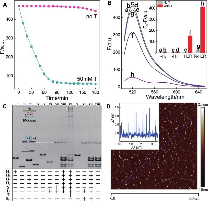
e60 Nucleic Acids Research, 2020, Vol. 48, No. 10 PAGE 4 OF 14
H5 , 2.5 nmol/kg), HCR (H1 +H2 # +H3 +H4 # +SM +L, 5 Feasibility study of the R-HCR machinery
nmol/kg each, H5 , 2.5 nmol/kg), H5 -expelled R-HCR For this proof-of-concept study, the as-achieved R-HCR
(H1 +H2 # +H3 +H4 # +S+L, 5 nmol/kg each) and R-HCR amplifier as illustrated in Figure 1A was first examined
with a chemically modified miR-21 inhibitor (miR-21 in- under optimal conditions (43) (Supplementary Figures S1
hibitor, H1 +H2 # +H3 +H4 # +S+L, 5 nmol/kg each, H5 , 2.5 and S2). As expected, the R-HCR mixture shows no flu-
nmol/kg) through intratumoral or intravenous injection. orescence change without initiator (Figure 2A), indicating
Mice were imaged using PE Spectrum & Quantum FX at these DNA probes are metastable without obvious signal
0, 3, 6, 9 h after intratumoral injection and at 4, 8, 12 h af- leakage (spontaneous reciprocal cross-catalytic reactions).
ter intravenous injection with excitation and emission wave- However, when initiator I was introduced into the R-HCR
lengths of 640 and 680 nm, respectively. Then they were sac- mixture, a dramatical fluorescence change was revealed and
rificed to collect the major organs and tumors for ex vivo it leveled off after 1.5 h (Figure 2A). More control experi-
imaging. Animal experiments were approved by the Animal ments were carried out for examining the detailed working
Care and Use Committee of Wuhan University.
Downloaded from https://academic.oup.com/nar/article/48/10/e60/5826808 by guest on 08 September 2020
mechanism of the R-HCR amplifier. Firstly, the indispens-
able role of HCR system was investigated by removing one
RESULTS of the non-fluorescent hairpins (H1 or H3 ) from the HCR
system. Obviously, the H1 - or H3 -excluded R-HCR am-
Principle of R-HCR machinery
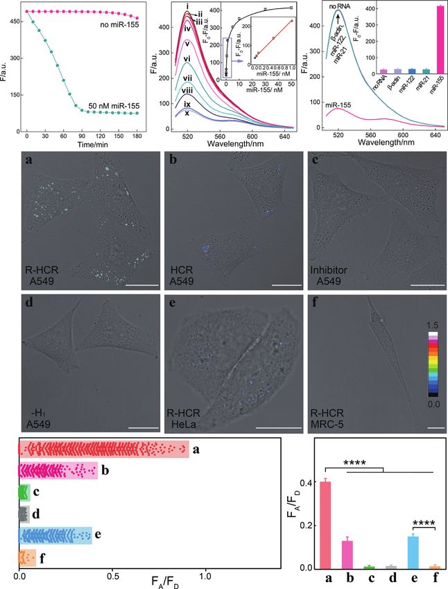
plifier disables the HCR-motivated dsDNA assembly and
The present R-HCR amplification platform is based on FRET generation (curves b, d of Figure 2B). These obser-
the design of appropriate HCR hairpin reactants (H1 , H2 , vations demonstrated that all participating HCR hairpins
H3 and H4 ) that are grafted with DNAzyme subunits, and are indispensable to propagate the HCR-amplified FRET
an additional DNAzyme substrate hybrid (partially hy- signal generation. Besides HCR, the DNAzyme biocatalysis
bridized S/L duplex) that is functionalized with the initia- was then explored by replacing the adenine ribonucleotide
tor sequence (Figure 1). The initiator activates the cross- of substrate (S) with an adenine deoxynucleotide, resulting
hybridization of HCR hairpins to generate long nicked ds- in the generation of a mutant substrate (SM ) that could not
DNA nanowires that carry chains of tandem RNA-cleaving be cleaved by DNAzyme. A comparably lower fluorescence
E6-type DNAzymes (42). The HCR-assembled DNAzymes response was obtained for the SM -substituted R-HCR ma-
then catalyze the efficient cleavage of DNAzyme substrate, chine (a characteristic traditional HCR amplifier) upon its
releasing numerous initiator sequence for reversely trigger- incubation with analyte (curve f of Figure 2B). Here the
ing the HCR amplifier. As shown in Figure 1A, the respec- HCR-assembled DNAzyme could not cleave the mutant
tive functionalization of DNAzyme subunits into the sepa- SM for triggering HCR system. This blocked DNAzyme-
rated hairpins (H1 and H3 ) prohibits the formation of an catalyzed HCR assembly leads to the assembly of a conven-
active DNAzyme while the different fluorophore-labelled tional HCR amplifier. As expected, a dramatic fluorescence
hairpins (H2 and H4 ) prohibits their efficient FRET proce- response was observed for the R-HCR machinery (curve
dure. The substrate (S), consisting of a ribonucleotide (rA)- h of Figure 2B), which is ascribed to the HCR-generated
containing DNAzyme substrate a*–h* and an initiator se- tandem DNAzyme copolymers that activate the production
quence (T), hybridizes partially with the blocker strand (L) of initiators for feeding back HCR system. The newly de-
to prohibit the undesired HCR-motivated signal leakage. veloped R-HCR amplifier shows an exponential amplifi-
The analyte (T) triggers the cyclic cross-opening of HCR cation efficiency (1:NN ), which is substantially higher than
reactants, leading to a sustained assembly of DNAzyme the multiple amplification efficiency (1:N) of conventional
nanowires that bring the two fluorophores (FAM and HCR. These results clearly demonstrate the successful cou-
TAMRA) into close proximity, thus enabling the Förster pling of HCR and DNAzyme without signal leakage, and
resonance energy transfer (FRET) process. Subsequently, the hierarchical integration of our reciprocal catalytic am-
each DNAzyme can catalyze the continuous scission of plifier with significant signal gain.
the substrate hybrid (S/L) to release new trigger strands Native gel electrophoresis and atomic force microscopy
for reversely stimulating the HCR transducer. During this (AFM) experiments were then carried out to explicit the
reciprocal catalytic process, each HCR-mediated H2 –H4 detailed reaction procedure of our R-HCR amplifier. The
hybridization event generates one FRET signal and each SM -substituted R-HCR system was examined as conven-
concomitant H1 –H3 hybridization event generates one ac- tional HCR amplifier. As indicated in Figure 2C, more
tive DNAzyme unit. Noted that each of these assembled bulky HCR products appeared for the target-activated R-
DNAzymes produces substantial amount of HCR trig- HCR system (sample ix) than that of the SM -substituted
gers for accelerating the whole reciprocal catalytic reac- conventional HCR system (sample xiii), owing to the con-
tion process. The amplifcation efficiency of conventional secutive accumulation of triggers for accelerating HCR sys-
HCR and DNAzyme amplifers was simplified as N, then tem. This promoted dsDNA assembly also implies a sig-
the newly developed R-HCR amplifier shows an exponen- nificant amplification capability of our reciprocal reaction
tial amplification efficiency (1:NN ) based on the recipro- process. Besides, the band of substrate hybrid (S/L) became
cal catalytic accumulation of HCR triggers and DNAzyme faint and even disappeared, while new product band of the
units. This leads to the promoted assembly of long tan- cleaved substrate emerged for the target-activated R-HCR
dem DNAzyme nanowires and the enhanced generation system (sample ix). Meanwhile, the band of mutant sub-
of FRET signal (detailed sequence, see Supplementary strate hybrid (SM /L) remained unchanged without gener-
Table S1). ating new product band of the cleaved substrate for the mu-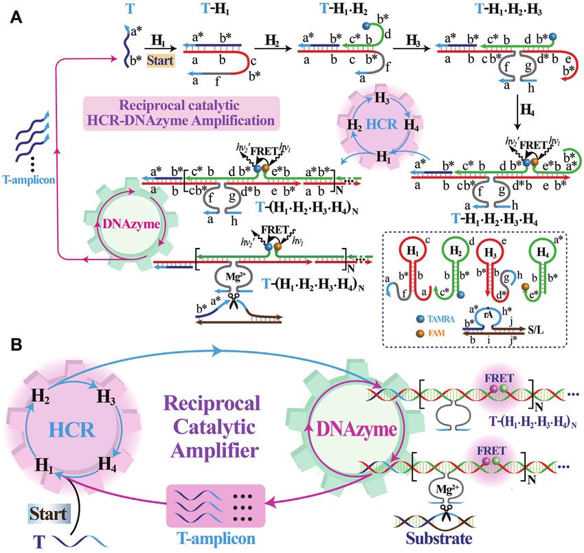
PAGE 5 OF 14                                                                             Nucleic Acids Research, 2020, Vol. 48, No. 10 e60

                                                                                                                                                          Downloaded from https://academic.oup.com/nar/article/48/10/e60/5826808 by guest on 08 September 2020
Figure 1. (A) Scheme reaction procedure of the proteinase-free replicating catalytic DNA machinery consisting of hybridization chain reaction (HCR) and
DNAzyme biocatalysis. (B) Simplified representation of the reciprocal HCR (R-HCR) amplification machinery.

tant SM -substituted HCR system (sample xiii). This was at-                   between the simulated curves (solid lines) and experimen-
tributed to the mutant substrate SM with adenine deoxynu-                     tal results (solid circles) was acquired as shown in Figure
cleotide which could not be cleaved by the present RNA-                       3A. Supplementary Table S4 summarizes the as-achieved
cleaving DNAzyme. Only the substrate (S) with adenine                         key parameters of each reaction scheme by using the re-
ribonucleotide could be be cleaved by the RNA-cleaving                        ciprocal catalytic reaction model. Obviously, the R-HCR
DNAzyme. In addition, the catalyzed cleavage of mere sub-                     machinery is dominated by the lead-in HCR process (as re-
strate (S) by HCR-assembled DNAzyme was furtherly ex-                         vealed by its reaction rate constant KT ) and the DNAzyme
plored by another gel electrophoresis (Supplementary Fig-                     biocatalysis process (as revealed by KM and Kcat ), which
ure S3). The morphology of our R-HCR product was then                         is reasonable since both of these two procedures are in-
examined by AFM. A substantial amount of micrometer-                          dispensable for carrying out the whole reaction processes.
long linear dsDNA nanowires (∼1.5 nm in diameter) was                         The comparably lower catalytic performance of the present
achieved for the analyte-triggered R-HCR machine (Figure                      HCR-reconstituted DNAzyme, as compared with the in-
2D) while merely tiny spots of HCR monomers emerged for                       tact DNAzyme* (42), is attributed to the slightly varied
the non-triggered R-HCR system (Supplementary Figure                          microenvironment of DNAzyme structure. In addition, the
S4), demonstrating our rational designed amplification sys-                   present R-HCR system was demonstrated by the marginal
tem.                                                                          signal leakage of HCR and DNAzyme, as demonstrated by
                                                                              their dramatically lower rate constants KD and KC . Inter-
Feasibility of R-HCR system for amplified DNA detection                       estingly, both of the sustained DNAzyme scission and effi-
                                                                              cient initiator exposure are needed to facilitate the high am-
Additionally, the underlying molecular reaction mechanism                     plification performance of the present reciprocal replicating
of our engineered R-HCR system was explored by using                          machinery. Noted that, even the side reaction or signal leak-
computer-aided simulations after a sophisticated model-                       age (crosstalk from the intrinsic imperfection of reactants)
ing of the system (details see the analytical modeling sec-                   is rather low, an obvious signal leakage could be predicted
tion of supporting information). The time-dependent fluo-                     theoretically after a certainly prolonged reaction time from
rescence readout of the R-HCR system was then collected                       the simulations. These theoretical simulations show good
and furtherly subjected to the MATLab program through                         coincidence with these experimental observations (Figure
a non-linear regression procedure. A satisfied consistency                    3A). This also implies that the present R-HCR amplifier, in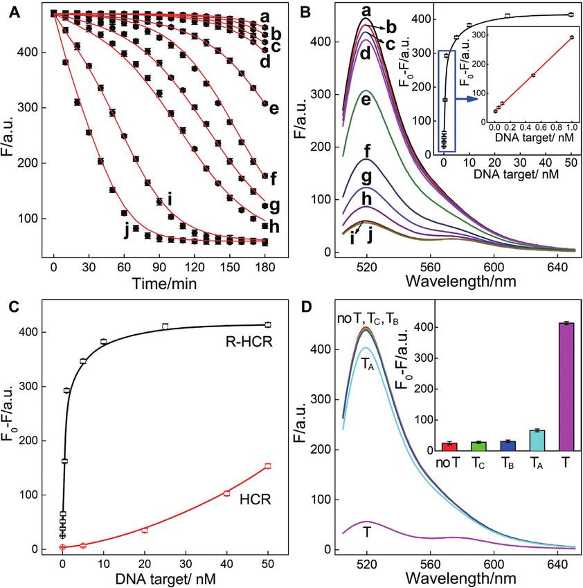
e60 Nucleic Acids Research, 2020, Vol. 48, No. 10 PAGE 6 OF 14
Downloaded from https://academic.oup.com/nar/article/48/10/e60/5826808 by guest on 08 September 2020
Figure 2. (A) Time-dependent fluorescence changes of the R-HCR system as shown in Figure 1 in the absence and presence of trigger T. (B) Fluorescence
spectra of the triggered R-HCR system that was subtracted by H1 alone: (a) no T, (b) T; or by H3 alone: (c) no T, (d) T; SM -substituted R-HCR system:
(e) no T, (f) T; and the intact R-HCR system: (g) no T, (h) T. Inset: Summary of the fluorescence intensity changes (at = 520 nm) shown in Figure
2B. (C) Native gel electrophoresis characterization of the R-HCR and conventional HCR systems. The ‘+’ and ‘-’ denote the presence and absence of the
corresponding components, respectively. (D) AFM image and the cross-section analysis of the R-HCR-generated dsDNA nanowires. F0 represents the
original fluorescence intensity. Error bars were derived from n = 5 experiments.
principle, can detect analyte at single-molecule level, illumi- tional HCR system, demonstrating the high signal gain of
nating its powerful potential applications in bioanalytical the present reciprocal catalytic amplifier. The selectivity of
fields. the R-HCR system was then evaluated by introducing one-,
Based on the aforementioned convincing results, it is two-, or three-base mutant targets (TA , TB and TC , respec-
clear that the R-HCR amplifier can be employed for sen- tively) (Figure 3D). The remarkable FRET readout of ana-
sitively analyzing trigger DNA. Accordingly, the fluores- lyte T was easily discriminated from TA , TB and TC , demon-
cence spectra of the R-HCR system were recorded after a strating the high selectivity of our R-HCR amplifier.
fixed time interval of 3 h for analyzing different concentra-
tions of trigger T (Figure 3B). Clearly, the fluorescence re-
sponse intensified with increasing concentration of T. A low Versatility of R-HCR-sustained miR-21 and miR-155 visual-
detection limit of 5.9 pM (3/slope) is acquired from the ization in living cells
calibration curve (Figure 3B inset), which is comparable to Here the versatile reciprocal catalytic HCR-DNAzyme am-
and even lower than most of the other proteinase-free signal plifier could be utilized as an amplification module, without
amplification methods (Supplementary Table S5). The per- further redesign and optimization of the entire system, for
formance of the SM -substituted R-HCR amplifier (as con- analyzing other targets through a facile integration with a
ventional HCR amplifier) was also employed for detecting sensing module. The modular feature of our sensing plat-
the same trigger T (Figure 3C and Supplementary Figure form was demonstrated by choosing microRNA-21 (miR-
S5). A tremendously lower FRET response was exhibited 21) as the model target. Abnormal miR-21 expressions are
for the conventional HCR system (Figure 3C). The R-HCR associated with a wide range of human cancers (44) and
amplifier shows a 78-fold signal enhancement than conven- have been recognized as important indicators for cancer di-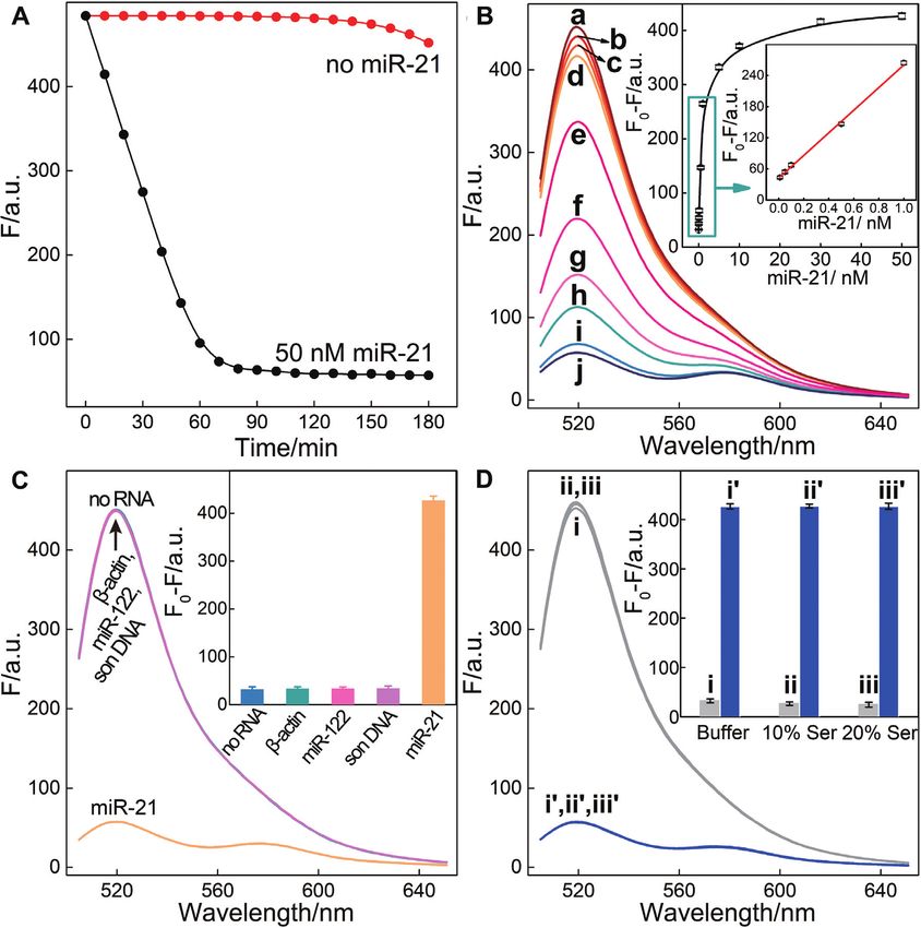
PAGE 7 OF 14 Nucleic Acids Research, 2020, Vol. 48, No. 10 e60
Downloaded from https://academic.oup.com/nar/article/48/10/e60/5826808 by guest on 08 September 2020
Figure 3. (A) Raw (solid circles) and simulated (line) time-dependent fluorescence changes of the R-HCR amplifier upon analysing different concentrations
of target T. (B) Fluorescence spectra of the R-HCR machine activated by different concentrations of T: (a) 0, (b) 1 × 10⁻¹¹ , (c) 5 × 10⁻¹¹ , (d) 1 × 10⁻¹⁰ ,
(e) 5 × 10⁻¹⁰ , (f) 1 × 10⁻⁹ , (g) 5 × 10⁻⁹ , (h) 1 × 10⁻⁸ , (i) 2.5 × 10⁻⁸ , and (j) 5 × 10⁻⁸ M. Inset: resulting calibration curve. (C) Comparison of the R-HCR
amplifier and conventional HCR system upon analyzing different concentrations of T. (D) Fluorescence spectra generated by the R-HCR amplifier upon
analyzing different mutant analytes (50 nM). Inset: Summary of the fluorescence spectra as shown in Figure 3D.
agnosis and prognosis (45). As shown in Figure 5A, the The satisfying anti-interference of the R-HCR system en-
sensing module includes only an auxiliary ‘helper’ hairpin couraged us to apply it as powerful intracellular imaging
H5 that is encoded with a miR-21-recognition sequence and tool for visualizing analytes in living cells. Then the R-HCR
a caged R-HCR initiator sequence (details, see Supplemen- amplifier was utilized to investigate the distinct miR-21 ex-
tary Figure S6). Target miR-21 can hybridize with H5 to ex- pressions of different cells by using confocal laser scanning
pose a HCR trigger for motivating the R-HCR-accelerated microscopy (CLSM). Here, all DNA probes were partially
FRET readout with satisfying low signal leakage (Figure modified with phosphorothioate bonds to prevent the po-
4A). The fluorescence spectra were acquired upon the anal- tential nuclease degradation in living cells (details see Sup-
ysis of different concentrations of miR-21 (Figure 4B). A plementary Table S2). The performance of phosphoroth-
detection limit of 6.8 pM was achieved for the R-HCR am- ioate R-HCR system shows a good agreement with that of
plifier (Figure 4B inset). The selectivity of the miR-21 detec- the intact R-HCR system (Supplementary Figure S7), in-
tion system was challenged by a series of interfering miR- dicating such modification has little effect on their sensing
NAs: -actin mRNA, son DNA and miR-122. A clear sig- performance. The low cytotoxicity of the R-HCR system
nal discrimination could be observed between miR-21 and was demonstrated by a conventional MTT assay in vitro
these interfering strands, demonstrating the high specificity (Supplementary Figure S8). Considering the unexpectedly
of the present miRNA assay (Figure 4C). Serum has little distinct and complex intracellular environment of different
effect on the performance of the reciprocal catalytic ampli- cells, the fluorescence emission ratio of acceptor to donor
fier, demonstrating its robust feature (Figure 4D). Thus the (FA /FD ) was employed as a visualization readout to min-
R-HCR machine can be developed as a general amplifica- imize the undesired system fluctuations. Our proposed R-
tion tool for detecting low abundant analyte. HCR was adapted for intracellular miR-21 imaging in threee60 Nucleic Acids Research, 2020, Vol. 48, No. 10 PAGE 8 OF 14
Downloaded from https://academic.oup.com/nar/article/48/10/e60/5826808 by guest on 08 September 2020
Figure 4. (A) Time-dependent fluorescence changes of the updated R-HCR system with and without miR-21. (B) Fluorescence spectra of the miR-21-
targeting R-HCR sensing platform generated by different concentrations of miR-21 after 3 h: (a) 0, (b) 1 × 10⁻¹¹ , (c) 5 × 10⁻¹¹ , (d) 1 × 10⁻¹⁰ , (e) 5 ×
10−10 , (f) 1 × 10⁻⁹ , (g) 5 × 10⁻⁹ , (h) 1 × 10⁻⁸ , (i) 3 × 10⁻⁸ , and (j) 5 × 10⁻⁸ M. Inset: resulting calibration curve. (C) Fluorescence spectra generated by
the updated R-HCR amplifier upon analysing different analytes (50 nM). Inset: Summary of the fluorescence spectra as shown in (C). (D) Fluorescence
spectra generated by the updated R-HCR system upon analyzing 50 nM of miR-21 in different serum solutions: buffer without (i) and with (i‘) miR-21,
10% serum without (ii) and with (ii‘) miR-21, 20% serum without (iii) and with (iii‘) miR-21. Inset: Summary of the fluorescence spectra as shown in (D).
Error bars were derived from n = 5 experiments.
different types of cell lines, including human breast cancer qRT-PCR assay (Supplementary Figure S10 and Table S3).
(MCF-7) with relatively high miR-21 expression profile, hu- Thus the reciprocal R-HCR imaging platform provided pre-
man cervical cancer cells (HeLa) with relatively low miR- cise localization of miR-21 in living cells. Moreover, a sub-
21 expression profile, and human embryonic lung fibroblast stantially declined FRET signal appeared in MCF-7 cells
cells (MRC-5) with no miR-21 expression. The R-HCR- that were pretreated with anti-miRNA antisense inhibitor
mediated intracellular imaging system was transfected into oligonucleotide (sample c of Figure 5B), implying that the
these different living cells via lipofectamine 3000 for 4 h (an miR-21 molecules were indeed presented in MCF-7 cells for
optimized incubation time, see Supplementary Figure S9). activating the R-HCR system. The corresponding scatter
An extremely intense FRET signal of miR-21 target was ob- plot of FRET signal furtherly evidenced the highly power-
served in MCF-7 cells (sample a, Figure 5B) while a com- ful intracellular biosensing performance of our R-HCR am-
paratively weaker FRET response emerged in HeLa cells plifier (Figure 5C). A statistical histogram analysis of the
(sample e, Figure 5B). In contrast, no detectable FRET sig- FRET signal (FA /FD ) of the R-HCR imaging system was
nal appeared in MRC-5 cells (sample f, Figure 5B), indi- collected as shown in Figure 5D. The FRET efficiency of
cating a higher expression of miR-21 in tumor cells than the R-HCR imaging system was acquired to be 0.63 by us-
normal cells, which was accordant with the previous re- ing conventional acceptor-photo-bleaching method (Sup-
port (24,46). The FRET signal of MCF-7 cells was 3.0- plementary Figure S11). Overall, the R-HCR imaging sys-
and 26.1-fold higher than that of HeLa cells and MRC-5 tem enables a reliable discrimination of different miR-21 ex-
cells (Figure 5D), which was consistent with conventional pressions in living cells.PAGE 9 OF 14 Nucleic Acids Research, 2020, Vol. 48, No. 10 e60
Downloaded from https://academic.oup.com/nar/article/48/10/e60/5826808 by guest on 08 September 2020
Figure 5. (A) Schematic representation of the miR-21-targeting R-HCR sensing platform by introducing a foreign helper H5 into the well-established
R-HCR amplifier. (B) Intracellular analysis of miR-21 based on the updated R-HCR imaging system and FRET transduction (in the form of FA /FD ).
CLSM probing of intracellular miR-21 by (a) R-HCR amplifier in MCF-7 cells, (b) conventional HCR amplifier in MCF-7 cells, (c) R-HCR amplifier
with a chemically modified miR-21 inhibitor in MCF-7 cells, (d) H1 -expelled R-HCR amplifier in MCF-7 cells, (e) R-HCR amplifier in HeLa cells, and (f)
R-HCR amplifier in MRC-5 cells. All scale bars correspond to 20 m. (C) The acquired FRET signal (FA /FD ) distributions from Figure 5B. (D) Statistical
histogram analysis of the FRET signal (FA /FD ) of these different cell samples through the R-HCR imaging system.e60 Nucleic Acids Research, 2020, Vol. 48, No. 10 PAGE 10 OF 14
Furthermore, the intracellular miR-21 localization was specificity for miRNA detection in complex cellular envi-
demonstrated by 3D z-stack projections through integrat- ronment.
ing a series of z-section images of the entire cells (Sup- Based on the above results, we applied the R-HCR am-
plementary Figure S12, Supplementary video S1). Clearly, plifier to investigate the varied expressions of miR-155 in
the endogenous miR-21 is distributed in the cytoplasm of human lung cancer (A549), human cervical cancer cells
MCF-7 cells, demonstrating that our R-HCR imaging sys- (HeLa) and human embryonic lung fibroblast cells (MRC-
tem realized the spatially resolved intracellular miRNA vi- 5) by using CLSM technique. It was found that the average
sualization. The mutually reinforcing replication between FRET signal per cell decreases in the order of A549 cells
the transducer HCR and DNAzyme biocatalyst was simi- > HeLa cells > MRC-5 cells (samples a, e and f of Figure
larly verified by excluding one of the non-fluorescent hair- 6D), which is consistent with previous research (48), sug-
pin reactants or by replacing the mutant substrate (SM ) gesting the good capability of the R-HCR imaging platform
from the R-HCR imaging mixture. As compared with for precise localization of low-abundance miR-155 in differ-
the intact R-HCR imaging system (sample a of Supple- ent living cells. Moreover, a significantly decreased FRET
Downloaded from https://academic.oup.com/nar/article/48/10/e60/5826808 by guest on 08 September 2020
mentary Figure S13A), almost no FRET readout was signal was exhibited in the anti-miRNA-treated A549 cells
shown in MCF-7 cells that were transfected with H1 - (sample c of Figure 6D), demonstrating that the R-HCR
or H3 -expelled R-HCR imaging system (sample c, d of imaging system enables a reliable sensing of miR-155 in liv-
Supplementary Figure S13A, respectively) while a weak ing cells. To verify the in situ enhanced amplification ca-
FRET response emerged in MCF-7 cells that were trans- pability of our R-HCR machinery in living cells, control
fected with the SM -substituted R-HCR system (sample experiments were performed by expelling one of the non-
b of Figure 5B and Supplementary Figure S13A). As fluorescent hairpin reactants or replacing the substrate (S)
compared with traditional HCR system, the R-HCR ma- with the mutant substrate (SM ). As shown in Figure 6D, a
chine lead to the generation of more dsDNA nanowires negligible FRET signal was observed in A549 cells that were
and more efficient FRET imaging signal for analyzing treated with H1 -expelled R-HCR imaging system (sample d,
miRNA in living cells. These demonstrated that each of Figure 6D) while a faint FRET response was exhibited in
these amplification constitutes was indispensable for car- A549 cells treated with the SM -substituted R-HCR system
rying out the synergistically amplified sensing platform, (conventional HCR amplifier, sample b, Figure 6D). Ob-
which was consistent with previous in vitro studies (Fig- viously, the R-HCR machinery leads to the generation of
ure 2B). In our design, the updated R-HCR system rec- more DNAzyme nanowires and more efficient FRET imag-
ognized and hybridized miR-21 to form stable complexes, ing signal for monitoring miRNA in living cells (sample a,
which would lead to the upregulation of the downstream Figure 6D). The corresponding scatter plot of FRET signal
target genes of PDCD4 and PTEN. A distinct up-regulated distribution was exhibited in Figure 6E and a statistical his-
PDCD4 and PTEN mRNAs/proteins was presented in togram analysis of the FRET signal (FA /FD ) of the R-HCR
R-HCR system or miR-21 inhibitor-treated MCF-7 cells imaging system was shown in Figure 6F. These results evi-
(Supplementary Figure S14), indicating the specific bind- denced the versatile and reliable intracellular imaging per-
ing of the R-HCR probe with miR-21 inside living formance of our R-HCR amplifier.
cells.
Encouraged by these results, we further validated the gen-
In vivo miR-21 imaging by the R-HCR system
erality of our R-HCR strategy only by updating the ac-
cessorial sensing module H6 for another microRNA, miR- Inspired by the excellent intracellular visualization per-
155, which is sufficient to induce tumorigenesis. High ex- formance in vitro, we further examined the updated R-
pression of miRNA-155 has been reported in various B HCR system for miRNA imaging in vivo. Hairpin H4 was
cell malignancies (47). Benefiting from the modular design, modified with a Cy5/quencher pair for miR-21 bioimag-
the miRNA-155-detecting R-HCR system can be readily ing. The biocompatibility of R-HCR system was further
constructed only by re-encoding hairpin H6 with a miR- demonstrated by hemolysis test (Supplementary Figure
155-recognition sequence and a caged R-HCR initiator se- S16), hematology and biochemical analysis (Supplemen-
quence (Supplementary Figure S15). As shown in Figure tary Figure S17), and H&E staining assay (Supplementary
6A, miR-155 opens H6 to release HCR trigger for stim- Figure S18). The intratumoral imaging performance of the
ulating the R-HCR-amplified FRET response. The fluo- R-HCR amplifier was first explored by intratumoral injec-
rescence spectra of the updated R-HCR system were ob- tion of nude mice bearing subcutaneous MCF-7 tumor on
tained upon its incubation with various concentrations of the right back. Upon exciting the mice with a fixed ex-
miRNA-155 (Figure 6B). A good linear relationship was ac- citation wavelength (ex = 640 nm), the fluorescence sig-
quired between fluorescence change and miRNA-155 con- nal reached its maximum value at 6 h postinjection of
centration ranging from 10 pM to 1 nM (Figure 6B inset). lipo3000-loaded R-HCR mixture and then decreased (Fig-
The limit of detection (LOD) was calculated to be 7.6 pM ure 7A). Quantitative analysis showed that R-HCR-treated
based on the 3 method. Three different interfering miR- mice presented 2.0- and 3.5-fold higher intratumoral Cy5
NAs (-actin mRNA, miR-122 and miR-21) were chosen signal than that treated with HCR and H5 -excluded R-
to further examine the specificity of the updated R-HCR HCR systems at 6 h (Figure 7B). Furthemore, the miRNA-
system toward miRNA-155. As shown in Figure 6C, the R- 21 inhibitor was injected into tumor site for knocking down
HCR system showed low FRET response toward these in- miR-21 expression, as expected, scarcely any Cy5 signal was
terfering miRNAs, which were close to the blank control. observed at miRNA-21 inhibitor-pretreated tumor, show-
These results indicated that this proposed method had high ing good agreement with the cellular experiments. Addi-PAGE 11 OF 14 Nucleic Acids Research, 2020, Vol. 48, No. 10 e60
Downloaded from https://academic.oup.com/nar/article/48/10/e60/5826808 by guest on 08 September 2020
Figure 6. (A) Time-dependent fluorescence changes of the miR-155-targeting R-HCR sensing platform without and with miR-155. (B) Fluorescence
spectra of the R-HCR system generated by different concentrations of miR-155 after 3 h: (i) 0, (ii) 1 × 10⁻¹¹ , (iii) 5 × 10⁻¹¹ , (iv) 1 × 10⁻¹⁰ , (v) 5 ×
10−10 , (vi) 1 × 10⁻⁹ , (vii) 5 × 10⁻⁹ , (viii) 1 × 10⁻⁸ , (ix) 3 × 10⁻⁸ and (x) 5 × 10⁻⁸ M. Inset: resulting calibration curve. (C) Fluorescence spectra of the
R-HCR amplifier upon analysing different interfering analytes (50 nM). Inset: Summary of the fluorescence spectra. Error bars were derived from n = 5
experiments. (D) Intracellular analysis of miR-155 through the R-HCR imaging system and FRET transduction (in the form of FA /FD ). CLSM probing of
intracellular miR-155 by (a) R-HCR in A549 cells, (b) conventional HCR in A549 cells, (c) R-HCR with a chemically modified miR-155 inhibitor in A549
cells, (d) H1 -expelled R-HCR in A549 cells, (e) R-HCR in HeLa cells and (f) R-HCR in MRC-5 cells. All scale bars correspond to 20 m. (E) The acquired
FRET signal (FA /FD ) distributions from (D). (F) Statistical histogram analysis of the FRET signal (FA /FD ) of these different cell samples through the
R-HCR imaging system.e60 Nucleic Acids Research, 2020, Vol. 48, No. 10 PAGE 12 OF 14
Downloaded from https://academic.oup.com/nar/article/48/10/e60/5826808 by guest on 08 September 2020
Figure 7. The R-HCR-activated miRNA imaging in tumors by intratumoral injection. (A) Whole-body fluorescence imaging of MCF-7 tumor-bearing
mice after injection of R-HCR, HCR, H5 -expelled R-HCR and R-HCR amplifier with a chemically modified miR-21 inhibitor. (B) Quantification of the
fluorescence of tumor sites in (A). (C) qRT-PCR and (D) western blot analysis of PDCD4 and PTEN in mice tumor after intratumoral injection of PBS,
miR-21 inhibitor and R-HCR system.
tionally, a significant enhancement of PDCD4 and PTEN Cy5 signal. Furthermore, the Cy5 of harvested tumors and
mRNAs/proteins was shown in mice tumor that was treated organs at 12 h post-injection was analyzed (Supplementary
with the updated R-HCR system or miR-21 inhibitor (Fig- Figures S19C and S19D). The intratumoral Cy5 signal in
ure 7C and 7D), which was consistent with the correspond- the R-HCR group was 3.3- and 4.6-fold higher than that
ing cellular studies. Thus the R-HCR system could realize in HCR and the H5 -excluded R-HCR groups, respectively.
miRNA-21 imaging in living mice. Additionally, the Cy5 of normal organs revealed no distinct
We further examined the imaging performance of the R- difference for all groups, proving the precise miRNA imag-
HCR imaging system in living mice bearing MCF-7 tu- ing in living mice.
mor by tail vein injection (Supplementary Figure S19A).
The lipo3000-loaded R-HCR system could gradually ac-
DISCUSSION
cumulate in tumor site via EPR effect. Quantitative anal-
ysis showed that the R-HCR group displayed 2.2- and 3.4- An unprecedented reciprocal replicating DNA machine
fold higher intratumoral Cy5 signal than the HCR and H5 - was engineered for high-performance in vivo miRNA imag-
excluded R-HCR groups at 12 h (Supplementary Figure ing based on the synergistic cross-activation of HCR and
S19B). The enhanced Cy5 signal of R-HCR in tumors was DNAzyme procedures. Target-activated transducer HCR
attributed to the reciprocal reinforcement feature of HCR assembles long tandem DNAzyme nanowires, which cat-
and DNAzyme biocatalysis that accelerated the entire reac- alyze the cyclic and continuous generation of triggers
tion procedure and promoted the generation of an amplified for reversely feeding back HCR transducer. The flexiblePAGE 13 OF 14 Nucleic Acids Research, 2020, Vol. 48, No. 10 e60
DNAzyme design dictates a more facile assembly of HCR 7. Notomi,T., Okayama,H., Masubuchi,H., Yonekawa,T., Watanabe,K.,
machine while the high catalytic efficiency of DNAzyme Amino,N. and Hase,T. (2000) Loop-mediated isothermal
amplification of DNA. Nucleic Acids Res., 28, e63.
contributes to the utmost in-depth amplification. Our 8. Walker,G.T., Fraiser,M.S., Schram,J.L., Little,M.C., Nadeau,J.G.
DNAzyme utilizes endogenous Mg2+ ions as the indispens- and Malinowski,D.P. (1992) Strand displacement amplification-an
able DNAzyme cofactors without additional DNAzyme isothermal, in vitro DNA amplification technique. Nucleic Acids Res.,
cofactors, which is more easier for machinery design and 20, 1691–1696.
operation. This cross-invasion of transducer HCR and 9. Willner,I., Shlyahovsky,B., Zayats,M. and Willner,B. (2008)
DNAzymes for sensing, nanobiotechnology and logic gate
DNAzyme biocatalysis could convert the limited molec- applications. Chem. Soc. Rev., 37, 1153–1165.
ular recognition event into an efficient accumulation of 10. Zhang,D.Y. and Seelig,G. (2011) Dynamic DNA nanotechnology
amplicons as well as the remarkable FRET motivation. using strand-displacement reactions. Nat. Chem., 3, 103–113.
The underlying molecular-level propagation mechanism of 11. Wang,Z.-G., Zhan,P. and Ding,B. (2013) Self-assembled catalytic
DNA nanostructures for synthesis of para-directed polyaniline. ACS
the R-HCR machine, especially the comprising HCR and Nano, 7, 1591–1598.
DNAzyme biocatalysis reactions, was systematically exam- 12. Liu,J. and Lu,Y. (2007) A DNAzyme catalytic beacon sensor for
Downloaded from https://academic.oup.com/nar/article/48/10/e60/5826808 by guest on 08 September 2020
ined by experimental studies and computer-aided simula- paramagnetic Cu2+ ions in aqueous solution with high sensitivity and
tions after an appropriate modeling of the system. Given the selectivity. J. Am. Chem. Soc., 129, 9838–9839.
accelerated reaction format and the multiple-guaranteed 13. Wang,Y. and Irudayaraj,J. (2011) A SERS DNAzyme biosensor for
lead ion detection. Chem. Commun., 47, 4394–4396.
recognitions, the proteinase-free DNA machine was suc- 14. Wang,F., Elbaz,J. and Willner,I. (2012) Enzyme-free amplified
cessfully applied to sensitively and selectively detect target. detection of DNA by an autonomous ligation DNAzyme machinery.
This robust amplifier could be easily integrated with a gen- J. Am. Chem. Soc., 134, 5504–55007.
eral and versatile sensing module for realizing an accurate 15. Liu,M., Zhang,Q., Chang,D., Gu,J., Brennan,J.D. and Li,Y. (2017) A
DNAzyme feedback amplification strategy for biosensing. Angew.
miRNA localization with high reliability in living cells and Chem. Int. Ed., 129, 6238–6242.
even living mice through a facile fluorescence imaging tech- 16. Wang,F., Elbaz,J., Teller,C. and Willner,I. (2011) Amplified detection
nique, thus provides a powerful sensing platform for trac- of DNA through an autocatalytic and catabolic DNAzyme-Mediated
ing low-amount biomarkers in vivo. As a nanomaterial-free process. Angew. Chem. Int. Ed., 50, 295–299.
sensing platform, our system could avoid the unexpected 17. Tian,Y., He,Y., Chen,Y., Yin,P. and Mao,C. (2005) A DNAzyme that
walks processively and autonomously along a One-Dimensional
adverse retention of transfecting inorganic nanomaterials track. Angew. Chem. Int. Ed., 44, 4355–4358.
in vivo and the accompanying unexpected biotoxicity. The 18. Peng,H., Li,X.-F., Zhang,H. and Le,X.C. (2017) A
R-HCR machine holds great potential for clinical diagno- microRNA-initiated DNAzyme motor operating in living cells. Nat.
sis and therapeutic evaluation. Commun., 8, 14378.
19. Dirks,R.M. and Pierce,N.A. (2004) Triggered amplification by
hybridization chain reaction. Proc. Natl. Acad. Sci. USA, 101,
SUPPLEMENTARY DATA 15275–15278.
20. Choi,H.M.T., Chang,J.Y., Trinth,L.A., Padilla,J.E., Frase,S.E. and
Supplementary Data are available at NAR Online. Pierce,N.A. (2010) Programmable in situ amplification for multiplexed
imaging of mRNA expression. Nat. Biotechnol., 28, 1208–1212.
21. Choi,H.M.T., Beck,V.A. and Pierce,N.A. (2014) Next-generation in
FUNDING situ hybridization chain reaction: higher gain, lower cost, greater
durability. ACS Nano, 8, 4284–4294.
Natural Science Foundation of China [21874103, 22. Wu,Z., Liu,G.Q., Yang,X.L. and Jiang,J.H. (2015) Electrostatic
81602610]; National Basic Research Program of China nucleic acid nanoassembly enables hybridization chain reaction in
living cells for ultrasensitive mRNA imaging. J. Am. Chem. Soc., 137,
(973 Program) [2015CB932601]; Fundamental Research 6829–6836.
Funds for the Central Universities [2042018kf0210, 23. Huang,J., Wu,Y., Chen,Y., Zhu,Z., Yang,X., Yang,C.J., Wang,K. and
2042019kf0206]. Funding for open access charge: Na- Tan,W. (2011) Pyrene-excimer probes based on the hybridization
tional Basic Research Program of China (973 Program) chain reaction for the detection of nucleic acids in complex biological
[2015CB932601]. fluids. Angew. Chem. Int. Ed., 50, 401–404.
24. Cheglakov,Z., Cronin,T.M., He,C. and Weizmann,Y. (2015) Live cell
Conflict of interest statement. None declared. microRNA imaging using cascade hybridization reaction. J. Am.
Chem. Soc., 137, 6116–6119.
25. Yang,L., Liu,C.H., Ren,W. and Li,Z.P. (2012) Graphene
REFERENCES surface-anchored fluorescence sensor for sensitive detection of
1. Zhang,H., Li,F., Dever,B., Li,X.F. and Le,X.C. (2013) microRNA coupled with enzyme-free signal amplification of
DNA-mediated homogeneous binding assays for nucleic acids and hybridization chain reaction. ACS Appl. Mater. Interfaces, 4,
proteins. Chem. Rev., 113, 2812–2841. 6450–6453.
2. Duan,R., Lou,X. and Xia,F. (2016) The development of 26. Wei,J., Gong,X., Wang,Q., Pan,M., Liu,X., Liu,J., Xia,F. and
nanostructure assisted isothermal amplification in biosensors. Chem. Wang,F. (2018) Construction of an autonomously concatenated
Soc. Rev., 45, 1738–1749. hybridization chain reaction for signal amplification and intracellular
3. Guo,S. and Wang,E. (2011) Functional micro/nanostructures: simple imaging. Chem. Sci., 9, 52–61.
synthesis and application in sensors, fuel cells, and gene delivery. Acc. 27. Wang,F., Elbaz,J., Orbach,R., Magen,N. and Willner,I. (2011)
Chem. Res., 44, 491–500. Amplified analysis of DNA by the autonomous assembly of polymers
4. Jung,C. and Ellington,A.D. (2014) Diagnostic applications of nucleic consisting of DNAzyme wires. J. Am. Chem. Soc., 133, 17149–17151.
acid circuits. Acc. Chem. Res., 47, 1825–1835. 28. Li,B., Jiang,Y., Chen,X. and Ellington,A.D. (2012) Probing spatial
5. Gill,P. and Ghaemi,A. (2008) Nucleic acid isothermal amplification organization of DNA strands using enzyme-free hairpin assembly
technologies-a review. Nucleosides Nucleotides Nucleic Acids, 27, circuits. J. Am. Chem. Soc., 134, 13918–13921.
224–243. 29. Zhou,Y., Yang,L., Wei,J., Ma,K., Gong,X., Shang,J., Yu,S. and
6. Banér,J., Nilsson,M., Mendel-Hartvig,M. and Landegren,U. (1998) Wang,F. (2019) An autonomous nonenzymatic concatenated DNA
Signal amplification of padlock probes by rolling circle replication. circuit for amplified imaging of intracellular ATP. Anal. Chem., 91,
Nucleic Acids Res., 26, 5073–5078. 15229–15234.e60 Nucleic Acids Research, 2020, Vol. 48, No. 10 PAGE 14 OF 14
30. Wang,H., Wang,H., Wu,Q., Liang,M., Liu,X. and Wang,F. (2019) A 39. Wang,H., Li,C., Liu,X., Zhou,X. and Wang,F. (2018) Construction
DNAzyme-amplified DNA circuit for highly accurate microRNA of an enzyme-free concatenated DNA circuit for signal amplification
detection and intracellular imaging. Chem. Sci., 10, 9597–9604. and intracellular imaging. Chem. Sci., 9, 5842–5849.
31. Gong,X., Wei,J., Liu,J., Li,R., Liu,X. and Wang,F. (2019) 40. Wei,J., Wang,H., Wu,Q., Gong,X., Ma,K., Liu,X. and Wang,F.
Programmable intracellular DNA biocomputing circuits for reliable (2020) A smart autocatalytic DNAzyme biocircuit for in vivo
cell recognitions. Chem. Sci., 10, 2989–2997. amplified MicroRNA imaging. Angew. Chem. Int. Ed., 59, 5965–5971.
32. Kausar,A., McKay,R.D., Lam,J., Bhogal,R.S., Tang,A.Y. and 41. Wu,C., Cansiz,S., Zhang,L., Teng,I.T., Qiu,L., Li,J., Liu,Y., Zhou,C.,
Gibbs-Davis,J.M. (2011) Tuning DNA stability to achieve turnover in Hu,R., Zhang,T. et al. (2015) A nonenzymatic hairpin DNA cascade
template for an enzymatic ligation reaction. Angew. Chem. Int. Ed., reaction provides high signal gain of mRNA imaging inside live cells.
50, 8922–8926. J. Am. Chem. Soc., 137, 4900–4903.
33. Kausar,A., Mitran,C.J., Li,Y. and Gibbs-Davis,J.M. (2013) Rapid, 42. Breaker,R.R. and Joyce,G.F. (1995) A DNA enzyme with
isothermal DNA self-replication induced by a destabilizing lesion. Mg2+ -Dependent RNA phosphoesterase activity. Chem. Biol., 2,
Angew. Chem. Int. Ed., 52, 10577–10581. 655–660.
34. Nie,J., Zhao,M. Z, Xie,W.J., Cai,L.Y., Zhou,Y.L. and Zhang,X.X. 43. Zadeh,J.N., Steenberg,C.D., Bois,J.S., Wolfe,B.R., Pierce,M.B.,
(2015) DNA cross-triggered cascading self-amplification artificial Khan,A.R., Dirks,R.M. and Pierce,N.A. (2011) NUPACK: analysis
biochemical circuit. Chem. Sci., 6, 1225–1229. and design of nucleic acid systems. J. Comput. Chem., 32, 170–173.
Downloaded from https://academic.oup.com/nar/article/48/10/e60/5826808 by guest on 08 September 2020
35. Lincoln,T.A. and Joyce,G.F. (2009) Self-sustained replication of an 44. Bartel,D.P. (2004) MicroRNAs: genomics, biogenesis, mechanism,
RNA enzyme. Science, 323, 1229–1232. and function. Cell, 116, 281–297.
36. Levy,M. and Ellington,A.D. (2003) Exponential growth by 45. Calin,G.A. and Croce,C.M. (2006) MicroRNA signatures in human
cross-catalytic cleavage of deoxyribozymogens. Proc. Natl. Acad. Sci. cancers. Nat. Rev. Cancer, 6, 857–866.
U.S.A., 100, 6416–6421. 46. Liang,C.P., Ma,P.Q., Liu,H., Guo,X.G., Yin,B.C. and Ye,B.C. (2017)
37. Yang,L., Wu,Q., Chen,Y., Liu,X., Wang,F. and Zhou,X. (2019) Rational engineering of a dynamic, entropy-driven DNA
Amplified MicroRNA detection and intracellular imaging based on nanomachine for intracellular microRNA imaging. Angew. Chem.
an autonomous and catalytic assembly of DNAzyme. ACS Sens., 4, Int. Ed., 56, 9077–9081.
110–117. 47. Eis,P.S., Tam,W., Sun,L., Chadburn,A., Li,Z., Gomez,M.F., Lund,E.
38. Wu,Q., Wang,H., Gong,K., Shang,J., Liu,X. and Wang,F. (2019) and Dahlberg,J.E. (2005) Accumulation of miR-155 and BIC RNA in
Construction of an autonomous nonlinear hybridization chain human B cell lymphomas. Proc. Natl. Acad. Sci. USA, 102,
reaction for extracellular Vesicles-Associated MicroRNAs 3627–3632.
discrimination. Anal. Chem., 91, 10172–10179. 48. Lee,H., Park,J.-E. and Nam,J.-M. (2014) Bio-barcode gel assay for
microRNA. Nat. Commun., 5, 3367.You can also read