The Rab11-binding protein REL CH/KIAA1468 controls intracellular cholesterol distribution
←
→
Page content transcription
If your browser does not render page correctly, please read the page content below
ARTICLE
The Rab11-binding protein RELCH/KIAA1468 controls
intracellular cholesterol distribution
Tomoaki Sobajima1,2, Shin‑ichiro Yoshimura1, Tomomi Maeda1,2, Haruhiko Miyata3, Eiji Miyoshi2, and Akihiro Harada1
Cholesterol, which is endocytosed to the late endosome (LE)/lysosome, is delivered to other organelles through vesicular
and nonvesicular transport mechanisms. In this study, we discuss a novel mechanism of cholesterol transport from recycling
endosomes (REs) to the trans-Golgi network (TGN) through RELCH/KIAA1468, which is newly identified in this study as a
Rab11-GTP– and OSBP-binding protein. After treating cells with 25-hydroxycholesterol to induce OSBP relocation from the
cytoplasm to the TGN, REs accumulated around the TGN area, but this accumulation was diminished in RELCH- or OSBP-
Downloaded from https://rupress.org/jcb/article-pdf/217/5/1777/441447/jcb_201709123.pdf by guest on 20 February 2020
depleted cells. Cholesterol content in the TGN was decreased in Rab11-, RELCH-, and OSBP-depleted cells and increased in the
LE/lysosome. According to in vitro reconstitution experiments, RELCH tethers Rab11-bound RE-like and OSBP-bound TGN-
like liposomes and promotes OSBP-dependent cholesterol transfer from RE-like to TGN-like liposomes. These data suggest
that RELCH promotes nonvesicular cholesterol transport from REs to the TGN through membrane tethering.
Introduction
Most mammalian cells acquire cholesterol through the endocyto- The Rab GTPase family, which comprises 60 members in
sis of plasma lipoproteins such as low-density lipoprotein (LDL). mammals, regulates various steps in intracellular protein trans-
After LDL is delivered to the lysosome, free cholesterol, which is port such as vesicle/tubule generation, motility along the cyto-
derived from hydrolyzed cholesterol ester liberated from LDL, is skeleton, tethering, and fusion by recruiting specific binding
transported from the lysosome to various subcellular membrane proteins to the membrane (Stenmark, 2009). Several studies
compartments (Ioannou, 2001; Ikonen, 2008). have suggested that certain Rab proteins, such as Rab8, Rab9,
Accumulating evidence suggests that intracellular cholesterol and Rab11, and their effector proteins are involved in intracellu-
transport is mediated by the following two mechanisms: vesic- lar cholesterol transport (Hölttä-Vuori et al., 2002; Narita et al.,
ular and nonvesicular transport. In vesicular transport, SNARE 2005; Kanerva et al., 2013).
proteins, which mediate vesicle/membrane fusion, are involved Rab11 is a highly conserved eukaryotic protein (Rab11a and
in cholesterol delivery from the endosome to the trans-Golgi Rab11b are the two paralogs encoded by the human genome) local-
network (TGN; Urano et al., 2008). In nonvesicular transport, ized to the recycling endosomes (REs). Rab11 has been implicated
oxysterol binding protein–related proteins (ORPs) are potential in the exocytic and endocytic recycling pathways to the plasma
key regulators. membrane (PM) and ciliary membrane biogenesis (Ullrich et al.,
Several ORPs are localized at membrane contact sites (MCSs) 1996; Chen et al., 1998; Knödler et al., 2010). In a previous study,
and mediate lipid transfer between organelle membranes the reesterification of cellular cholesterol, which is catalyzed by
(Olkkonen, 2015). In addition, the oxysterol-binding protein ER-resident acyl-coenzyme A-cholesterol acyltransferase, was
(OSBP)-related ligand binding domain (ORP-related domain reduced in Rab11-overexpressing cells, indicating that Rab11 or
[ORD]) of ORPs binds lipids such as oxysterol, ergosterol, cho- RE function is involved in intracellular cholesterol transport
lesterol, phosphatidylinositol (PI), and phosphatidylserine (PS; (Hölttä-Vuori et al., 2002). However, the precise molecular role
Im et al., 2005; Maeda et al., 2013; Mesmin et al., 2013; Liu and of Rab11 in cholesterol transport is poorly understood.
Ridgway, 2014), suggesting that ORPs function as lipid sensors or In this article, we present a novel role of Rab11 in cholesterol
lipid transfer proteins at MCSs. OSBP, which is a TGN-localized transfer from REs to the TGN; RELCH/KIAA1468, which is a newly
protein, is among the best characterized ORPs. OSBP transfers identified Rab11 effector protein, tethers the RE and TGN mem-
cholesterol from the ER to the TGN through the countertransfer branes by binding TGN-localized OSBP and promotes OSBP-de-
of PI 4-phosphate (PI4P) at ER–Golgi MCSs (Mesmin et al., 2013). pendent nonvesicular cholesterol transport from REs to the TGN.
1Department of Cell Biology, Graduate School of Medicine, Osaka University, Osaka, Japan; 2Department of Molecular Biochemistry and Clinical Investigation,
Graduate School of Medicine, Osaka University, Osaka, Japan; 3Department of Experimental Genome Research, Research Institute for Microbial Diseases, Osaka, Japan.
Correspondence to Shin-ichiro Yoshimura: shyoshimura@acb.med.osaka-u.ac.jp; Akihiro Harada: aharada@acb.med.osaka-u.ac.jp.
© 2018 Sobajima et al. This article is available under a Creative Commons License (Attribution 4.0 International, as described at https://creativecommons.org/licenses/by/4
.0/).
Rockefeller University Press https://doi.org/10.1083/jcb.201709123 1777
J. Cell Biol. 2018 Vol. 217 No. 5 1777–1796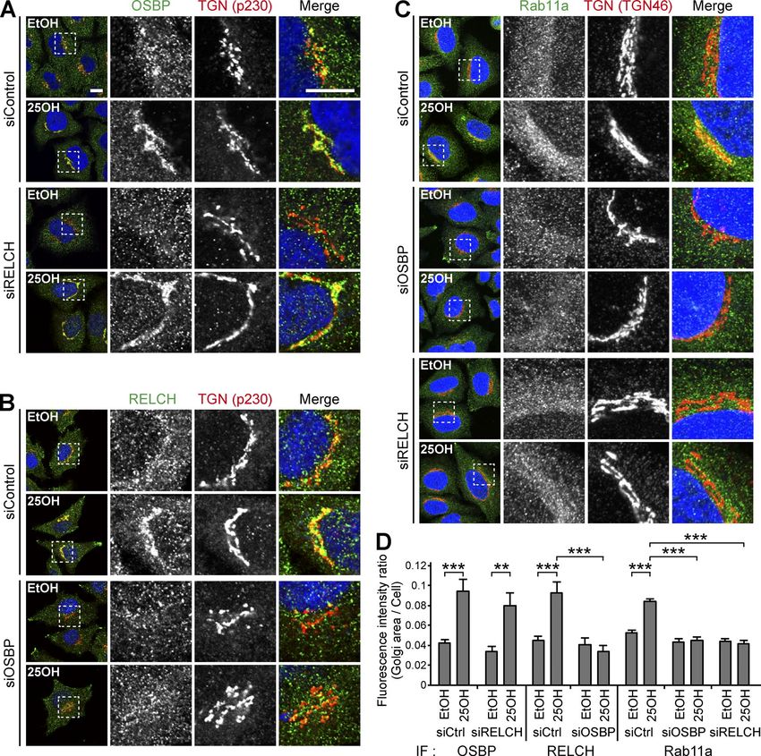
Results (Fig. S2, A and D). These results indicate that the RELCH–Rab11
RELCH/KIAA1468 is a novel Rab11-binding protein complex plays an unknown role. Therefore, to obtain insight into
We performed a GST pulldown assay to identify novel Rab11 its function, we attempted to identify the RELCH-interacting
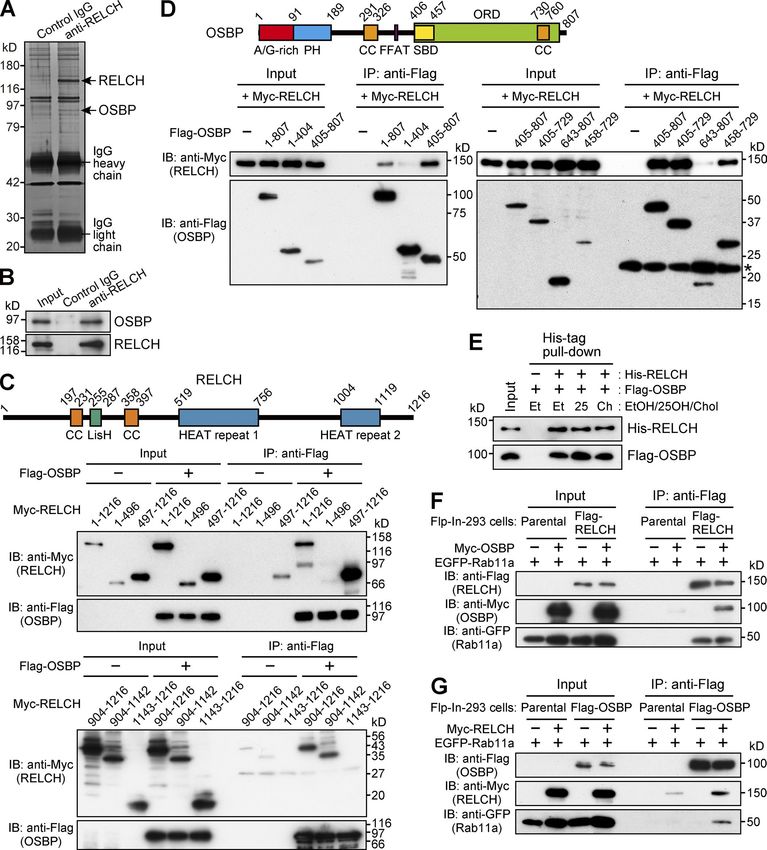
binding proteins. A specific interacting protein of ∼130 kD was protein. Immunoprecipitation with RELCH from mouse brain
obtained by incubating mouse brain lysate with GTP-loaded lysate demonstrated the coprecipitation of an ∼90-kD protein.
Rab11a (Fig. 1, A and B). The mass spectrometry analysis identi- Mass spectrometry analysis revealed that this protein was iden-
fied seven peptides corresponding with KIAA1468 (also called the tical to OSBP (Fig. 2, A and B). To identify the region of RELCH
Institute of Physical and Chemical Research cDNA 2310035C23 that binds OSBP, Flag-tagged OSBP and Myc-tagged RELCH serial
gene). This protein possesses the Lis1 homology (LisH) domain, deletion mutants were coexpressed in HEK293FT cells, and the
coiled-coil (CC) domains, and HEAT repeat motifs (Fig. 1 E). Here- proteins were immunoprecipitated using an anti-Flag antibody.
inafter, this protein is designated RELCH (Rab 11–binding protein Full-length RELCH and C-terminal region fragments containing
containing LisH, CC, and HEAT repeats). The direct interaction the second HEAT repeat motif (residues 497–1,216, 904–1,216,
between RELCH and GTP-bound Rab11 was confirmed using and 904–1,142) were coprecipitated with OSBP, but the N-termi-
recombinant proteins (Fig. 1 C). To assess the RELCH-binding nal region (1–496) and a C-terminal mutant (1,143–1,216) were
specificity among the Rab family proteins, we performed a yeast not (Fig. 2 C). These data indicate that OSBP binds the HEAT
two-hybrid assay. RELCH bound Rab11a and Rab11b and weakly repeat 2–containing region of RELCH. We further examined the
Downloaded from https://rupress.org/jcb/article-pdf/217/5/1777/441447/jcb_201709123.pdf by guest on 20 February 2020
bound Rab25 but did not bind the other 33 Rab proteins (Fig. 1 D). RELCH-binding region of OSBP. The full-length and C-terminal
According to a two-hybrid assay using serial deletion mutants of regions (405–807, 405–729, and 458–729) of OSBP coprecipitated
RELCH, the region between residues 497 and 779 containing the with RELCH, but the N-terminal region (1–404), C-terminal CC,
first HEAT repeat motif was necessary for the binding of RELCH and a part of ORD (643–807) did not. These data indicate that
to Rab11 (Figs. 1 E and S1, A and B). Furthermore, we tested this RELCH binds the C-terminal region of OSBP, which contains
binding in vitro using a GST-fused 497–779 fragment of RELCH the ORD (Fig. 2 D). The binding experiments using the purified
and GDP- or GTP-bound His6×-tagged Rab11a. The fragment spe- proteins confirmed the presence of a direct interaction between
cifically bound Rab11a-GTP (Fig. 1 F). By performing immuno- RELCH and OSBP (Fig. 2 E). Moreover, the immunoprecipita-
fluorescence microscopy, we observed that RELCH colocalized tion experiments using cell lysates coexpressing epitope-tagged
with Rab11- and transferrin receptor (TfnR)-positive REs but not Rab11a, RELCH, and OSBP showed that these proteins could form
with the early/sorting endosomal protein EEA1, the TGN pro- a ternary complex (Fig. 2, F and G).
tein p230, or the late endosome (LE)/lysosome proteins cation-
dependent mannose-6-phosphate receptor (CD-MPR) and Lamp2 RELCH links Rab11 to OSBP for RE relocation to the TGN area
(Figs. 1 G and S1 C). These results indicate that RELCH specifically OSBP is a cholesterol transfer or cholesterol-sensing protein that
binds Rab11-GTP. functions at the TGN (Wang et al., 2005; Mesmin et al., 2013).
In cells treated with 25-hydroxycholesterol (25-OH), which may
Identification of OSBP as a RELCH-interacting protein mimic a cholesterol-starved state, OSBP bound 25-OH and trans-
Because RELCH is a Rab11 effector protein, we hypothesized that located to the TGN from the cytoplasm through its pleckstrin
the depletion of RELCH could cause defects in protein trafficking homology (PH) domain (Ridgway et al., 1992). Under this condi-
in the endocytic recycling or exocytic transport pathways, which tion, RELCH and Rab11 also translocated to the juxtanuclear TGN
have been previously reported in studies using cells expressing area (Fig. 3, A–D; and Fig. S3 A). Furthermore, superresolution
a Rab11 dominant-negative mutant (Ullrich et al., 1996; Chen et structured illumination microscopy (SR-SIM) revealed that the
al., 1998) or Rab11-depleted cells (Takahashi et al., 2012). To verify RELCH- and Rab11-positive endosomes were positioned close to,
this hypothesis, we performed fluorescence-labeled transferrin but were distinguishable from, the PHOSBP-labeled TGN after the
internalization/recycling and exocytic vesicular stomatitis virus 25-OH treatment (Fig. 3, E–G). By performing immunoelectron
(VSVG) transport assays (Diao et al., 2008; Linford et al., 2012). microscopy, we confirmed that the gold-labeled Rab11a-positive
However, neither transferrin endocytosis nor recycling defects structures and Golgi apparatus were located close to each other
were observed in the RELCH-depleted cells (Fig. S2, A and B). In after the 25-OH treatment (Fig. 3 H). To better understand the role
the exocytic VSVG transport assay, compared with the Rab11a/b- played by these proteins in RE relocation close to the TGN area,
depleted cells, we observed a minor effect in the RELCH-depleted we examined the effect of 25-OH treatment in OSBP-, RELCH-,
cells (Fig. S2, A and C). Furthermore, we performed SDS-PAGE and Rab11a/b-depleted cells. These proteins were depleted by
to examine the mobility of the transmembrane cargo proteins >90% using specific siRNAs (Fig. S2 A). RELCH depletion did not
exiting the TGN because a previous study reported that certain affect the OSBP translocation to the TGN after the 25-OH treat-
cargo proteins from Rab11a-deficient small intestinal cells had ment (Fig. 4, A and D). In contrast, 25-OH–induced RELCH trans-
slower mobility than proteins from control cells as a result of location to the TGN area was diminished in the OSBP-depleted
abnormal glycosylation, which likely reflects decelerated pro- cells (Fig. 4, B and D). These data suggest that 25-OH–induced
tein export from the TGN (Sobajima et al., 2014). In the Rab11- RELCH translocation to the TGN area is dependent on OSBP.
depleted HeLa cells, we also observed slower SDS-PAGE mobility Moreover, translocation of the Rab11a-positive endosomes to the
in TfnR, Lamp2, and TGN46 (Fig. S2, A and D). In contrast, no TGN area in response to the 25-OH treatment was diminished
obvious differences were observed in the mobility of these glyco- in the OSBP- and RELCH-depleted cells (Fig. 4, C and D). In the
proteins between the RELCH-depleted cells and the control cells Rab11a/b-depleted cells, translocation of OSBP and RELCH to the
Sobajima et al. Journal of Cell Biology 1778
RELCH/KIAA1468 regulates cholesterol transport https://doi.org/10.1083/jcb.201709123
Downloaded from https://rupress.org/jcb/article-pdf/217/5/1777/441447/jcb_201709123.pdf by guest on 20 February 2020 Figure 1. Identification of RELCH as a Rab11-interacting protein. (A) The bead-bound GST-Rab11a WT or Q70A protein was incubated with a mouse brain lysate and GDP or GTP, respectively. The samples were separated on 4–12% gradient gels and silver-stained. The band with the asterisk was analyzed by per- forming mass spectrometry. (B) The pulled-down samples shown in A were immunoblotted with a RELCH antibody. (C) In vitro pulldown assay using purified recombinant RELCH and GST-Rab11-GTP or -GDP. The samples were immunoblotted with RELCH and GST antibodies. (D) Yeast two-hybrid assay using RELCH and GTP-locked Rabs. Yeast cotransformed with Gal4AD (AD) and Gal4BD (BD) plasmids encoding RELCH and Rab-GTP, respectively, was grown on SC−LW plates. Five independent colonies were selected and restreaked on SC−LW and QDO plates. Growth on the QDO plate indicates an interaction between the two proteins. (E) The interaction between Rab11 (Q70A) and a series of RELCH deletion mutants was tested by performing a yeast two-hybrid assay. The original data are shown in Fig. S1 A. (F) In vitro binding assay using the GST-tagged RBD of RELCH (GST-RELCH [RBD]) and C-terminal His-tagged Rab11a S25N or Q70A loaded with GDP or GTP, respectively. The samples were immunoblotted using Rab11 and GST antibodies. (G) HeLa cells expressing EGFP-Rab11a were immunostained with the RELCH antibody. The nuclei were stained with DAPI (blue). The arrowheads indicate RELCH-positive structures overlapped with EGFP-Rab11a–positive structures. Bar, 10 µm. Sobajima et al. Journal of Cell Biology 1779 RELCH/KIAA1468 regulates cholesterol transport https://doi.org/10.1083/jcb.201709123
Downloaded from https://rupress.org/jcb/article-pdf/217/5/1777/441447/jcb_201709123.pdf by guest on 20 February 2020 Figure 2. OSBP is a RELCH-binding protein. (A and B) Protein G Sepharose beads bound to control IgG or the RELCH antibody were incubated with a mouse brain lysate. The precipitated samples were analyzed by SDS-PAGE and silver staining and further analyzed by mass spectrometry (A). The mass spectrometry analysis identified RELCH and OSBP, which are indicated by the arrows. The precipitated samples were analyzed by immunoblotting (IB) using the OSBP and RELCH antibodies (B). (C and D) The lysates of HEK293FT cells coexpressing a series of Flag-tagged OSBP and Myc-tagged RELCH fragments were immunopre- cipitated (IP) using a Flag antibody. The samples were immunoblotted with the Myc and Flag antibodies. SBD, sterol-binding domain. In total, 0.5% and 1.5% of the input samples were loaded in upper and lower panels, respectively (C). The asterisk indicates the nonspecific bands (D). (E) A His-tag pulldown assay was performed using His-RELCH and Flag-OSBP in the presence of solvent (ethanol, EtOH), 25-OH, or cholesterol (Chol). The samples were immunoblotted with the His and Flag antibodies. (F and G) The Flag-, EGFP-, and Myc-tagged proteins were coexpressed in the Flp-In–293 cells. The lysates were immunoprecipitated using a Flag antibody, and the samples were immunoblotted with the EGFP, Myc, and Flag antibodies. Sobajima et al. Journal of Cell Biology 1780 RELCH/KIAA1468 regulates cholesterol transport https://doi.org/10.1083/jcb.201709123
TGN area was strongly induced regardless of 25-OH treatment against calnexin, TGN46, and Lamp1, respectively. First, we mea-
(Fig. S3, B, C, and E). Although the mechanism spontaneously sured the cholesterol content using membranes isolated from
targeting the OSBP and RELCH to the TGN area in the absence of cells treated with U18666A, which is an inhibitor of the NPC1
Rab11 is unknown, these data indicate that Rab11 determines the protein (Lange et al., 2000; Lu et al., 2015). Cholesterol content in
localization of RELCH on the RE. In addition, the triple knock- the LEs/lysosomes from the U18666A-treated cells was increased,
down of Rab11a/Rab11b/OSBP blocked translocation of RELCH to which is consistent with the fluorescence microscopy observa-
the TGN (Fig. S3, D and E). Altogether, these data indicate that tion (Fig. 7, C–E). This result indicates that the immunoisolation
RELCH links Rab11 to OSBP for RE relocation to the TGN area. technique can be used to measure cholesterol content in the
organelle membrane. Then, we measured the cholesterol con-
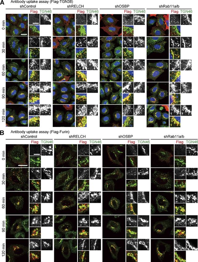
Intracellular cholesterol distribution was altered in RELCH-, tent in membranes isolated from RELCH-, OSBP-, and Rab11a/b-
OSBP-, and Rab11-depleted cells depleted cells. Cholesterol content in the LEs/lysosomes from the
Because RELCH directly bound OSBP and was translocated to the RELCH-, OSBP-, and Rab11a/b-depleted cells was higher than that
TGN area in an OSBP-dependent manner (Figs. 2, 3, 4, and S3), in the control cells. However, cholesterol content in the TGN and
we hypothesized that RELCH and Rab11 function with OSBP in ER membranes from the knockdown cells was lower than that in
cholesterol transport. Therefore, we examined the subcellular the control cells (Fig. 7, F and G). To determine whether choles-
distribution of cholesterol in RELCH-, OSBP-, and Rab11a/b- terol accumulation in the LEs/lysosomes was caused by defects in
Downloaded from https://rupress.org/jcb/article-pdf/217/5/1777/441447/jcb_201709123.pdf by guest on 20 February 2020
depleted cells. Compared with the control cells, cholesterol accu- vesicle-mediated transport, we performed a retrograde transport
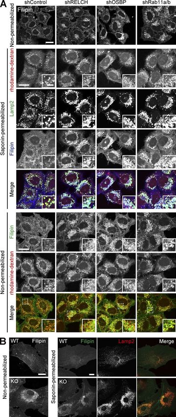
mulated in the LEs/lysosomes in depleted cells as indicated by assay using Flag-tagged TGN38 and Furin, which traffics from
Filipin or BODIPY-cholesterol staining (Figs. 5 A and S4, A and B). the PM to the TGN through the RE or LE/lysosome, respectively
However, no obvious accumulation of cholesterol was observed (Shin et al., 2004; Chia et al., 2011). The trafficking of TGN38 and
in the TGN area or Rab11- and TfnR-positive REs using fluores- Furin from the PM to the TGN was not altered in Rab11-, RELCH-,
cence microscopy (Fig. S4, C–E). Moreover, compared with the and OSBP-depleted cells (Fig. 8). Therefore, these data suggest
control cells, in the RELCH-, Rab11a/b-, and OSBP-depleted cells, that RELCH, OSBP, and Rab11a/b are involved in nonvesicular
the LEs/lysosomes were enlarged, positioned at the juxtanuclear cholesterol transport from the endocytic compartment to the
region, and resembled the LEs/lysosomes in fibroblasts derived TGN and ER; thus, cholesterol accumulated in the lysosomes.
from patients with lipid storage disorders such as Niemann–Pick
syndrome type C (Sokol et al., 1988). Cholesterol accumulation RELCH, OSBP, and Rab11 mediate cholesterol transfer between
in enlarged and clustered LEs/lysosomes at the juxtanuclear the RE and TGN in vitro
region was also observed in fibroblasts derived from RELCH Several studies have indicated that lipid transfer between organ-
knockout (KO) mice (Figs. 5 B and S2 E). Subsequently, we per- elle membranes occurs at MCSs and is mediated by a tethering
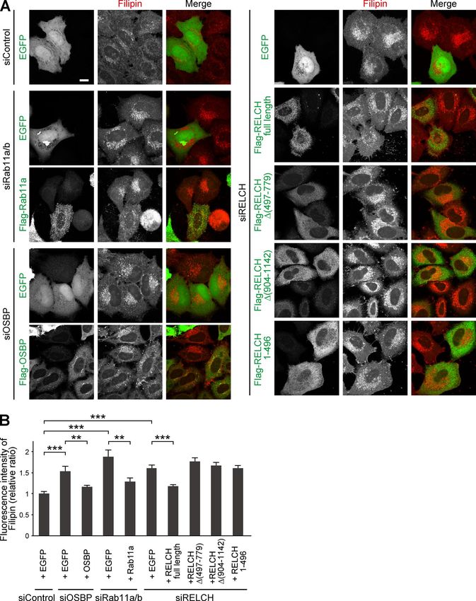
formed rescue experiments by expressing Flag-tagged OSBP, mechanism (Prinz, 2014; Eisenberg-Bord et al., 2016). There-
Rab11, or RELCH. The expression of these proteins rescued the fore, we hypothesized that RELCH, OSBP, and Rab11 might be
effect on cholesterol accumulation in the OSBP-, Rab11a/b-, and tethered between the TGN and RE membranes. Consistently, our
RELCH-depleted cells (Fig. 6, A and B). In contrast, the expres- results showed that REs accumulated around the TGN area in the
sion of the RELCH mutants Δ (497–779), Δ (904–1,143), and 25-OH–treated cells in an OSBP- and RELCH-dependent manner,
(1–496), which fail to bind Rab11, OSBP, and both, respectively, suggesting that these proteins function in tethering between the
did not rescue the effect on cholesterol accumulation in the LEs/ TGN and RE (Fig. 4). To further examine this mechanism, we per-
lysosomes in the RELCH-depleted cells (Fig. 6, A and B). These formed an in vitro assay using purified recombinant proteins and
data suggest that Rab11, RELCH, and OSBP function together in artificial liposomes following a protocol slightly modified from
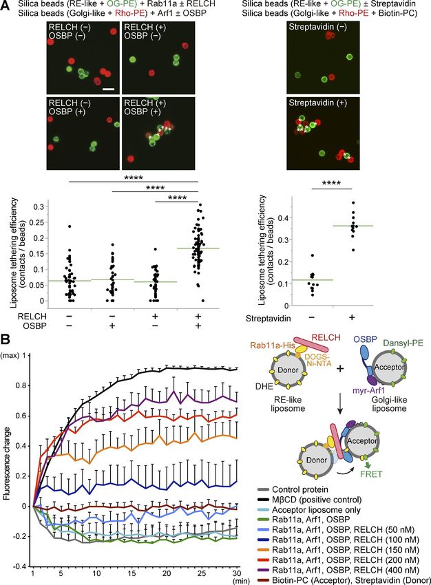
cholesterol transport. To examine the cholesterol distribution in that proposed by Mesmin et al. (2013). First, we confirmed that
the organelles in greater detail, we used a subcellular fraction- OSBP bound Golgi-like lipid-coated silica beads prebound with
ation technique with OSBP-, RELCH-, and Rab11a/b-depleted myristoylated Arf1-GTP protein (myr-Arf1(Q71L)-Flag; Fig. S5, A
cells. The cell homogenates were separated by density gradient and B). Then, we demonstrated that RELCH bound silica beads
centrifugation, and the cholesterol content in each fraction was coated with an RE-like nickel-containing lipid (DOGS-NTA(Ni))
measured. The TGN membrane was marked by TGN46 and con- prebound with His6×-tagged Rab11a-GTP (Rab11a(Q70A)-His; Fig.
centrated in fractions 4–6, the LEs/lysosomes were marked by S5, C and D). The binding of these proteins to the lipids was further
Lamp2 and concentrated in fractions 7–9, and the ER was marked confirmed using a liposome flotation or sedimentation assay (Fig.
by calnexin and concentrated in fractions 10–11 (Fig. 7 A). The S5, E and F). Then, we mixed Arf1-GTP–bound beads coated with
cholesterol content was increased in the LE/lysosome fractions a rhodamine-phosphatidylethanolamine (PE)–labeled Golgi-
of the RELCH-, OSBP-, and Rab11a/b-depleted cells (Fig. 7 B). In like lipid and Rab11-GTP-bound beads coated with an Oregon
contrast, compared with the control cells, the cholesterol levels in green 488–DHPE (OG-PE)–labeled RE-like lipid in the presence
the TGN and ER fractions were slightly decreased in the RELCH-, or absence of OSBP and RELCH. Compared with reactions lack-
OSBP-, and Rab11a/b-depleted cells (Fig. 7 B). However, it is dif- ing both proteins or reactions with only RELCH or OSBP, aggre-
ficult to exclude entirely the contamination of other organelle gate formation between the Golgi-like and RE-like liposomes
membranes between fractions using this method; therefore, we occurred more frequently after adding both RELCH and OSBP
further measured the cholesterol content in the ER, TGN, and (Fig. 9 A, left, and Fig. S5 G). Aggregation between the streptavi-
LE/lysosome membranes, which were isolated using antibodies din-bound liposomes and biotin-conjugated phosphatidylcholine
Sobajima et al. Journal of Cell Biology 1781
RELCH/KIAA1468 regulates cholesterol transport https://doi.org/10.1083/jcb.201709123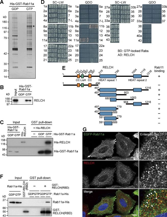
Downloaded from https://rupress.org/jcb/article-pdf/217/5/1777/441447/jcb_201709123.pdf by guest on 20 February 2020 Figure 3. Localization of RELCH, OSBP, and Rab11 after 25-OH treatment. (A–D) HeLa cells were incubated with 6.2 µM 25-OH or solvent (ethanol, EtOH) for 24 h and immunostained with antibodies against TGN46 and OSBP (A), RELCH (B), or Rab11a (C). The nuclei were stained with DAPI (blue). The enlarged images show the TGN area. Bar, 10 µm. (D) Quantification of the ratio of the fluorescence intensity in the Golgi area to the intensity in the whole intracellular area (n = 10–25 cells). (E–G) HeLa cells transfected with the Flag-tagged PH domain of OSBP (PHOSBP) were incubated with EtOH or 25-OH for 24 h before analysis. The cells were immunostained with antibodies against Flag and either RELCH (E) or Rab11a (F) and imaged by SR-SIM. The fluorescence intensity Sobajima et al. Journal of Cell Biology 1782 RELCH/KIAA1468 regulates cholesterol transport https://doi.org/10.1083/jcb.201709123
(PC)–containing liposomes was also observed (Fig. 9 A, right). enhanced at the TGN, although the precise mechanism of this
Therefore, these data suggest that RELCH tethers between OSBP- enhancement remains unknown. However, we can exclude the
bound and Rab11-bound membranes. possibility that the sterol-binding state of OSBP affects its bind-
Finally, we examined cholesterol transfer using an in vitro ing to RELCH because the in vitro pulldown experiment using
system. Golgi-like liposomes containing dansyl-PE and RE-like purified proteins indicated that RELCH binds OSBP regardless of
liposomes containing dehydroergosterol (DHE) were mixed, and sterol binding (Fig. 2 E). We speculated that the proteins are post-
the fluorescence resonance energy transfer (FRET) between dan- translationally modified at the TGN and that these modifications
syl and DHE was measured, which revealed sterol transfer from may strengthen the interaction between these proteins. Consis-
the RE-like liposomes to the Golgi-like liposomes. After adding tently, OSBP is phosphorylated at the TGN by Golgi-localized
Rab11a, Arf1, OSBP, and different concentrations of RELCH, the protein kinase D (Nhek et al., 2010). Moreover, RELCH possesses
DHE transfer occurred in a RELCH dose-dependent manner, but numerous phosphorylation sites (Hornbeck et al., 2015); there-
no significant DHE transfer activity was observed in the reac- fore, determining whether the phosphorylation of these proteins
tions lacking RELCH (Fig. 9 B). However, DHE transfer activity controls their binding could be interesting.
between the streptavidin-bound liposomes and biotin-PC lipo- Filipin or BODIPY-cholesterol staining indicated that choles-
somes was not observed (Fig. 9 B), indicating that the cholesterol terol accumulated in the LEs/lysosomes in the RELCH-, OSBP-,
transfer did not simply reflect the aggregation of liposomes. and Rab11-depleted cells (Figs. 5 and S4). Furthermore, the TGN
Downloaded from https://rupress.org/jcb/article-pdf/217/5/1777/441447/jcb_201709123.pdf by guest on 20 February 2020
These results suggest that RELCH functions in membrane teth- and ER cholesterol contents in the RELCH-, OSBP-, and Rab11-de-
ering and promotes OSBP-mediated cholesterol transfer between pleted cells were reduced compared with those in the control cells
Rab11-bound RE and OSBP-bound Golgi-like membranes. (Fig. 7). We did not detect any obvious alteration in the vesicular
trafficking from the LE/lysosome or RE to the TGN in these pro-
tein-depleted cells (Fig. 8). In addition, the in vitro reconstitu-
Discussion tion experiments showed that RELCH promotes OSBP-dependent
We identified that RELCH is a Rab11-GTP binding protein and nonvesicular cholesterol transport to the TGN-like membrane
revealed the interaction between RELCH and OSBP. In cells (Fig. 9). Altogether, these results suggest that the Rab11–REL
treated with 25-OH, Rab11-positive REs were gathered around CH–OSBP complex is involved in cholesterol transport from the
the TGN area in a RELCH- and OSBP-dependent manner, suggest- LE/lysosome to the TGN and ER. However, the LE/lysosome-TGN
ing that membrane tethering between the RE and TGN occurred MCSs at which lipid transfer occurs have not been identified. Nev-
through the intracellular sterol-sensing mechanism of OSBP. Our ertheless, in previous studies, the MCS formation between the LE/
experiments using artificial liposomes indicated that the tether- lysosome and ER was observed, but whether cholesterol is directly
ing mediated by these proteins is crucial for efficient cholesterol transferred from the LE/lysosome to the ER was not determined
transfer between the membranes. (Rocha et al., 2009; Alpy et al., 2013; Raiborg et al., 2015). Because
RELCH contains two HEAT repeat motifs. The HEAT motif the RELCH and Rab11 localization is limited to the RE or TGN-
comprises α-helical structures and functions in protein–protein adjacent area (Figs. 1 G and S1 C), the Rab11–RELCH–OSBP com-
interactions (Yoshimura and Hirano, 2016). The first N-terminal plex is unlikely to directly mediate the cholesterol transport from
HEAT repeat motif in RELCH likely binds Rab11-GTP in a man- the LE/lysosome to the TGN or ER. The most likely scenario is
ner similar to that of other Rab proteins that preferentially bind that cholesterol traffics from the LE/lysosome to the TGN via the
α-helices (Fig. 1, E and F; and Fig. S1, A and B; Khan and Ménétrey, RE, and this complex mediates nonvesicular cholesterol trans-
2013). In addition, the second C-terminal HEAT repeat motif in port from the RE to the TGN and, consequently, affects choles-
RELCH interacts with OSBP (Fig. 2 C). In a previous study, OSBP terol transport from the TGN to the ER. However, we did not
was shown to directly interact with the protein phosphatase 2A detect significant cholesterol accumulation in the RE by Filipin
(PP2A) complex, which has subunits containing HEAT repeats or or BODIPY-cholesterol staining in the RELCH-, OSBP-, and Rab11-
HEAT-like α-helical structures (Wang et al., 2008). Although the depleted cells (Fig. S4, D and E). Thus, we speculate that choles-
precise region of each PP2A subunit necessary for OSBP binding terol transiently exports from the LEs/lysosomes and rapidly traf-
was not examined, the authors speculated that the HEAT repeats fics through the REs, and the undelivered cholesterol from the RE,
in both subunits are involved in the interaction. Therefore, REL reflecting defects in the RE–TGN pathway caused by the depletion
CH may interact with OSBP via a similar mechanism as PP2A. of RELCH, OSBP, and Rab11, is retrieved to the LEs/lysosomes.
After the 25-OH treatment, RELCH was strongly recruited Importantly, Urano et al. (2008) demonstrated that the deple-
around the TGN area in an OSBP-dependent manner (Figs. 3 and tion of three SNARE proteins, i.e., VAMP4, syntaxin6, and syn-
4), suggesting that the interaction between the two proteins is taxin16, that are involved in vesicular traffic between the early
profiles of PHOSBP and either RELCH or Rab11a were measured at the position marked by the arrows in the merged insets. The red and green arrows in the
graphs indicate the intensity peak of PHOSBP and RELCH or Rab11a, respectively. N, nucleus. Bars: (main images) 5 µm; (enlarged images) 1 µm. (G) Quantifica-
tion of the distance between the intensity peak of PHOSBP and RELCH or Rab11a (n = 15–34 cells). (H) Immunoelectron microscopy of HeLa cells treated with
25-OH and nontreated control cells after the EGFP-Rab11a transfection. Silver-enhanced gold particles were used to label the EGFP-Rab11a–positive vesicles
and tubules (arrows). Bar, 500 nm. Data are expressed as means ± SEM. Significance was calculated by performing two-tailed Student’s t tests (*, P < 0.05;
***, P < 0.001). IF, immunofluorescence.
Sobajima et al. Journal of Cell Biology 1783
RELCH/KIAA1468 regulates cholesterol transport https://doi.org/10.1083/jcb.201709123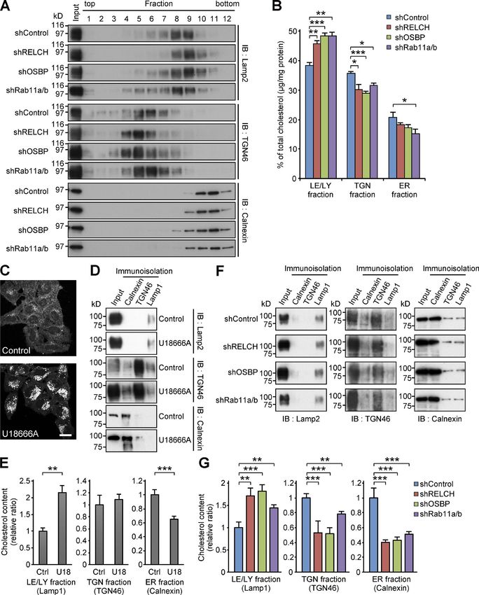
Downloaded from https://rupress.org/jcb/article-pdf/217/5/1777/441447/jcb_201709123.pdf by guest on 20 February 2020 Figure 4. Interdependence among RELCH, OSBP, and Rab11 in TGN localization after 25-OH treatment. (A–C) siRNA-transfected HeLa cells were incu- bated with solvent (ethanol, EtOH) or 25-OH for 24 h before analysis. The cells were immunostained with antibodies against OSBP (A), RELCH (B), and Rab11a (C). The TGN was labeled with the TGN46 or p230 antibodies. The nuclei were stained with DAPI (blue). The enlarged images show the TGN area. Bar, 10 µm. (D) Quantification of the ratio of the fluorescence intensity in the Golgi area to the intensity in the intracellular area (n = 10–39 cells). Data are expressed as means ± SEM. Significance was calculated by performing two-tailed Student’s t tests (**, P < 0.01; ***, P < 0.001). IF, immunofluorescence. endosome/RE and the TGN (Mallard et al., 2002) inhibited cho- transport mechanism mediated by GARP and SNARE complexes lesterol transport from the lysosome to the TGN and resulted in and a nonvesicular transport mechanism mediated by Rab11– the partial inhibition of cholesterol reesterification in the ER. RELCH–OSBP. We assume that the two mechanisms coopera- Moreover, the depletion of a subunit of Golgi-associated retro- tively function in cholesterol transport from REs to the TGN. grade protein (GARP), which is a tethering complex that regu- The lipid raft hypothesis highlights the importance of cho- lates endosome–Golgi retrograde vesicular transport, resulted lesterol content in the TGN (Surma et al., 2012; Guo et al., 2014). in cholesterol accumulation in the LEs/lysosomes (Fröhlich et Previous findings suggest that sphingolipid/sterol-rich lipid al., 2015). In conjunction with these findings, the results of this microdomains at the TGN (often considered a lipid raft) are study suggest that two different cholesterol transport mecha- involved in sorting specific sets of cargo proteins, e.g., glycosyl- nisms exist in LE/lysosome-RE-TGN trafficking, i.e., a vesicular phosphatidylinositol (GPI)-anchored protein, from the TGN to Sobajima et al. Journal of Cell Biology 1784 RELCH/KIAA1468 regulates cholesterol transport https://doi.org/10.1083/jcb.201709123
the PM (Hansen et al., 2000; Castillon et al., 2013). Interestingly,
cells deficient in subunits of the GARP complex exhibit defects in
the exit of GPI-anchored protein from the TGN to the PM (Hirata
et al., 2015). These findings suggest that the transport defect in
GARP-KO cells might reflect a disruption in lipid microdomain
formation at the TGN. Therefore, it will be important to address
the contribution of Rab11–RELCH–OSBP–mediated nonvesicular
cholesterol transport to lipid microdomain formation and lipid
microdomain-dependent protein-cargo sorting at the TGN, in
addition to vesicular transport mechanisms.
Materials and methods
Plasmids
A pFAT2 vector encoding the His-GST-Rab constructs, a pFBT9
vector encoding the Gal4BD-GTP–locked Rab constructs, and
Downloaded from https://rupress.org/jcb/article-pdf/217/5/1777/441447/jcb_201709123.pdf by guest on 20 February 2020
a pEGFP-C2 vector encoding the EGFP-fused Rab constructs
were generated as previously described (Haas et al., 2005;
Yoshimura et al., 2007). Mouse RELCH/KIAA1468 (accession
no. NM_173187.3) and mouse Rab11a were PCR amplified from
the Mouse 17-d Embryo Marathon Ready cDNA library (Takara
Bio Inc.) using KOD DNA polymerase (Toyobo). Then, the cDNA
was subcloned into the pSCB vector using the StrataClone Blunt
PCR cloning kit (Agilent Technologies). A series of RELCH dele-
tion mutants and a RELCH fragment containing an N-terminal
PreScission protease recognition site were generated by PCR
using pSCB-RELCH. The RELCH constructs were inserted into
the pQE32tev, pFAT2, pACT2, pcDNA3.1-Myc, and pcDNA5/FRT/
TO-Flag vectors according to their intended use. C-terminal
His-tagged mouse Rab11a S25N and Q70A mutants (GDP- and
GTP-locked mutants, respectively) without a stop codon were
generated by PCR using pSCB-mouse Rab11a and then ligated
into the NcoI and XhoI sites of the pET28c vector. Human
OSBP, a C-terminal Flag-tagged human Arf1 Q71L mutant, and
yeast NMT1 were PCR amplified from pOTB7-OSBP (4560111;
GE Healthcare), pcDNA3.1-Arf1 (Q71L)-HA (10832; Addgene),
and pRSF-1-NMT (42578; Addgene), respectively. OSBP, PHOSBP
(OSBP91-189), and OSBP deletion mutants and mouse Rab11a were
inserted into pcDNA5/FRT/TO-Flag or pcDNA3.1-Myc for mam-
malian expression. The human Rab11a Q70A mutant was gener-
ated by PCR and then inserted into pcDNA5/FRT/TO-EGFP. For
the bacterial expression of His-OSBP321-409-His, OSBP321-409 was
inserted into the pET28c vector. For the coexpression of NMT1
and Arf1 (Q71L)-Flag in bacteria, the constructs were inserted
into multiple cloning sites 1 and 2, respectively, of the pETDuet-1
vector (Novagen). The pET21a-streptavidin-Alive construct was
purchased from Addgene (20860).
Antibodies
Figure 5. Cholesterol accumulation in LEs/lysosomes after RELCH, The rabbit polyclonal anti-Rab11a, anti-GFP, and anti-GST anti-
OSBP, or Rab11 depletion. (A) The lysosomes were labeled with bodies were prepared as previously described (Atik et al., 2014;
rhodamine-dextran in the shRNA-treated cells. The cells were stained with Sobajima et al., 2014; Kunii et al., 2016). The other primary
Filipin under the nonpermeabilized condition and Filipin and Lamp2 under
antibodies included mouse monoclonal Rab11 (clone 47; BD),
the permeabilized condition. (B) Mouse tail–tip fibroblasts derived from WT
and RELCH KO mice were stained with the Lamp2 antibody and/or Filipin. EEA1 (clone 14; BD), p230 (clone 15; BD), CD-MPR, and Lamp2
Bars: (main images) 20 µm; (insets) 2 µm. (clones 22d4 and H4B4, respectively; Developmental Studies
Hybridoma Bank), TfnR (clone H68.4; Zymed), Myc (9B11; Cell
Signaling Technology), GAPDH (6C5; EMD Millipore), Flag (M2;
Sobajima et al. Journal of Cell Biology 1785
RELCH/KIAA1468 regulates cholesterol transport https://doi.org/10.1083/jcb.201709123Downloaded from https://rupress.org/jcb/article-pdf/217/5/1777/441447/jcb_201709123.pdf by guest on 20 February 2020 Figure 6. Expression of RELCH, OSBP, or Rab11 rescues the effect on cholesterol accumulation in the RELCH-, OSBP-, or Rab11-depleted cells. (A) HeLa cells transfected with the indicated siRNA and plasmids were costained with Filipin and the Flag antibody. Bar, 10 µm. (B) Quantification of the fluorescence intensity of Filipin in the plasmid-transfected cells (relative to siControl + EGFP cells; n = 18–89 cells). Data are expressed as means ± SEM. Significance was calculated by performing two-tailed Student’s t tests (**, P < 0.01; ***, P < 0.001). Sigma-Aldrich), His (GE Healthcare), sheep TGN46 (Serotec), rabbit Lamp1 (PA1-654A; Invitrogen). Species-specific secondary rabbit calnexin (for immunoblotting, clone C5C9 from Cell Sig- antibodies labeled with Alexa Fluor 488, 568, and 594; Cy5; and naling Technology; for immunoisolation, ADI-SPA-860 from HRP were purchased from Thermo Fisher Scientific and Jackson Enzo Life Sciences), rat Lamp2 (for mouse, clone Abl-93; Devel- ImmunoResearch Laboratories, Inc. The rabbit polyclonal anti- opmental Studies Hybridoma Bank), goat lamin B (C-20; Santa RELCH and anti-OSBP antibodies were raised against bacterially Cruz Biotechnology), rabbit TGN46 (ab50595; Abcam), and expressed His-RELCH and His-OSBP321-409-His, respectively. The Sobajima et al. Journal of Cell Biology 1786 RELCH/KIAA1468 regulates cholesterol transport https://doi.org/10.1083/jcb.201709123
Downloaded from https://rupress.org/jcb/article-pdf/217/5/1777/441447/jcb_201709123.pdf by guest on 20 February 2020 Figure 7. RELCH, OSBP, and Rab11 depletion results in less cholesterol accumulation in the TGN and ER. (A and B) The homogenates from the shRNA- expressing HeLa cells were fractionated using a Histodenz step density gradient. (A) The fractions were immunoblotted (IB) with antibodies against TGN46, calnexin, and Lamp2. (B) Percentages of cholesterol (µg/mg protein) in the TGN (fractions 4–6), LEs/lysosomes (LE/LY; 7–9), or ER (10 and 11) in the total fractions are shown in the bar graph. (C) HeLa cells were treated with 2 µg/ml U18666A for 16 h and stained with Filipin. Bar, 20 µm. (D–G) Immunoisolation of ER, TGN, and LE/lysosome from the PNS derived from the U18666A-treated (D and E) or RELCH-, Rab11a/b-, and OSBP-depleted cells by shRNAs (F and G). ER, TGN, and LE/lysosome membranes were isolated using calnexin, TGN46, and Lamp1 antibodies, respectively. (D and F) The isolated samples were immu- noblotted with calnexin, TGN46, and Lamp2 antibodies. (E and G) Quantification of the cholesterol content in the isolated membranes (relative to the control samples). Data are expressed as means ± SEM from at least three independent experiments. *, P < 0.05; **, P < 0.01; ***, P < 0.001 (relative to the control; two tailed Student’s t tests). Representative data from three independent experiments are shown in A, D, and F. Sobajima et al. Journal of Cell Biology 1787 RELCH/KIAA1468 regulates cholesterol transport https://doi.org/10.1083/jcb.201709123
Downloaded from https://rupress.org/jcb/article-pdf/217/5/1777/441447/jcb_201709123.pdf by guest on 20 February 2020 Figure 8. Retrograde transports of TGN38 and Furin in the RELCH-, OSBP-, and Rab11-depleted HeLa cells. (A and B) Antibody uptake assay in HeLa cells stably expressing the indicated shRNAs. The cells transfected with Flag-tagged TGN38 (A) or Furin (B) were incubated with a Flag antibody on ice for 45 min and then chased at 37°C in growth medium. The cells were fixed at the indicated times and immunostained with the Flag and TGN46 antibodies. The nuclei were stained with DAPI (blue). Bars: (main images) 20 µm; (enlarged images) 5 µm. Sobajima et al. Journal of Cell Biology 1788 RELCH/KIAA1468 regulates cholesterol transport https://doi.org/10.1083/jcb.201709123
Downloaded from https://rupress.org/jcb/article-pdf/217/5/1777/441447/jcb_201709123.pdf by guest on 20 February 2020 Figure 9. RELCH controls the OSBP-dependent cholesterol transfer from the RE-like membrane to the Golgi-like membrane via tethering activity. (A) Bead-based liposome tethering assay between Golgi- and RE-like liposomes containing rhodamine-PE (Rho-PE; red) and OG-PE (green), respectively. The protein-bound beads (the binding of myr-Arf1, Rab11a, OSBP, and RELCH is demonstrated in Fig. S5 [A–D]) were mixed and imaged under a fluorescence microscope (top left). In the top right panel, the tethering assay using the streptavidin-His protein and biotin-PC–containing liposomes is shown. The asterisks indicate the contacts between two beads. The liposome tethering efficiency was calculated as the ratio of the number of contacts to the total number of beads. The data were collected from randomly selected fields per group. Representative examples of the counts and calculations are shown in Fig. S5 G. The results are presented in the dot-plot graphs (bottom). The green lines indicate the mean of each group. ANOVA, Wilcoxon, and Kruskal–Wallis tests were performed (****, P < 0.0001). Bar, 10 µm. (B) Lipid transfer assay between RE-like donor and Golgi-like acceptor liposomes containing DHE and dansyl-PE, respectively. Recombinant proteins were bound to each type of liposome (right). The transfer reaction was initiated by mixing the two types of protein-bound liposomes. MβCD was used as a positive control to determine the maximum FRET signal. Maltose binding protein was used as a control protein. The data from each experiment were corrected by setting the initial and maximum values to 0 and 1, respectively, and are presented in the line graph (left). Data are expressed as means ± SEM from at least three independent experiments. Sobajima et al. Journal of Cell Biology 1789 RELCH/KIAA1468 regulates cholesterol transport https://doi.org/10.1083/jcb.201709123
antisera were affinity purified and subsequently used for the the deletion of the His-tag. To purify Flag-OSBP from mamma-
immunofluorescence and immunoblotting analyses. lian cells, Flp-In–293 cells (Thermo Fisher Scientific) were used
to generate a stable Flag-OSBP cell line. The stably transfected
Protein expression and purification cells from fifty 15-cm dishes were harvested and lysed with 0.2%
XL-1 blue cells expressing pQE32- or pMAL-based constructs and Triton X-100 and 1 mM methyl-β-cyclodextrin (MβCD; Wako
Rosetta II cells expressing pFAT2- or pET-based constructs were Pure Chemical Industries) in PBS. The lysate was clarified and
cultured in LB medium at 18°C for 16–18 h with 0.25 mM IPTG and mixed with anti-Flag M2 affinity gel. After incubation for 2 h at
subsequently purified using Ni-NTA agarose (QIAGEN) or amy- 4°C, the beads were washed once with NL100, once with NL500,
lose resin (New England Biolabs, Inc.) as previously described and twice with PBS. Then, the bound protein was eluted with 400
(Nakajo et al., 2016). The protein-bound Ni-NTA agarose or amy- µg/ml Flag-peptide in PBS. The eluted protein was analyzed on an
lose resin was washed four times with IMAC20 (20 mM Tris- SDS-PAGE gel stained with CBB. The concentration of the peak
HCl, pH 8.0, 300 mM NaCl, and 20 mM imidazole) or PBS and fractions was estimated by comparison to a series of BSA stan-
then eluted with IMAC200 (IMAC20 with 200 mM imidazole) dards using SDS-PAGE and CBB staining. Small aliquots of puri-
or PBS containing 10 mM maltose, respectively. The proteins fied proteins were snap-frozen in liquid N2 for storage at −80°C.
were dialyzed in PBS. To purify the nucleotide-free Rab11a (S25N
and Q70A)-His, the clarified lysates from bacteria expressing Pulldown assay
Downloaded from https://rupress.org/jcb/article-pdf/217/5/1777/441447/jcb_201709123.pdf by guest on 20 February 2020
Rab11a S25N and Q70A in pET28c were mixed with cOmplete For the GST pulldown assay using the GST-Rab11a proteins and a
His-tag purification resin (Roche). After incubating for 2 h at mouse brain lysate, 500 µg of the GST-Rab11a (WT or Q70A) pro-
4°C, the beads were washed twice with IMAC20 and twice with tein were bound to 50 µl Glutathione Sepharose 4B in a 4-ml total
IMAC20 containing 10 mM EDTA, and then the bound proteins volume of NE100 (20 mM Hepes-NaOH, pH 7.5, 100 mM NaCl,
were eluted with IMAC200. The eluted proteins were analyzed 20 mM EDTA, and 0.1% Triton X-100) for 1 h at 4°C. The beads
on SDS-PAGE gels stained with Coomassie brilliant blue (CBB). were washed three times with NE100 and once with NL100 and
The peak fractions were dialyzed in PBS. The myristoylated Arf1 incubated for 1 h at 4°C in a 5-ml total volume of 20 mg mouse
(Q71L)-Flag protein was produced and purified as previously brain lysate prepared with NL100 containing 10 mM MgCl2, 1%
described (Franco et al., 1995) with slight modifications. To pre- protease inhibitor mixture (Wako Pure Chemical Industries), and
pare the myristate–BSA complex, sodium myristate (198-09512; 10 mM GDP or GTP, pH 7.0 (Wako Pure Chemical Industries). The
Wako Pure Chemical Industries) was mixed with 5% BSA (A7906; beads were washed three times with NL100 and once with NL200
Sigma-Aldrich) in PBS and dissolved by sonication (Branson Son- (NL100 with 200 mM NaCl), and the bound proteins were subse-
ifier 250) at 60–70°C. Rosetta II cells expressing Arf1 (Q71L)-Flag quently eluted with 500 µl NE200 (NE100 with 200 mM NaCl).
and NMT1 in pETDuet-1 were grown at 37°C to OD600 0.6–0.7, The eluted proteins were precipitated with TCA. The proteins
and prewarmed myristate-BSA was slowly added to a final con- were dissolved in 40 µl SDS-PAGE sample buffer, separated on a
centration of 100 µM. After incubating with myristate–BSA for 4–12% gradient gel, and silver stained. For the GST pulldown assay
10 min at 37°C, the bacteria were cultured at 27°C for 18 h with using the GST-Rab11a and His-RELCH proteins, 10 µg GST-Rab11a
0.3 mM IPTG. The bacterial pellet was lysed with 1 mg/ml lyso- WT or Q70A was bound to 20 µl Glutathione Sepharose 4B, and the
zyme (Wako Pure Chemical Industries) in PBS for 30 min at 37°C beads were incubated with 20 µg His-RELCH protein in a 500-µl
and sonicated on ice. The lysate was clarified by centrifugation total volume. For the GST pulldown assay using the recombinant
at 12,000 g for 5 min. Myristoylated Arf1 was precipitated with GST-RELCH497-779 (Rab-binding domain [RBD]) and Rab11a-His
35% ammonium sulfate. The precipitated pellet was dissolved in proteins, 10 µg GST-RELCH (RBD) protein were bound to 20 µl
PBS and incubated with anti-Flag M2 affinity gel (Sigma-Aldrich) Glutathione Sepharose 4B, and the beads were incubated with
for 18 h at 4°C. Then, the beads were washed with NL100 (20 mM 10 µg Rab11a (S25N or Q70A)-His in a 500-µl total volume. The
Hepes-NaOH, pH 7.5, 100 mM NaCl, 5 mM MgCl2, and 0.1% Triton eluted protein and bead-bound protein were analyzed by immu-
X-100), NL500 (NL100 with 500 mM NaCl), NE200 (NE100 with noblotting. For the His-tag pulldown assay using the His-RELCH
200 mM NaCl), and twice with TBS. The protein was eluted with and Flag-OSBP proteins, 0.5 µg His-RELCH protein was bound
200 µg/ml Flag-peptide (Sigma-Aldrich) in TBS and analyzed to 20 µl Ni-NTA beads in a 500-µl total volume of buffer (20 mM
on an SDS-PAGE gel stained with CBB. To prepare the untagged Hepes-NaOH, pH 7.5, 100 mM NaCl, and 5 mM MgCl2) for 1 h at
RELCH, XL-1 blue cells expressing pQE32-RELCH containing 4°C. The beads were washed five times with the same buffer and
an N-terminal PreScission protease recognition site were cul- incubated with 0.5 µg Flag-OSBP protein in a 500-µl total volume
tured in LB medium at 18°C for 16 h with 0.25 mM IPTG and then of the buffer mixed with 10 µl ethanol, 1 mM cholesterol (08722-
purified using Ni-NTA agarose as described above. The bound 94; Nacalai Tesque), or 1 mM 25-OH (H1015; Sigma-Aldrich) for 1 h
protein was eluted with IMAC200, and the eluted protein was at 4°C. The beads were washed five times with buffer containing
incubated with 20 U/ml PreScission protease (GE Healthcare) for ethanol, 20 µM cholesterol, or 20 µM 25-OH and then eluted with
3 h at 4°C. Then, the protein was mixed with Ni-NTA beads and 30 µl IMAC200. The eluted proteins were immunoblotted.
Glutathione Sepharose 4B (GE Healthcare) in a dialysis tube and
incubated in PBS for 16 h at 4°C to remove the cleaved fragment Immunoprecipitation
and the GST-fused PreScission protease. After the beads were For the immunoprecipitation assay using the anti-RELCH anti-
removed, the supernatant containing the protein was analyzed body and a mouse brain lysate, preimmunized rabbit serum or
by immunoblotting using His and RELCH antibodies to confirm rabbit antiserum against the recombinant RELCH protein was
Sobajima et al. Journal of Cell Biology 1790
RELCH/KIAA1468 regulates cholesterol transport https://doi.org/10.1083/jcb.201709123mixed with 40 µl protein G Sepharose 4 Fast Flow (GE Health- according to the manufacturer’s instructions. The siRNA oligonu-
care) in a 150-µl total volume of NE100. After incubation for 1 h cleotides used in this study included Hs_RAB11A_5 (SI00301553;
at 4°C, the beads were washed five times with 500 µl NE100 and QIAGEN), Hs_RAB11B_6 (SI02662695; QIAGEN), Hs_OSBP_5
once with NL100 and subsequently incubated for 2 h at 4°C in a (SI02628920; QIA GEN), and Hs_KIAA1468_4116_s and _as
500-µl total volume of 10 mg of a mouse brain lysate prepared (11861007 and 11861008; Sigma-Aldrich). The RNAi depletion
with NL100 containing 0.1% protease inhibitor. The beads were efficiencies were confirmed by immunoblotting.
washed with NL100, and the bead-bound proteins were dissolved
in 40 µl SDS-PAGE sample buffer. For the immunoprecipitation Generation of shRNA lentivirus and stable cell lines
assay using the lysate from the HEK293FT or Flp-In–293 cells shRNA lentiviruses expressing Rab11a, RELCH, and OSBP shRNA
coexpressing the indicated proteins, the cells were plated at a were produced using a pLKO.1 vector, and the lentivirus express-
density of 5 × 10⁵ cells per well in a six-well plate and transfected ing Rab11b-shRNA was produced using a pLKO.1-neo vector
with 1 µg of the indicated plasmids. After 24 h, the cells were lysed (10878 and 13425; Addgene) according to the manual. The fol-
with 500 µl lysis buffer (NL100 or NL200) containing 0.1% prote- lowing oligonucleotides were designed: 5′-CCGGAAGAGTAATCT
ase inhibitor for 20 min on ice. The lysates were clarified by cen- CCTGTCTCGACTCGAGTCGAGACAGGAGATTACTCTTTTTTTG-3′
trifugation, and the cleared lysates were preabsorbed with 30 µl (Hs_Rab11a_shRNA_F) and 5′-AATTCAAAAAAAGAGTAATCTCCT
of Pierce control agarose resin (26150; Thermo Fisher Scientific) GTCTCGACTCGAGTCGAGACAGGAGATTACTCTT-3′ (Hs_Rab11a_
Downloaded from https://rupress.org/jcb/article-pdf/217/5/1777/441447/jcb_201709123.pdf by guest on 20 February 2020
for 1 h at 4°C. After removal of beads by centrifugation, the super- shRNA_R); 5′-CCGGCCGCATCGTGTCACAGAAACACTCGAGTG
natant was incubated with 30 µl anti-Flag M2 affinity gel for 2 h TTTCTGTGACACGATGCGGTTTTTG-3′ (Hs_Rab11b_shRNA_F)
at 4°C. The beads were washed with lysis buffer, and the bound and 5′-AATTCAAAAACCGCATCGTGTCACAGAAACACTCGAGTG
proteins were eluted with 30 µl of 200 µg/ml Flag-peptide in lysis TTTCTGTGACACGATGCGG-3′ (Hs_Rab11b_shRNA_R); 5′-CCG
buffer. The eluted proteins were analyzed by immunoblotting. GCCAATCAAACCTCTTGAAACTCGAGTTTCAAGAGGTTTGAT
TGGTTTTTG-3′ (Hs_RELCH_shRNA_F) and 5′-AATTCAAAAACC
Mass spectrometric analysis AATCAAACCTCTTGAAACTCGAGTTTCAAGAGGTTTGATTGG-3′
The proteins were separated on a NuPAGE Novex Bis-Tris gel (Hs_RELCH_shRNA_R); and 5′-CCGGCCCGCTAATGGAAGAAG
(NP0321; Thermo Fisher Scientific) and silver stained using a TTTACTCGAGTAAACTTCTTCCATTAGCGGGTTTTTG-3′ (Hs_
silver stain mass spectrometry kit (299-58901; Wako Pure Chem- OSBP_shRNA_F) and 5′-AATTCAAAAACCCGCTAATGGAAGAAG
ical Industries) according to the manufacturer’s instructions. TTTACTCGAGTAAACTTCTTCCATTAGCGGG-3′ (Hs_OSBP_shR-
The bands were cut from the gel, and the proteins were reduced, NA_R). HeLa cells were infected with the produced viruses as
alkylated, and digested with trypsin in Tris buffer for 16 h at 37°C. previously described (Nakajo et al., 2016). The cells were cultured
The samples were analyzed using a SYNAPT G2 (Waters Corp.) with growth medium (DMEM plus 10% FBS) containing 2 µg/ml
or QExactive (Thermo Fisher Scientific) mass spectrometer at puromycin for RELCH or OSBP shRNA or medium containing
Osaka University Center for Medical Research and Education. 2 µg/ml puromycin and 400 µg/ml G418 for Rab11a/b shRNA for
The database search was conducted using ProteinLynx Global 7 d. The cells were seeded at a density of a single cell per well in
Server (v.2.4; Waters Corp.) or MASCOT Server (v.2.3; Matrix 96-well plates to obtain single clones. The depletion efficiency
Science) software and the International Protein Index database of each clone was examined by performing immunofluorescence
(mouse; v.3.77 or 3.87; EMBL-EBI). and immunoblotting analyses.
Yeast two-hybrid assay Immunofluorescence and transport assay
The yeast two-hybrid assay was performed as previously HeLa cells were seeded on glass coverslips. The cells were washed
described (Haas et al., 2005). The Saccharomyces cerevisiae with PBS, fixed with 3% PFA in PBS for 20 min at RT, and washed
strain PJ69-4A was cotransformed with Gal4BD (pFBT9-Rabs) again with PBS. The fixed cells were permeabilized with 0.2%
and Gal4AD (pACT2) plasmids harboring full-length or dele- saponin in PBS for 4 min at RT and incubated with the primary
tion constructs of RELCH and grown on SC-LW (SC/−Leu/−Trp) antibodies in PBS containing 0.2% saponin for 1 h at RT. The
plates for 3 d at 30°C; five independent colonies were selected cells were washed with PBS and incubated with the secondary
and restreaked on SC-LW and QDO (SC/−Leu/−Trp/−His/−Ade) antibodies and/or DAPI in PBS containing 0.2% saponin for 1 h
plates, and the cells were grown for 3 d at 30°C. at RT. After washing with PBS, the coverslips were mounted in
Mowiol mounting medium (0.1 M Tris-HCl, pH 9.5, 25% glyc-
Cell culture and transfection erol, 10% Mowiol, and 5% DABCO). The VSVG transport assay
HeLa, HEK293FT, and Flp-In–293 cells were cultured in DMEM and surface biotinylation assay were performed as previously
with high glucose, l-glutamine, phenol red, and sodium pyruvate described (Diao et al., 2008). 5 × 10⁵ HeLa cells were incubated
(Wako Pure Chemical Industries) containing 10% FBS at 37°C and with the growth medium containing the adenovirus encoding
5% CO2. Flp-In–293 cells stably expressing Flag-OSBP and Flag- VSVGtsO45-GFP for 1 h at 37°C and then further cultured for 24 h
RELCH were maintained in medium containing 100 µg/ml hygro- at 40°C. VSVGtsO45-GFP was released from the ER at 32°C in the
mycin B. For the plasmid transfection, TransIT-LT1 transfection presence of 0.1 mg/ml cycloheximide (C1988; Sigma-Aldrich). At
reagent (Mirus Bio) was used as previously described (Nakajo the indicated times, the cells were washed twice with ice-cold PBS
et al., 2016). For the siRNA transfection, Lipofectamine RNAi- and incubated with 500 µl PBS supplemented with 5 mg/ml sulfo-
MAX transfection reagent (Thermo Fisher Scientific) was used NHS-LC-biotin (21327; Thermo Fisher Scientific) for 30 min at
Sobajima et al. Journal of Cell Biology 1791
RELCH/KIAA1468 regulates cholesterol transport https://doi.org/10.1083/jcb.201709123You can also read