High-speed, three-dimensional imaging reveals chemotactic behaviour specific to human-infective Leishmania parasites
←
→
Page content transcription
If your browser does not render page correctly, please read the page content below
RESEARCH ARTICLE

                                    High-speed, three-dimensional imaging
                                    reveals chemotactic behaviour specific to
                                    human-infective Leishmania parasites
                                    Rachel C Findlay1,2, Mohamed Osman3, Kirstin A Spence1, Paul M Kaye3,
                                    Pegine B Walrad1*, Laurence G Wilson2*
                                    1
                                     York Biomedical Research Institute, Department of Biology, University of York,
                                    York, United Kingdom; 2Department of Physics, University of York, York, United
                                    Kingdom; 3York Biomedical Research Institute, Hull York Medical School, University
                                    of York, York, United Kingdom

                                    Abstract Cellular motility is an ancient eukaryotic trait, ubiquitous across phyla with roles in
                                    predator avoidance, resource access, and competition. Flagellar motility is seen in various parasitic
                                    protozoans, and morphological changes in flagella during the parasite life cycle have been
                                    observed. We studied the impact of these changes on motility across life cycle stages, and how
                                    such changes might serve to facilitate human infection. We used holographic microscopy to image
                                    swimming cells of different Leishmania mexicana life cycle stages in three dimensions. We find that
                                    the human-infective (metacyclic promastigote) forms display ‘run and tumble’ behaviour in the
                                    absence of stimulus, reminiscent of bacterial motion, and that they specifically modify swimming
                                    direction and speed to target host immune cells in response to a macrophage-derived stimulus.
                                    Non-infective (procyclic promastigote) cells swim more slowly, along meandering helical paths.
                                    These findings demonstrate adaptation of swimming phenotype and chemotaxis towards human
                                    cells.

*For correspondence:
pegine.walrad@york.ac.uk (PBW);
laurence.wilson@york.ac.uk (LGW)    Introduction
                                    Flagellar-dependent motility (Bray, 2001; Beneke et al., 2019) is key for transmission of unicellular
Competing interests: The
                                    Leishmania parasites, causative agents of the leishmaniases. These infections represent the world’s
authors declare that no
                                    ninth largest infectious disease burden with an expanding territory endemic to 98 countries that
competing interests exist.
                                    threatens 350 million people globally (Alvar et al., 2012). Like many vector-borne parasites,
Funding: See page 11                life cycle stage-specific differentiation is observed, affecting both Leishmania body shape and flagel-
Received: 20 November 2020          lar morphology (Pulvertaft and Hoyle, 1960; Bates and Rogers, 2004; Ambit et al., 2011). Nota-
Accepted: 08 June 2021              bly, flagellar length changes dramatically in Leishmania parasites relative to other flagellated
Published: 28 June 2021             microorganisms. Procyclic promastigotes’ (PCF) cell bodies are 10–12 mm long, with a flagellum of
                                    approximately the same length, while human-infective metacyclic promastigotes’ (META) cell bodies
Reviewing editor: Malcolm J
McConville, The University of
                                    are 8–10 mm in length, with a flagellum 20 mm long (Sacks and Perkins, 1984; Video 1). We summa-
Melbourne, Australia                rise the key stages in the Leishmania mexicana life cycle in Figure 1—figure supplement 1.
                                        Studying the three-dimensional swimming patterns of motile flagellates gives insight into regula-
   Copyright Findlay et al. This
                                    tion of the flagellar beat and details of the cells’ navigation strategy. The physics of microorganism
article is distributed under the
                                    swimming and navigation has been considered extensively in the context of bacterial motility, where
terms of the Creative Commons
Attribution License, which          biased random walks are used to counter the randomisation of Brownian motion (Berg and Brown,
permits unrestricted use and        1972; Taktikos et al., 2013; Son et al., 2016). Leishmania cells are an order of magnitude larger in
redistribution provided that the    each dimension than typical model bacteria, and rotational diffusivity scales inversely with the cell’s
original author and source are      volume (Berg, 1993). In a medium with viscosity close to that of water, it takes a few seconds to ran-
credited.                           domise the orientation of an Escherichia coli cell, but over 200 s to randomise the orientation of

Findlay et al. eLife 2021;10:e65051. DOI: https://doi.org/10.7554/eLife.65051                                                       1 of 15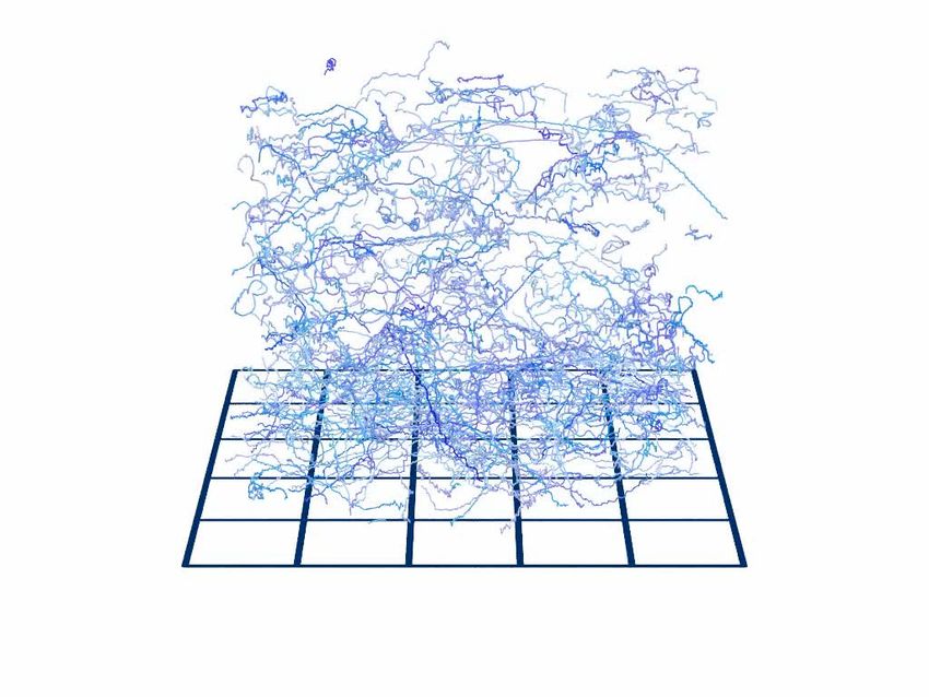
Research article                                                         Microbiology and Infectious Disease Physics of Living Systems

                                                                                              Leishmania. The physical constraints that shape
                                                                                              the response of these two micoorganisms are
                                                                                              therefore fundamentally different. From a fluid
                                                                                              dynamics perspective, both operate at low Rey-
                                                                                              nolds number, but Brownian motion dominates
                                                                                              life for the bacterial system, while it is negligible
                                                                                              for Leishmania. Nevertheless, there are intriguing
                                                                                              signs that the run-tumble locomotion characteris-
                                                                                              tic of bacteria like E. coli has analogues in motile
                                                                                              single-cell eukaryotes: Chlamydomonas reinhard-
                                                                                              tii algae exhibit run and tumble behaviour
                                                                                              (Polin et al., 2009), and sperm from the sea
                                                                                              urchin Arbacia punctulata exhibit sharp reorienta-
                                                                                              tion events amidst periods of helical swimming
                                    Video 1. Differential interference contrast movie of an
                                                                                              about a straight ‘run’ axis (Jikeli et al., 2015).
                                    unenriched sample of Leishmania mexicana cells,
                                                                                                  Chemotaxis has been observed in trypanoso-
                                    including procyclic promastigotes and metacyclic
                                    promastigotes, as well as intermediate forms. The cells matids, although the results have often been
                                    were attached to the chamber surface by pre-treating      somewhat nuanced. In an early study of chemo-
                                    the glass slide with polylysine. The scale bar represents taxis in L. mexicana, Leishmania major, and Leish-
                                    25 mm.                                                    mania donovani (Bray, 1983), it was found that
                                    https://elifesciences.org/articles/65051#video1           promastigote cells move up gradients of sugars,
                                                                                              serum, and some serum components. This work
                                                                                              also found some evidence that peritoneal macro-
                                                                                              phages from mice were attracted by serum that
                                    had been ‘activated’ by exposure to L. mexicana. The hypothesis that the host and motile parasites
                                    interact by diffusing messengers is supported by a study that shows that Leishmania promastigotes
                                    are repelled by ATP, possibly allowing them to evade neutrophils and adapt for phagocytosis
                                    (Detke and Elsabrouty, 2008). Other authors have found evidence that promastigotes of Leish-
                                    mania spp. chemotax towards sugars (Oliveira et al., 2000), though another contemporary study
                                    suggested that the directional behaviour is more appositely ascribed to osmotaxis, and is therefore
                                    chemically non-specific (Leslie et al., 2002). Intriguingly, the latter study speculates that chemotactic
                                    or osmotactic stimulus could play an important role in providing an orientational stimulus for the par-
                                    asites that prevents them being excreted and allows them to navigate within the sandfly host.
                                    Barros et al., 2006 performed experiments in which they timed the duration between reorientation
                                    events in swimming cells, showing that Leishmania amazonensis promastigote cells respond to both
                                    osmo- and chemotaxis, and find that chemotaxis dominates the response for all but the highest stim-
                                    ulant concentrations. More recently, it was shown that Leishmania braziliensis and L. amazonensis
                                    show chemotactic responses towards stimulants including a repurposed cancer drug (methotrexate)
                                    (Dı́az et al., 2013) and amino acids (Diaz et al., 2015). Optical tweezers studies on L. amazonensis
                                    (de Thomaz et al., 2011; Pozzo et al., 2009) showed that individual cells are attracted to higher
                                    glucose concentrations, and in the related trypanosomatids Trypanosoma cruzi and Trypanosoma
                                    rangeli, cells orient themselves towards tissue extracted from tsetse flies (their insect host) . Most
                                    recently, there have been exciting developments in the area of ‘social motility’ (collective motion) in
                                    Trypanosomes (Oberholzer et al., 2010; Schuster et al., 2017; Shaw et al., 2019), where it was
                                    shown that chemotactic behaviour is not just a single-cell phenomenon, but one that is exhibited in
                                    collective cell behaviour as well (DeMarco et al., 2020). Millimetre-scale groups of Trypanosoma
                                    brucei cells displaying social motility can change their swimming direction to avoid other colonies of
                                    their own species, but are attracted by chemicals diffusing from neighbouring colonies of E. coli.
                                        Holographic microscopy has proven to be a versatile and powerful tool for investigating the
                                    dynamics of swimming microorganisms at high speed and diffraction-limited resolution (Katz and
                                    Sheng, 2010; Romero et al., 2012; Weiße et al., 2020; Heddergott et al., 2012; Wilson et al.,
                                    2013; Molaei et al., 2014; Jikeli et al., 2015; Zhang et al., 2018; Thornton et al., 2020). Three-
                                    dimensional tracking, using holographic microscopy or other techniques, unambiguously resolves
                                    chirality in the shape of objects and in the geometry of swimming paths. A long-standing question in
                                    the biological physics of the organelle at the heart of the flagellum focuses on the breaking of sym-
                                    metry. The axoneme has a chiral structure in which dynein molecules on each doublet can attach to

Findlay et al. eLife 2021;10:e65051. DOI: https://doi.org/10.7554/eLife.65051                                                               2 of 15
Research article Microbiology and Infectious Disease Physics of Living Systems
A relative mRNA expression
B C
8 H4 SHERP
6
4
2
0
1.5 mm
PCF
PCF
META
META PCF META 1.5 mm
(n=500) (n=500)
D PCF E META F
(Mean = 18.3 ± 0.2 m/s) (Mean = 29.5 ± 0.2 m/s) PCF
0.12
META
0.08
0.08
P(v)
0.04
0.04
0 0
0 20 40 60 0 20 40 60 250 m
v ( m/s) v ( m/s)
G H 4 Cell body (PCF) J PCF
Main frq.s META
2
x ( m)
23 1
0
2 18 1
PSD ( m /Hz)
-2 10
-4
2
12 14 16
I 2
Cell body (META)
1 Main frq.s
x ( m)
0
1 0.57 0.01
-1 10
0.65 0.01
-2
10 11 12 0.1 1 10
Time (s) Frequency (Hz)
Figure 1. Swimming phenotypes in infective (metacyclic promastigote [META]) and non-infective (procyclic promastigote [PCF]) Leishmania mexicana
cells. (A) qPCRs of H4 (PCF molecular marker) and SHERP (META molecular marker) levels relative to N-myristyltransferase (nmt) (constant transcript
control) demonstrate that enriched, but not wholly distinct populations of cells were isolated and characterised. (B) Three-dimensional swimming
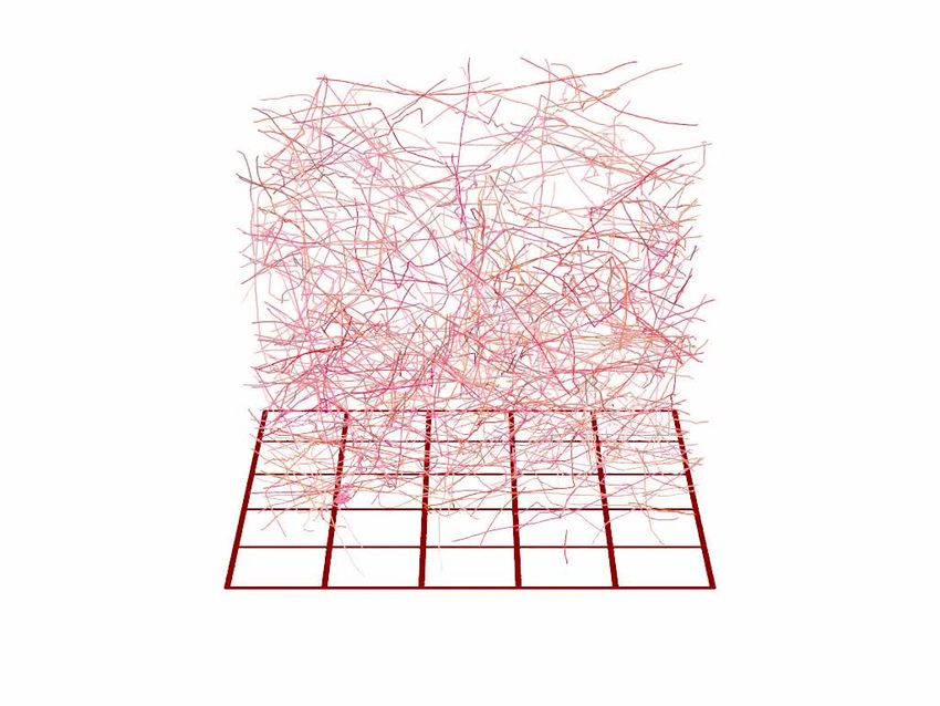
trajectories from 500 PCF cells of L. mexicana, showing stereotypical meandering helical paths (Video 2). (C) Trajectories of 500 META cells of L.
mexicana. These show a marked transition to a ‘run and tumble’ phenotype of rapid, straight trajectories interspersed by sharp reorientations
(Video 3). (D,E) Distribution of instantaneous swimming speeds in PCF (n = 3231) and META (n = 2202) cells, respectively. (F) Individual tracks of PCF
and META cells at a smaller scale, illustrating the different swimming phenotypes (Video 4). (G) Cartoon illustrating the dominant swimming phenotype
for PCF (top) and META (bottom). (H) The x-component of cell body motion as a function of time for a PCF cell. The black line represents the raw data,
and the blue line is the motion reconstituted using the principal frequency components only. (I) The x-component of motion for a META cell, both raw
data (black) and principal frequencies only (red). The flagellar beat frequency is more pronounced in META cells compared to PCF cells because the
cell bodies of the former are significantly smaller, and their flagella are longer. (J) Averaged power spectra of PCF (blue, n = 3231) and META (red,
n = 2202) motion, when the lowest frequencies have been removed (see text). The peaks at around 0.6 Hz correspond to the rotation of the flagellar
beat plane and the peaks at around 20 Hz to the flagellar beat in both cell types. The peak values and estimated uncertainties are indicated.
The online version of this article includes the following figure supplement(s) for figure 1:
Figure supplement 1. The Leishmania life cycle.
Findlay et al. eLife 2021;10:e65051. DOI: https://doi.org/10.7554/eLife.65051 3 of 15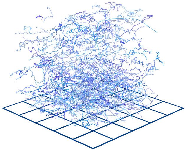
Research article                                                           Microbiology and Infectious Disease Physics of Living Systems

                                    Video 2. Computer rendering of 500                        Video 3. Computer rendering of 500
                                    Leishmania mexicana procyclic promastigote tracks,        Leishmania mexicana metacyclic promastigote tracks,
                                    showing stereotypical meandering helical paths. The       showing the stereotypical run and tumble phenotype of
                                    squares on the ground represent 300 mm on a side,         unstimulated cells. The squares on the ground
                                    and the tracks are between 20 and 60 s in duration.       represent 300 mm on a side, and the tracks are
                                    https://elifesciences.org/articles/65051#video2           between 20 and 60 s in duration.
                                                                                              https://elifesciences.org/articles/65051#video3

                                    their clockwise neighbour, as viewed from the
                                    basal end of the axoneme (Afzelius et al., 1995).
                                    Several groups have examined whether this structural chirality of the axoneme results in chiral flagel-
                                    lar waveforms. Rodrı́guez et al., 2009 showed that the flagellum of T. brucei could produce waves
                                    of either left- or right-handed chirality, in both procyclic-form and bloodstream-form cells.
                                    Bargul et al., 2016 examined several species and strains of Trypanosoma and demonstrated that
                                    the chirality of the flagellar attachment can vary, even between strains of the same species, but the
                                    direction of the flagellum’s rotation is independent of these morphological features. This has also
                                    been seen in the absence of an accompanying cell body attached to a flagellum; the male microga-
                                    mete of Plasmodium shows both right- and left-handed chiralities (Wilson et al., 2013).
                                        Here, we compare and quantitatively define the swimming dynamics and parameters of two key
                                    L. mexicana life cycle stages and find evidence of
                                    unbiased chirality in swimming behaviour. We fur-
                                    ther demonstrate a macrophage-driven chemo-
                                    tactic strategy involving novel run and tumble
                                    behaviour in Leishmania, where the parasites
                                    change direction, speed, and tumbling behaviour
                                    in the presence of human blood-derived
                                    macrophages.

                                    Results and discussion
                                    To characterise motile L. mexicana, we harvested
                                    PCF and META stages using molecularly verified
                                    culture techniques and verified each population
                                    pool via morphological distinctions and expres-
                                    sion of stage-specific markers, Histone H4 and
                                                                                              Video 4. Computer rendering of two exemplar cell
                                    SHERP (de Pablos et al., 2019; Knuepfer et al.,
                                                                                              tracks: a procyclic promastigote (blue) and a metacyclic
                                    2001; Figure 1A). We then used holographic
                                                                                              promastigote (red) of Leishmania mexicana. The
                                    microscopy to analyse the three-dimensional               squares on the ground represent 250 mm on a side.
                                    motility of suspensions of PCF and META. Sub-             The procyclic promastigote track displays the
                                    sets of this data are presented as composite ren-         stereotypical meandering helical trajectory, while the
                                    derings of 500 cell tracks in a sample volume             metacyclic cell shows longer, straight runs interspersed
                                    ~ 1:5  1:5  1:2      mm3       (Figure      1B,         with tumble events.
                                    C, Video 2, Video 3). In liquid culture, observed         https://elifesciences.org/articles/65051#video4

Findlay et al. eLife 2021;10:e65051. DOI: https://doi.org/10.7554/eLife.65051                                                                   4 of 15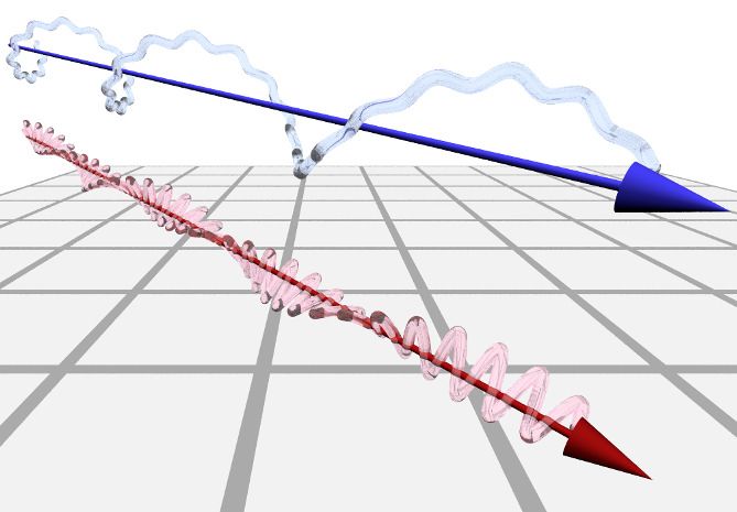
Research article                                                             Microbiology and Infectious Disease Physics of Living Systems

Figure 2. Three-dimensional geometry of metacyclic promastigote (META) and procyclic promastigote (PCF) cell tracks in Leishmania mexicana. (A)
Cartoon depicting left- and right-handed tracks, corresponding to negative and positive helicity, respectively (H ¼ 0 is an achiral line). (B) Normalised
probability density of instantaneous helicity in a population of PCF (n = 1.1  105). (C) Normalised probability density of helicity among META
(n = 7.9  104). (D) An example META trajectory in which the colour code shows instantaneous speed. The cell was imaged at 50 Hz, with every 30th
point plotted for clarity and five changes in direction (‘tumbles’), as indicated. (E) The normalised probability density of tumble angles among META.
The mean tumble angle is 72˚ ± 0.5˚ (SEM) and the mode is 21˚. (F) The distribution of run durations among META, showing two distinct exponential
regimes, at short and long times as indicated by the piecewise straight-line fits (black lines). The inset shows the complete range of run durations. (G)
Correlation between the duration of a run and the tumble angle following it among META (n = 5027 runs). Shorter runs (population 1) are followed by a
wider range of angles than longer runs (population 2) indicative of two characteristic run phenotypes.

                                     swimming speeds vary both within and between these two groups. We find the mean swimming
                                     speed of PCF cells (Figure 1D) to be 18.3 ± 0.2 mm/s (SEM), significantly lower than that of META
                                     cells (Figure 1E), which have a mean speed of 29.5 ± 0.2 mm/s (SEM). Although the mean swimming
                                     speeds are significantly different, the distributions of swimming speeds are broad and overlap: the
                                     standard deviations in swimming speed are 8.0 mm/s (PCF) and 10.4 mm/s (META). Such overlap may
                                     result from enriched, but not wholly distinct, PCF and META populations being characterised (per
                                     gene marker expression) (Figure 1A,D,E).
                                        Our results indicate PCF cells swim with a characteristic slow, corkscrew-like motion around a
                                     gently curving axis, a similar pattern to that of sea urchin sperm cells (Jikeli et al., 2015). In contrast,
                                     culture-derived META cells display a distinct swimming phenotype with straight path segments
                                     punctuated by sharp turning events, a ‘run and tumble’ motif reminiscent of enteric bacteria such as
                                     E. coli (Figure 1C). The ‘tumble’ events were marked by a decrease in the processive speed of the
                                     cells, consistent with a reversal of the flagellar beat and similar to pauses in motion observed in try-
                                     panosomes (Heddergott et al., 2012).
                                        The chirality of swimming motion can be challenging to access experimentally (Rodrı́guez et al.,
                                     2009; Heddergott et al., 2012; Wheeler, 2017; Schuster et al., 2017), especially in cells with a
                                     complicated morphology. We assess the chirality of the tracks by using three-dimensional tracking

Findlay et al. eLife 2021;10:e65051. DOI: https://doi.org/10.7554/eLife.65051                                                                     5 of 15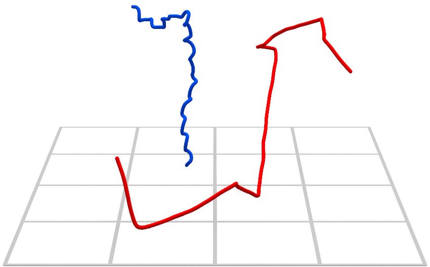
Research article                                                       Microbiology and Infectious Disease Physics of Living Systems

                                    to measure the helicity H (Wilson et al., 2013) as illustrated in Figure 2A (H ¼ 0 implies an achiral
                                    line, identical to its mirror image). Importantly, neither PCF (Figure 2B) nor META (Figure 2C) cell
                                    tracks display a systematic chirality on the population level. Individual tracks display a right- or left-
                                    handed character, and we do not observe any switches from left- to right-handed chirality (or
                                    vice versa) in any of our tracks.
                                        To analyse the characteristic run and tumble nature of META tracks in more detail, we separate
                                    the tracks into ‘runs’ of duration t n ¼ tn tn 1 punctuated by ‘tumbles’ through an angle
                                    (Figure 2D). Tumble angles appear biased towards small angles (Figure 2E), so several tumbles
                                    would typically be required to completely randomise a cell’s swimming direction. In contrast to com-
                                    monly studied bacterial species that display a heuristically similar swimming pattern, the distribution
                                    of Leishmania META run durations is not a simple exponential. We observe at least two distinct run
                                    behavioural ‘regimes’, as shown in Figure 2F. Runs of t < 1.5 s are the most common, though the
                                    distribution has a long exponential tail. Data on the longest run durations are noisy owing to the
                                    finite size of the sample volume, which limits the longest runs that may be captured. The two distinct
                                    run regimes are denoted by exponential lines of best fit (non-linear least squares regressions over
                                    the domain as indicated) in the main panel of Figure 2F, while the inset shows all data down to very
                                    low event frequencies at run durations up to 25 s. Figure 2G shows the relationship between run
                                    duration (t ) and subsequent tumble angle (). We note two distinct populations of events, implying
                                    two distinct processes: population 1 represents larger reorientations following shorter runs, and
                                    population 2 represents slight ‘course corrections’ during longer runs. This implies that sharper turns
                                    are suppressed during longer runs.
                                        In bacterial systems (Berg, 1993; Turner et al., 2016), an exponential distribution of runs is the
                                    hallmark of an underlying Poisson process with a constant probability of tumbling per unit time.
                                    Fine-tuning of this tumble probability in response to a biological sensing mechanism enables cells to
                                    navigate towards or away from an external stimulus. To determine whether the behaviour of META
                                    cells was fixed or could be regulated in conditions that might favour intracellular infection, we
                                    repeated our analysis using META cells exposed to J774.2 immortalised mouse macrophages (mMj)
                                    and human monocyte-derived macrophages (hMj), as well as medium alone (the latter as a negative
                                    control), with results shown in Figure 3. We compare the number of cells found close to the chemo-
                                    tactic bait (‘Tip’) to the number found in a region 2 cm from the bait (‘Away’), as well as their respec-
                                    tive swimming behaviour, in Figure 3A. Cells were significantly drawn toward the pipette tip in the
                                    presence of primary human blood-derived macrophages, but not to murine macrophages (p < 0.05).
                                    Unlike META cells, PCF cells showed a general attraction towards the unrefined agar in the pipette
                                    tip even in the absence of a particular stimulus (data shown in Figure 3—figure supplement 1).
                                        The number of tumbles exhibited by META cells decreases threefold in the presence of hMj
                                    (Figure 3B), independent of the number of tracks observed or track length. A more detailed exami-
                                    nation of the geometry of the tracks (quantities recapitulated in Figure 3C) yields unaltered distribu-
                                    tions of tumble angles (Figure 3D) and run durations (Figure 3E) between control and hMj
                                    response. This apparently contradictory observation – number of tumbles decreases, but run dura-
                                    tion is unaffected – is explained by a sub-population retaining the unstimulated behaviour, while the
                                    majority of cells suppress tumbles almost entirely. As described in the Materials and methods sec-
                                    tion, at least two tumbles per track are required to establish tumble frequency. Therefore, if a cell
                                    performs one or zero tumbles, it will not contribute to the run duration statistics. The average dis-
                                    placement per run is enhanced in both ‘Tip’ and ‘Away’ cases in the presence of hMj (Figure 3F).
                                    Importantly, this indicates the presence of a biologically relevant chemotactic stimulus from hMj
                                    that causes META cells to swim faster and straighter.
                                        Speed distributions for each experimental condition are shown in Figure 3G–L. There is no dis-
                                    tinction between the ‘Tip’ and ‘Away’ speed in the presence of DMEM alone. In contrast, the pres-
                                    ence of mMj somewhat enriches the fraction of cells swimming at low speeds (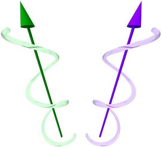
Research article Microbiology and Infectious Disease Physics of Living Systems
A 0.8
B C
Away Away
t2 = t2 - t1
No. Tumbles ( 1000)
Tip 30 Tip 2
400
Fraction of tracks
0.6
2
y ( m)
20 27
0.4 380
Time (s)
25
1
n=10609
n=12569
10
n=9869
n=5732
0.2 t1
n=418
n=589
360 23
0 0
DMEM hMφ 740 760 780 800
DMEM mMφ hMφ
x ( m)
D E -1
10
F 60
Mean run displacment ( m)
hMφ, away Away
DMEM, away hMφ, tip Tip
0.08 DMEM, away
DMEM, tip DMEM, tip
-2 40
hMφ, away
P( )
10
P(θ)
hMφ, tip
0.04
n=21625
n=24264
n=7458
n=8649
20
-3
10
0
0 45 90 135 180 0 3 6 9 12 0
DMEM hMφ
Tumble angle θ (degrees) Run duration (sec)
G DMEM
H mM
I hM
0.15 0.10
n = 10609 0.30 n = 418 n = 5732
v = 21.2 m/s v = 13.6 m/s v = 30.5 m/s
Away
0.10
v= 8.6 m/s 0.20 v= 8.1 m/s v= 16.5 m/s
P(v)
0.05
0.05 0.10
0.00 0.00 0.00
0 20 40 60 80 100 0 20 40 60 80 100 0 20 40 60 80 100
J K L
0.15 0.10
n = 9869 n = 589 n = 12569
0.30
v = 20.5 m/s v = 10.6 m/s v = 39.5 m/s
0.10
v= 8.1 m/s v= 8.1 m/s v= 17.2 m/s
Tip
0.20
P(v)
0.05
0.05 0.10
0.00 0.00 0.00
0 20 40 60 80 100 0 20 40 60 80 100 0 20 40 60 80 100
Speed v ( m/s) Speed v ( m/s) Speed v ( m/s)
Figure 3. The run and tumble behaviour of metacyclic promastigote (META) cells in the presence of an immunological stimulus. (A) Fractions of tracks
from META cells observed in a bulk suspension of cells 1 cm away from (‘Away’, solid bars) and immediately adjacent to a loaded pipette tip that had
been immersed in the sample medium (‘Tip’, filled bars) for 30 min. The negative controls, in which tips were filled with DMEM agar, show negligible
differences in the number of tracks proximal to the tip, versus the distal bulk. Pipette tips filled with either cultured J774.2 immortalised, mouse
stomach-derived macrophages (mMj) or fresh human blood-derived macrophages (hMj) showed accumulations of cells around the pipette tips (error
bars = 95% CI). (B) The number of ‘tumble’ events for cells in control (DMEM) and test (hMj) conditions. Exposure to hMj reduces the number of
tumble events considerably, consistent with a more persistent swimming direction. This effect impacts not only the cells that are in the immediate
proximity of the human-derived macrophages (hMj, Tip) but also those in the rest of the sample chamber. As the cells are free to explore the whole
sample volume, any ‘activation’ of cells exposed to a macrophage stimulus may persist even when the cells themselves have left the immediate
environment of the pipette tip. (C) The x-y projection of a section of a META track, showing tumble times (t1 ; t2 ), reorientation angles (1 ; 2 ), and
Figure 3 continued on next page
Findlay et al. eLife 2021;10:e65051. DOI: https://doi.org/10.7554/eLife.65051 7 of 15Research article Microbiology and Infectious Disease Physics of Living Systems
Figure 3 continued
intervening run duration (t 2 ). (D) The distribution of tumble angles remains unchanged in the presence of macrophage stimulus, suggesting an
unbiased stochastic reorientation process. (E) Distribution of run durations. Contrary to what would be expected in the equivalent situation in
chemotaxis in model bacterial systems, there is no increase in run duration in the presence of stimulus (hMj ‘Tip’), compared to controls (hMj ‘Away’,
DMEM ‘Tip’, DMEM ‘Away’); the distribution of run durations is almost identical in all cases. (F) The mean displacement per run, showing an increase in
the run displacement as a consequence of the faster run speed near the macrophage-loaded pipette tip (error bars = error bars=95% CI). (G–L)
Normalised instantaneous cell speed distributions for cells distal versus proximal to stimulus. Control samples (DMEM; G,J) show minimal difference,
but the presence of mouse- or human-derived macrophages causes a decrease or increase in swimming speed, respectively.
The online version of this article includes the following figure supplement(s) for figure 3:
Figure supplement 1. Fluorescein modelling of chemoeffector gradient.
Figure supplement 2. Chemotaxis results from procyclic promastigotes (PCF).
environment in parasitic protozoans has been well documented in trypanosomes (Hill, 2003;
Rico et al., 2013). For Leishmania, this has been described in terms of changes in metabolism, gene
expression (De Pablos et al., 2016), and the acquisition of a complement-resistant lipophosphogly-
can coat (McConville et al., 1992; Dostálová and Volf, 2012). We have shown that META have a
distinct swimming phenotype and chemotactic response compared to the precursor PCF forms.
In terms of the swimming phenotype, the similarity between L. mexicana swimming patterns and
those of bacteria is superficial. Bacterial runs and tumbles are a strategy to enable navigation in an
environment that randomises their swimming direction through rotational Brownian motion. Leish-
mania cells are at least five to eight times the (linear) size of E. coli bacteria, and rotational diffusivity
is inversely proportional to particle volume, therefore Leishmania will have their orientation rando-
mised by Brownian motion around 100 times more slowly than E. coli (Berg, 1993). It follows that
Brownian motion is negligible when considering Leishmania cell orientation, and that the run and
tumble behaviour observed here originates from a different imperative, serving a distinct function
specific to Leishmania. More broadly, we note that foraging strategies among larger animals also fol-
low superficially similar movement patterns (Viswanathan et al., 1999).
We find no bias in the chirality of swimming tracks in large populations. This leads us to conclude
that helical swimming patterns resulting from small lateral asymmetries or irregularities in cell body
shape that are sufficient to cause a rotation through asymmetric fluid drag on the cell body. This nat-
ural variation in the shape of cells can be seen in Video 1, in which a collection of META cells with
beating flagella have been affixed to a glass surface using polylysine. The helical character of the
tracks is more obvious in the case of PCF than in META, as seen in Figure 1F, Video 4. This is borne
out in the broader histogram seen in Figure 2B compared to that in Figure 2C, and is likely a reflec-
tion of the larger cell body size relative to flagellar length, compared to META.
The attraction of META to hMj is perhaps unsurprising given the biological relevance of macro-
phage infection in the life cycle of this parasite. Both hMj (the definitive host cells) and neutrophils
are drawn to sites of infection during inflammation, while resident dermal macrophages may act as
host cells facilitating persistence (Lee et al., 2020). Chemical gradients are known to occur and per-
sist in cutaneous infections, as damage to tissue, sandfly saliva and Leishmania-derived molecules
have been shown to recruit immune cells by this mechanism (Dey et al., 2018; Giraud et al., 2018;
Alcoforado Diniz et al., 2021). META cells have also been observed to move within the skin, away
from their site of introduction (Peters et al., 2008). Under our assay conditions, we did not see a sig-
nificant attraction to the murine macrophage line J774.2. Previous studies by others have shown that
transcriptional responses to pathogens may differ significantly between primary cells and immortal-
ised cells, for example, in the response to Mycobacterium (Andreu et al., 2017). In addition, the
human macrophages used here are relatively quiescent compared to the rapidly cycling J774.2 cells,
with likely altered production of metabolites that may affect chemotaxis. Further studies aimed at
defining the nature of the chemotactic stimuli involved may help to clarify the observed difference in
response.
The control experiments showing an attraction between PCF and DMEM-infused agar are consis-
tent with previous studies on promastigote cultures that had not been subject to META sub-popula-
tion enrichment (Oliveira et al., 2000; Leslie et al., 2002; Barros et al., 2006). We speculate that
this attraction is due to the presence of small carbohydrates associated with the unrefined agar, but
note that any subsequent experiments in which PCF might be exposed to hMj would be biologically
Findlay et al. eLife 2021;10:e65051. DOI: https://doi.org/10.7554/eLife.65051 8 of 15Research article Microbiology and Infectious Disease Physics of Living Systems
Video 5. Artist’s impression of a mixed population of Video 6. Artist’s impression of a mixed population of
swimming metacyclic and procyclic promastigotes of swimming metacyclic and procyclic promastigotes of
Leishmania mexicana. The cells are rendered with the Leishmania mexicana, presented as a three-
cell bodies in proportion to the flagella, and the cells’ dimensional (3D) red-cyan anaglyph. The cells are
swimming trajectories are those obtained from tracking rendered with the cell bodies in proportion to the
data. flagella, and the cells’ swimming trajectories are those
https://elifesciences.org/articles/65051#video5 obtained from tracking data.
https://elifesciences.org/articles/65051#video6
irrelevant. PCF do not naturally encounter and
cannot infect hMj, and attraction towards sugars
or other inert matrix components would be difficult to decouple from attraction to cells.
It is of obvious interest to examine how more biologically relevant viscosities might impact these
swimming patterns (Heddergott et al., 2012). Our in vitro analyses provide a framework to examine
Leishmania spp. life cycle-dependent motility distinctions in medium, but these parasites encounter
dynamic in vivo environments including sandfly bloodmeals, midgut and proboscis and mammalian
dermis, bloodstream, and potentially lymph (Moll et al., 1993). In addition, T. brucei are known to
sense each other’s presence and this directs movement as evidenced by in vitro social motility stud-
ies (Lopez et al., 2015; Imhof et al., 2015). While the in vivo tsetse fly environment is far more com-
partmentalised for T. brucei life cycle stages than that of the sandfly for Leishmania, investigating
whether Leishmania parasites also display the capacity to sense each other would be an exciting and
pertinent tangent.
Our studies show that flagellar motility is modified in cells adapted for infection of the human
host. We show that run and tumble behaviour is a phenotypic marker of human-infective META
stage parasites, and that this behaviour is suppressed in the presence of a chemotactic stimulus,
causing cells to swim faster and straighter towards their target. We speculate that this provides an
evolutionary advantage which promotes successful phagocytic uptake of non-replicative META cells,
fundamental to human infection and parasite life cycle progression. Through optimised flagellar
motility and an as-yet uncharacterised sensing mechanism, Leishmania proactively drive their uptake
by human immune cells.
Materials and methods
L. mexicana cell culture
Promastigote parasites of L. mexicana (strain M379) were cultured in M199 and Grace’s media at 26˚
C as described previously (de Pablos et al., 2019). All cells were maintained within low passage
numbers (Research article Microbiology and Infectious Disease Physics of Living Systems
Molecular validation of L. mexicana promastigote stages via qRTPCR
RNA was isolated from PCF and META populations concurrent to image capture, purified, reverse
transcribed, and analysed for Histone H4 and SHERP mRNA levels relative to constitutive nmt levels
as described previously (de Pablos et al., 2019).
Macrophage cell culture
Immortalised mouse stomach macrophages (J774.2) and human blood-derived macrophages from
resident donors were isolated and cultured in vitro for 7 days in Dulbecco’s modified eagle medium
(DMEM) at 37˚C prior to centrifugation. Approximately 1104 cells were resuspended in 10 ml
DMEM:1% agar for each chemotactic assay sample. Macrophage viability was confirmed via alamar
blue staining 2 hr after sample preparation.
Sample chambers
To compare life cycle stages, glass sample chambers were constructed from slides and UV-curing
glue, giving a final sample volume measuring approximately 20 15 1.2 mm3. These were loaded
with L. mexicana in M199 and sealed with petroleum jelly before observation.
Chemotaxis assay
The stability of the chemotactic apparatus and resultant gradient was verified via a fluorescein-
infused agar gradient establishment test (see supplementary data to Figure 3). The fluorescein agar
was then replaced in the 10 ml pipette tip by different ‘chemotactic bait’: DMEM 1% agar alone,
DMEM 1% agar with immortalised mouse macrophages or with human blood-derived macrophages.
Chemotaxis sample chambers were constructed as above with the addition of the 10 ml pipette tip
sealed into the sample chamber with petroleum jelly. The assembly was then placed on a tempera-
ture-controlled microscope stage at 34˚C in a room set at 34˚C to equilibrate for 30 min, allowing
transient convection currents to dissipate and a chemical gradient to become established. Cells
were imaged immediately adjacent to the pipette tip, but with the tip itself approximately 50 mm
outside the field of view to prevent imaging artifacts (‘Tip’), and at a distance of approximately 1 cm
from the tip (‘Away’). Five movies were acquired in each location (‘Tip’ and ‘Away’), alternating
between the two locations between individual movie acquisitions.
Holographic video microscopy
The holographic microscopy setup was similar to that used previously (Singh et al., 2018;
Kühn et al., 2018; Thornton et al., 2020), with a few modifications. The samples were imaged on a
Nikon Eclipse E600 upright microscope. The illumination source was a single-mode fibre-coupled
laser diode with peak emission at 642 nm. The end of the fibre was mounted below the specimen
stage using a custom adaptor and delivered a total of 15 mW of optical power to the sample. A Mik-
rotron MC-1362 monochrome camera was used to acquire videos of 3000 frames, at a frame rate of
50 Hz and with an exposure time of 100 ms. A 10 magnification bright-field lens with numerical
aperture of 0.3 was used to acquire data at a video resolution of 1024 1024 pixels2, corresponding
to a field of view measuring 1.44 1.44 mm2. The raw videos were saved as uncompressed, 8-bit
AVI files. Three biological replicates of each condition were prepared, with five technical replicates
from each of these.
Holographic data reconstruction
From each individual video frame, we calculated a stack of 130 images, spaced at 10 mm along the
optical axis, sampling the optical field within a volume of 1.44 1.44 1.3 mm3. These sequences
of images were calculated using the Rayleigh-Sommerfeld back-propagation scheme (Lee and Grier,
2007). We localised the cells using a method based on the Gouy phase anomaly, as described in
more detail elsewhere (Wilson and Zhang, 2012; Farthing et al., 2017; Gibson et al., 2021). This
method segments features based on axial optical intensity gradients within a sample, allowing us to
extract three-dimensional coordinates for individual cells in each frame. Lateral position uncertainties
are approximately 0.5 mm, while the axial performance is slightly worse at approximately 1.5 mm.
The latter is limited by the angular resolution of the microscope objective and the scattering proper-
ties of the anisotropic cell bodies. A separate software routine was used to compare the putative
Findlay et al. eLife 2021;10:e65051. DOI: https://doi.org/10.7554/eLife.65051 10 of 15Research article Microbiology and Infectious Disease Physics of Living Systems
cell coordinates extracted in each frame and associate those most likely to constitute cell tracks. The
tracks were smoothed using piecewise cubic splines in order to remove noise in the cell coordinates
and provide better estimates of cell velocity as described in previous work (Kühn et al., 2018).
Examining the mean-squared displacement of the cells’ smoothed trajectories allowed us to discrimi-
nate between swimming and diffusing cells, and to discard the latter. The smoothing process also
allowed for linear interpolation of missing data points, up to five consecutive points (equivalent to
0.1 s). Tracks with a duration shorter than 3 s were discarded. To give a more intuitive sense of the
swimming trajectories, we have provided an ‘artist’s impression’ rendering of the swimming cells, in
Video 5, and in a red-cyan anaglyph version in Video 6.
Frequency-domain analysis
To extract the flagellar beat and body roll frequencies, the smoothed trajectories were subtracted
from the raw values, and the power spectrum of the residuals was calculated. The power spectra
from all cells in the sample were summed to give the data in Figure 1J. To illustrate the dominance
of the two main frequency components, the track residuals were filtered in the frequency domain
using a filter with two Gaussian pass bands with mean (standard deviation) frequencies of 1 Hz (1 Hz)
and 20 Hz (2 Hz). The resulting signal contains the body roll and flagellar beat, removing extraneous
noise, as shown in Figure 1H,I.
Track chirality analysis
The PCF and META cell tracks were analysed to extract their chirality (handedness). This was done
according to a scheme similar to that previously used for determining the chirality of flagellar wave-
forms (Wilson et al., 2013). We first constructed an array of displacement vectors Tj from a cell’s
coordinates rj ðtÞ by calculating Tj ¼ rj rj 1 , and obtained the local track handedness as the angle
formed between vector Tj and the plane formed by the two previous segments (defined by
Tj 2 ^ Tj 2 ), divided by the total contour length Tj 2 þ Tj 1 þ Tj . We used this instead of (e.g.)
the more conventional Frenet-Serret apparatus because the handedness (H) is mathematically
well behaved for straight trajectories.
Run-tumble analysis
Tumbles were identified as large changes in swimming direction coupled with a drop in swimming
speed. Quantitatively, we calculated
!
ðvj þ vj 1 Þ Tj Tj 1
¼1 arccos ; (1)
2hvij Tj Tj 1
where we denote the local speed during segment Tj as vj and the average speed of track j as hvij .
This quantity shows clear peaks when the cells change direction (‘tumbles’), as identified in
Figure 2D. Run duration t was then defined as the time elapsed between two subsequent tumble
events. The tumble events take a finite time, during which the cell is essentially stationary. We there-
fore define the tumble angle as the change in the cell’s swimming direction between two time points
1 s apart, with the tumble event halfway in between.
Acknowledgements
This work was supported by Wellcome Trust grant nos. WT10472 and WT105502MA, EPSRC grant
no. EP/N014731/1, and MRC grant no. MR/L00092X/1. We thank Dr Luis M de Pablos for cell culture
assistance.
Additional information
Funding
Funder Grant reference number Author
Engineering and Physical EP/N014731/1 Laurence G Wilson
Findlay et al. eLife 2021;10:e65051. DOI: https://doi.org/10.7554/eLife.65051 11 of 15Research article Microbiology and Infectious Disease Physics of Living Systems
Sciences Research Council
Wellcome Trust WT104726 Paul M Kaye
Medical Research Council MR/L00092X/1 Pegine B Walrad
Wellcome Trust WT105502MA Rachel C Findlay
BBSRC BB/M011151/1 Kirstin A Spence
The funders had no role in study design, data collection and interpretation, or the
decision to submit the work for publication.
Author contributions
Rachel C Findlay, Conceptualization, Data curation, Formal analysis, Validation, Investigation, Meth-
odology, Writing - review and editing; Mohamed Osman, Resources, Investigation; Kirstin A Spence,
Visualization; Paul M Kaye, Resources, Formal analysis, Writing - review and editing; Pegine B Wal-
rad, Conceptualization, Resources, Data curation, Formal analysis, Supervision, Funding acquisition,
Validation, Investigation, Methodology, Writing - original draft, Project administration, Writing -
review and editing; Laurence G Wilson, Conceptualization, Resources, Data curation, Software, For-
mal analysis, Supervision, Funding acquisition, Investigation, Visualization, Methodology, Writing -
original draft, Project administration, Writing - review and editing
Author ORCIDs
Rachel C Findlay https://orcid.org/0000-0003-3235-130X
Kirstin A Spence https://orcid.org/0000-0002-1220-4698
Paul M Kaye https://orcid.org/0000-0002-8796-4755
Pegine B Walrad https://orcid.org/0000-0002-2302-0720
Laurence G Wilson https://orcid.org/0000-0001-6659-6001
Ethics
Human subjects: Unrelated adults of both genders and diverse racial backgrounds local to Yorkshire,
United Kingdom volunteered for tissue sampling for this research. After health screening by medical
staff, informed consent was obtained from donors in compliance with ICH GCP, the UK Data Protec-
tion Act, and other regulatory requirements, as appropriate and under the University of York Depart-
ment of Biology Ethics Committee (BEC)-approved study CL 201201 v2.3 to donate blood for a
BEC-approved biomedical research under Project License PK 201702.
Decision letter and Author response
Decision letter https://doi.org/10.7554/eLife.65051.sa1
Author response https://doi.org/10.7554/eLife.65051.sa2
Additional files
Supplementary files
. Transparent reporting form
Data availability
Data underlying the conclusions in this study are are available at the York Research Database
(https://doi.org/10.15124/a8eabcd4-66c1-41a3-aa4b-423921b06568).
The following dataset was generated:
Database and
Author(s) Year Dataset title Dataset URL Identifier
Wilson LG 2021 Leishmania motility and chemotaxis https://doi.org/10.15124/ The York Research
data a8eabcd4-66c1-41a3- Database, 10.15124/
aa4b-423921b06568 a8eabcd4-66c1-41a3-
aa4b-423921b06568
Findlay et al. eLife 2021;10:e65051. DOI: https://doi.org/10.7554/eLife.65051 12 of 15Research article Microbiology and Infectious Disease Physics of Living Systems
References
Afzelius BA, Dallai R, Lanzavecchia S, Bellon PL. 1995. Flagellar structure in normal human spermatozoa and in
spermatozoa that lack dynein arms. Tissue and Cell 27:241–247. DOI: https://doi.org/10.1016/S0040-8166(95)
80044-1, PMID: 7645004
Alcoforado Diniz J, Chaves MM, Vaselek S, Miserani Magalhães RD, Ricci-Azevedo R, de Carvalho RVH,
Lorenzon LB, Ferreira TR, Zamboni D, Walrad PB, Volf P, Sacks DL, Cruz AK. 2021. Protein methyltransferase 7
deficiency in Leishmania major increases neutrophil associated pathology in murine model. PLOS Neglected
Tropical Diseases 15:e0009230. DOI: https://doi.org/10.1371/journal.pntd.0009230, PMID: 33651805
Alvar J, Vélez ID, Bern C, Herrero M, Desjeux P, Cano J, Jannin J, den Boer M, WHO Leishmaniasis Control
Team. 2012. Leishmaniasis worldwide and global estimates of its incidence. PLOS ONE 7:e35671. DOI: https://
doi.org/10.1371/journal.pone.0035671, PMID: 22693548
Ambit A, Woods KL, Cull B, Coombs GH, Mottram JC. 2011. Morphological events during the cell cycle of
Leishmania major. Eukaryotic Cell 10:1429–1438. DOI: https://doi.org/10.1128/EC.05118-11, PMID: 21926331
Andreu N, Phelan J, de Sessions PF, Cliff JM, Clark TG, Hibberd ML. 2017. Primary macrophages and J774 cells
respond differently to infection with Mycobacterium tuberculosis. Scientific Reports 7:42225. DOI: https://doi.
org/10.1038/srep42225, PMID: 28176867
Bargul JL, Jung J, McOdimba FA, Omogo CO, Adung’a VO, Krüger T, Masiga DK, Engstler M. 2016. Species-
Specific Adaptations of Trypanosome Morphology and Motility to the Mammalian Host. PLOS Pathogens 12:
e1005448. DOI: https://doi.org/10.1371/journal.ppat.1005448, PMID: 26871910
Barros VC, Oliveira JS, Melo MN, Gontijo NF. 2006. Leishmania amazonensis: chemotaxic and osmotaxic
responses in promastigotes and their probable role in development in the phlebotomine gut. Experimental
Parasitology 112:152–157. DOI: https://doi.org/10.1016/j.exppara.2005.10.005, PMID: 16313904
Bates PA, Rogers ME. 2004. New insights into the developmental biology and transmission mechanisms of
Leishmania. Current molecular medicine 4:601–609. DOI: https://doi.org/10.2174/1566524043360285,
PMID: 15357211
Beneke T, Demay F, Hookway E, Ashman N, Jeffery H, Smith J, Valli J, Becvar T, Myskova J, Lestinova T, Shafiq
S, Sadlova J, Volf P, Wheeler RJ, Gluenz E. 2019. Genetic dissection of a Leishmania flagellar proteome
demonstrates requirement for directional motility in sand fly infections. PLOS Pathogens 15:e1007828.
DOI: https://doi.org/10.1371/journal.ppat.1007828, PMID: 31242261
Berg HC. 1993. Random Walks in Biology. Princeton, NJ: Princeton University Press.
Berg HC, Brown DA. 1972. Chemotaxis in Escherichia coli analysed by three-dimensional tracking. Nature 239:
500–504. DOI: https://doi.org/10.1038/239500a0, PMID: 4563019
Bray RS. 1983. Leishmania: chemotaxic responses of promastigotes and macrophages in vitro. The Journal of
Protozoology 30:322–329. DOI: https://doi.org/10.1111/j.1550-7408.1983.tb02923.x, PMID: 6631775
Bray D. 2001. Cell Movements: From Molecules to Motility. 2nd ed. New York: Garland Science. DOI: https://
doi.org/10.4324/9780203833582
De Pablos LM, Ferreira TR, Walrad PB. 2016. Developmental differentiation in Leishmania lifecycle progression:
post-transcriptional control conducts the orchestra. Current Opinion in Microbiology 34:82–89. DOI: https://
doi.org/10.1016/j.mib.2016.08.004, PMID: 27565628
de Pablos LM, Ferreira TR, Dowle AA, Forrester S, Parry E, Newling K, Walrad PB. 2019. The mRNA-bound
Proteome of Leishmania mexicana: Novel Genetic Insight into an Ancient Parasite. Molecular & Cellular
Proteomics 18:1271–1284. DOI: https://doi.org/10.1074/mcp.RA118.001307, PMID: 30948621
de Thomaz AA, Fontes A, Stahl CV, Pozzo LY, Ayres DC, Almeida DB, Farias PMA, Santos BS, Santos-Mallet J,
Gomes SAO, Giorgio S, Feder D, Cesar CL. 2011. Optical tweezers for studying taxis in parasites. Journal of
Optics 13:044015. DOI: https://doi.org/10.1088/2040-8978/13/4/044015
DeMarco SF, Saada EA, Lopez MA, Hill KL. 2020. Identification of Positive Chemotaxis in the Protozoan
Pathogen Trypanosoma brucei. mSphere 5:e00685–20. DOI: https://doi.org/10.1128/mSphere.00685-20,
PMID: 32817459
Detke S, Elsabrouty R. 2008. Leishmania mexicana amazonensis: plasma membrane adenine nucleotide
translocator and chemotaxis. Experimental Parasitology 118:408–419. DOI: https://doi.org/10.1016/j.exppara.
2007.10.010, PMID: 18031742
Dey R, Joshi AB, Oliveira F, Pereira L, Guimarães-Costa AB, Serafim TD, de Castro W, Coutinho-Abreu IV,
Bhattacharya P, Townsend S, Aslan H, Perkins A, Karmakar S, Ismail N, Karetnick M, Meneses C, Duncan R,
Nakhasi HL, Valenzuela JG, Kamhawi S. 2018. Gut microbes egested during bites of infected sand flies
augment severity of Leishmaniasis via Inflammasome-Derived IL-1b. Cell Host & Microbe 23:134–143.
DOI: https://doi.org/10.1016/j.chom.2017.12.002, PMID: 29290574
Dı́az E, Köhidai L, Rı́os A, Vanegas O, Silva A, Szabó R, Mező G, Hudecz F, Ponte-Sucre A. 2013. Leishmania
braziliensis: cytotoxic, cytostatic and chemotactic effects of poly-lysine-methotrexate-conjugates. Experimental
Parasitology 135:134–141. DOI: https://doi.org/10.1016/j.exppara.2013.06.007, PMID: 23816643
Diaz E, Zacarias AK, Pérez S, Vanegas O, Köhidai L, Padrón-Nieves M, Ponte-Sucre A. 2015. Effect of aliphatic,
monocarboxylic, dicarboxylic, heterocyclic and sulphur-containing amino acids on Leishmania spp chemotaxis.
Parasitology 142:1621–1630. DOI: https://doi.org/10.1017/S003118201500116X, PMID: 26396059
Dostálová A, Volf P. 2012. Leishmania development in sand flies: parasite-vector interactions overview. Parasites
& Vectors 5:276. DOI: https://doi.org/10.1186/1756-3305-5-276, PMID: 23206339
Findlay et al. eLife 2021;10:e65051. DOI: https://doi.org/10.7554/eLife.65051 13 of 15Research article Microbiology and Infectious Disease Physics of Living Systems
Farthing NE, Findlay RC, Jikeli JF, Walrad PB, Bees MA, Wilson LG. 2017. Simultaneous two-color imaging in
digital holographic microscopy. Optics Express 25:28489–28500. DOI: https://doi.org/10.1364/OE.25.028489,
PMID: 31956278
Gibson T, Bedrossian M, Serabyn E, Lindensmith C, Nadeau JL. 2021. Using the Gouy phase anomaly to localize
and track bacteria in digital holographic microscopy 4D images. Journal of the Optical Society of America A
38:A11–A18. DOI: https://doi.org/10.1364/JOSAA.404004, PMID: 33690523
Giraud E, Lestinova T, Derrick T, Martin O, Dillon RJ, Volf P, Műller I, Bates PA, Rogers ME. 2018. Leishmania
proteophosphoglycans regurgitated from infected sand flies accelerate dermal wound repair and exacerbate
leishmaniasis via insulin-like growth factor 1-dependent signalling. PLOS Pathogens 14:e1006794. DOI: https://
doi.org/10.1371/journal.ppat.1006794
Heddergott N, Krüger T, Babu SB, Wei A, Stellamanns E, Uppaluri S, Pfohl T, Stark H, Engstler M. 2012.
Trypanosome Motion Represents an Adaptation to the Crowded Environment of the Vertebrate Bloodstream.
PLOS Pathogens 8:e1003023. DOI: https://doi.org/10.1371/journal.ppat.1003023
Hill KL. 2003. Biology and mechanism of trypanosome cell motility. Eukaryotic Cell 2:200–208. DOI: https://doi.
org/10.1128/EC.2.2.200-208.2003, PMID: 12684369
Imhof S, Vu XL, Bütikofer P, Roditi I. 2015. A glycosylation mutant of Trypanosoma brucei Links Social Motility
Defects In Vitro to Impaired Colonization of Tsetse Flies In Vivo. Eukaryotic Cell 14:588–592. DOI: https://doi.
org/10.1128/EC.00023-15, PMID: 25862152
Jikeli JF, Alvarez L, Friedrich BM, Wilson LG, Pascal R, Colin R, Pichlo M, Rennhack A, Brenker C, Kaupp UB.
2015. Sperm navigation along helical paths in 3D chemoattractant landscapes. Nature Communications 6:7985.
DOI: https://doi.org/10.1038/ncomms8985, PMID: 26278469
Katz J, Sheng J. 2010. Applications of Holography in Fluid Mechanics and Particle Dynamics. Annual Review of
Fluid Mechanics 42:531–555. DOI: https://doi.org/10.1146/annurev-fluid-121108-145508
Knuepfer E, Stierhof YD, McKean PG, Smith DF. 2001. Characterization of a differentially expressed protein that
shows an unusual localization to intracellular membranes in Leishmania major. Biochemical Journal 356:335–
344. DOI: https://doi.org/10.1042/bj3560335, PMID: 11368759
Kühn MJ, Schmidt FK, Farthing NE, Rossmann FM, Helm B, Wilson LG, Eckhardt B, Thormann KM. 2018. Spatial
arrangement of several flagellins within bacterial flagella improves motility in different environments. Nature
Communications 9:5369. DOI: https://doi.org/10.1038/s41467-018-07802-w, PMID: 30560868
Lee SH, Chaves MM, Kamenyeva O, Gazzinelli-Guimaraes PH, Kang B, Pessenda G, Passelli K, Tacchini-Cottier F,
Kabat J, Jacobsen EA, Nutman TB, Sacks DL. 2020. M2-like, dermal macrophages are maintained via IL-4/
CCL24–mediated cooperative interaction with eosinophils in cutaneous leishmaniasis. Science Immunology 5:
eaaz4415. DOI: https://doi.org/10.1126/sciimmunol.aaz4415
Lee SH, Grier DG. 2007. Holographic microscopy of holographically trapped three-dimensional structures. Optics
Express 15:1505–1512. DOI: https://doi.org/10.1364/OE.15.001505, PMID: 19532383
Leslie G, Barrett M, Burchmore R. 2002. Leishmania mexicana: promastigotes migrate through osmotic
gradients. Experimental Parasitology 102:117–120. DOI: https://doi.org/10.1016/S0014-4894(03)00031-6,
PMID: 12706748
Lopez MA, Saada EA, Hill KL. 2015. Insect stage-specific adenylate cyclases regulate social motility in African
trypanosomes. Eukaryotic Cell 14:104–112. DOI: https://doi.org/10.1128/EC.00217-14, PMID: 25416239
McConville MJ, Turco SJ, Ferguson MA, Sacks DL. 1992. Developmental modification of lipophosphoglycan
during the differentiation of Leishmania major promastigotes to an infectious stage. The EMBO Journal 11:
3593–3600. DOI: https://doi.org/10.1002/j.1460-2075.1992.tb05443.x, PMID: 1396559
Molaei M, Barry M, Stocker R, Sheng J. 2014. Failed escape: solid surfaces prevent tumbling of Escherichia coli.
Physical Review Letters 113:068103. DOI: https://doi.org/10.1103/PhysRevLett.113.068103, PMID: 25148353
Moll H, Fuchs H, Blank C, Röllinghoff M. 1993. Langerhans cells transport Leishmania major from the infected
skin to the draining lymph node for presentation to antigen-specific T cells. European Journal of Immunology
23:1595–1601. DOI: https://doi.org/10.1002/eji.1830230730, PMID: 8325337
Oberholzer M, Lopez MA, McLelland BT, Hill KL. 2010. Social motility in african trypanosomes. PLOS Pathogens
6:e1000739. DOI: https://doi.org/10.1371/journal.ppat.1000739, PMID: 20126443
Oliveira JS, Melo MN, Gontijo NF. 2000. A sensitive method for assaying chemotaxic responses of Leishmania
promastigotes. Experimental Parasitology 96:187–189. DOI: https://doi.org/10.1006/expr.2000.4569,
PMID: 11162370
Peters NC, Egen JG, Secundino N, Debrabant A, Kimblin N, Kamhawi S, Lawyer P, Fay MP, Germain RN, Sacks
D. 2008. In vivo imaging reveals an essential role for neutrophils in leishmaniasis transmitted by sand flies.
Science 321:970–974. DOI: https://doi.org/10.1126/science.1159194, PMID: 18703742
Polin M, Tuval I, Drescher K, Gollub JP, Goldstein RE. 2009. Chlamydomonas swims with two "gears" in a
eukaryotic version of run-and-tumble locomotion. Science 325:487–490. DOI: https://doi.org/10.1126/science.
1172667, PMID: 19628868
Pozzo LY, Fontes A, de Thomaz AA, Santos BS, Farias PM, Ayres DC, Giorgio S, Cesar CL. 2009. Studying taxis
in real time using optical tweezers: applications for Leishmania amazonensis parasites. Micron 40:617–620.
DOI: https://doi.org/10.1016/j.micron.2009.02.008, PMID: 19345110
Pulvertaft RJV, Hoyle GF. 1960. Stages in the life-cycle of Leishmania donovani. Transactions of the Royal
Society of Tropical Medicine and Hygiene 54:191–196. DOI: https://doi.org/10.1016/0035-9203(60)90057-2
Rico E, Rojas F, Mony BM, Szoor B, MacGregor P, Matthews KR. 2013. Bloodstream form pre-adaptation to the
tsetse fly in Trypanosoma brucei. Frontiers in Cellular and Infection Microbiology 3:78. DOI: https://doi.org/10.
3389/fcimb.2013.00078
Findlay et al. eLife 2021;10:e65051. DOI: https://doi.org/10.7554/eLife.65051 14 of 15You can also read