Repressing Ago2 mRNA translation by Trim71 maintains pluripotency through inhibiting let-7 microRNAs
←
→
Page content transcription
If your browser does not render page correctly, please read the page content below
RESEARCH ARTICLE

                                        Repressing Ago2 mRNA translation by
                                        Trim71 maintains pluripotency through
                                        inhibiting let-7 microRNAs
                                        Qiuying Liu1, Xiaoli Chen2, Mariah K Novak1, Shaojie Zhang2, Wenqian Hu1*
                                        1
                                         Department of Biochemistry and Molecular Biology, Mayo Clinic, Rochester, United
                                        States; 2Department of Computer Science, University of Central Florida, Orlando,
                                        United States

                                        Abstract The regulation of stem cell fate is poorly understood. Genetic studies in
                                        Caenorhabditis elegans lead to the hypothesis that a conserved cytoplasmic double-negative
                                        feedback loop consisting of the RNA-binding protein Trim71 and the let-7 microRNA controls the
                                        pluripotency and differentiation of stem cells. Although let-7-microRNA-mediated inhibition of
                                        Trim71 promotes differentiation, whether and how Trim71 regulates pluripotency and inhibits the
                                        let-7 microRNA are still unknown. Here, we show that Trim71 represses Ago2 mRNA translation in
                                        mouse embryonic stem cells. Blocking this repression leads to a specific post-transcriptional
                                        increase of mature let-7 microRNAs, resulting in let-7-dependent stemness defects and accelerated
                                        differentiation in the stem cells. These results not only support the Trim71-let-7-microRNA bi-stable
                                        switch model in controlling stem cell fate, but also reveal that repressing the conserved pro-
                                        differentiation let-7 microRNAs at the mature microRNA level by Ago2 availability is critical to
                                        maintaining pluripotency.

                                        Introduction
*For correspondence:                    The switch between pluripotency and differentiation in embryonic stem cells (ESCs) remains incom-
hu.wenqian@mayo.edu                     pletely understood. Although nuclear events controlling stemness are becoming increasingly clear,
                                        how cytoplasmic pathways of gene expression regulate ESCs’ fates between pluripotency and differ-
Competing interests: The
                                        entiation are still poorly understood (Ye and Blelloch, 2014).
authors declare that no
                                            Genetic studies in C. elegans led to the postulation that a conserved cytoplasmic bi-stable switch
competing interests exist.
                                        controls the pluripotency and differentiation of stem cells (Ecsedi and Grosshans, 2013). This switch
Funding: See page 24                    is proposed to involve reciprocal negative regulation between the conserved pro-differentiation let-
Received: 06 January 2021               7 microRNA (miRNA) and Trim71 (Lin41 in C. elegans), a conserved and ESC-specific RNA-binding
Accepted: 12 February 2021              protein (RBP). The following observations support this model. First, the let-7 miRNA negatively cor-
Published: 18 February 2021             relates with Trim71 during stem cell differentiation: the let-7 miRNA level increases, while Trim71
                                        decreases during differentiation. Second, Trim71 is a conserved target of the let-7 miRNA, and
Reviewing editor: Timothy W
Nilsen, Case Western Reserve
                                        repressing Trim71 by let-7 promotes stem cell differentiation (Aeschimann et al., 2019;
University, United States               Ecsedi et al., 2015; Grishok et al., 2001; Roush and Slack, 2008). Third, inhibiting Trim71 sup-
                                        presses developmental defects caused by mutations in the core components of the miRNA pathway
    Copyright Liu et al. This article
                                        in C. elegans (Büssing et al., 2010; Grishok et al., 2001), suggesting that Trim71 may negatively
is distributed under the terms of
                                        regulate the miRNA pathway. Thus, it is hypothesized that let-7 miRNA and Trim71 reciprocally
the Creative Commons
Attribution License, which              repress each other. This double-negative feedback loop forms a molecular bi-stable switch, in which
permits unrestricted use and            stem-cell differentiation is controlled by the let-7-miRNA-mediated inhibition of Trim71 and pluripo-
redistribution provided that the        tency is controlled by the hypothetical Trim71-mediated inhibition of the let-7 miRNA (Ecsedi and
original author and source are          Grosshans, 2013). Due to the conservation of let-7 miRNA, Trim71, and the let-7-mediated inhibi-
credited.                               tion of Trim71, the cytoplasmic bi-stable switch controlling stem cell fate is thought to be conserved

Liu et al. eLife 2021;10:e66288. DOI: https://doi.org/10.7554/eLife.66288                                                              1 of 27
Research article                                                                   Cell Biology Chromosomes and Gene Expression

                                    in animals. A lingering question in this bi-stable switch model, however, is whether and how Trim71
                                    inhibits the let-7 miRNA and regulates pluripotency in stem cells.
                                        Trim71 was proposed to interact with and ubiquitylate Ago2, a critical component of the miRNA
                                    pathway, resulting in Ago2 degradation in mammalian cells (Rybak et al., 2009). Although the func-
                                    tional significance of this interaction to stem cell biology was not examined, this observation seemed
                                    to support the bi-stable switch model. Later studies, however, indicated that the Trim71-Ago2 inter-
                                    action is RNA dependent (Chang et al., 2012; Loedige et al., 2013), and the proposed Trim71-
                                    mediated Ago2 degradation is absent in vivo (Chen et al., 2012; Welte et al., 2019). Thus, it is
                                    unclear how Trim71 modulates the let-7 miRNA. In terms of biological functions, Trim71 knockout
                                    mice are embryonic lethal (Cuevas et al., 2015), while Trim71 knockout mouse ESCs (mESCs) have
                                    no proliferation or stemness defects (Chang et al., 2012; Mitschka et al., 2015; Welte et al., 2019;
                                    Worringer et al., 2014), indicating an enigmatic role of Trim71 in stem cell biology. Collectively,
                                    these results highlight the hypothetical status of Trim71’s function and mechanisms in the bi-stable
                                    switch model and beg for investigations on how Trim71 regulates the let-7 miRNAs and whether this
                                    regulation plays a role in controlling pluripotency in stem cells.
                                        Here, we show that Trim71 maintains pluripotency through inhibiting the let-7 miRNAs. We iden-
                                    tified the transcriptome-wide targets of Trim71 in mESCs and determined that Trim71 binds and
                                    represses Ago2 mRNA translation. Specific disruption of this repression leads to an elevated Ago2
                                    level, which results in a specific post-transcriptional increase of the mature let-7 miRNAs, decreased
                                    stemness, and accelerated differentiation in mESCs. These stem cell defects are dependent on the
                                    let-7 miRNAs, as specific inhibition of the let-7 miRNAs abolishes the stemness defects caused by
                                    the loss of Trim71-mediated repression of Ago2 mRNA translation in mESCs. Collectively, these
                                    results provide direct support for the cytoplasmic bi-stable switch model of stem cell fate decision.
                                    Moreover, this study reveals that repressing the conserved pro-differentiation let-7 microRNAs at
                                    the mature miRNA level by Ago2 availability is critical to maintaining pluripotency.

                                    Results
                                    Transcriptome-wide identification of Trim71’s target mRNAs in mESCs
                                    To study Trim71’s function in mESCs, we created bi-allelic FLAG-tagged Trim71 in mESCs. Using
                                    CRISPR/Cas9-mediated genomic editing, we inserted a FLAG-tag at the N-terminus of Trim71
                                    (Figure 1A) and identified bi-allelic FLAG-tag knock-in mESC clones (Figure 1B). The knock-in
                                    sequence changes neither Trim71’s native promoter nor the 3’UTR (untranslated region), where tran-
                                    scriptional and post-transcriptional regulations mainly occur, respectively, and the FLAG-Trim71 is
                                    expressed at the endogenous level (Figure 1—figure supplement 1A). Moreover, the FLAG-Trim71
                                    mESC is phenotypically identical to the wild type (WT) mESC: they have similar morphology, growth
                                    rates, self-renewal abilities, and express similar levels of core pluripotency transcription factors (Fig-
                                    ure 1—figure supplement 1B–F). Thus, we refer to the FLAG-Trim71 mESCs as the WT mESCs.
                                        The FLAG-tag facilitates unambiguous detection and efficient isolation of the endogenous Trim71
                                    in mESCs. Using an anti-FLAG monoclonal antibody, we could specifically detect Trim71 in the
                                    FLAG-Trim71 mESCs (Figure 1C). Moreover, most Trim71 could be immunoprecipitated (IP) from
                                    the FLAG-Trim71 mESCs lysate via the anti-FLAG antibody (Figure 1D). This IP is specific because:
                                    (a) in the IP using IgG, Trim71 remained in the supernatant; and (b) when the IP was performed in
                                    the control mESC without the FLAG-tag, the IP sample generated little signal (Figure 1D).
                                        To determine whether Trim71 regulates mESCs, we identified transcriptome-wide targets of
                                    Trim71 in mESCs using cross-linking immunoprecipitation and sequencing (CLIP-seq)
                                    (Figure 1E; Darnell, 2010). This method not only revealed which mRNAs Trim71 binds but also iden-
                                    tified the binding sites on those mRNAs. Trim71-binding sites are mainly located in the introns and
                                    3’UTRs of the target mRNAs (Figure 1F; Supplementary file 1). Sequence analysis identified an
                                    over-represented stem-loop structure, but no enriched primary sequence motifs, in the Trim71-bind-
                                    ing sites compared to randomized sequences (Figure 1G). This observation suggests that Trim71
                                    recognizes RNA secondary structures, but not a primary sequence, which is consistent with recent in
                                    vitro and in vivo studies on Trim71:RNA interactions (Kumari et al., 2018; Welte et al., 2019).
                                    Cdkn1a mRNA (Figure 1H), a validated Trim71 target (Chang et al., 2012), is among the identified

Liu et al. eLife 2021;10:e66288. DOI: https://doi.org/10.7554/eLife.66288                                                              2 of 27
Research article Cell Biology Chromosomes and Gene Expression
A Mouse Trim71 Genomic Locus B Genotyping C FLAG
WT
sgRNA oligoA + oligoB Trim71
FLAG
M WT 98kd
ATG Stop Trim71
Cas9 + sgRNA 400bp 64kd
Anti-FLAG
Donor oligo
oligoA oligoB 50kd
300bp
ATG Stop 36kd
Exonic coding region
FLAG-tag 27kd
200bp
Exonic noncoding region
Anti-Gapdh
D IP: anti-FLAG IP: IgG
WT FLAG-Trim71 E
Reads for Unique Mapped
Dataset Raw Reads
25% Input
25% Input
25% Input
Mapping Reads
25% Sup.
25% Sup.
25% Sup.
SM-Input 157,298,472 135,838,745 78,087,829 (57.49%)
CLIPseqA 108,166,439 102,736,328 72,146,251 (70.22%)
IP
IP
IP
191kD CLIPseqB 89,633,607 86,145,908 51,705,646 (60.02%)
F 400
97kD 332
Anti-FLAG
300
64kD
51kD * 200 161
39kD #
100
100
28kD 59
19kD 13
0
Anti-Gapdh Intergenic
3’UTR 5’UTR CDS Intron
Region
G H Ago2 Locus
4
3 5000
SM-Input
bits
2
0
1 5000
0 CLIPseq
1 2 3 4 5 6 7 8 9 10 11
0
Position
Cdkn1a (p21) Locus
I
5000
SM-Input
0
5000
CLIPseq
0
Base-pair possibility
Figure 1. Transcriptome-wide identification of Trim71 target mRNAs in mouse embryonic stem cells (mESCs). (A) Workflow for knock-in the FLAG-tag
to the endogenous Trim71 locus in mESCs. (B) Genotyping of the FLAG-Trim71 mESCs using the two primers in (A). (C) Specific detection of the
endogenous Trim71 via the FLAG-tag. Western blotting in the WT and the FLAG-Trim71 mESCs using an anti-FLAG monoclonal antibody. (D) Efficient
and specific isolation of the endogenous Trim71. An anti-FLAG monoclonal antibody and mouse IgG were used to immunoprecipitate (IP) the
Figure 1 continued on next page
Liu et al. eLife 2021;10:e66288. DOI: https://doi.org/10.7554/eLife.66288 3 of 27
Research article                                                                       Cell Biology Chromosomes and Gene Expression

Figure 1 continued
endogenous Trim71 from the lysates of the WT and the FLAG-Trim71 mESCs. The inputs, supernatants (Sup.), and IP samples were subject to SDS-
PAGE and western blotting using the indicated antibodies. * IgG heavy chain; # a non-specific band. (E) A table summarizing the number of reads from
the Trim71 CLIP-seq experiments (F) Distribution of Trim71 binding regions in the mouse genome. (G) RNA secondary structures over-represented in
the Trim71 binding regions within the 3’UTRs of mRNAs. (H) UCSC genome browser snapshots for the two Trim71 target mRNAs. The red arrows
indicate the Trim71 binding regions in 3’UTRs. (I) Predicted RNA secondary structure in the Trim71 binding region in Ago2 mRNA’s 3’UTR.
The online version of this article includes the following figure supplement(s) for figure 1:
Figure supplement 1. The FLAG-Trim71 mouse embryonic stem cells (mESCs) are phenotypically indistinguishable from the WT mESCs.

                                    mRNAs with Trim71-binding sites in the 3’UTR. This observation argued for the validity of the 3’UTR
                                    Trim71-binding sites we identified.
                                       In this study, we focused on the Trim71:Ago2–mRNA interaction because: (a) the Ago2’s 3’UTR
                                    contains only one Trim71-binding site with the predicted stem-loop structure (Figure 1H and I); (b)
                                    this binding site is also present in a recent study on identifying transcriptomic-wide targets of
                                    Trim71 Welte et al., 2019; (c) genetic studies in C. elegans suggest that Trim71 has links to the
                                    miRNA pathway (Ecsedi and Grosshans, 2013), in which Ago2 is a key component.

                                    Specific inhibition of the Trim71’s binding on Ago2 mRNA
                                    Previous studies indicated that knocking out/down Trim71 had no impact on Ago2 (Chang et al.,
                                    2012; Welte et al., 2019), which we recapitulated in our mESCs (Figure 2—figure supplement 1A).
                                    One caveat of this loss-of-function approach, however, is that hundreds of Trim71:mRNA interac-
                                    tions and potential Trim71-mediated protein interactions are lost in Trim71 knockout cells, making it
                                    difficult to evaluate the functional significance of a specific Trim71:mRNA interaction (e.g., Trim71:
                                    Ago2–mRNA interaction in this study).
                                        To specifically investigate the function of the Trim71:Ago2–mRNA interaction, we deleted the
                                    Trim71-binding region (115 bp), defined from the CLIP-seq (Figure 1H), in the 3’-UTR of Ago2
                                    mRNA using genome editing. We identified two independent mESC clones with bi-allelic deletions,
                                    which we named CLIPD clones (Figure 2A, Figure 2—figure supplement 1B). RNA-seq revealed
                                    similar reads intensity and distribution across Ago2 3’UTR except the deleted Trim71-binding region
                                    among the WT and the two CLIPD clones (Figure 2—figure supplement 1C), indicating no large
                                    DNA fragment deletion caused by the genome editing in the target region. CLIP-qRT-PCR indicated
                                    that Trim71 in the CLIPD mESCs does not bind Ago2 mRNA, but still specifically interacts with other
                                    target mRNAs, such as Cdkn1a mRNA (Figure 2—figure supplement 1D and E). Thus, the CLIPD
                                    cells enabled us to specifically examine the function of the Trim71:Ago2–mRNA interaction in
                                    mESCs.

                                    Trim71 represses Ago2 mRNA translation in mESCs
                                    Multiple lines of evidence indicated that Trim71 represses Ago2 mRNA translation in mESCs.
                                       First, Ago2 protein level increased approximately twofold without an increase of the mRNA in
                                    two independent CLIPD mESC clones compared to WT mESCs (Figure 2B and C). In the Trim71
                                    knockout (KO) genetic background, however, the CLIPD in the 3’UTR of Ago2 mRNA did not alter
                                    Ago2 level (Figure 2—figure supplement 1F), indicating that this Trim71-binding site does not reg-
                                    ulate Ago2 mRNA translation in cis and is dependent on Trim71 to regulate Ago2 expression.
                                       Second, polysome analysis indicated that Ago2 mRNA, but not other Trim71 target mRNAs (e.g.,
                                    Cdkn1a mRNA) nor a control mRNA (Gapdh mRNA), showed increased ribosome association in the
                                    CLIPD mESCs compared to WT mESCs (Figure 2D and E), indicating translational upregulation.
                                       Third, when ectopically expressed in mESCs, Trim71 did not decrease Ago2 mRNA level, but
                                    reduced Ago2 protein level (Figure 2F and H). Moreover, the ectopically expressed Trim71 shifted
                                    Ago2 mRNA from the polysome region to the RNP region on the sucrose density gradient
                                    (Figure 2G), indicating translation inhibition. This repression is specific to Ago2 mRNA, as neither
                                    Ago1 level (Figure 2H) nor the ribosome association of Gapdh mRNA (Figure 2G) altered when
                                    Trim71 was overexpressed.
                                       Fourth, the repression of Ago2 is dependent on Trim71’s binding to Ago2 mRNA, as this repres-
                                    sion was lost in CLIPD mESCs (Figure 2I), where Trim71 does not bind Ago2 mRNA (Figure 2—

Liu et al. eLife 2021;10:e66288. DOI: https://doi.org/10.7554/eLife.66288                                                                    4 of 27
Research article Cell Biology Chromosomes and Gene Expression
A B WT CLIPΔ1 CLIPΔ2
C WT
Trim71 CLIP peak CLIPΔ1
FLAG-Trim71 CLIPΔ2
P1 P2 Relative Level 1 0.82 0.68 n.s.
n.s. n.s.
Ago2 CDS 3’UTR n.s.
Ago2 1.0
Relative Level
Relative Level 1 2.02 2.16
Marker WT CLIPΔ1 CLIPΔ2 Ago1 0.5
1000bp Relative Level 1 1.02 1.13
Gapdh 0
500bp
Ago2 mRNA Cdkn1a mRNA
D 0.25 E * n.s. n.s. F Relative Ago2 mRNA Level
RNP 80S Polysomes 100 0 0.5 1.0 1.5
Polysome
Absorbance (254nm)
Percentage of Total
0.20 80S
WT Vector
RNP
0.15 CLIPΔ
50 n.s.
FLAG-Trim71
0.10 n.s.
FLAG-Trim71 n.s.
0.05 (C12A/C15A)
0 FLAG-Trim71
0 WT CLIPΔ WT CLIPΔ WT CLIPΔ (R738A)
0 20 40 60 80
Distance (mm) Ago2 Cdkn1a Gapdh
mRNA mRNA mRNA
G Ago2 mRNA
* Gapdh mRNA
60 60
* *
Percentage of Total
Percentage of Total
* Vector
40 40
FLAG-Trim71
FLAG-Trim71
20 20 (C12A/C15A)
FLAG-Trim71
(R738A)
0 0
RNP 80S Polysomes RNP 80S Polysomes
H WT I CLIPΔ
FLAG-Trim71
FLAG-Trim71
FLAG-Trim71
FLAG-Trim71
FLAG-Trim71
FLAG-Trim71
(C12A/C15A)
(C12A/C15A)
(R738A)
(R738A)
Vector
Vector
FLAG-Trim71 FLAG-Trim71
Ago2 Ago2
Ago1 Ago1
Gapdh Gapdh
Figure 2. Trim71 represses Ago2 mRNA translation in mouse embryonic stem cells (mESCs). (A) Deletion of Trim71 binding region in Ago2 mRNA’s
3’UTR. Genotyping PCR was performed using the indicated P1 and P2 primers. CLIPD1 and CLIPD2 are two independent clones from the genomic
editing. (B) Western blotting in the WT, CLIPD1, and CLIPD2 mESCs. (C) qRT-PCR quantification of two Trim71 target mRNAs, Ago2 mRNA, and Cdkn1a
mRNA, in the WT, CLIPD1, and CLIPD2 mESCs. 18S rRNA was used for normalization. (D) Polysome analysis in WT and CLIPD mESCs. (E) Inhibiting
Figure 2 continued on next page
Liu et al. eLife 2021;10:e66288. DOI: https://doi.org/10.7554/eLife.66288 5 of 27
Research article                                                                       Cell Biology Chromosomes and Gene Expression

Figure 2 continued
Trim71’s binding on Ago2 mRNA specifically upregulates its translation. The mRNA distribution in the RNP, the 80S, and the polysome fractions (shown
in C) were quantified by qRT-PCR in the WT and the CLIPD mESCs, respectively. (F) Overexpression of Trim71 and its mutants does not change Ago2
mRNA level in the WT mESCs. The expression level of Ago2 mRNA in the WT mESCs with an empty vector, FLAG-Trim71, FLAG-Trim71(C12A/C15A),
and FLAG-Trim71(R738A) was quantified by qRT-PCR. 18S rRNA was used for normalization. (G) Quantification of the indicated mRNA distributions in
the RNP, 80S, and polysome fractions in the cell lysates from the WT mESCs expressing an empty vector, FLAG-Trim71, FLAG-Trim71(C12A/C15A), or
FLAG-Trim71(R738A). (H) Western blotting in WT mESCs expressing an empty vector, FLAG-Trim71, a Trim71 ubiquitination mutant (C12A/C15A), and a
Trim71 RNA-binding mutant (R738A). (I) Western blotting in CLIPD mESCs expressing an empty vector, FLAG-Trim71, a Trim71 ubiquitination mutant
(C12A/C15A), and a Trim71 RNA-binding mutant (R738A). The qPCR results in (C) and (E–G) represent the means (± SD) of three independent
experiments. *p0.05) by the Student’s t-test.
The online version of this article includes the following figure supplement(s) for figure 2:
Figure supplement 1. Specific disruption of the interaction between Trim71 and Ago2 mRNA.

                                    figure supplement 1D). Moreover, an RNA-binding mutation (R738A) of Trim71 abolished its ability
                                    to repress Ago2 mRNA translation (Figure 2F–H).
                                       Lastly, the E3 ligase mutations in Trim71 (C12A/C15A) did not abolish the translation repression
                                    of Ago2 mRNA (Figure 2F–H), arguing that Trim71 does not regulate Ago2 through protein degra-
                                    dation in mESCs.
                                       Collectively, these results reveal that the Trim71 represses Ago2 mRNA translation in mESCs.

                                    Repressing Ago2 mRNA translation by Trim71 is required for
                                    maintaining stemness
                                    To determine the significance of the Trim71:Ago2–mRNA interaction to ESC biology, we compared
                                    the WT and the CLIPD mESCs’ capacities in proliferation, self-renewal, and differentiation.
                                        WT and CLIPD mESCs had no morphological difference and proliferated at similar rates (Fig-
                                    ure 2—figure supplement 1G). However, when self-renewal was evaluated using the colony forma-
                                    tion assay, CLIPD mESCs displayed a defect in maintaining stemness (Figure 3A). When subjected to
                                    the exit pluripotency assay, which determines the rate ESCs exit the pluripotent state
                                    (Betschinger et al., 2013), CLIPD mESCs had an increased rate of losing pluripotency (Figure 3B).
                                    These observations indicated that CLIPD mESCs have stemness defects and are prone to
                                    differentiation.
                                        To measure differentiation kinetics, we harvested mESCs at various time points during embryonic
                                    body (EB) formation. Western blotting revealed that CLIPD mESCs showed a faster decline in the lev-
                                    els of all three core pluripotency transcription factors, Nanog, Oct4, and Sox2, compared with WT
                                    mESCs (Figure 3C). When mESCs were subject to spontaneous monolayer differentiation, structural
                                    markers for lineage-committed cells from the three germ layers were detected first and at higher lev-
                                    els in cells from CLIPD mESCs compared to WT mESCs (Figure 3D). These results indicated that the
                                    CLIPD mESCs undergo differentiation more rapidly.
                                        The stemness and differentiation defects in the CLIPD mESCs are dependent on Ago2, as they
                                    were lost in the Ago2 KO genetic background (Figure 3). These observations indicate that Trim71-
                                    mediated repression of Ago2 mRNA translation, which is lost in the CLIPD mESCs, is required for
                                    maintaining stemness in mESCs.

                                    Inhibiting Trim71-mediated repression of Ago2 mRNA translation
                                    results in a specific post-transcriptional increase of let-7 miRNAs
                                    Ago2 is a key component in the miRNA pathway (Bartel, 2018; Carthew and Sontheimer, 2009).
                                    To determine whether the stemness defects in the CLIPD mESCs are dependent on the miRNA path-
                                    way, we blocked the miRNA pathway by knocking out Dicer or Dgcr8 (Figure 3—figure supplement
                                    1A), which are required for processing pre-miRNAs and pri-miRNAs, respectively (Ha and Kim,
                                    2014). In either Dicer KO or Dgcr8 KO mESCs, both mature miRNA levels and miRNA activities
                                    were significantly reduced (Figure 3—figure supplement 1B and C). In either the Dicer KO or the
                                    Dgcr8 KO genetic background, inhibiting the Trim71:Ago2–mRNA interaction did not alter mESC
                                    self-renewal or differentiation, as determined by colony formation assay and EB differentiation,
                                    respectively (Figure 3—figure supplement 1D–F). These results indicate that the stemness defects
                                    in the CLIPD mESCs are dependent on the miRNA pathway.

Liu et al. eLife 2021;10:e66288. DOI: https://doi.org/10.7554/eLife.66288                                                                    6 of 27
Research article                                                                                        Cell Biology Chromosomes and Gene Expression

   A                     15%FBS + Lif                                               B                   15%FBS - Lif        2i+lif
          500 mESCs                                  AP Staining                        1000 mESCs                                           AP Staining
                               7 days                                                                     2 days            5 days
                 Percentage of Undifferentiated Colonies                                      Percentage of Undifferentiated Colonies
                  0       10         20             30     40          50                     0           20           40           60                  80

         WT                                                                             WT
                                                           *                                                                *
       CLIPΔ1                                                   *                   CLIPΔ1                                      *

       CLIPΔ2                                                                       CLIPΔ2

       Ago2 KO                                                                      Ago2 KO
                                                                     n.s.                                                                        n.s.
       Ago2 KO                                                                      Ago2 KO
        CLIPΔ                                                                        CLIPΔ

   C                                                                15%FBS - Lif
                                                mESCs                                                    EB Formation
                                                               30 rpm/bacterial plates
                                    Nanog                              Oct4                              Sox2                       β-Tubulin
                   Day(s):      0       3       5                0          3       5               0     3     5               0        3          5
              WT

            CLIPΔ1

            CLIPΔ2

           Ago2 KO

       Ago2 KO/CLIPΔ

   D                                                                15%FBS - Lif
                                                mESCs                                                    Monolayer Differentiation
                                                                tissue culture plates
                                  Claudin6                             SMA                        Keratin17/19
                                                                                                                                β-Tubulin
                                (Endoderm)                          (Mesoderm)                     (Ectoderm)
                   Day(s): 0        2       5        7          0      2        5       7     0     2      5     7          0       2        5      7
              WT

            CLIPΔ1

            CLIPΔ2

           Ago2 KO

       Ago2 KO/CLIPΔ

Figure 3. Trim71-mediated repression of Ago2 mRNA translation is required for maintaining pluripotency. (A) Colony formation assay for
mouse embryonic stem cells (mESCs). The mESCs were cultured in 15%FBS + Lif for 7 days, and the resultant colonies were fixed and stained for AP.
(B) Exit pluripotency assay for mESCs. The mESCs were induced to exit pluripotency in medium without Lif for 2 days and then switched to 2i+Lif
medium for 5 days. The resultant colonies were fixed and stained for AP. In (A) and (B), the colony morphology and AP intensity were evaluated
Figure 3 continued on next page

Liu et al. eLife 2021;10:e66288. DOI: https://doi.org/10.7554/eLife.66288                                                                                    7 of 27
Research article                                                                          Cell Biology Chromosomes and Gene Expression

Figure 3 continued
through microscopy. 100–200 colonies were examined each time to determine the percentage of undifferentiated colonies. The results represent the
means (± SD) of four independent experiments. *p0.05) by the Student’s t-test. (C) Western blotting of pluripotency
factors during EB formation. (D) Western blotting of markers of lineage-committed cells during mESC monolayer differentiation.
The online version of this article includes the following figure supplement(s) for figure 3:
Figure supplement 1. The stemness defects caused by the loss of Trim71-mediated repression of Ago2 mRNA translation is dependent on the miRNA
pathway.

                                         To determine how miRNAs were altered in the CLIPD mESCs, we performed small RNA sequenc-
                                     ing. We found that WT and CLIPD mESCs have similar miRNA expression patterns (Figure 4A, Fig-
                                     ure 4—figure supplement 1A and B). Of the 515 detected miRNAs, only 59 were differentially
                                     expressed (Figure 4A, Supplementary file 2). Interestingly, however, the let-7 miRNAs were the
                                     most dramatically increased miRNAs in the CLIPD mESCs (Figure 4A). We verified this result by qRT-
                                     PCR. In the CLIPD mESCs, most let-7 miRNAs increased greater than fourfold compared to those in
                                     the WT mESCs, while the levels of several non-let-7 miRNAs did not increase (Figure 4B). This spe-
                                     cific increase of let-7 miRNAs occurs at the post-transcriptional level, as several pri-let-7 miRNAs
                                     were not elevated in the CLIPD mESCs (Figure 4C). Although several pre-let-7 miRNAs were ele-
                                     vated in the CLIPD mESCs (Figure 4C), the twofold to threefold increase in pre-miRNAs was not at
                                     the same magnitude as the increased mature let-7 miRNAs (Figure 4B and C), suggesting that let-7
                                     miRNAs are also regulated at the mature miRNA level.
                                         Let-7 miRNAs are conserved pro-differentiation miRNAs that are induced during ESC differentia-
                                     tion (Büssing et al., 2008). The following observations, however, indicated that the differentiation
                                     program was not activated in the CLIPD mESCs. First, all the mESCs for these gene profiling experi-
                                     ments were cultured in 2i+lif medium, a stringent condition for suppressing differentiation and main-
                                     taining stemness (Ying et al., 2008). Second, except for the let-7 miRNAs, the miRNA expression
                                     patterns were highly similar between the WT and the CLIPD mESCs, and the expression of mESC-
                                     specific miR-290–295 members was not altered (Figure 4—figure supplement 1C). Third, the CLIPD
                                     mESCs expressed similar levels of the pluripotency factors as the WT mESCs (Figure 4D), and the
                                     markers of the lineage-committed cells were absent at the start of differentiation (Figure 3D). Col-
                                     lectively, these results indicated that the increased let-7 miRNAs in the CLIPD mESCs were not
                                     caused by differentiation.
                                         Consistent with the increased let-7 miRNA levels, the endogenous targets of let-7 miRNAs were
                                     repressed in the CLIPD mESCs. Western blotting indicated that CLIPD mESCs had decreased Trim71
                                     and Lin28a, two conserved targets of the let-7 miRNAs, compared to the WT mESCs; the pluripo-
                                     tency factors (Nanog, Oct4, Sox2), which are not targeted by the let-7 miRNAs (Melton et al.,
                                     2010), however, were expressed at similar levels (Figure 4D). Transcriptomic profiling of the WT
                                     and the CLIPD mESCs via RNAseq revealed that let-7 target mRNAs, as determined by TargetScan
                                     (Agarwal et al., 2015), were significantly repressed in the CLIPD mESCs compared to non-let-7 miR-
                                     NAs’ targets or mRNAs not targeted by the expressed miRNAs (Figure 4E). These results indicated
                                     a specific increase of let-7 miRNA activity in the CLIPD mESCs.
                                         The increased let-7 miRNA levels and activity are dependent on Ago2, because both the
                                     increased let-7 miRNA levels and the repression of let-7 targets were abolished in the CLIPD mESCs
                                     in the Ago2 KO genetic background (Figure 4B and D).

                                     Increasing Ago2 levels results in a specific elevation of let-7 miRNAs
                                     and stemness defects in mESCs
                                     To determine whether the increased Ago2 leads to the specific increase of let-7 miRNAs and the
                                     stemness defects as we observed in the CLIPD mESCs, we made stable WT mESC lines, in which
                                     Ago2 can be induced by doxycycline (dox) in a dosage-dependent manner (Figure 5A). To examine
                                     the early effects of increased Ago2 levels on let-7 miRNAs, we performed qRT-PCR on the WT
                                     mESCs treated with increasing amounts of dox for 16–20 hr. Most of the let-7 miRNAs showed an
                                     Ago2-level-dependent increase, and some of these miRNAs (e.g., let-7f and miR-98) increased
                                     approximately 10-fold when Ago2 level was increased approximately fivefold (Figure 5A and B, Fig-
                                     ure 5—figure supplement 1A). The levels of a group of non-let-7 miRNAs, however, were not

Liu et al. eLife 2021;10:e66288. DOI: https://doi.org/10.7554/eLife.66288                                                                         8 of 27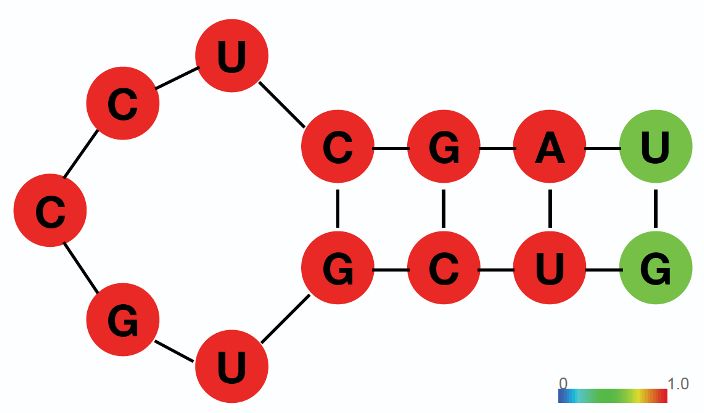
Research article Cell Biology Chromosomes and Gene Expression
A 18 C WT CLIPΔ1 CLIPΔ2
16 1.5 pri-miRNAs
14 1
Relative Expression Level
CLIPΔ (log2RPM)
12 0.5
10 0
pri-let-7f-2
8 pri-let-7e pri-let-7d pri-let-7g pri-let-7i
10 pri-miR-98
pre-miRNAs
6
4 5
2
2 4 6 8 10 12 14 16 18 0
WT (log2RPM)
pre-let-7d pre-let-7e pre-let-7g pre-let-7i pre-miR-98
B
Relative Expression Level
0 5 10 D Ago2 KO
WT CLIPΔ1 CLIPΔ2 Ago2 KO CLIPΔ
let-7a
Ago2
let-7b Relative Level 1 2.03 1.97 0 0
Flag-Trim71
let-7 targets
let-7c Relative Level 1 0.68 0.54 0.98 1.05
let-7 microRNAs
let-7d Lin28a
Relative Level 1 0.61 0.48 1.97 2.26
let-7e Nanog
Relative Level 1 0.97 1.15 0.93 0.95
let-7f
Oct4
let-7g Relative Level 1 0.96 1.08 1.00 1.05
Sox2
let-7i
Relative Level 1 1.01 1.05 1.47 1.53
miR-98 Gapdh
miR-290a E 1
Let-7 miRNAs’ targets
Cummulative Fraction
0.8 (n = 474)
miR-291 WT non-let-7 miRNAs’ targets
CLIPΔ1 0.6 (n = 2651)
miR-293 not targeted by miRNAs -8
CLIPΔ2 vs p = 5 x 10
(n = 8151)
miR-183 Ago2Δ 0.4 vs p = 0.007
-10
Ago2Δ/CLIPΔ vs p = 7 x 10
miR-30a 0.2
miR-16 0
-1 -0.75 -0.5 -0.25 0 0.25 0.5 0.75
Log2(CLIPΔ2/WT)
Figure 4. Loss of Trim71-mediated repression of Ago2 mRNA translation results in significant post-transcriptional increase of let-7 miRNAs. (A)
Comparison of global miRNA expression in WT and CLIPD mouse embryonic stem cells (mESCs). The results are the average of four independent small
RNA-seqs in the WT and the CLIPD mESCs. Blue dots: let-7 miRNAs; red dot: differentially expressed miRNAs; black dots: non-differentially expressed
miRNAs. (B) qRT-PCR on let-7 miRNAs and non-let-7 miRNAs. For each miRNA, the expression level in WT cells was set as 1 for relative comparison. U6
Figure 4 continued on next page
Liu et al. eLife 2021;10:e66288. DOI: https://doi.org/10.7554/eLife.66288 9 of 27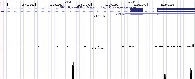
Research article                                                                         Cell Biology Chromosomes and Gene Expression

Figure 4 continued
RNA was used for normalization. (C) qRT-PCR on the let-7 pri-miRNAs and pre-miRNAs. For pri-miRNAs and pre-miRNAs, the expression level in the
WT cells was set as 1 for relative comparison. 18S rRNA and U6 RNA were used for pri-miRNA and pre-miRNA normalization, respectively. The results
in (B) and (C) are from three independent replicates. (D) Western blotting of Ago2, conserved let-7 targets, and non-let-7 targets. Gapdh was used for
normalization in calculating the relative expression levels. (E) Cumulative distributions of expression level changes of let-7 targets, miRNA targets
without let-7 binding sites, and mRNAs not targeted by miRNAs in WT and CLIPD mESCs.
The online version of this article includes the following figure supplement(s) for figure 4:
Figure supplement 1. The loss of Trim71-mediated repression of Ago2 mRNA translation does not alter global miRNA in mouse embryonic stem
cells (mESCs).

                                     elevated at these increasing amounts of Ago2 (Figure 5B and Figure 5—figure supplement 1A),
                                     indicating that increasing Ago2 level in mESCs results in a specific increase of let-7 miRNAs.
                                        In mammals, miRNAs can associate with all the four Ago proteins. To examine whether the
                                     increase of let-7 miRNAs is specific to Ago2, we increased the level of another Ago protein, Ago1,
                                     which is expressed in mESCs (Figure 5—figure supplement 1B and C). Similar to the results from
                                     Ago2, increasing Ago1 level also resulted in a specific post-transcriptional increase of let-7 miRNAs
                                     in mESCs (Figure 5—figure supplement 1D and E).
                                        Consistent with increased let-7 miRNAs, the two conserved let-7 targets, Trim71 and Lin28a, dis-
                                     played decreasing levels in the mESCs with increasing amounts of Ago2, while the levels of non-let-7
                                     targets, such as Nanog, Oct4, and Sox2, were not altered in these mESCs (Figure 5A). This Ago2-
                                     mediated increase of let-7 miRNAs occurred at the post-transcriptional level because the let-7 pri-
                                     miRNAs were not elevated in the mESCs with increasing amounts of Ago2 (Figure 5C).
                                        To evaluate whether increasing Ago2 results in stemness defects, we examined the ability to
                                     maintain stemness and the rate of exit pluripotency by the colony formation assay and the exit pluri-
                                     potency assay, respectively, in the mESCs with increasing amounts of Ago2. When the Ago2 level
                                     was elevated, the mESCs had decreased ability in maintaining stemness and increased rates in exit
                                     pluripotency (Figure 5D and E). Consistent with these, increased Ago2 resulted in a faster decline in
                                     the levels of the pluripotency factors (e.g., Nanog and Oct4) during EB formation (Figure 5F).
                                        Collectively, these results argued that the specific increase of let-7 miRNAs and the stemness
                                     defects in the CLIPD mESCs are caused by the increased Ago2.

                                     The increased let-7 miRNAs are bound and stabilized by Ago2 in mESCs
                                     Ago2 binds all miRNAs. Why does the elevation of Ago2 result in a specific increase of let-7 miRNAs
                                     in mESCs? A unique aspect of the pro-differentiation let-7 miRNAs in mESCs is that although genes
                                     encoding let-7 miRNAs are actively transcribed (Suh et al., 2004; Thomson et al., 2006), the let-7
                                     miRNA levels are low, indicating post-transcriptional regulations of let-7 miRNAs. Indeed, the proc-
                                     essing of let-7 pre-miRNAs are repressed by Lin28a in mESCs (Hagan et al., 2009; Heo et al.,
                                     2008). Since forming miRNPs (miRNA–protein complex) with Ago2 stabilizes mature miRNAs
                                     (Diederichs and Haber, 2007; Winter and Diederichs, 2011), we speculated that increased Ago2 in
                                     mESCs stabilizes the over-produced let-7 miRNAs that are degraded when Ago2 level is limiting. To
                                     test this, we performed two experiments.
                                         We first determined that the increased let-7 miRNAs are bound by Ago2 in the Ago2 elevated
                                     mESCs. We generated mESCs with a FLAG-tag at the N-terminus of the endogenous Ago2, which
                                     enabled specific isolation of both the endogenous Ago2 and the dox-induced FLAG-Ago2 via the
                                     anti-FLAG antibody (Figure 5—figure supplement 2A). RNA immunoprecipitation and qRT-PCR
                                     revealed that among the Ago2-bound miRNAs, let-7 miRNAs are specifically increased when Ago2
                                     is elevated (Figure 5—figure supplement 2B and C). Then, we measured RNA stability through acti-
                                     nomycin-D-mediated transcriptional shut-off (Figure 5—figure supplement 2D). We found that
                                     increased Ago2 did not alter the decay of let-7 pri-miRNAs, but specifically stabilized the let-7 miR-
                                     NAs (Figure 5—figure supplement 2E and F).
                                         Collectively, these observations not only indicate that the increased Ago2 directly binds and pro-
                                     tects let-7 miRNAs from degradation in mESCs, but also argue that besides the previously character-
                                     ized Lin28a-mediated inhibition of pre-miRNA processing, let-7 miRNAs are also repressed at the

Liu et al. eLife 2021;10:e66288. DOI: https://doi.org/10.7554/eLife.66288                                                                      10 of 27
Research article Cell Biology Chromosomes and Gene Expression
A WT + GFP WT + Ago2 B Relative miRNA Level
0 5 10 15
Dox (ng/ml): 0 10 20 30 40 50 0 10 20 30 40 50
Ago2 let-7a
Relative Level 1 0.99 0.93 0.81 0.91 0.90 1.08 1.18 2.36 3.59 4.32 4.67
let-7b
Trim71
let-7 let-7c
targets
Lin28a
let-7d
let-7 miRNAs
Nanog
let-7e
Oct4
Sox2 let-7f
Gapdh let-7g
C let-7i
Relative RNA Level
1.5
1 miR-98
0.5
miR-290a
0
pri-let-7f/
pri-let-7d pri-let-7e pri-let-7g pri-let-7i miR-293 Dox (ng/ml)
pri-miR-98
D 15%FBS + Lif 0
500 mESCs AP Staining miR-183
n.s. 7 days 10
Colonies (% of Total)
n.s. * 20
25 *
Undifferentiated
miR-182
30
20
40
15 miR-30a
50
10
5 miR-16
0
Dox (ng/ml) 0
30 50 0 30 50 F 15%FBS - Lif
mESCs EBs
WT + GFP WT + Ago2 30 rpm/bacterial plates
E 15%FBS - Lif 2i+lif WT + GFP WT + Ago2
1000 mESCs AP Staining
2 days 5 days Days: 0 3 5 0 3 5
Colonies (% of Total)
n.s. *
30 n.s. Nanog
Undifferentiated
*
20
Oct4
10
Sox2
0
Dox (ng/ml) 0 30 50 0 30 50
β-Tubulin
WT + GFP WT + Ago2
Figure 5. Increased Ago2 leads to significant increase of let-7 miRNAs and accelerated differentiation in mouse embryonic stem cells (mESCs). (A)
Western blotting in mESCs with dox-inducible expression of Ago2. Gapdh was used for normalization in calculating the relative expression of Ago2. (B)
Relative levels of miRNAs in mESCs with dox-inducible expression of Ago2. U6 RNA was used for normalization. (C) Relative levels of pri-miRNAs in
mESCs with dox-inducible expression of Ago2. 18S rRNA was used for normalization. In (B) and (C), the miRNA and pri-miRNA expression levels in
Figure 5 continued on next page
Liu et al. eLife 2021;10:e66288. DOI: https://doi.org/10.7554/eLife.66288 11 of 27Research article Cell Biology Chromosomes and Gene Expression
Figure 5 continued
mESCs without dox treatment were set as 1 for determining relative levels. The results are from four biological replicates. (D) Colony formation assay
for mESCs with dox-inducible expression of either GFP or Ago2. (E) Exit pluripotency assay for mESCs with dox-inducible expression of either GFP or
Ago2. The results in (D) and (E) represent the means (± SD) of six independent experiments. *p0.05) by the Student’s
t-test. (F) Western blot analysis on pluripotency factors during EB formation from the GFP-expressing mESCs and Ago2-expressing mESCs (treated with
50 ng/ml dox).
The online version of this article includes the following figure supplement(s) for figure 5:
Figure supplement 1. Increased Ago proteins in mouse embryonic stem cells (mESCs) result in a specific increase of let-7 miRNAs.
Figure supplement 2. The increased let-7 miRNAs are bound and stabilized by the elevated Ago2 in mouse embryonic stem cells (mESCs).
mature miRNA level in mESCs by the limiting Ago2 level (e.g., caused by the Trim71-mediated
repression of Ago2 mRNA translation).
The stemness defects in the CLIPD mESCs are dependent on the let-7
miRNAs
Let-7 miRNAs have a conserved function in promoting cell differentiation (Büssing et al., 2008;
Lee et al., 2016; Roush and Slack, 2008). To determine whether the stemness defects caused by
the increased Ago2 in the CLIPD mESCs is dependent on the let-7 miRNAs, we performed the fol-
lowing experiments.
First, we repressed let-7 miRNAs through generating stable mESC lines in which the expression
of exogenous Lin28a, Lin28b, or GFP can be induced by dox. Lin28a and Lin28b specifically repress
the maturation of let-7 miRNAs at the pre-miRNA and pri-miRNA levels, respectively (Hagan et al.,
2009; Heo et al., 2008; Piskounova et al., 2011). Lin28a, but not Lin28b, is highly expressed in
mESCs (Figure 6—figure supplement 1A). When either Lin28a or Lin28b was induced (Figure 6A),
the let-7 miRNAs in the CLIPD mESCs were reduced to levels similar to those in the WT mESCs with
no significant alterations in a group of non-let-7 miRNAs (Figure 6B). Consistent with this, the activi-
ties of let-7 miRNAs were also specifically repressed. The levels of the conserved let-7 targets,
Trim71 and Lin28a, increased in the CLIPD mESCs when the exogenous Lin28a or Lin28b was
expressed, but the levels of non-let-7 targets (e.g., Nanog, Oct4, and Sox2) were not altered
(Figure 6A). The ectopically expressed Lin28a or Lin28b alleviated the decreased ability to maintain
stemness and inhibited the increased rate of exit pluripotency in the CLIPD mESCs, as determined
by the colony formation assay and the exit pluripotency assay, respectively (Figure 6C and D). More-
over, western blotting revealed that the ectopically expressed Lin28a or Lin28b also inhibited the
rapid decrease of pluripotency factors during EB formation in the CLIPD mESCs (Figure 6E). These
results argued that the stemness defects in the CLIPD mESCs are dependent on the increased let-7
miRNAs.
One caveat of the Lin28 ectopic expression is the potential pleiotropic effects (reviewed in
Tsialikas and Romer-Seibert, 2015). To address this and to specifically determine whether let-7
miRNAs are responsible for the stemness defects, in a parallel experiment, we used locked nucleic
acid antisense oligonucleotides (LNA) targeting the conserved seed sequence of let-7 miRNAs to
attenuate their activities (Figure 6—figure supplement 1B). In the presence of the anti-let-7 LNA,
the CLIPD mESCs had an increase (threefold) in the ability of maintaining stemness as determined by
the colony formation assay (Figure 6F), indicating that the decreased stemness in the CLIPD mESCs
is dependent on let-7 miRNAs.
Altogether, the results from the ectopic expression of Lin28a/b and LNA-mediated inhibition of
let-7 miRNAs indicated that the stemness defects caused by the loss of Trim71-mediated repression
of Ago2 mRNA translation are dependent on the increased let-7 miRNAs.
Trim71 represses mRNA translation at post-initiation step(s) in mESCs
Trim71 can repress mRNA translation (Aeschimann et al., 2017; Loedige et al., 2013). Recent
observations, however, argued that Trim71 inhibits gene expression through mRNA degradation in
mESCs (Welte et al., 2019). To determine whether or not the Trim71-mediated translation repres-
sion is mRNA-specific in mESCs (e.g., Ago2 mRNA in Figure 2), we performed the tethering assay.
When tethered to a FLuc mRNA via the specific interaction between the bacteriophage lN
Liu et al. eLife 2021;10:e66288. DOI: https://doi.org/10.7554/eLife.66288 12 of 27Research article Cell Biology Chromosomes and Gene Expression
A GFP V5-Lin28a V5-Lin28b B Relative miRNA Level
0 2 4 6 8
WT CLIPΔ WT CLIPΔ WT CLIPΔ
let-7a
anti-V5 let-7b
let-7c
Lin28a
let-7 miRNAs
let-7d
let-7 Relative Level 1 0.63 1.26 1.32
targets let-7e
Trim71
let-7f
Relative Level 1 0.71 1.05 1.15 0.98 1.05
let-7g
Nanog
let-7i
Oct4 miR-98
Sox2 miR-290a
WT + GFP
miR-293
β-Tubulin WT + V5-Lin28a
miR-183 WT + V5-Lin28b
C miR-182 CLIPΔ + GFP
15%FBS + Lif
500 mESCs AP Staining WT CLIPΔ + V5-Lin28a
7 days miR-30a
CLIPΔ CLIPΔ + V5-Lin28b
50 miR-16
Colonies (% of Total)
n.s.
Undifferentiated
40 n.s. D 15%FBS - Lif 2i+lif
1000 mESCs AP Staining
30 * 50 2 days 5 days
n.s.
Colonies (% of Total)
20
Undifferentiated
40
n.s.
10 *
30
0
GFP V5-Lin28a V5-Lin28b 20
10
0
E GFP V5-Lin28a V5-Lin28b
WT CLIPΔ
F 15%FBS + Lif + LNA
b
b
a
a
28
28
28
28
500 mESCs AP Staining
in
in
in
in
-L
-L
-L
-L
7 days
P
P
V5
V5
GF
GF
V5
V5
Undifferentiated Colonies (% of Total)
Days: 0 5 0 5 0 5 0 5 0 5 0 5 0 10 20 30 40
Nanog
control WT
Oct4 LNA
CLIPΔ
Sox2 WT
anti-let-7 *
LNA
β-Tubulin CLIPΔ
Figure 6. Inhibiting let-7 miRNAs blocks the stemness defects caused by the loss of Trim71-mediated repression of Ago2 mRNA translation. (A)
Western blotting in WT and CLIPD mouse embryonic stem cells (mESCs) expressing GFP, V5-Lin28a, or Lin28b. Beta-tubulin was used for normalization
in determining the relative expression level of let-7 targets Lin28a and Trim71. (B) Relative levels of miRNAs. U6 RNA was used for normalization. The
results represent the means (± SD) of four biological replicates. (C) Colony formation assay for WT and CLIPD mESCs expressing GFP, V5-Lin28a, or
Figure 6 continued on next page
Liu et al. eLife 2021;10:e66288. DOI: https://doi.org/10.7554/eLife.66288 13 of 27Research article Cell Biology Chromosomes and Gene Expression
Figure 6 continued
Lin28b. (D) Exit pluripotency assay for WT and CLIPD mESCs expressing GFP, V5-Lin28a, or Lin28b. The results in (C) and (D) represent the means (± SD)
of six independent experiments. (E) Western blotting of pluripotency factors during EB formation at Day 0 and Day 5 of WT and CLIPD mESCs
expressing GFP, V5-Lin28a, or Lin28b. (F) Colony formation assay for WT and CLIPD mESCs cultured in the presence of 500 nM anti-let-7 LNA or a
control LNA. The results represent three independent experiments. *p0.05) by the Student’s t-test.
The online version of this article includes the following figure supplement(s) for figure 6:
Figure supplement 1. Inhibition of let-7 miRNAs in mouse embryonic stem cells (mESCs).
polypeptide and the BoxB RNA motif, Trim71 reduced the FLuc activity but not the FLuc mRNA level
(Figure 7A and B), indicating translation repression. This repression is specific, as Trim71 does not
repress the control mRNA without the BoxB sites (Figure 7B). Thus, repressing mRNA translation
can be a general mechanism for Trim71 in mESCs.
To determine how Trim71 represses translation in mESCs, we used bicistronic reporters contain-
ing either the HCV-IRES (internal ribosome entry site), which requires all the initiation factors except
eIF4G and eIF4E, or the CrPV-IRES, which only requires the 40S ribosomal subunit for initiating trans-
lation (Fraser and Doudna, 2007; Figure 7C). In these reporters, FLuc was produced by the canoni-
cal translation, and the RLuc was generated by the IRES-mediated translation. Tethering Trim71 to
either of these two reporter mRNAs led to a decrease of both FLuc and RLuc activities, while no
changes in mRNA levels (Figure 7D and E). This result indicated that Trim71 either interferes with
40S ribosomal subunit recruitment or inhibits an event at or after the 60S subunit joining step during
mRNA translation. Moreover, we found that Trim71-mediated translation repression does not
require 3’ end poly(A) tail. When tethered to a FLuc mRNA that is devoid of both poly(A) tail and
the poly(A) tail binding protein, Pabpc1 (Figure 7F; Zhang et al., 2017), Trim71 also specifically
represses the reporter mRNA translation (Figure 7G and H). Since poly(A) tail and Pabpc1 can pro-
mote mRNA translation at multiple steps, including 40S ribosomal subunit recruitment and the 60S
ribosomal subunit joining step during the initiation process (Kahvejian et al., 2005; Mangus et al.,
2003), these observations, combined with the result from the IRES reporters, argue that Trim71 reg-
ulates mRNA translation at a post-initiation step(s) in mESCs.
Discussion
Our data reveal that Trim71 maintains pluripotency in stem cells by specifically inhibiting the con-
served let-7 miRNAs through repressing Ago2 mRNA translation. These results not only provide
direct support for the conserved cytoplasmic bi-stable switch model (Ecsedi and Grosshans, 2013)
in stem-cell fate decisions, but also revealed that a new layer of regulation on the conserved pro-dif-
ferentiation let-7 miRNAs: repressing the mature miRNA by Ago2 availability. This regulation is criti-
cal for pluripotency in stem cells. Our findings raise several interesting aspects in stem cell biology
and RNA biology.
Ago2 and let-7 miRNAs
Previous studies indicate that overexpressing Ago2 in certain cells (e.g., 293T and NIH3T3) elevated
global miRNA levels by stabilizing mature miRNAs (Diederichs and Haber, 2007; Winter and Die-
derichs, 2011). Our results, however, indicate that elevated Ago2 specifically increased the let-7
miRNAs in mESCs at the post-transcriptional level. Although different cell types may contribute to
these different results, an important variable is the Ago2 level. Transfection-based assays tend to
result in high expression of exogenous genes. In our CLIPD mESCs or the WT mESCs with the dox-
induced exogenous Ago2, however, the Ago2 level increased approximately twofold or
maximally approximately fivefold, respectively, compared to that in the WT mESCs. This modest
increase is biologically relevant because the Trim71-mediated repression of Ago2 mRNA translation
only has an approximately twofold effect on the Ago2 level in mESCs (Figure 2). When
this approximately twofold repression on Ago2 was specifically disrupted, the mESCs displayed let-
7-miRNA-dependent defects in maintaining pluripotency (Figures 3 and 6). Thus, although previous
studies (Diederichs and Haber, 2007; Winter and Diederichs, 2011) and ours all indicate that
Ago2 is the limiting factor in forming the effector miRNPs, modulating Ago2 levels under
Liu et al. eLife 2021;10:e66288. DOI: https://doi.org/10.7554/eLife.66288 14 of 27Research article Cell Biology Chromosomes and Gene Expression
A B Normalized FLuc Activity Relative FLuc mRNA
Reporter: 0 0.5 1.0 0 0.5 1.0 1.5
FLuc
5BoxB pA Reporter Reporter n.s.
*
λN-GFP
Control:
λN-Trim71
FLuc
pA Control n.s. Control n.s.
C D Relative Bi-cistronic mRNA Level
0 0.5 1.0
FLuc RLuc
IRES 5BoxB pA
HCV n.s.
Reporter
IRES Required Initiation Factors λN-GFP
λN-Trim71
HCV eIF3, eIF2, Met tRNA, GTP, 40S
CrPV 40S only CrPV
n.s.
Reporter
E
* * * *
Relative Luciferase Activity
1.0
λN-GFP F Reporter:
λN-Trim71
MALAT1
FLuc
3’-end
0.5
5BoxB
Control:
MALAT1
0
FLuc
3’-end
FLuc RLuc FLuc RLuc
HCV-IRES CrPV-IRES
G H
Relative FLuc mRNA Level Normalized Luciferase Activity
0 0.5 1.0 0 0.5 1.0
Poly(A)- n.s. Poly(A)-
Reporter Reporter *
λN-GFP
λN-GFP
λN-Trim71
λN-Trim71
Poly(A)- Poly(A)- n.s.
n.s.
Control Control
Figure 7. Trim71 represses mRNA translation at post-initiation step(s) in mouse embryonic stem cells (mESCs). (A) FLuc reporters for the tethering
assay. (B) The FLuc activity and mRNA level determined in the tethering assay. (C) The IRES-containing bicistronic reporters. (D) mRNA levels from the
IRES-containing reporters. (E) Luciferase activities from the IRES-containing reporters. (F) The poly(A) minus FLuc reporters. (G) mRNA levels from the
Figure 7 continued on next page
Liu et al. eLife 2021;10:e66288. DOI: https://doi.org/10.7554/eLife.66288 15 of 27Research article Cell Biology Chromosomes and Gene Expression
Figure 7 continued
poly(A) minus reporters. (H) FLuc activities from the poly(A) minus reporters. The results represent the means (± SD) of three independent experiments.
*p0.05) by the Student’s t-test.
biologically relevant settings is likely to result in changes of specific miRNAs, such as the let-7 miR-
NAs. Ago2 and let-7 miRNAs levels are frequently dysregulated in cancers (Ye et al., 2015). We
speculate that modulating Ago2 levels may also regulate tumorigenesis by altering the activities of
specific miRNAs (e.g., the tumor-suppressive let-7 miRNAs).
Two inter-connected layers of regulations on let-7 miRNAs in stem cells
Let-7 is a conserved pro-differentiation miRNA that is abundantly expressed in differentiated cells
(Roush and Slack, 2008). In stem cells, although the genes encoding let-7 miRNAs are actively tran-
scribed, the levels of mature let-7 miRNAs are low (Suh et al., 2004; Thomson et al., 2006), indicat-
ing post-transcriptional inhibition of let-7 miRNAs. Previous studies characterized that the conserved
RBPs Lin28a and Lin28b inhibit let-7 miRNAs’ maturation at the pre- and pri-miRNA levels
(Tsialikas and Romer-Seibert, 2015). Here, we revealed an additional layer of regulation of let-7
miRNAs at the mature miRNA level by Ago2 availability.
Interestingly, these two layers of regulation on let-7 miRNAs are intertwined through Lin28a, a
conserved let-7 target that promotes let-7 pre-miRNA degradation, in mESCs. When the let-7 miR-
NAs were elevated by the increased Ago2, in either the CLIPD mESCs or the WT with induced
Ago2, there was a corresponding decrease of the endogenous Lin28a (Figure 4D and Figure 5A).
Moreover, the decreased Lin28a resulted in a compromise in the degradation of let-7 pre-miRNAs,
as indicated by twofold to threefold increase of let-7 pre-miRNAs in the CLIPD mESCs (Figure 4C).
This compromise explains the significant increase of mature let-7 miRNAs upon a modest increase of
Ago2 (Figure 4 and Figure 5): the initial increase of the let-7 miRNAs caused by the elevated Ago2
decreases Lin28a and alleviates Lin28a-mediated inhibition on the maturation of let-7 miRNAs,
resulting in more let-7 pre-miRNAs become mature let-7 miRNAs. This positive regulatory loop
amplifies let-7 miRNAs and makes the pro-differentiation let-7 miRNAs sensitive to Ago2 levels in
stem cells.
The two inter-connected layers of inhibition on the potent pro-differentiation let-7 miRNAs are
important to maintaining stemness. During self-renewal of mESCs, although genes encoding let-7
miRNAs are transcribed, the production of these potent pro-differentiation miRNAs is inhibited at
both the pre-miRNA level by Lin28a and the mature miRNA level by Ago2. These two layers of regu-
lation may work together to ensure low levels of let-7 miRNPs in stem cells. Considering that let-7
miRNAs are repressed in cancers (Büssing et al., 2008), and Lin28a or Lin28b are highly expressed
in many cancers (Piskounova et al., 2011), we speculate that the Ago2-mediated regulation of let-7
miRNPs may also be employed by cancer cells for their dysregulated proliferation.
Trim71 and Ago2
The Trim71-mediated downregulation of Ago2 occurs through repressing Ago2 mRNA translation in
mESCs (Figure 2), but not the proposed E3-ligase-mediated protein degradation (Rybak et al.,
2009). This result is consistent with previous observations that the potential E3 ligase activity is not
required for Trim71-mediated gene regulation in vivo (Chen et al., 2012; Welte et al., 2019). In
human cells, TRIM71 level negatively correlates with AGO2 level (Chen et al., 2013). Thus, we spec-
ulate that the Trim71-mediated repression of Ago2 mRNA translation is conserved between mouse
and human. Although the Trim71-binding region in the 3’UTR of mouse Ago2 mRNA is not con-
served in sequence, Trim71 recognizes its RNA targets via structural features but not sequence
motifs (Figure 1G; Kumari et al., 2018; Welte et al., 2019). Both human and mouse Ago2 have
long 3’UTRs (5.2 kb and 11.8 kb, respectively) with multiple in silico predicted hairpins that Trim71
can bind. However, our CLIP-seq data indicated that Trim71 only binds one such predicted hairpin in
mESCs (Figure 1I), suggesting that either not all the predicted hairpins form in vivo or Trim71 uses
additional features for target recognition. Trim71 can both repress mRNA translation and induce
mRNA degradation (Aeschimann et al., 2017; Loedige et al., 2013; Welte et al., 2019;
Liu et al. eLife 2021;10:e66288. DOI: https://doi.org/10.7554/eLife.66288 16 of 27Research article Cell Biology Chromosomes and Gene Expression
Worringer et al., 2014). Thus, it will be interesting to determine what features determine whether
Trim71 represses translation or destabilizes mRNA.
A new approach for functional characterization of RBPs
Trim71 is essential for animal development (Cuevas et al., 2015; Ecsedi and Grosshans, 2013). Pre-
vious studies, however, reported no stemness defects in Trim71 knockout mESCs (Mitschka et al.,
2015; Welte et al., 2019). Moreover, Ago2 level was not altered in Trim71 knockdown or knockout
mESCs (Chang et al., 2012; Welte et al., 2019), which we also observed in our mESCs (Figure 2—
figure supplement 1A). In contrast to these results, our study indicates that Trim71 plays a critical
role in regulating pluripotency in mESCs through repressing Ago2 mRNA translation. How to recon-
cile these results?
The main difference is the number of disrupted Trim71:mRNA interactions. In the CLIPD mESCs,
we specifically inhibited one: the Trim71:Ago2–mRNA interaction; while in Trim71 knockout/knock-
down mESCs, hundreds of Trim71:mRNA interactions and potential Trim71-mediated protein–pro-
tein interactions are lost/attenuated. The phenotype of the Trim71 knockout mESCs is the functional
additions of all the disrupted interactions. If Trim71’s target mRNAs contain both positive and nega-
tive regulators of pluripotency, knocking out Trim71 may result in no phenotypical changes. Interest-
ingly, our Trim71 CLIP-seq indicated that besides Ago2 mRNA, Trim71 also binds mRNAs encoding
pluripotency factors, such as Nanog (Supplementary file 1). Thus, it is possible that in the Trim71
knockout mESCs, the defects caused by the increased Ago2 may be compensated by an increased
Nanog from the lost Trim71:Nanog–mRNA interaction. Alternatively, Ago2 protein level does not
change in the Trim71 knockout/knockdown mESCs due to combinatory results of the disrupted
Trim71:Ago2 mRNA interaction and secondary effects from other lost Trim71:mRNA interactions,
leading to no stemness defects in the knockout mESCs. Thus, an important caveat in interpreting
results from the knockout studies on RBPs is that no phenotypical changes does not necessarily
mean that the target RBP is not functionally significant. Then, how to effectively characterize biologi-
cal functions of RBPs?
Thanks to the wide applications of CLIP-based methods, many RBPs’ target RNAs and the bind-
ing regions in these RNAs are being well characterized. In addition to the loss-of-function methods,
we believe specific inhibition of candidate RBP:mRNA interaction(s) via mutating/deleting binding
sites on the target mRNA(s) will reveal more exciting roles of RBPs and significant RBP:mRNA inter-
actions under many biological processes.
Materials and methods
Key resources table
Reagent type
(species) or resource Designation Source or reference Identifiers Additional information
Antibody Mouse monoclonal Sigma-Aldrich Cat# F1804 WB (1:5000)
anti-FLAG M2 IP
Antibody Normal mouse IgG Santa Cruz Cat# sc-2025 IP
Biotechnology
Antibody Mouse monoclonal Santa Cruz Cat# sc-32233 WB (1:5000)
anti-GAPDH (6C5) Biotechnology
Antibody Rabbit monoclonal Selleckchem Cat# A5032 WB (1:5000)
anti-beta-Tubulin
Antibody Rabbit monoclonal Cell Signaling Cat# 5053 WB (1:1000)
anti-Ago1 (D84G10) Technology
Antibody Rabbit monoclonal Cell Signaling Cat# 2897 WB (1:1000)
anti-Ago2 (C34C6) Technology
Antibody Mouse monoclonal BD Transduction Cat# 611202 WB (1:5000)
anti-Oct-4 Laboratories
Antibody Rabbit monoclonal Cell Signaling Cat# 8822 WB (1:3000)
anti-Nanog (D2A3) Technology
Continued on next page
Liu et al. eLife 2021;10:e66288. DOI: https://doi.org/10.7554/eLife.66288 17 of 27You can also read