Human primary epidermal organoids enable modeling of dermatophyte infections
←
→
Page content transcription
If your browser does not render page correctly, please read the page content below
Wang et al. Cell Death and Disease (2021)12:35
                                    https://doi.org/10.1038/s41419-020-03330-y                                                                                                             Cell Death & Disease

                                     ARTICLE                                                                                                                                                    Open Access

                                    Human primary epidermal organoids enable
                                    modeling of dermatophyte infections
                                    Xuan Wang1,2, Shuyong Wang3, Baolin Guo2, Yuxin Su1,2, Zuolong Tan2, Mingyang Chang2, Jinmei Diao1, Yi Zhao4 and
                                    Yunfang Wang1,2

                                      Abstract
                                      Technology of generating human epidermal derivatives with physiological relevance to in vivo epidermis is
                                      continuously investigated for improving their effects on modeling of human natural dermatological status in basic and
                                      clinical studies. Here, we report a method of robust establishment and expansion of human primary epidermal
                                      organoids (hPEOs) under a chemically defined condition. hPEOs reconstruct morphological, molecular, and functional
                                      features of human epidermis and can expand for 6 weeks. Remarkably, hPEOs are permissive for dermatophyte
                                      infections caused by Trichophyton Rubrum (T. rubrum). The T. rubrum infections on hPEOs reflect many aspects of
                                      known clinical pathological reactions and reveal that the repression on IL-1 signaling may contribute to chronic and
                                      recurrent infections with the slight inflammation caused by T. rubrum in human skin. Thus, our present study provides
                                      a new insight into the pathogenesis of T. rubrum infections and indicates that hPEOs are a potential ex vivo model for
                                      both basic studies of skin diseases and clinical studies of testing potential antifungal drugs.
                  1234567890():,;
1234567890():,;
                  1234567890():,;
1234567890():,;

                                    Introduction                                                                                        xenobiotic substances that can bring unfavorable impacts
                                      The mammalian skin epidermis comprises a highly                                                   on clinical safety and can also interfere with the
                                    specialized and multi-layered epithelium that includes the                                          mechanistic studies on skin diseases. Hence, safer and
                                    stratum basale, stratum spinosum, stratum granulosum,                                               more-effective culture systems according to clinical set-
                                    and stratum corneum, which protects the body against                                                tings have been investigated for decades8. Until recently,
                                    harmful factors1. In the past few decades, classical two-                                           the chemically defined and xeno-free culture methods
                                    dimensional (2D) culture methods2–4 have been used as                                               have been successfully developed for long-term culture of
                                    invaluable tools to supply primary epidermal cells for                                              human epidermal cells with the use of small molecules9 or
                                    studies on skin stem cell biology as well as for stem cell-                                         laminin-based matrices10. However, these expanding
                                    based therapies of extensive and severe skin injuries5–7.                                           epidermal cells in 2D format cannot recapitulate the
                                    However, these 2D culture techniques for human epi-                                                 in vivo cellular composition and the physiological struc-
                                    dermal cells rely on the presence of feeder layers of                                               ture of human epidermis. Over the past decades, several
                                    murine fibroblasts (3T3 cells) and media supplemented                                                protocols have been developed to efficiently generate
                                    with fetal bovine serum or bovine pituitary extract2–4.                                             three-dimensional (3D) skin equivalent with physiological
                                    These conditions introduce unidentified variables or                                                 relevance to human skin11, especially the reconstructed
                                                                                                                                        multi-layered epidermis under the air–liquid interphase
                                                                                                                                        (ALI) condition12–14, which have provided elaborate skin
                                    Correspondence: Yunfang Wang (wangyf1972@gmail.com)
                                    1                                                                                                   3D models for skin research and regenerative medicine.
                                     Translational Medicine Research Center, Beijing Tsinghua Chang Gung
                                    Hospital, Beijing 102218, China                                                                     However, the reconstructed epidermis using ALI method
                                    2
                                     Department of Stem Cell and Regenerative Medicine, Beijing Institute of                            have a limited lifespan and passage ability, which may
                                    Health Service and Transfusion Medicine, Beijing 100850, China
                                                                                                                                        limit large-scale application of this model system for
                                    Full list of author information is available at the end of the article
                                    These authors contributed equally: Xuan Wang, Shuyong Wang, Baolin Guo                              industry or clinical programs.
                                    Edited by E. Candi

                                    © The Author(s) 2021
                                                        Open Access This article is licensed under a Creative Commons Attribution 4.0 International License, which permits use, sharing, adaptation, distribution and reproduction
                                                        in any medium or format, as long as you give appropriate credit to the original author(s) and the source, provide a link to the Creative Commons license, and indicate if
                                    changes were made. The images or other third party material in this article are included in the article’s Creative Commons license, unless indicated otherwise in a credit line to the material. If
                                    material is not included in the article’s Creative Commons license and your intended use is not permitted by statutory regulation or exceeds the permitted use, you will need to obtain
                                    permission directly from the copyright holder. To view a copy of this license, visit http://creativecommons.org/licenses/by/4.0/.

                                    Official journal of the Cell Death Differentiation Association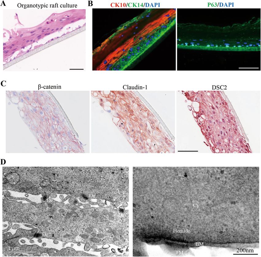
Wang et al. Cell Death and Disease (2021)12:35 Page 2 of 17
Recently, the established technologies for 3D organoid and may led to decreased cell viability and lowered viable
cultures have enabled the robust generations of various cell yields32. In addition, there also existed reports of fast
mini-organs that resemble their tissue-of origin, and have methods to isolate keratinocytes7,33. However, all of these
also allowed for the long-term expansions of these mini- previously reported methods have specifically applied
organs under the conditions of chemically defined sys- scenarios. For instance, the traditional method usually
tems15,16. These organoid-associated techniques show works well for neonatal tissues, but it becomes very dif-
many great advantages for generating model systems of ficult to use when used to isolate cells from adult tissues32.
tissues including small intestine17, stomach18, brain19, The reported fast method used in RECELL system is
liver20, kidney21, and esophagus22,23. Likewise, the orga- suitable for isolating epidermal cells from the thin split-
noid technology provides an opportunity for generating thickness cutaneous biopsy (0.2–0.3 mm) harvested from
3D model system of skin tissue. Notably, we and others an uninvolved area using a dermatome33. And other
have successfully established several different type of skin- trypsin-ethylenediaminetetraacetic acid (EDTA)-based
associated organoids, such as mouse and human hair- fast method usually produces a heterogeneous cell
bearing organoids24,25, canine and mouse epidermal population, including keratinocytes and fibroblasts7. It is
organoids26,27, mouse sweat gland organoids28, and unclear whether these methods function well enough to
sebaceous gland organoids29. Until now, however, a efficiently isolate epidermal cells from human skin spe-
method for the efficient establishment and effective cul- cimens with irregular thickness and size from various
ture of human epidermal organoids as a model of human clinic scenario, such as the specimen from circumcision,
epidermis tissue has not yet been reported. In addition, abdominal operation or plastic surgery. We therefore
there is still no report of human epidermal organoids started to develop a fast and robust protocol via an
applied in the studies on skin diseases. organized procedure with the sequential usages of various
Here, we report the extensive and reproducible estab- enzymes for specifically digesting skin tissue extracellular
lishment of human primary epidermal organoids (hPEOs) matrix protein in order to isolate pure human epidermal
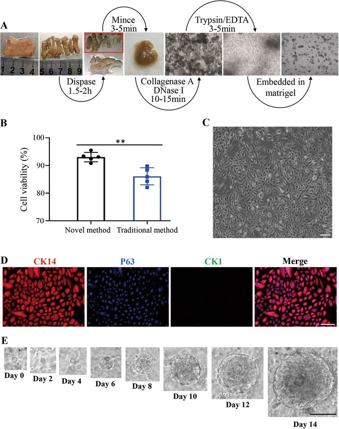
from human interfollicular epidermis. hPEOs are cells from foreskin tissues within 3 h (Fig. 1A). Based on
expandable and contain both proliferative epidermal our novel method with significant time saving, the cell
stem/progenitor cells and differentiated epidermal cells. viability was improved with statistical significance, when
They have a highly organized structure resembling human compared with the reported traditional method32 (Fig. 1B
epidermis. Remarkably, hPEOs can be established from and Supplementary Fig. 1). In 2D culture at day 7, the
single Integrin α6high cells through clonal expansion and isolated human epidermal cells displayed a typical
cell differentiation. For proving their applications in cobblestone-like morphology under the condition of a
analyses of skin infectious diseases, hPEOs were used to commercially available medium (EpiLife medium) with-
model T. rubrum infections that are the most prevalent out support from feeder cells (Fig. 1C). Most of the
dermatophyte to cause human nail and skin infections attached cells were positive for basal stem/progenitor cell
worldwide30,31. The T. rubrum infections on hPEOs markers, CK14 and P63, and negative for suprabasal cell
reflected many aspects of known clinical pathological differentiation marker CK1 (Fig. 1D). Taken together, all
reactions. Furthermore, we found that T. rubrum trig- of above results indicated that our newly established
gered the upregulated expression of the anti- isolation protocol had the advantage of improved effi-
inflammatory factor, IL-1RN (IL-1 receptor antagonists) ciency for isolation of viable epidermal cell populations
at transcriptional, translational, and secretion levels in the containing basal stem/progenitor cells.
infected cells. These induced antiinflammation effects by Next, we tried to use the freshly isolated human primary
T. rubrum may be responsible for its high degree of epidermal cells to establish 3D cultures of hPEOs. Because
adaptation to human skin and for its tendency to cause the inter-species differences exist between human skin
chronic infections with slight inflammation. Thus, our and canine or mouse skin34, all of previously reported
study provides the first-hand evidences for application of culture methods for mouse and canine epidermal orga-
hPEOs as a novel system to model skin infectious diseases noids26–28 could not be satisfactorily used for human
and also provides a theoretical basis for antifungal treat- epidermal organoid culture. Therefore, a new culture
ments in future. method with required components for culturing hPEOs
was totally established. Candidate components were
Results identified from five primary categories consisting of 10
Establishment of hPEOs as ex vivo human epidermal factors: N-acetylcysteine (NAC, here abbreviated as Na),
derivatives N2 (N), B27 (B), EGF (E), FGF-10 (F), Noggin (No), R-
Previously, the commonly used protocol (or traditional Spondin1 (R), Wnt3a (W), A83-01 (A), and Forskolin (Fs),
method) to isolate epidermal cells includes an overnight which were collectively referred to as NaNBEFNoRWAFs.
procedure for tissue digestion, which is time-consuming The major reasons to select these factors were based on
Official journal of the Cell Death Differentiation Association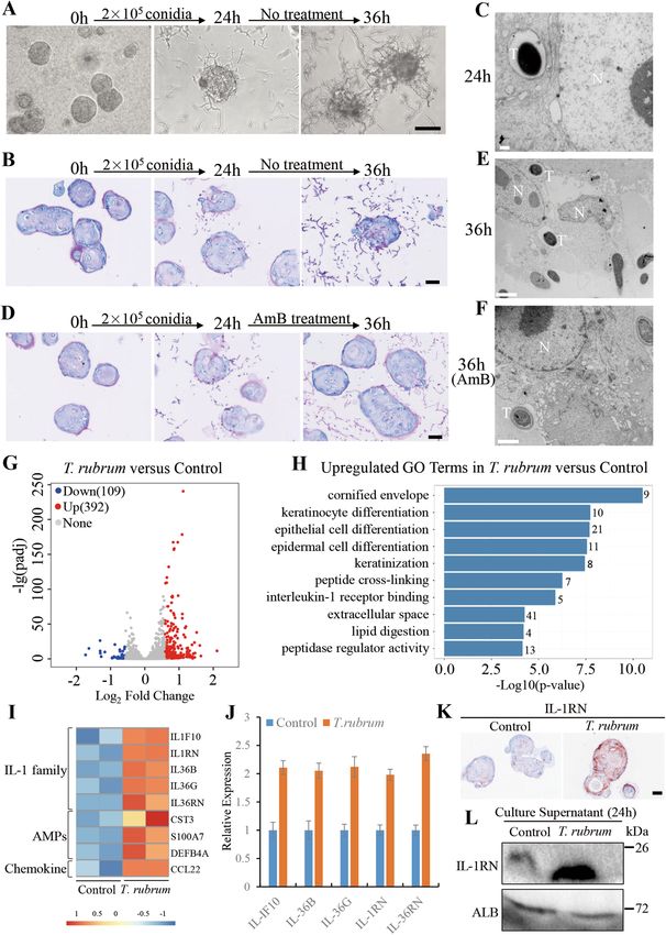
Wang et al. Cell Death and Disease (2021)12:35                                                                                             Page 3 of 17

  Fig. 1 A novel human epidermal cells isolation system and growth of hPEOs. A Schematic representing isolation of epidermal cells from human
  foreskin tissue. B Comparison of the viability of cells derived from the traditional method and the novel method. Results are the mean ± SD from five
  independent repeated experiments. n.s., not significant (p > 0.05), *p < 0.05, **p < 0.01, ***p < 0.001. C Morphology of isolated human epidermal cells
  after attachment in a low-calcium, serum-free medium (EpiLife). D Immunofluorescence analysis of epidermal progenitor cell markers, CK14 and P63,
  in the attached epidermal cells. E Representative serial images of hPEOs growing at the indicated time points in the NaNBEFNoRWAFs medium. SD,
  standard deviation. Scale bar: 50 µm (C, D), 100 µm (E).

Official journal of the Cell Death Differentiation Association
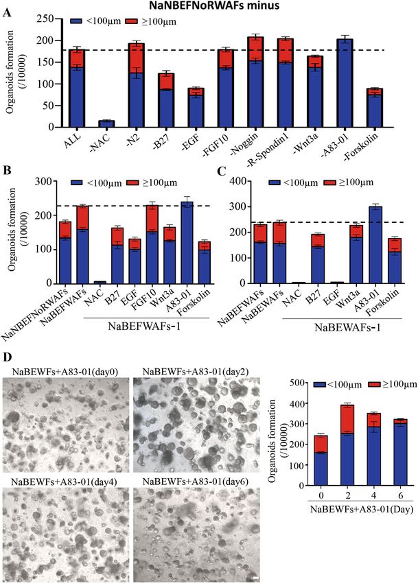
Wang et al. Cell Death and Disease (2021)12:35                                                                                      Page 4 of 17

  Fig. 2 Optimization of hPEOs cultures. A The numbers of hPEOs after removal of individual factors from the NaNBEFNoRWAFs pool. B, C The
  numbers of hPEOs were counted after the removal of individual factors from the pool of NaBEFWAFs (B) and NaBEWAFs (C). D Representative images
  of hPEOs generated by various timings of A83-01 treatment in NaBEWFs medium. The hPEOs were counted on day 10. Results are the mean ± SD
  from three replicates from three independent repeated screening experiments. All screening experiments were performed with keratinocytes from
  three different donors. Scale bar: 100 µm (D).

Official journal of the Cell Death Differentiation Association
Wang et al. Cell Death and Disease (2021)12:35                                                                  Page 5 of 17

our specially designed principles: 1. antioxidants have         condition of treatment with NaBEFWAFs, both the total
been reported to protect epidermal stem cells from aging        number of hPEOs and the ratio of large-size hPEOs were
and to maintain their stemness phenotype35. N-acet-             increased, as compared to those hPEOs under the con-
ylcysteine was chosen because of its antioxidant activity       dition of treatment with NaNBEFNoRWAFs (Fig. 2B).
and its inhibitory effects on apoptosis of epithelial cells36   Two additional rounds of removal test finally proved that
and on epithelial-mesenchymal transition37; 2. the serum-       the collected candidates of NAC, B27, EGF, Wnt3a, A83-
free supplements, both N2 and B27, were selected for            01, and Forskolin were essential for hPEOs formation (Fig.
their roles to support organoid cultures without addition       2B, C and Supplementary Fig. 3 and 4). Particularly,
of serum38; 3. the activation of morphogen signaling            removal of A83-01 increased total number of hPEOs, but
pathways such as epidermal growth factor receptor, ker-         mainly generated the small-size hPEOs in all dropout
atinocyte growth factor receptor, and Wnt/β-catenin             experiments. Therefore, we hypothesized whether A83-01
signaling have been demonstrated to promote epidermal           could have an effect on the generation of hPEOs in a time-
morphogenesis and proliferation39–41. The relevant acti-        dependent manner. To determine this, A83-01 was added
vators of these pathways like EGF, FGF-10, Wnt3a, and R-        into the pool of NaBEWFs on Days 0, 2, 4, and 6,
Spondin 1 were therefore selected; 4. dual SMAD sig-            respectively. Significantly, when A83-01 was added on
naling inhibition enabled expansion of epidermal stem           Day 2, the combined pool induced the maximum effi-
cells in 2D culture9, therefore A83-01 (TGFβ inhibitor)         ciency on increases of numbers for both total and large-
and Noggin (BMP antagonist) were included. 5. the               size hPEOs, though still with some heterogeneity in size
increased level of cellular cAMP (cyclic adenosine              between organoids (Fig. 2D). We, therefore, kept on
monophosphate) promoted the proliferation of epidermal          employing this optimized combination to generate hPEOs
cells42. Forskolin is capable of up-regulating intracellular    for the subsequent studies.
cAMP by activating adenylyl cyclase and so was selected.
Under the specially designed conditions with the ten            hPEOs maintained normal expressions of epidermis-
factors, the freshly isolated human epidermal cells that        specific markers
were embedded in basement membrane extract (BME) or                Next, the histological characteristics of hPEOs were
matrigel proliferated rapidly, and formed solid organoids       analyzed to compare with those of human primary skin
~200 µm in diameter with a concentric cell arrangement          tissue. Results of Hematoxylin and Eosin (H&E) staining
within ~14 days (Fig. 1E).                                      indicated that hPEOs were morphologically similar to
                                                                normal skin epidermis after 10 days in culture with small
Optimization of culture conditions for hPEOs                    basal-like cells in contact with the extracellular matrix;
   The results from above experiments suggested the clues       large flat suprabasal-like cells in the interior; and har-
for optimizing the combination of small molecules or            dened keratinized material in the center of organoid
factors for generation and culture of hPEOs. Therefore,         structures (Fig. 3A). Afterwards, the cellular composition
each of above-selected candidate was further analyzed           of hPEOs was compared with that of human primary skin
individually and dynamically. During our studies, we            tissue by using both basal cell and suprabasal cell-specific
removed factors one by one from the pool of NaNBEF-             markers. Results indicated that the interior of hPEOs
NoRWAFs to test its effect individually. Removal of each        consisted of the differentiated cells shown by CK1+
of N2 (N), Noggin (No), or R-Spondin 1 (R) resulted in          immunostaining, as well as the abundant keratinization
the increased number of total hPEOs and large-size              evidenced by Involucrin+ immunostaining (Fig. 3B). The
hPEOs (≥100 µm in diameter). Removal of A83-01 also             outermost cell layer of hPEOs was positive for all analyzed
increased the total number but decreased the size for all of    stem/progenitor cell markers (CK5, CK14, integrin α6
generated hPEOs (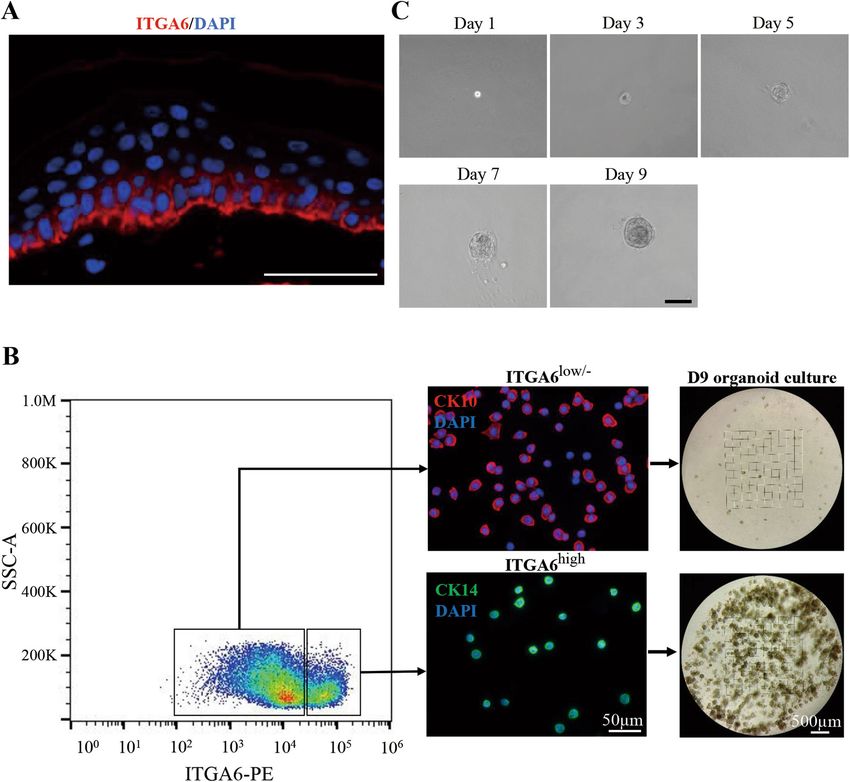
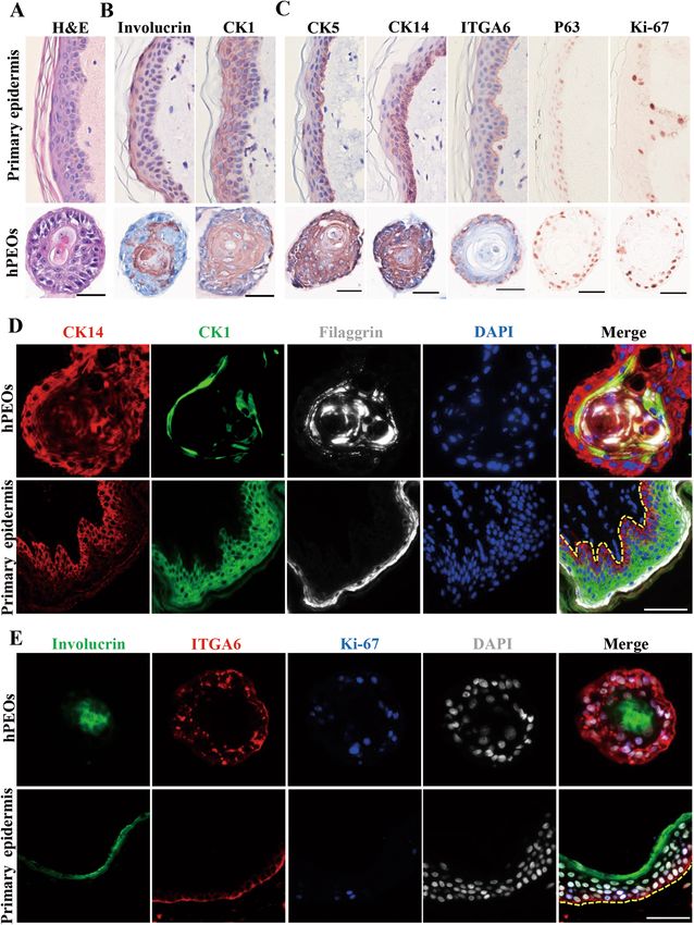
Wang et al. Cell Death and Disease (2021)12:35                                                                                        Page 6 of 17

  Fig. 3 Marker expression of hPEOs. A H&E staining of the sections of hPEOs and human primary epidermis tissue. B, C Immunohistochemical
  staining for epidermal differentiation (B), stem cell-associated (C), markers of sections of hPEOs and human primary epidermis tissue. D, E Co-
  immunostaining analysis of various basal and differentiation markers in hPEOs and human primary epidermis tissue. Dotted line marks the
  epidermis–dermis junction. Scale bar: 50 µm.

Official journal of the Cell Death Differentiation Association
Wang et al. Cell Death and Disease (2021)12:35 Page 7 of 17
collagen, and type VII collagen) were found in the base of hPEOs under present established conditions remained a
hPEOs (Supplementary Fig. 5C). As expected, in all possible limitation. During the five continuous passages
hPEOs, there were no detectable expressions for mela- ex vivo, the chromosomal stability was also evaluated by
nocyte marker, Gp100, Langerhans cell marker, CD1a, performing karyotype analysis, which showed that hPEOs
vascular cell marker, CD31, and the marker for fibro- maintained a normal karyotype continuously (Fig. 5D).
blasts, Vimentin (Supplementary Fig. 5D). Together, these Together, these results suggested that the present culture
data demonstrated that hPEOs, as the human primary conditions for hPEOs were reliable and could be opti-
skin tissue-derived epidermal organoids, have a well-dif- mized for additional expansion after further
ferentiated, keratinized stratified squamous epithelium. improvements.
hPEOs primarily originated from basal cells Reconstruction of pluri-stratified epidermis equivalents
Furthermore, we pursued the study to investigate from hPEOs
whether some special subpopulations of human epidermal Mammalian skin epidermis contains the basal stem/
cells could act as the cell origin for hPEOs. In fact, progenitors that can give rise to all of the differentiated
mammalian skin epidermis in vivo contain the hetero- stratified layers during homeostasis46. Here, we tested
geneous populations of epidermal cells including basal whether the hPEOs containing basal cells could have the
progenitors, differentiated suprabasal cells and granular capability to generate reconstructed human epidermis
cells. In line with previous finding45, our result of in situ in vitro. Previously, the epidermis equivalents as organo-
immunostaining showed that ITGA6, as a surface marker typic rafts were all established from cultured epidermal
of stem/progenitor cells in human skin epidermis, was basal cells in vitro under the ALI conditions47. Here, our
located primarily in their basal layer (Fig. 4A). Therefore, hPEOs were enzymatically dissociated into single cells
ITGA6 was further employed for differentially sorting the and exposed to ALI differentiation for 10–14 days. Results
freshly isolated human primary epidermal cells. Results of of H&E staining showed the generation of a pluri-
cell sorting analyses indicated that the ITGA6high cells stratified epithelium resembling primary epidermis (Fig.
were primarily the CK14+ cells with a small, round shape, 6A). Results of immunostaining further showed that dif-
displaying the properties of basal progenitors, whereas the ferentiation markers were normally expressed and located
ITGA6low/− cells were mainly the CK10+ cells with dif- in layers. CK14+, P63+ cells were seen in the basal layer,
ferentiation characters of a big and flat morphology (Fig. whereas CK10+ cells were seen in the suprabasal layers
4B). After fluorescence-activated cell sorting (FACS)- (Fig. 6B). Moreover, an integrated network of cellular
based cell sorting, the isolated ITGA6high cells or junctions is essential to support a stable environmental
ITGA6 low/− cells were cultured under the same condition barrier and maintains the normal polarization of epi-
for hPEOs formation and maintenance. Results indicated dermis48. Notably, our results from immunostaining and
that the ITGA6high cells developed into hPEOs much ultrastructural analysis demonstrated that hPEO-derived
more efficiently than the ITGA6low/− cells (Fig. 4B). epidermis equivalent possessed the correct distribution of
Remarkably, after FACS isolation, a single ITGA6high cell adhesion junctions (β-catenin), tight junction (Claudin-1),
was fully competent to develop into a hPEO within 9 days desmosomes (DSC2), and hemidesmosomes (Fig. 6C, D).
(Fig. 4C). Together, these results strongly supported that Together, these results suggested that hPEOs have the
hPEOs could originate primarily from the basal cells of capacity to generate the stratified epithelium with the
human epidermis. appropriate cellular architecture and to express markers
resembling primary epidermis under ALI conditions.
The hPEOs showed moderate proliferative potential and
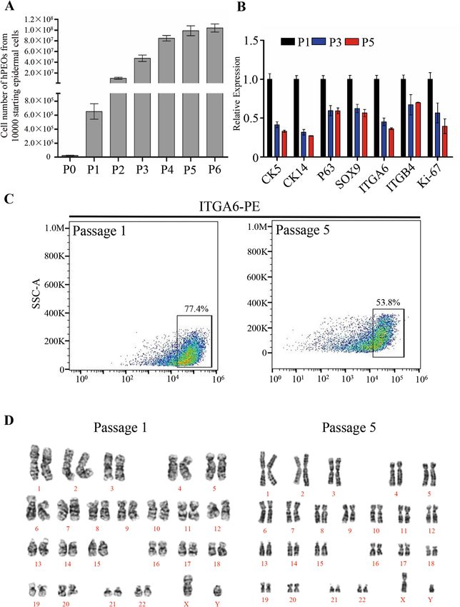
maintained genetic stability during passaging The hPEOs enabled for modeling dermatophyte infections
Next, additional characteristics of hPEOs were further To further evaluate the potential application of hPEOs
determined during long-term cultures. Remarkably, our in studies of clinical dermatology, their capacities of host
hPEOs were found to expand for up to six passages, and responses were tested after infections of T. rubrum. For
they reached to a total amplification of 10,000-fold within inducing the infections, conidia of T. rubrum were used to
6 weeks (Fig. 5A). However, over sequential passaging, inoculate hPEOs prepared in Matrigel. The conidia ger-
results of serial qRT–PCR analyses showed that the minated gradually after inoculation, and a fraction of
expression levels of basal cell markers (CK5, CK14, P63, hyphal growth was observed successfully. Remarkably,
SOX9, ITGA6, and ITGB4) decreased, and the level of some of hyphae adhered to hPEOs at 24 h and the
proliferating cell marker Ki-67 was also downregulated extensive hyphae covered hPEOs at 36 h’ later (Fig. 7A).
(Fig. 5B). Reduction of the proportion of ITGA6high cells At the time of 24 h, results of PAS staining on the sections
was also observed during continuous passaging (Fig. 5C). of infected hPEOs showed that some of hyphae invaded
Therefore, these results explained that the proliferation of from the basal cells of hPEOs and penetrated into the
Official journal of the Cell Death Differentiation AssociationWang et al. Cell Death and Disease (2021)12:35 Page 8 of 17 Fig. 4 The hPEOs derived from basal layer cells. A ITGA6 immunostaining of human primary epidermis tissue counterstained with DAPI. B Human epidermal cell suspensions were separated into ITGA6high cells and ITGA6low/− cells. Identity of the populations was confirmed by staining for CK14 and CK10. Sorted cells were grown in organoid cultures for 9 days. C Representative serial images of an epidermal organoid generated from single ITGA6high cells. Scale bar: 50 µm (A), 100 µm (C). hPEOs, whereas the organoid structure of hPEOs still We next investigated important genes involved in the remained relatively intact. At the time of 36 h post host responses to T. rubrum using high-throughput RNA infection, the growing hyphae caused serious destructions sequencing (RNA-Seq) of the infected organoids and of the structure of hPEOs (Fig. 7B). From results of control organoids. Given the situations of hPEOs integ- transmission electron microscopy (TEM) imaging, the rity, and avoidance as much as possible for the con- penetration of small numbers of T. rubrum hyphae into tamination of the genome of T. rubrum, a period of 24 h hPEOs was observed at 24 h, and the significant cell dis- of infection was considered appropriate for the evaluation ruption was found at 36 h (Fig. 7C, E). In addition, inhi- by RNA-Seq. Therefore, transcriptomes of hPEOs at bitory effects shown by the limited extent of fungal times of 24 h post infection, and at those on 24 h without invasion were observed after 12-hour treatment with infection were analyzed. Upon the infections of hPEOs, amphotericin B (AmB), a well-known antifungal agent 392 genes were upregulated and 109 genes were down- (Fig. 7D, F). regulated (Fig. 7G). Gene ontology-term analysis of the Official journal of the Cell Death Differentiation Association
Wang et al. Cell Death and Disease (2021)12:35                                                                                     Page 9 of 17

  Fig. 5 Expansion potential of hPEOs. A Cell numbers of hPEOs at each passage derived from ten thousand epidermal cells. Results are mean ± SD
  from three replicates from three independent repeated experiments. B qRT–PCR of the indicated markers in hPEOs of passage 1 and 5.
  C Representative FACS plots of ITGA6high cells at passage 1 and 5. D Karyotype of hPEOs at passages 1 and 5 is shown.

Official journal of the Cell Death Differentiation Association
Wang et al. Cell Death and Disease (2021)12:35                                                                                     Page 10 of 17

  Fig. 6 Development of a full-thickness human epidermis equivalent from hPEOs. A H&E staining of organotypic rafts generated from hPEOs.
  B Immunofluorescence analysis of the expression and localization of CK10, CK14, and P63 in hPEOs-derived organotypic rafts.
  C Immunohistochemistry of cell–cell junction markers on hPEO-derived organotypic rafts. DSC2, desmocollin 2. D Section TEM of organotypic rafts
  derived from hPEOs. De desmosomes, Hemide hemidesmosomes, BM basement membrane, Cy cytoplasm, TM transwell membrane. Scale bar: 50 μm
  (A–C).

upregulated genes revealed that a substantial number of                    KRT77, KRT78, KRT80, KRT84), late cornified envelope
genes were related to the cornified envelope, keratinocyte                  proteins (LCE1A, LCE1D, LCE1F, LCE2A, LCE2B, LCE2C,
differentiation, keratinization, peptide cross-linking and                 LCE2D, LCE3A, LCE3C, LCE3D, LCE3E, LCE6A), small
lipid digestion, which contribute to the formation of the                  proline-rich protein (SPRR2A, SPRR2B, SPRR2C, SPRR2D,
epidermal barrier (Fig. 7H). Representative upregulated                    SPRR2E, SPRR2F, SPRR2G, SPRR3, SPRR4, PRR9), kal-
genes belonging to the formation of a highly cross-linked,                 likrein related-peptidases (KLK12, KLK13, KLK14, KLK6,
cornified envelope included involucrin (IVL), transgluta-                   KLK9), serine protease inhibitors (SERPINB12, SER-
minases (TGM1, TGM3), corneodesmosin (CDSN), cor-                          PINB4, SERPINB7), and desmoglein-4 (DSG4) (Supple-
nifelin (CNFN), keratins (KRT33A, KRT34, KRT4, KRT7,                       mentary Fig. 6A). These data suggested that T. rubrum

Official journal of the Cell Death Differentiation Association
Wang et al. Cell Death and Disease (2021)12:35                                                                                            Page 11 of 17

  Fig. 7 Modeling dermatophytosis in hPEOs. A Phase-contrast images showing progression of fungal elements on hPEOs infected by T. rubrum
  conidia over time. B Periodic-Acid Schiff (PAS) staining of infected hPEOs without antibiotic treatment. C The TEM of infected hPEOs after 24 h. D PAS
  staining of infected hPEOs with antibiotic treatment from 24 h. E, F The TEM of infected (E), antibiotic-treated (F) hPEOs after 36 h. G Volcano plot
  showing differential expression of host genes between T. rubrum-infected and control hPEOs at 24 h. Each dot represents a gene. The red and blue
  dots represent differentially expressed genes with P < 0.05. H GO analyses of upregulated genes in T. rubrum group versus control group. I Heatmap
  showing the expression of induced innate immunity-related genes. J qRT–PCR analysis of the expression of genes belonging to IL-1 receptor binding
  in T. rubrum group and the controls. K Immunohistochemical staining for IL-1RN in hPEOs of T. rubrum group and the controls. L Secretion lever of IL-
  1RN in culture supernatant of T. rubrum group and the control group at 24 h. AmB: amphotericin B. Scale bar: 100 μm (A), 50 μm (B, D, and K), 0.5 μm
  (C), 2 μm (E, F).

Official journal of the Cell Death Differentiation Association
Wang et al. Cell Death and Disease (2021)12:35                                                                    Page 12 of 17

triggered the stem/progenitor cells in hPEOs to undergo          slightly, whereas the expression of TLR4 was down-
terminal differentiation and resulted in the formation of        regulated significantly (Supplementary Fig. 6D), which
more mature stratum corneum, tight junctions and bar-            were consistent with previous reports56,57. By contrast, the
rier lipids49,50, which are essential for the skin to minimize   expression of TLR1 increased significantly after infection,
the effects of the infections. These phenomena are in            indicating that it may be involved in initiating the cellular
accordance with clinical signs of thickened stratum cor-         immune responses of organoids to T. rubrum infections.
neum and desquamation observed in patients with T.               In addition, we also tested the expression of human beta
rubrum infections.                                               defensins (hBD) including hBD1–4 in the organoids,
  In a parallel analysis, it was also known that infections of   which played important role in epidermal defense against
hPEOs also induced the increased expression of innate            dermatophyte. The results showed that the expression of
immune effectors in epidermal cells, which included              all hBDs tested were upregulated after T. rubrum infec-
upregulated antimicrobial peptides (AMPs)51 (CST3,               tions, especially the hBD-2 with the most significant
DEFB4A, S100A7) and inflammation cytokines (CCL22,                upregulation (Supplementary Fig. 6E), suggesting that
IL1F10, IL-1RN, IL36B, IL36G, IL-36RN) (Fig. 7I). Inter-         they also participated in epidermal defense against to T.
estingly, many of those increased genes are associated           rubrum.
with interleukin-1 (IL-1) receptor binding (Fig. 7H,I).             In conclusion, our established infection model can well
These genes belonging to the IL-1 family52 further proved        recapitulate the in vivo epidermal responses including
to be upregulated in hPEOs after T. rubrum infections by         epidermal differentiation and innate immune responses to
qRT–PCR (Fig. 7J). Active IL-1 signaling has been con-           T. rubrum infections and also revealed that the IL-RN
sidered as a protective factor in skin responses to fungal       produced by epidermal cells would be a contributing
infections53. As expected, our results showed the upre-          factor to the development of chronic and low inflamma-
gulation of pro-inflammatory cytokines of IL-1 signaling          tion in the infections caused by T. rubrum.
(e.g., IL1F10, IL36B, IL36G). Unexpectedly, we also found
the increased expression of several anti-inflammatory             Discussion
cytokines, IL-1 receptor antagonists (IL-36RN and IL-               In this study, we developed a new method to success-
1RN) (Fig. 7I, J). To reinforce this finding, protein             fully expand human epidermis as organoids for several
expression of IL-1 receptor antagonists, IL-36RN, and IL-        passages, which provided a novel skin 3D model for skin
1RN, were tested further.                                        research. Remarkably, hPEOs can be generated directly
  When compared with controls, results of both immu-             from freshly isolated keratinocytes within 10 days using
nohistochemical analyses of organoid sections and wes-           our culture strategy, whereas classic reconstructed epi-
tern blot analyses of cell lysates of the infected hPEOs         dermis using ALI method need more time to be estab-
revealed that IL-1RN was significantly induced after T.           lished because the freshly isolated epidermal cells need to
rubrum infections, whereas IL-36RN did not change sig-           expand to obtain large cell numbers for seeding on sub-
nificantly (Fig. 7K and Supplementary Fig. 6B, C).                strates before the start of ALI differentiation12,13,58.
Remarkably, T. rubrum also caused increased secretion of         Therefore, hPEOs can be more efficiently produced than
IL-1RN in the culture supernatant (Fig. 7L), suggesting          reconstructed epidermis for large-scale and rapid appli-
that IL-1 signaling can be competitively inhibited at the        cations in industry or clinical programs. Furthermore,
receptor level by an antagonist(s) in T. rubrum infections.      hPEOs can be generated even from the single, isolated
Our data suggested that T. rubrum may suppress the skin          ITGA6high basal cells, and allow the epidermal prolifera-
inflammation by releasing antagonists to compromise the           tion and stratification at the same time, which cannot be
pro-inflammatory IL-1 signaling, preventing inflamma-              achieved under ALI conditions. The clonogenic expansion
tory cascade reactions and evading the immune responses          of hPEOs from single cells can therefore provide an ideal
and elimination by human hosts. Generally, dermato-              model to study the epidermal stem cell homeostasis and
phytes recognition is considered to be carried out by            mechanism of stratification at the single cells level.
pathogen-associated molecular patterns in epidermal              Nevertheless, we recognize that the reconstructed epi-
cells, especially cell surface toll-like receptors (TLRs)        dermis at ALI share more resemblance to the architecture
including TLR1, TLR2, TLR4, TLR5, TLR6, and                      of in vivo epidermis than our hPEOs.
TLR1054,55. We thereby started to investigate whether               Based on a newly established and chemically defined
these receptors were potentially involved in mediating the       culture system without using bovine serum, murine feeder
interaction between T. rubrum and our organoids                  cells, and the bovine pituitary extract that are essential for
through qRT–PCR analysis of their expression before and          classical 2D cultures2–4, our strategy and conditions for
after infection. As a result, the expression of TLR2 and         generating and expanding hPEOs are more conducive to
TLR10 experienced no obvious alterations during infec-           those needed for clinical applications. Under our designed
tion, and the expression of TLR5 and TLR6 increased              conditions, it was shown that a large quantity of human

Official journal of the Cell Death Differentiation Association
Wang et al. Cell Death and Disease (2021)12:35                                                                  Page 13 of 17

epidermal cells can be generated with a 10,000-fold              epidermal barrier caused by T. rubrum infections. We
expansion in 6 weeks. Generally, from a 1-cm2 biopsy,            must admit that the infectious process is not as physio-
containing ~1−3 × 10⁶ cells, one can obtain at least 1010        logical as in vivo situation and this is a limitation to our
human epidermal cells in
Wang et al. Cell Death and Disease (2021)12:35                                                               Page 14 of 17

shaken every 3–5 min. After centrifugation at 1000 rpm          cultured and expanded in the organoid formation med-
for 5 min, the pellets were subjected to 0.25% Trypsin-         ium as described above.
EDTA (Gibco) for a further 3–5 min. All of these diges-
tions were maintained at 37 °C. After filtering through a        Cell viability assays
70 μm Nylon cell strainer (BIOLGIX) and centrifugation,           BD Horizon™ Fixable Viability Stain 510 (FVS510) was
we finally obtained human single, primary epidermal cells.       used to evaluate cell viability according to the manu-
The isolation of primary epidermal cells from foreskin          facturer’s instructions. The isolated keratinocytes
tissue using traditional method was done according to           obtained from different digestion methods were incubated
protocols in a previous report32.                               with staining agent for 15–20 min at room temperature,
                                                                protected from light, then washed three times with
Generation and expansion of hPEOs                               phosphate-buffered saline (PBS). The stained cells can be
  The isolated epidermal cells were seeded in 30–50 μL          fixed with 4% PFA and sorted with a BD Horizon TM
BME 2 (R&D) or Matrigel (Corning) at a density of 1 ×           V500. The data were analyzed using the Flowjo software
102−1 × 10³ cells/μL on ultra-low attachment surfaces of        (TreeStar).
24-well plates (Corning). After incubating the plates at
37 °C for 5–10 min for gelation, the droplets were cul-         Immunostaining
tured in Advanced Dulbecco’s Modified Eagle Medium/                The 4% PFA-fixed skin tissues and organoids were
F12 medium supplemented with 0.1% bovine serum                  embedded in paraffin, and 4 μm sections were prepared
albumin (BSA), 1% N2, 1% B27, 10 mM HEPES, 1%                   for immunostaining or H&E using standard protocols. For
GlutaMAX, 100 U/mL Penicillin, 0.1 mg/mL Streptomy-             immunohistochemistry, slides were permeabilized with
cin (all from Gibco), 1 mM N-Acetyl-L-cysteine (Sigma-          0.25% Triton X-100 and blocked with 0.3% H2O2 solution,
Aldrich), 1 μM A83-01 (StemCell), 10 μM Forskolin               incubated with primary antibodies overnight at 4 °C, fol-
(Selleck), 50 ng/mL EGF, 100 ng/mL FGF-10, 100 ng/mL            lowing with incubation with a secondary antibody for 1 h
Noggin, 250ng/mL R-Spondin 1, 100 ng/mL Wnt3a (all              at room temperature. Subsequently, ABC and NovaRED
from R&D).                                                      staining were performed according to the manufacturer’s
  After testing each component in the culture medium,           instruction (both from Vector Labs). For immuno-
hPEOs were found to grow best in the optimized medium           fluorescent staining, slides were permeabilized with 0.25%
which was supplemented with 0.1% BSA, 1% B27, 10 mM             Triton X-100 and blocked with 10% Goat or Donkey
HEPES, 1% GlutaMAX, 1 mM N-Acetyl-L-cysteine,                   serum in PBS for 1 h at room temperature. Then the slides
100 U/mL Penicillin, 0.1 mg/mL Streptomycin, 10 μM              were incubated with a primary antibody at 4 °C overnight.
Forskolin, 50 ng/mL EGF, 100 ng/mL Wnt3a, and the               Secondary antibodies were incubated 1 h in the dark at
1 μM A83-01 that was added after 2 day’s culture. hPEOs         room temperature. After counterstaining with 4′,6-dia-
could be removed from BME 2 or Matrigel by incubating           midino-2-phenylindole (Sigma-Aldrich), the images were
the 3D drops on ice for 30–60 min and further dissociated       captured using Vectra 3.0.5 and processed using the
into small clumps of cells or single cells with 0.25%           Inform 2.2.0 (PerkinElmer). A complete list of the primary
Trypsin-EDTA. hPEOs were passaged at a 1:3–1:4 ratio            and secondary antibodies used is provided in Supple-
every 5–7 days. hPEOs could be dissociated into small           mentary Table 1.
clumps or single cells and cryopreserved in serum-free
cryopreservation medium (StemCell) and placed in                Quantitative real-time PCR
−80 °C or liquid nitrogen, and also could be recovered            Total RNA was isolated using RNeasy Micro or Mini
with the optimized organoid medium.                             Extraction Kit (Qiagen). Then 1 µg RNA was reverse-
                                                                transcribed into cDNA using ReverTra Ace qPCR RT
Isolation of ITGA6high cells from primary human foreskin        master mix (Toyobo, Japan) according to the manu-
and organoid culture                                            facturer’s instructions. Quantitative real-time PCR
  Human epidermal cells were isolated as described              (qRT–PCR) was performed on a Bio-Rad iQ5 System
above, and the cell suspensions were incubated with PE          using the SYBR Green PCR Master Mix (Toyobo, Japan).
Rat Anti-Human ITGA6 (BD Biosciences) at 4 °C for               Expression levels were normalized to the GAPDH. A
30–45 min. The stained cells were analyzed or sorted with       complete list of the primers used is provided in Supple-
BD FACSCalibur (BD Biosciences). The sorted cells were          mentary Table 2.
re-suspended in BME 2 and cultured with optimized
organoid medium as described above. For the generation          Karyotype analysis
of hPEOs from a single ITGA6high cell, the sorted                 The hPEOs at passage 1 and passage 5 were incubated
ITGA6high cells were embedded in Matrigel and seeded in         with 40 ng/mL colchicine (Sigma-Aldrich) for 4 h. Then
96-well plates at a density of 1 cell/well. Cells were          the cells were dissociated into single cells with 0.25%

Official journal of the Cell Death Differentiation Association
Wang et al. Cell Death and Disease (2021)12:35 Page 15 of 17
Trypsin-EDTA and processed using a standard kar- infected hPEOs were extracted using Trizol according to
yotyping protocol. the manufacturer’s instructions. RNA-Seq libraries were
generated using NEBNext Ultra RNA Library Prep Kit for
Generation of reconstructed human epidermis from cells Illumina (NEB, USA). Sequencing was performed by
of hPEOs using ALI conditions Annoroad (China). RNA-Seq was sequenced on
The hPEOs were dissociated into single cells, re- Noves6000 platform. The reads were mapped to the
suspended in organoid medium, and subsequently pla- human reference genome (hg4) using HISAT2. The low-
ted onto 12-well Millicell Hanging Cell Culture Inserts quality parts of raw reads were filtered. Clean Data was
(0.4 μm PET, Millipore) pre-coated with collagen I (rat aligned to the reference genome using HISAT2 v2.1.0.
tail, Corning) (5 µg/cm2) or Matrigel at a cell density of Reads Count for each gene in each sample was counted by
5–8 × 10⁵/cm2. After 5 days of incubation, the cells were HTSeq v0.6.0, and FPKM (Fragments Per Kilobase Millon
exposed to ALI by aspirating the medium from inside the Mapped Reads) was then calculated to estimate the
culture inserts and then adding 1.2 mL of fresh medium expression level of genes in each sample. Differentially
consisting of complete EpiLife medium (Gibco) supple- expressed genes (DEGs) were analyzed by DESeq2 using
mented with 1.5 mM calcium chloride and 50 µg/mL counts. Genes with P < 0.05 and FC > 1.5 are identified as
vitamin C (Sigma-Aldrich) to the outside of each culture DEGs. Original data were uploaded to the Gene Expres-
insert. The medium was refreshed every 2 days by sion Omnibus database (accession number GSE134403).
removing the medium from the lower compartment and
adding the fresh medium. The ALI cultures were main- Transmission electron microscopy
tained for 7–10 days. The T. rubrum-infected or non-infected hPEOs were
fixed with 2.5% glutaraldehyde at 4 °C overnight. Then the
Trichophyton rubrum and production of conidia cells were post-fixed by incubation for 2 h with 1%
Typical strain of T. rubrum were isolated from naturally osmium tetroxide/0.1 sodium cacodylate and dehydrated
infected skin of human who was previously diagnosed as in a graded series of acetone solutions. The cells were
tinea pedis and has been given written consent. The T. embedded in Polybed 812 epoxy resin (Polysciences, Inc,
rubrum were grown on sabouraud dextrose agar at 25 °C Warrington, PA). Ultrathin sections were cut and col-
for 2 weeks to reach confluence. Then the fungi were lected on 50 mesh copper grids, stained with 4% aqueous
scraped and seeded over 3% oats/1.5% agar at 25 °C for uranyl for 15 min, and then with Reynolds’ lead citrate for
3 weeks. The surface of the plates was scraped and the 7 min. Stained sections were examined with a JEM-1400
scraping content was added into sterile PBS. The sus- Plus electron microscope (JEOL).
pension was then filtered through 40 μm Nylon cell
strainer in order to collect unicellular fungal elements Western blots
corresponding to conidia. Then the conidia were cen- Protein concentrations of cell lysates lysed in RIPA
trifuged at 3000 rpm for 20 min at 4 °C, washed two times (Beyotime Biotechnology) or concentrated conditional
by PBS. Finally, the conidia were suspended in fresh cold medium derived from T. rubrum group and control group
PBS, stored at 4 °C, and used within 1 month. were measured using a Pierce BCA Protein Assay Kit
(Thermo Fisher Scientific). Proteins were subjected to
Conidia infection of hPEOs electrophoresis on 15% Bis-Tris gels and transferred to
Because the gel can act as a diffusion barrier for T. polyvinylidene difluoride (PVDF) membranes (Millipore),
rubrum, to improve their access to the organoids, the which were incubated with the primary antibodies, fol-
hPEOs (at 7–9 days of culture), were firstly released from lowed by a secondary antibody. The antibodies are listed
BME 2 or Matrigel through incubating the 3D drops on in Supplementary Table 1.
ice for 30–60 min. Then, the isolated hPEOs were re-
suspended in fresh BME 2 or Matrigel that contained Statistical analysis
conidia at a density of 2 × 10⁵ per 30 µL gel. After the gel The data were shown as mean ± SD and P value were
solidification, they were covered by organoids growth calculated by two-tailed Student’s t test by the GraphPad
medium and incubated at 37 °C. Prism 8.0 software. n.s., not significant (p > 0.05), *p <
0.05, **p < 0.01, ***p < 0.001, ****p < 0.0001. Each quanti-
mRNA sequencing and analysis tative experiment was repeated at least three times.
The hPEOs at 24 h after infection or in controls, with-
out infection, were released from BME 2 or Matrigel by Acknowledgements
incubating the 3D drops on ice for 30–60 min and washed We thank Dr. Zhenpeng Sun for his help with clinical samples; Dr. Fangyan
Chen for providing help and guidance for the isolation of T. rubrum and the
by PBS as much as possible to remove the adhering production of conidia; Mr. Jian Zhang and Chuanwen Wang for the
hyphae. The total RNA of the infected hPEOs and non- bioinformatics analysis; and Dr. L.M. Reid and Hui Zhong who helped edit the
Official journal of the Cell Death Differentiation AssociationWang et al. Cell Death and Disease (2021)12:35 Page 16 of 17
manuscript. This work was supported by the National Natural Science 13. De Vuyst, E. et al. Reconstruction of Normal and Pathological Human Epi-
Foundations of China (no. 81730052), the National Major Scientific and dermis on Polycarbonate Filter. Epidermal Cells: Methods and Protocols (ed.
Technological Special Project for “Significant New Drugs Development” Kursad Turksen) 191–201 (Springer, New York, 2014).
(2018ZX09711003-001-002), the Interdisciplinary Cooperation Project of Beijing 14. Auxenfans, C. et al. Evolution of three dimensional skin equivalent models
Nova Program (Z1811100006218127), the National Key Research and reconstructed in vitro by tissue engineering. Eur. J. Dermatol. 19, 107–113
Development Program of China (no. 2016YFC1101305), and the Natural (2009).
Science Foundations of Beijing (no. L182003). 15. Clevers, H. Modeling development and disease with organoids. Cell 165,
1586–1597 (2016).
Author details 16. Li, M. & Izpisua Belmonte, J. C. Organoids - preclinical models of human
1 disease. N. Engl. J. Med. 380, 569–579 (2019).
Translational Medicine Research Center, Beijing Tsinghua Chang Gung
Hospital, Beijing 102218, China. 2Department of Stem Cell and Regenerative 17. Sato, T. et al. Single Lgr5 stem cells build crypt-villus structures in vitro without
Medicine, Beijing Institute of Health Service and Transfusion Medicine, Beijing a mesenchymal niche. Nature 459, 262–265 (2009).
100850, China. 3Army Tuberculosis Prevention and Control Key Laboratory, 18. Bartfeld, S. et al. In vitro expansion of human gastric epithelial stem cells and
Beijing Key Laboratory of New Techniques of Tuberculosis Diagnosis and their responses to bacterial infection. Gastroenterology 148, 126–136.e126
Treatment, Institute for Tuberculosis Research, the 8th Medical Center of (2015).
Chinese PLA General Hospital, Beijing 100091, China. 4Department of 19. Lancaster, M. A. et al. Cerebral organoids model human brain development
Dermatology, Beijing Tsinghua Chang Gung Hospital, School of Clinical and microcephaly. Nature 501, 373 (2013).
Medicine, Tsinghua University, Beijing 102218, China 20. Huch, M. et al. Long-term culture of genome-stable bipotent stem cells from
adult human liver. Cell 160, 299–312 (2015).
Conflict of interest 21. Takasato, M. et al. Kidney organoids from human iPS cells contain multiple
The authors declare that they have no conflict of interest. lineages and model human nephrogenesis. Nature 536, 238 (2016).
22. Trisno, S. L. et al. Esophageal organoids from human pluripotent stem cells
delineate sox2 functions during esophageal specification. Cell Stem Cell 23,
501–515 e507 (2018).
Publisher’s note 23. DeWard, A. D., Cramer, J. & Lagasse, E. Cellular heterogeneity in the mouse
Springer Nature remains neutral with regard to jurisdictional claims in esophagus implicates the presence of a nonquiescent epithelial stem cell
published maps and institutional affiliations. population. Cell Rep. 9, 701–711 (2014).
24. Lee, J. et al. Hair follicle development in mouse pluripotent stem cell-derived
Supplementary Information accompanies this paper at (https://doi.org/ skin organoids. Cell Rep. 22, 242–254 (2018).
10.1038/s41419-020-03330-y). 25. Lee, J. et al. Hair-bearing human skin generated entirely from pluripotent stem
cells. Nature 582, 399–404 (2020).
Received: 21 July 2020 Revised: 15 November 2020 Accepted: 17 26. Wiener, D. J. et al. Establishment and characterization of a canine keratinocyte
November 2020 organoid culture system. Vet. Dermatol. 29, 375–e126 (2018).
27. Boonekamp, K. E. et al. Long-term expansion and differentiation of adult
murine epidermal stem cells in 3D organoid cultures. Proc. Natl. Acad. Sci. 116,
14630–14638 (2019).
28. Diao, J. et al. Sweat gland organoids contribute to cutaneous wound healing
References and sweat gland regeneration. Cell Death Dis. 10, 238–238 (2019).
1. Fuchs, E. & Raghavan, S. Getting under the skin of epidermal morphogenesis. 29. Feldman, A. et al. Blimp1(+) cells generate functional mouse sebaceous gland
Nat. Rev. Genet. 3, 199–209 (2002). organoids in vitro. Nat. Commun. 10, 2348 (2019).
2. Rheinwatd, J. G. & Green, H. Seria cultivation of strains of human epidemal 30. Nenoff, P., Krüger, C., Ginter-Hanselmayer, G. & Tietz, H.-J. Mycology – an
keratinocytes: the formation keratinizin colonies from single cell is. Cell 6, update. Part 1: dermatomycoses: causative agents, epidemiology and
331–343 (1975). pathogenesis. J. der Dtsch. Dermatol. Ges. 12, 188–210 (2014).
3. Tsao, M. C., Walthall, B. J. & Ham, R. G. Clonal growth of normal human 31. Vermout, S. et al. Pathogenesis of dermatophytosis. Mycopathologia 166,
epidermal keratinocytes in a defined medium. J. Cell Physiol. 110, 219–229 267–275 (2008).
(1982). 32. Aasen, T. & Izpisua Belmonte, J. C. Isolation and cultivation of human kerati-
4. Wille, J. J., Pittelkow, M. R., Shipley, G. D. & Scott, R. E. Integrated control of nocytes from skin or plucked hair for the generation of induced pluripotent
growth and differentiation of normal human prokeratinocytes cultured in stem cells. Nat. Protoc. 5, 371–382 (2010).
serum-free medium: clonal analyses, growth kinetics, and cell cycle studies. J. 33. Gravante, G. et al. A randomized trial comparing ReCell® system of epidermal
Cell Physiol. 121, 31–44 (1984). cells delivery versus classic skin grafts for the treatment of deep partial
5. Green, H., Kehinde, O. & Thomas, J. Growth of cultured human epidermal cells thickness burns. Burns 33, 966–972 (2007).
into multiple epithelia suitable for grafting. Proc. Natl. Acad. Sci. USA 76, 34. Rittié, L. Cellular mechanisms of skin repair in humans and other mammals. J.
5665–5668 (1979). Cell Commun. Signal. 10, 103–120 (2016).
6. Gallico, G. G., O’Connor, N. E., Compton, C. C., Kehinde, O. & Green, H. Per- 35. Jobeili, L. et al. Selenium preserves keratinocyte stemness and delays senes-
manent coverage of large burn wounds with autologous cultured human cence by maintaining epidermal adhesion. Aging 9, 2302–2315 (2017).
epithelium. N. Engl. J. Med. 311, 448–451 (1984). 36. Park, J. H., Kang, S.-S., Kim, J. Y. & Tchah, H. The antioxidant N-acetylcysteine
7. Hirsch, T. et al. Regeneration of the entire human epidermis using transgenic inhibits inflammatory and apoptotic processes in human conjunctival epi-
stem cells. Nature 551, 327–332 (2017). thelial cells in a high-glucose environment effect of NAC on conjunctiva in a
8. Hynds, R. E., Bonfanti, P. & Janes, S. M. Regenerating human epithelia with high-glucose environment. Invest. Ophthalmol. Vis. Sci. 56, 5614–5621 (2015).
cultured stem cells: feeder cells, organoids and beyond. EMBO Mol. Med. 10, 37. Felton, V. M., Borok, Z. & Willis, B. C. N-acetylcysteine inhibits alveolar epithelial-
139–150 (2018). mesenchymal transition. Am. J. Physiol. Lung Cell Mol. Physiol. 297, L805–L812
9. Mou, H. et al. Dual SMAD signaling inhibition enables long-term expansion of (2009).
diverse epithelial basal cells. Cell Stem Cell 19, 217–231 (2016). 38. Huch, M. et al. In vitro expansion of single Lgr5+ liver stem cells induced by
10. Tjin, M. S. et al. Biologically relevant laminin as chemically defined and fully Wnt-driven regeneration. Nature 494, 247–250 (2013).
human platform for human epidermal keratinocyte culture. Nat. Commun. 9, 39. Ferby, I. et al. Mig6 is a negative regulator of EGF receptor–mediated skin
4432 (2018). morphogenesis and tumor formation. Nat. Med. 12, 568–573 (2006).
11. Boyce, S. T. & Lalley, A. L. Tissue engineering of skin and regenerative medicine 40. Guo, L., Yu, Q. C. & Fuchs, E. Targeting expression of keratinocyte growth factor
for wound care. Burns Trauma. 6, 4 (2018). to keratinocytes elicits striking changes in epithelial differentiation in trans-
12. Poumay, Y. et al. A simple reconstructed human epidermis: preparation of the genic mice. EMBO J. 12, 973–986 (1993).
culture model and utilization in in vitro studies. Arch. Dermatol. Res. 296, 41. Kretzschmar, K. & Clevers, H. Wnt/β-catenin signaling in adult mammalian
203–211 (2004). epithelial stem cells. Dev. Biol. 428, 273–282 (2017).
Official journal of the Cell Death Differentiation AssociationWang et al. Cell Death and Disease (2021)12:35 Page 17 of 17
42. Green, H. Cyclic AMP in relation to proliferation of the epidermal cell: a new 56. Oliveira, C. B. et al. Toll-like receptors (TLR) 2 and 4 expression of keratinocytes
view. Cell 15, 801–811 (1978). from patients with localized and disseminated dermatophytosis. Rev. Inst. Med.
43. Igarashi, M., Finch, P. W. & Aaronson, S. A. Characterization of recombinant Trop. Sao Paulo. 57, 57–61 (2015).
human fibroblast growth factor (FGF)-10 reveals functional similarities with 57. Faway, E. et al. Responses of reconstructed human epidermis to trichophyton
keratinocyte growth factor (FGF-7). J. Biol. Chem. 273, 13230–13235 (1998). rubrum infection and impairment of infection by the inhibitor PD169316. J.
44. Hsu, Y. C., Li, L. & Fuchs, E. Emerging interactions between skin stem cells and Invest Dermatol. 139, 2080–2089.e6 (2019).
their niches. Nat. Med. 20, 847–856 (2014). 58. Boyce, S. T. et al. Cultured skin substitutes reduce donor skin harvesting for
45. Watt, F. M. Role of integrins in regulating epidermal adhesion, growth and closure of excised, full-thickness burns. Ann. Surg. 235, 269–279 (2002).
differentiation. EMBO J. 21, 3919 (2002). 59. Petrucelli, M. F. et al. Dual RNA-seq Analysis of Trichophyton rubrum and
46. Mascré, G. et al. Distinct contribution of stem and progenitor cells to epi- HaCat keratinocyte co-culture highlights important genes for fungal-host
dermal maintenance. Nature 489, 257–262 (2012). interaction. Genes 9, 362 (2018).
47. Prunieras, M., Regnier, M. & Woodley, D. Methods for cultivation of keratino- 60. Achterman, R. R. et al. Dermatophytes activate skin keratinocytes via mitogen-
cytes with an air-liquid interface. J. Invest. Dermatol. 81, 28s–33s (1983). activated protein kinase signaling and induce immune responses. Infect.
48. Simpson, C. L., Patel, D. M. & Green, K. J. Deconstructing the skin: cytoarchi- Immun. 83, 1705–1714 (2015).
tectural determinants of epidermal morphogenesis. Nat. Rev. Mol. Cell Biol. 12, 61. Tani, K. et al. The effect of dermatophytes on cytokine production by human
565–580 (2011). keratinocytes. Arch. Dermatol. Res. 299, 381–387 (2007).
49. Goleva, E., Berdyshev, E. & Leung, D. Y. Epithelial barrier repair and prevention 62. Faway, E., Cambier, L., Mignon, B., Poumay, Y. & Lambert de Rouvroit, C.
of allergy. J. Clin. Invest. 129, 1463–1474 (2019). Modeling dermatophytosis in reconstructed human epidermis: A new tool to
50. Candi, E., Schmidt, R. & Melino, G. The cornified envelope: a model of cell study infection mechanisms and to test antifungal agents. Med. Mycol. 55,
death in the skin. Nat. Rev. Mol. Cell Biol. 6, 328–340 (2005). 485–494 (2017).
51. Schauber, J. & Gallo, R. L. Antimicrobial peptides and the skin immune defense 63. Aksentijevich, I. et al. An autoinflammatory disease with deficiency of the
system. J. Allergy Clin. Immunol. 122, 261–266 (2008). interleukin-1-receptor antagonist. N. Engl. J. Med. 360, 2426–2437 (2009).
52. Sims, J. E. & Smith, D. E. The IL-1 family: regulators of immunity. Nat. Rev. 64. Marrakchi, S. et al. Interleukin-36–receptor antagonist deficiency and gen-
Immunol. 10, 89–102 (2010). eralized pustular psoriasis. N. Engl. J. Med. 365, 620–628 (2011).
53. Yoshikawa, F. S., Ferreira, L. G. & de Almeida, S. R. IL-1 signaling inhibits 65. Jesus, A. A. & Goldbach-Mansky, R. IL-1 blockade in autoinflammatory syn-
Trichophyton rubrum conidia development and modulates the IL-17 dromes. Annu. Rev. Med. 65, 223–244 (2014).
response in vivo. Virulence 6, 449–457 (2015). 66. Sato, K. et al. Dectin-2 is a pattern recognition receptor for fungi that couples
54. Heinen, M. P., Cambier, L., Fievez, L. & Mignon, B. Are Th17 cells playing a role with the Fc receptor gamma chain to induce innate immune responses. J.
in immunity to dermatophytosis? Mycopathologia 182, 251–261 (2017). Biol. Chem. 281, 38854–38866 (2006).
55. Kollisch, G. et al. Various members of the Toll-like receptor family contribute to 67. Faway, E., Lambert de Rouvroit, C. & Poumay, Y. In vitro models of derma-
the innate immune response of human epidermal keratinocytes. Immunology tophyte infection to investigate epidermal barrier alterations. Exp. Dermatol.
114, 531–541 (2005). 27, 915–922 (2018).
Official journal of the Cell Death Differentiation AssociationYou can also read