The Rise of Retinal Organoids for Vision Research - MDPI
←
→
Page content transcription
If your browser does not render page correctly, please read the page content below
International Journal of
            Molecular Sciences

Review
The Rise of Retinal Organoids for Vision Research
Kritika Sharma 1 , Tim U. Krohne 2                   and Volker Busskamp 1,3, *
 1    Department of Ophthalmology, Faculty of Medicine, University of Bonn, 53127 Bonn, Germany;
      Kritika.Sharma@ukbonn.de
 2    Department of Ophthalmology, Faculty of Medicine and University Hospital Cologne,
      University of Cologne, 50937 Cologne, Germany; tim.krohne@uk-koeln.de
 3    Center for Regenerative Therapies (CRTD), Technical University Dresden, 01307 Dresden, Germany
 *    Correspondence: volker.busskamp@tu-dresden.de

 Received: 30 September 2020; Accepted: 7 November 2020; Published: 11 November 2020

 Abstract: Retinal degenerative diseases lead to irreversible blindness. Decades of research into
 the cellular and molecular mechanisms of retinal diseases, using either animal models or human
 cell-derived 2D systems, facilitated the development of several therapeutic interventions. Recently,
 human stem cell-derived 3D retinal organoids have been developed. These self-organizing 3D organ
 systems have shown to recapitulate the in vivo human retinogenesis resulting in morphological
 and functionally similar retinal cell types in vitro. In less than a decade, retinal organoids have
 assisted in modeling several retinal diseases that were rather difficult to mimic in rodent models.
 Retinal organoids are also considered as a photoreceptor source for cell transplantation therapies
 to counteract blindness. Here, we highlight the development and field’s improvements of retinal
 organoids and discuss their application aspects as human disease models, pharmaceutical testbeds,
 and cell sources for transplantations.

 Keywords: human retina; PSCs; 3D retinal organoids; retinal neurodegeneration; disease modeling

1. Introduction
     Vision is our most dominant sense, and by far the most complex and highly developed [1]. Most of
our experiences of the world, and our memories of it, are based on this perceptual modality [2].
Image perception begins with the detection and processing of light, wherein a set number of photons
focused by the cornea and lens enter the proximal surface of the retina. The retina is the innermost
neuronal region at the back of the eye: it facilitates the conversion of light into electrochemical signals,
processes images, and transmits this visual information to higher brain areas via the optic nerve.
The retina consists of different neuronal and glial cell types, such as the light-sensitive rod and cone
photoreceptors (PRs) that catch photons via their photopigments and mediate the retinal function
described above. Further, these photons are readily converted into electrical signals through a complex
signaling cascade. These signals are then further processed by inner retinal neurons and relayed
through the optic nerve to several areas of the brain, mainly the primary visual cortex in the occipital
lobe [3]. After processing, this information becomes associated with any previous innate or learned
memories, and is stored for future reference [1].
     Impairment at different steps of image or information processing can lead to different eye disorders,
graded from restorable to non-correctable unilateral or bilateral vision loss, thereby significantly
reducing the quality of life [4]. As life spans increase, and the global population rises exponentially,
these eye disorders are causing increasing problems. Age-related macular degeneration (AMD) is the
most common cause of blindness in the western world [5–7] and is expected to affect 388 million people
globally by 2040 [8]. Retinitis pigmentosa (RP) is the leading cause of inherited blindness, showing a

Int. J. Mol. Sci. 2020, 21, 8484; doi:10.3390/ijms21228484                        www.mdpi.com/journal/ijms
Int. J. Mol. Sci. 2020, 21, 8484 2 of 13
prevalence of 1 in 4000, with over 70 genes responsible for vision loss [9]. RP results in degradation
of rod PRs, followed by the loss of cone function, resulting in complete blindness. Significant rod
degeneration has already occurred before the patient exhibits the first symptoms. There are significant
therapeutic advances for treating the neovascular subtype of AMD, such as intravitreally administered
anti-vascular endothelial growth factor substances [10]; however, there are currently no effective,
targeted and efficient treatment options for retinal cell degeneration in the atrophic subtype of AMD
and in RP [11]. In recent years, considerable progress has been made in understanding these retinal
diseases and in identifying potential therapeutic targets for intervention [12]. Most of this research is
based on animal models as human post-mortem retinal tissue is extremely fragile [13], not expandable,
shows donor and preparation-dependent batch effects and is often unavailable. Stem cells offer new
opportunities for modelling retinal diseases, and stem cell-derived 3D retinal organoids have recently
become the focus for therapeutics and disease research. Here we summarize these developments in 3D
retinal organoid technology for disease modeling and personalized medicine.
Examining the Retina In Vitro
Investigating the underlying morphological, physiological, and functional aspects of a pathology is
a prerequisite for finding a cure. Many retinopathies have been illustrated by imaging surgically excised
retinal tissue. However, the survival of human retinal tissue in vitro depends on fast isolation and a
continuous oxygen supply to keep the tissue electrically functional [14]. In addition, there is extremely
limited availability of donated human retinal tissues which have specific retinal pathologies and their
progression states. The disease states of donated post-mortem retinal tissues are unclear at the time of
extraction, and the tissues come mostly from healthy donors. Therefore, widespread use of human
retinal tissues as functional in vitro testbeds for vision research is impractical. Hence, researchers
have examined the next best option, animal retinas with retinal pathologies secondary to genetic
or interventional modifications. Even though animal models have expanded our understanding of
developmental, physiological, functional, and regenerative attributes of the retina, there are noticeable
disadvantages, primarily challenges in extrapolating the findings of rodents to humans due to a
different physiology such as absence of a macula and differences in color vision. Moreover, there are
ethical concerns when using non-human primate models. One solution would be to generate functional
retinal tissues in vitro from an unlimited and ethically acceptable human cell source.
Pluripotent stem cells represent such an unlimited cell source. Mouse embryonic stem cells (ESCs)
were discovered in 1981 [15] and human ESCs in 1998 [16]. Human ESCs were established as efficient,
reproducible, and easy to handle human-based cellular models for disease investigation, as well as for
drug testing. However, due to ethical concerns resulting from the disruption of embryos to generate
ESCs, the groundbreaking discovery of induced pluripotent stem cells (iPSCs) in 2006 (mouse) [17] and
2007 (human) [18] overcame these ethical roadblocks and boosted biomedical stem-cell research. These
autologous cells can be generated from the patient’s own cells and thus avoid immune rejection caused
by the human leukocyte antigen (HLA) [19], and have encouraged an era of personalized cellular
medicine following transplantations of stem cell-derived somatic cells or tissues. Currently, banks of
iPSC cell lines are being evaluated for clinical application: these are derived from homozygous HLA
donors with immunological compatibility for a larger population set, also called the super donors.
These banks are called haplobanks and they have great potential for broadening the horizons for
therapeutics and regenerative medicine. Like all pluripotent stem cells, iPSCs are able to form all cell
types of cell lineages including retinal cells when incubated with appropriate fate-specification factors
in an in vitro cell culture environment [20]. These infinitely expanding, reprogrammed pluripotent
stem cell-derived 2D cultures have established their footing in unfolding complex developmental
pathways; and have entered the field of drug testing and discovery, disease modelling, and even
cell replacement therapies to functionally and physiologically compensate for lost or degenerated
cells [21–28]. These stem-cell-derived 2D cultures, however, fail to entirely replicate the structural,
physiological and functional aspects of the retina. To reinstate cell-cell interactions and the naturalInt. J. Mol. Sci. 2020, 21, 8484 3 of 13
course of cell signaling-assisted development as seen in retinogenesis in vivo, 3D organoids offer a
stable, efficient model for analysis of retinal pathologies.
2. The Rise of Retinal Organoids–Technical Development and Advances
First efforts to generate 3D retinal tissues in vitro began by promoting and enhancing cell-cell
interactions that mimicked the 3D in vivo environment. This laid the foundation for 3D organ-like
structures, or so-called organoids. Organoids are autologous tissues derived from pluripotent stem
cells (PSCs) in vitro, either via self-organization or guided by a scaffold. Therefore, they structurally
and functionally mimic the in vivo environment. From Wilson’s sponge aggregation experiment
in 1907 [29] to today’s complex, functional in vitro architecture, research in organoids has come a
long way. The 1980s was the decade when human organoids were determined [30] and, ever since,
various groups have extended their expertise in generating different organoids representing numerous
mini-organs in the lab environment. These have included intestinal, prostate, brain and retinal
organoids from embryonic stem cells (ESCs), adult stem cells (ASCs) and from induced pluripotent
stem cells (iPSCs) [31–37].
The era of retinal organoids started with serum-free and modulating factor-free differentiation
of mouse ESCs [38] and human ESCs [37] that generated eye-field precursors in 3D retinal clusters,
expressing cone-rod homeoboxes (CRX) and opsins. However, stratified cell-specific layered
organization was first generated from mESCs in 2011 [38]. These free-floating, serum-free aggregates
of embryoid bodies with matrix components from Matrigel generated prominent optic-cup vesicles
after undergoing a series of evagination and subsequent invagination steps. These vesicles underwent
morphogenesis after self-patterning, self-directed organization, and stepwise domain-specific
regulation of cellular architecture, like in vivo retinogenesis. Soon this retinal organoid protocol
was adapted for hESCs by adding extrinsic factors such as temporally-regulated antagonists for Wnt,
sonic hedgehog, and fibroblast growth factor [38,39]. These retinal organoids successfully exhibited
stratified neural retina (Figure 1), generating nascent chemical and electrical retinal synapses [40].
Many different protocols have been developed based on the initial findings: they use different
exogenous factors and long-term high-oxygen culturing conditions. To increase the quantity of
organoids formed per batch, a protocol was developed which applied trisectioning to the forming
organoids. This led to the efficient generation of large stratified retinal organoids, not requiring
any evagination of optic-vesicle-like structures. Thereby, the overall quantity of retinal organoids,
including the number of PRs, was significantly increased [41]. In 2014, the formation of outer segments
(the light-sensitive subcellular components of PRs) was shown in human-cell-derived 3D retinal
organoids. Functional light responses were recorded from 27-week-old retinal organoids [42] for the
first time. These morphologically functional organoids expressed markers for the phototransduction
protein (recoverin) and the synaptic vesicle protein found in rods. This probably occurred because
the organoids survived for longer, enabling PRs to mature both morphologically and topologically,
and thus eliciting a downstream functional output. It should be noted, however, that these functional
events were relatively rare and occurred only in 2 out of 13 randomly selected PRs.Int. J. Mol. Sci. 2020, 21, 8484 4 of 13
Int. J. Mol. Sci. 2020, 21, 8484 4 of 13
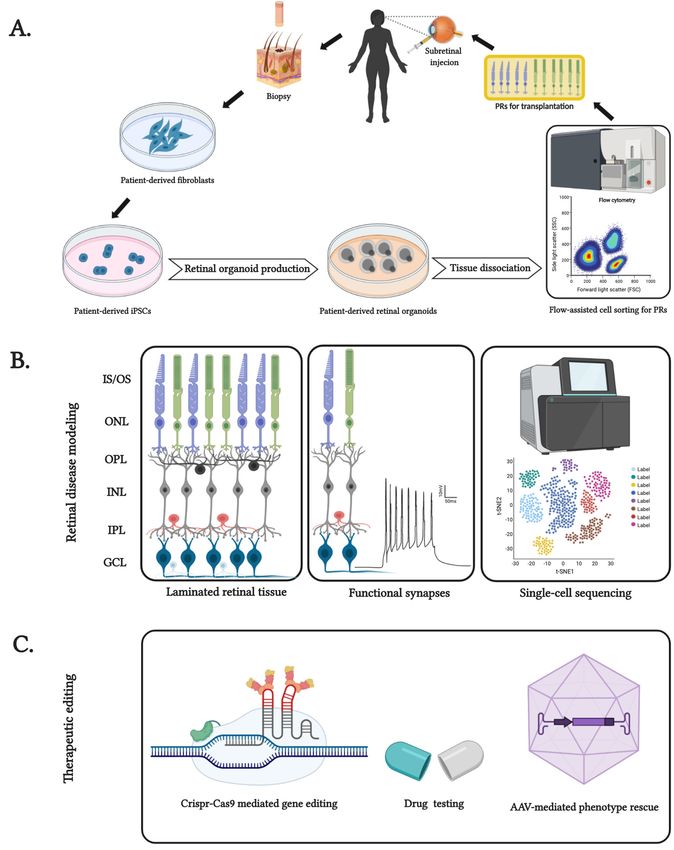
Figure 1. Features
Figure andand
1. Features applications of patient-specific
applications induced
of patient-specific pluripotent
induced pluripotentstem cellcell
stem (iPSC)-derived
(iPSC)-derived
retinal organoids. (A) Generation of patient genotype-specific autologous PRs extracted
retinal organoids. (A) Generation of patient genotype-specific autologous PRs extracted from mature
from mature
retinal organoids. (B) Features of patient-derived retinal organoids, from its laminated
retinal organoids. (B) Features of patient-derived retinal organoids, from its laminated cytoarchitecture
containing inner andcontaining
cytoarchitecture outer segments
inner(IS
andand OS),segments
outer inner and(IS outer
andplexiform
OS), innerlayer
and(IPL andplexiform
outer OPL), inner
layer
and(IPL
outer nuclear layer (INL and ONL), ganglion cell layer (GCL) and functionally recapitulated
and OPL), inner and outer nuclear layer (INL and ONL), ganglion cell layer (GCL) and form
to well-distributed cell types of the human retina for patient-specific disease modeling. (C) Use of
functionally recapitulated form to well-distributed cell types of the human retina for patient-specific
retinal organoids as test beds for pharmaceutical interventions and genome editing techniques such as
disease modeling. (C) Use of retinal organoids as test beds for pharmaceutical interventions and
Crispr-Cas9-mediated and viral vector-mediated rescue of blindness phenotype for inherited retinal
genome editing techniques such as Crispr-Cas9-mediated and viral vector-mediated rescue of
degeneration. Created with BioRender.com.
blindness phenotype for inherited retinal degeneration. Created with BioRender.com.
To prolong the survival of these organoids, culture conditions were enhanced using a pre-defined
To prolong the survival of these organoids, culture conditions were enhanced using a pre-
bioreactor that significantly improved the laminar stratification of the retinal layers, generating a high
defined bioreactor that significantly improved the laminar stratification of the retinal layers,
generating a high number of mature PRs with clearly visible cilia and nascent OS-like structures with
stacked membranous disks [43,44]. The yield of PRs was also significantly improved, with 1 millionInt. J. Mol. Sci. 2020, 21, 8484 5 of 13
number of mature PRs with clearly visible cilia and nascent OS-like structures with stacked membranous
disks [43,44]. The yield of PRs was also significantly improved, with 1 million cells extracted from
120 organoids, enough to transplant into mice [44]. However, the lack of a vascular system within the
organoids often led to inefficient oxygen and nutrient supply, which to some extent also explains the
limited size and high level of necrosis [45,46]. Impressive new approaches include improving the yield
of organoids using hydrogels [47], and speeding up maturation of PRs with intact retinal ganglion
cells (RGCs) by supplementing with IGF1 [48] By enhanced vascular-like perfusion, the morphological
maturation of PRs and their interactions with co-cultured retinal pigmented epithelium (RPE) improved.
Postsynaptic density 95 and c-terminal binding protein antibodies identified the evenly distributed
PRs in the outer retina at days (D120-D160). This photoreceptor development corresponds to in vivo
human retinogenesis. Photoreceptor outer segments (POS) structures emerge in vivo at around week
(W23-W25), and POS structures established in these organoids appeared between W18 and W28, a
remarkably similar developmental pattern [49]. Appearance of cilia emerging from mitochondria
which inner segments and cilia emerging from them present around W13 and outer segments appeared
around W21 similar to previously assessed protocols [50,51]. Many improvements in generating retinal
organoids from iPSCs have demonstrated that the addition of retinoic acid and taurine between D90
and D120 of differentiation enhances the formation of rod and S-cone PRs [52]. Very recently, an
improved 3D retinal organoid protocol was published, which demonstrated a proper stratification
of all retinal layers, including light-sensitive PRs that were functionally connected to inner retinal
neurons [14]. Importantly, this protocol also highlighted differences in human iPSC lines regarding their
competence in generating layered retinal tissues. These cell-line-specific differences are an important
parameter to be considered when establishing different protocols for retinal organoid production.
Normally, every research lab uses its own default set of lines, which might be suboptimal for protocols
from other laboratories.
2.1. Retinal Organoids for Disease Modeling
Connecting cilia are important subcellular structures within the light-sensitive outer segments of
photoreceptors. Ciliary function has been studied extensively, from its biogenesis to its detrimental
dysfunction in rods and cones, which can cause blindness. The underlying pathological mechanisms
are being investigated [53]. Patient-derived human iPSCs, including retinal organoids, offer a new
option for uncovering the underlying mechanism of the disease (Figure 1). For example, the mutation
of CEP290, a primary ciliary protein, was studied in patient-derived retinal organoid structures [54].
Retinal organoids derived from CEP290-mutated LCA (Leber congenital amaurosis) and JSRD (Joubert
syndrome and related disorders) were examined to understand these ciliopathies. The results
corroborate the previously proposed function of CEP290 in gating-specific ciliary proteins, impacting
the biogenesis and transport of cell types [55]. Disease-specific retinal dysfunction, and its dysregulated
molecular counterpart, mirrored an early-onset retinal degeneration phenotype that had previously
been discovered clinically [54]. Recently, a late-onset RP model was also established in an in vitro 3D
organoid system underlying the PDE6B mutated phenotype [56]. Another project developed retinitis
pigmentosa GTPase regulator (RPGR) mutation-specific RP retinal organoids that assist in examining
the molecular dysregulation of this specific phenotype. It was shown that the RPGR mutation
resulted in defective PR morphology and localization, shortened cilia, an altered transcriptional profile,
and even dysregulated electrophysiological output when compared to a healthy control. As a proof
of concept, CRISPR-Cas9 (summarized in Figure 1C) genomic engineering was applied to repair the
RPGR mutation, which led to a significant improvement [57]. A similar study has been established
with an X-linked juvenile retinoschisis retinal organoid model [58]. Ciliary F-actin assembly was
recently studied in retinal organoids, concluding that the role of the PR cilium actin regulator (PCARE)
and Arp2/3 complex activator is to regulate the formation of primary cilia that drive disc formation in
the outer segments (OS) of functional PRs. As a proof of concept, pharmacological inhibition of actinInt. J. Mol. Sci. 2020, 21, 8484 6 of 13
polymerization has also been shown to mirror the PCARE mutation phenotype, and that of PCARE-/-
mice [59].
Retinal organoids are essential for exploring highly heterogeneous diseases such as RP. One
of several RP mutations is the hypomorphic mutation in the tRNA nucleotidyl transferase CCA
adding 1 (TRNT1) gene, which severely affects the PRs and causes early-onset RP. To replicate
the disease in its patient-specific genotype, patient iPSCs were used to generate retinal organoids.
These organoids displayed similar TRNT1 protein augmentation, deficits in autophagy, and general
pathophysiology of cell types that are otherwise inaccessible in living patients [60]. Similarly, retinal
organoids were also derived from a patient-specific iPSC line with compound heterozygous CRB1
mutations (c.1892A > G and c.2548G > A) [61]. To replicate the RPE65-associated LCA and AlPL1-LCA
phenotype in a 3D in vitro system, patient-specific iPSCs were generated that were later used to
generate organoids [62,63]. A similar study explored pathogenic splicing variants of the ABCA4 gene,
a transporter protein responsible for Stargardt’s disease [64]. These studies have emphasized the
importance of patient-specific, disease-targeted 3D model systems. A PR degeneration model was
also generated using the NRL (neural retina leucine zipper) null phenotype, further mimicking S-cone
syndrome and RP. This NRL-/- human-based 3D organoid system uncovered the possible role of MEF2C
as a candidate regulator in cone development [65]. Since animal models restrict the understanding
of the pathophysiology of X-linked RP (XLRP), human 3D retinal organoids provide an efficient,
stable, and reproducible model system for exploring RP2 mutation. As a proof of concept, a study
was conducted to rescue the XLRP phenotype using AAV-mediated gene augmentation, preserving
PRs [66].
2.2. Model for Validation of New Treatment Strategies
Furthermore, the protective effects of ophthalmic supplements such as 4-hydroxytamoxifen and
diethylstilbestrol which were already established in retinal explants [67], were tested in in vitro 3D
model systems [68]. 3D retinal organoids were generated to replicate general PR degeneration and used
as testbeds to confirm their protective effects on the overall health of PRs. Recently, a protocol for retinal
organoid generation underlined the essential conditions for optimal PR development. Growth factors,
small molecules, and cell seeding density seem to significantly affect the numerical and functional
efficiency in generating light-sensitive PRs. For the first time, these organoids were used as testbeds for
evaluating the pharmacological effects of moxifloxacin (a retinotoxic agent at higher doses), resulting
in successful replication of in vivo-like retinal cell damage that included loss of PRs and amacrine
cells [49].
Retinal organoids are also helpful for exploring gene therapies for several retinopathies, for
example by studying the gene delivery in a human system. One such study conducted recently indicates
that AAV2-7m8 has superior transduction, possibly due to higher infectiousness and effective activation
of secondary receptors [69]. Most genome editing techniques rely on highly specific endonucleases
and the capacity of a cell to repair double-stranded breaks (DSB): since DSBs are cell-cycle dependent,
it is even more important to examine the present gene editing options in vitro before starting clinical
trials [70].
Modeling late-onset degenerative diseases could be difficult using stem cell-derived retinal
organoids: stem cells rejuvenate during the reprogramming step and aging-associated epigenetic
signatures are therefore lost [71]. It was previously thought that age-dependent aspects of diseases
could not be replicated in human retinal organoids. However, a side-by-side comparison of retinal
organoids with post-mortem human retinas based on single-cell transcriptomics has demonstrated that
the different cell types within the organoids reach stable cellular states converging towards the ones
from adult peripheral retinal cell types [14].Int. J. Mol. Sci. 2020, 21, 8484 7 of 13
2.3. Retinal Organoids as Cell Sources for Therapeutic Transplantation
Photoreceptors are the vision-forming light-sensitive sensory neurons of the retina: their loss causes
blindness. Since the human retina is unable to regenerate PRs or RPE intrinsically, the transplantation
of donor cells has been intensively explored as a therapeutic option (Figure 1). Huge efforts have been
made to generate human PR and RPE cells which can be successfully transplanted. Pluripotent stem
cell-derived RPE cell replacement therapies have entered clinical trials for the treatment of diseases
such as AMD and Stargardt’s disease [72–75]. In addition to RPE transplantation, there has also
been increasing research efforts to develop strategies and techniques for restoring visual function
by transplanting PRs [20,73–83]. However, the biomedical application of PRs still requires extensive
research. The biggest problem for experimental PR transplantation approaches is finding a high-quality
and quantity source of human PRs and demonstrating robust and functional PR integration into the host
retina. Photoreceptors derived from retinal organoids have great potential for therapeutic photoreceptor
transplantation [84,85]. Stem cell-derived photoreceptors can be further engineered, for example,
by expressing optogenetic tools for studying functional integration into the host retina [86]. Obtaining
sufficient quantities of transplantable cells is, however, still a challenge, despite the development
of several retinal organoid protocols. As an alternative, controversial evidence has been presented
suggesting that cytoplasmic transfer from grafted PR cells to remaining host PR cells can be beneficial,
instead of physical and functional graft integration [86–88]. These findings require additional research,
which will involve retinal organoids.
3. Conclusions
Retinal organoids are complex in structure and cellular organization. Therefore, it is challenging
to recapitulate all morphological and functional features in vitro. Nevertheless, research in the last few
years has revolutionized the field. From the pioneering work [28], several groups have focused on
generating simple, efficient, less labor intensive, and less time-consuming protocols across various cell
lines and cell types. Most importantly, new protocols must be reproducible. Testing combinations
of several innovative approaches has made the generation of highly efficient and stable organoids
easier. In summary, the relatively new field of human retinal organoids is dynamic and still growing.
Several technical roadblocks and difficulties have already been overcome. Many research teams have
significantly contributed to the detail-oriented, ongoing refinement of protocols for the generation of
retinal organoids [89]. As a result, light-sensitive and functionally stratified human retinal organoids
are now available. These are useful tools for basic research and disease modeling with ability
for high throughput pharmaceutical drug screening and are complementary to the use of animal
models. In addition, retinal organoids are being researched as a source of photoreceptors for cell
transplantation therapy.
Author Contributions: Conceptualization, K.S., T.U.K. and V.B.; writing—original draft preparation, K.S.;
writing—review and editing, K.S., T.U.K. and V.B.; visualization, K.S.; supervision, V.B.; funding acquisition, V.B.
All authors have read and agreed to the published version of the manuscript.
Funding: V.B. acknowledges funding by the Volkswagen Foundation (Freigeist—A110720), the European
Research Council (ERC-StG-678071-ProNeurons) and by the Deutsche Forschungsgemeinschaft (SPP2127,
EXC-2068-390729961—Cluster of Excellence—Physics of Life at TU Dresden and EXC-2151-390873048—Cluster of
Excellence—ImmunoSensation2 at the University of Bonn).
Acknowledgments: The authors thank Sara Oakeley for critical feedback on the manuscript. Open Access
Funding by the Publication Fund of the TU Dresden.
Conflicts of Interest: The authors declare no conflict of interest.Int. J. Mol. Sci. 2020, 21, 8484 8 of 13
Abbreviations
PR Photoreceptors
RP Retinitis pigmentosa
AMD Age-related macular degeneration
mESCs Mouse embryonic stem cells
hESCs Human embryonic stem cells
CRX Cone-rod homeobox
iPSCs Induced pluripotent stem cells
IS Inner segment
IPL Inner plexiform layer
INL Inner nuclear layer
OPL Outer plexiform layer
ONL Outer nuclear layer
GCL Ganglion cell layer
OS Outer segment
POS Photoreceptor outer segment
RGC Retinal ganglion cells
RPE Retinal pigmented epithelium
LCA Leber’s congenital amaurosis
JSRD Joubert syndrome and related disorders
RPGR Retinitis pigmentosa GTPase regulator
TRNT1 tRNA nucleotidyl transferase CCA adding 1
NRL Neural leucine zipper
DSB Double stranded break
PCARE Photoreceptor cilium actin regulator
XLRP X-linked retinitis pigmentosa
References
1. Gerrig, R.J.; Zimbardo, P.G.; Campbell, A.J.; Cumming, S.R.; Wilkes, F.J. Psychology and Life; Pearson 1001/151
Castlereagh St: Sydney, Australia, 2015.
2. Kandel, E.R.; Schwartz, J.; Jessell, T. Principles of Neural Science, 4th ed.; McGraw-Hill Companies, Incorporated:
New York, NY, USA, 2000.
3. Chapman, B.; Stryker, M.P.; Bonhoeffer, T. Development of Orientation Preference Maps in Ferret Primary
Visual Cortex. J. Neurosci. 1996, 16, 6443–6453. [CrossRef] [PubMed]
4. Vu, H.T.V.; Keeffe, J.E.; McCarty, C.A.; Taylor, H.R. Impact of Unilateral and Bilateral Vision Loss on Quality
of Life. Br. J. Ophthalmol. 2005, 89, 360–363. [CrossRef] [PubMed]
5. Resnikoff, S.; Pascolini, D.; Etya’ale, D.; Kocur, I.; Pararajasegaram, R.; Pokharel, G.P.; Mariotti, S.P. Global Data
on Visual Impairment in the Year 2002. Bull. World Health Organ. 2004, 82, 844–851. [CrossRef] [PubMed]
6. Finger, R.P.; Fimmers, R.; Holz, F.G.; Scholl, H.P.N. Prevalence and Causes of Registered Blindness in the
Largest Federal State of Germany. Br. J. Ophthalmol. 2011, 95, 1061–1067. [CrossRef]
7. Li, J.Q.; Welchowski, T.; Schmid, M.; Mauschitz, M.M.; Holz, F.G.; Finger, R.P. Prevalence and Incidence of
Age-Related Macular Degeneration in Europe: A Systematic Review and Meta-Analysis. Br. J. Ophthalmol.
2019, 104. [CrossRef]
8. Flaxman, S.R.; Bourne, R.R.A.; Resnikoff, S.; Ackland, P.; Braithwaite, T.; Cicinelli, M.V.; Das, A.; Jonas, J.B.;
Keeffe, J.; Kempen, J.; et al. Global Causes of Blindness and Distance Vision Impairment 1990–2020:
A Systematic Review and Meta-Analysis. Lancet Glob. Health 2017, 5, e1221–e1234. [CrossRef]
9. Huang, X.F. Current Pharmacological Concepts in the Treatment of the Retinitis Pigmentosa. Adv. Exp.
Med. Biol. 2018, 1074, 439–445. [CrossRef]
10. Krzystolik, M.G.; Afshari, M.A.; Adamis, A.P.; Gaudreault, J.; Gragoudas, E.S.; Michaud, N.A.; Li, W.;
Connolly, E.; O’Neill, C.A.; Miller, J.W. Prevention of Experimental Choroidal Neovascularization with
Intravitreal Anti-Vascular Endothelial Growth Factor Antibody Fragment. Arch. Ophthalmol. 2002, 120,
338–346. [CrossRef]Int. J. Mol. Sci. 2020, 21, 8484 9 of 13
11. Miller, J.W.; Bagheri, S.; Vavvas, D.G. Advances in Age-Related Macular Degeneration Understanding and
Therapy. US Ophthalmic Rev. 2017, 10, 119. [CrossRef]
12. Scholl, H.P.N.; Strauss, R.W.; Singh, M.S.; Dalkara, D.; Roska, B.; Picaud, S.; Sahel, J.A. Emerging Therapies
for Inherited Retinal Degeneration. Sci. Transl. Med. 2016, 8, 368rv6. [CrossRef]
13. Fradot, M.; Busskamp, V.; Forster, V.; Cronin, T.; Léveillard, T.; Bennett, J.; Sahel, J.A.; Roska, B.; Picaud, S.
Gene Therapy in Ophthalmology: Validation on Cultured Retinal Cells and Explants from Postmortem
Human Eyes. Hum. Gene Ther. 2011, 22, 587–593. [CrossRef] [PubMed]
14. Cowan, C.S.; Renner, M.; De Gennaro, M.; Gross-Scherf, B.; Goldblum, D.; Hou, Y.; Munz, M.; Rodrigues, T.M.;
Krol, J.; Szikra, T.; et al. Cell Types of the Human Retina and Its Organoids at Single-Cell Resolution. Cell
2020, 182, 1623–1640.e34. [CrossRef] [PubMed]
15. Evans, M.J.; Kaufman, M.H. Establishment in Culture of Pluripotential Cells from Mouse Embryos. Nature
1981, 292, 154–156. [CrossRef] [PubMed]
16. Thomson, J.A.; Itskovitz-Eldor, J.; Shapiro, S.S.; Waaknitz, M.A.; Swiergiel, J.J.; Marshall, V.S.; Jones, J.M.
Embryonic Stem Cell Lines Derived from Human Blastocysts. Science 1998, 282, 1145–1147. [CrossRef]
17. Takahashi, K.; Yamanaka, S. Induction of Pluripotent Stem Cells from Mouse Embryonic and Adult Fibroblast
Cultures by Defined Factors. Cell 2006, 126, 663–676. [CrossRef]
18. Takahashi, K.; Tanabe, K.; Ohnuki, M.; Narita, M.; Ichisaka, T.; Tomoda, K.; Yamanaka, S. Induction of
Pluripotent Stem Cells from Adult Human Fibroblasts by Defined Factors. Cell 2007, 131, 861–872. [CrossRef]
19. Nakatsuji, N.; Nakajima, F.; Tokunaga, K. HLA-Haplotype Banking and IPS Cells. Nat. Biotechnol. 2008,
739–740. [CrossRef]
20. Krohne, T.U.; Westenskow, P.D.; Kurihara, T.; Friedlander, D.F.; Lehmann, M.; Dorsey, A.L.; Li, W.; Zhu, S.;
Schultz, A.; Wang, J.; et al. Generation of Retinal Pigment Epithelial Cells from Small Molecules and OCT4
Reprogrammed Human Induced Pluripotent Stem Cells. Stem Cells Transl. Med. 2012, 1, 96–109. [CrossRef]
21. Tucker, B.A.; Solivan-Timpe, F.; Roos, B.R.; Anfinson, K.R.; Robin, A.L.; Wiley, L.A.; Mullins, R.F.; Fingert, J.H.
Duplication of TBK1 Stimulates Autophagy in IPSC-Derived Retinal Cells from a Patient with Normal
Tension Glaucoma. J. Stem Cell Res. Ther. 2014, 4, 161. [CrossRef]
22. Alonso-Alonso, M.L.; Srivastava, G.K. Current Focus of Stem Cell Application in Retinal Repair. World J.
Stem Cells 2015, 7, 641. [CrossRef]
23. Barber, A.C.; Hippert, C.; Duran, Y.; West, E.L.; Bainbridge, J.W.B.; Warre-Cornish, K.; Luhmann, U.F.O.;
Lakowski, J.; Sowden, J.C.; Ali, R.R.; et al. Repair of the Degenerate Retina by Photoreceptor Transplantation.
Proc. Natl. Acad. Sci. USA 2013, 110, 354–359. [CrossRef] [PubMed]
24. Hambright, D.; Park, K.-Y.; Brooks, M.; Mckay, R.; Swaroop, A.; Nasonkin, I.O. Long-Term
Survival and Differentiation of Retinal Neurons Derived from Human Embryonic Stem Cell Lines in
Un-Immunosuppressed Mouse Retina. Mol. Vis. 2012, 18, 920–936. [PubMed]
25. Mekala, S.R.; Vauhini, V.; Nagarajan, U.; Maddileti, S.; Gaddipati, S.; Mariappan, I. Derivation,
Characterization and Retinal Differentiation of Induced Pluripotent Stem Cells. J. Biosci. 2013, 38, 123–134.
[CrossRef] [PubMed]
26. Meng, F.; Wang, X.; Gu, P.; Wang, Z.; Guo, W. Induction of Retinal Ganglion-like Cells from Fibroblasts by
Adenoviral Gene Delivery. Neuroscience 2013, 250, 381–393. [CrossRef] [PubMed]
27. Phillips, M.J.; Perez, E.T.; Martin, J.M.; Reshel, S.T.; Wallace, K.A.; Capowski, E.E.; Singh, R.; Wright, L.S.;
Clark, E.M.; Barney, P.M.; et al. Modeling Human Retinal Development with Patient-Specific Induced
Pluripotent Stem Cells Reveals Multiple Roles for Visual System Homeobox 2. Stem Cells 2014, 32, 1480–1492.
[CrossRef] [PubMed]
28. Singh, V.K.; Kalsan, M.; Kumar, N.; Saini, A.; Chandra, R. Induced Pluripotent Stem Cells: Applications in
Regenerative Medicine, Disease Modeling, and Drug Discovery. Front. Cell Dev. Biol. 2015, 3, 2. [CrossRef]
29. Wilson, H.V.; Penny, J.T. On Some Phenomena of Coalescence and Regeneration in Sponges. J. Exp. Zool.
1907, 5, 245–258. [CrossRef]
30. Shannon, J.M.; Mason, R.J.; Jennings, S.D. Functional Differentiation of Alveolar Type II Epithelial Cells in
Vitro: Effects of Cell Shape, Cell-Matrix Interactions and Cell-Cell Interactions. Biochim. Biophys. Acta 1987,
931, 143–156. [CrossRef]
31. Eiraku, M.; Takata, N.; Ishibashi, H.; Kawada, M.; Sakakura, E.; Okuda, S.; Sekiguchi, K.; Adachi, T.;
Sasai, Y. Self-Organizing Optic-Cup Morphogenesis in Three-Dimensional Culture. Nature 2011, 472, 51–58.
[CrossRef]Int. J. Mol. Sci. 2020, 21, 8484 10 of 13
32. Gao, D.; Vela, I.; Sboner, A.; Iaquinta, P.J.; Karthaus, W.R.; Gopalan, A.; Dowling, C.; Wanjala, J.N.;
Undvall, E.A.; Arora, V.K.; et al. Organoid Cultures Derived from Patients with Advanced Prostate Cancer.
Cell 2014, 159, 176–187. [CrossRef]
33. Huch, M.; Dorrell, C.; Boj, S.F.; Van Es, J.H.; Li, V.S.W.; Van De Wetering, M.; Sato, T.; Hamer, K.; Sasaki, N.;
Finegold, M.J.; et al. In Vitro Expansion of Single Lgr5 + Liver Stem Cells Induced by Wnt-Driven
Regeneration. Nature 2013, 494, 247–250. [CrossRef] [PubMed]
34. Jung, P.; Sato, T.; Merlos-Suárez, A.; Barriga, F.M.; Iglesias, M.; Rossell, D.; Auer, H.; Gallardo, M.; Blasco, M.A.;
Sancho, E.; et al. Isolation and in Vitro Expansion of Human Colonic Stem Cells. Nat. Med. 2011, 17,
1225–1227. [CrossRef] [PubMed]
35. Sato, T.; Vries, R.G.; Snippert, H.J.; Van De Wetering, M.; Barker, N.; Stange, D.E.; Van Es, J.H.; Abo, A.;
Kujala, P.; Peters, P.J.; et al. Single Lgr5 Stem Cells Build Crypt-Villus Structures in Vitro without a
Mesenchymal Niche. Nature 2009, 459, 262–265. [CrossRef] [PubMed]
36. Lancaster, M.A.; Renner, M.; Martin, C.A.; Wenzel, D.; Bicknell, L.S.; Hurles, M.E.; Homfray, T.; Penninger, J.M.;
Jackson, A.P.; Knoblich, J.A. Cerebral Organoids Model Human Brain Development and Microcephaly.
Nature 2013, 501, 373–379. [CrossRef]
37. Meyer, J.S.; Shearer, R.L.; Capowski, E.E.; Wright, L.S.; Wallace, K.A.; McMillan, E.L.; Zhang, S.C.; Gamm, D.M.
Modeling Early Retinal Development with Human Embryonic and Induced Pluripotent Stem Cells. Proc.
Natl. Acad. Sci. USA 2009, 106, 16698–16703. [CrossRef]
38. Nakano, T.; Ando, S.; Takata, N.; Kawada, M.; Muguruma, K.; Sekiguchi, K.; Saito, K.; Yonemura, S.;
Eiraku, M.; Sasai, Y. Self-Formation of Optic Cups and Storable Stratified Neural Retina from Human ESCs.
Cell Stem Cell 2012, 10, 771–785. [CrossRef]
39. Eguizabal, C.; Vassena, R.; Garreta, E. Embryonic stem cells induced pluripotent stem cells Complete Meiosis
from Human Induced Pluripotent Stem Cells. Stem Cells 2015, 29, 1186–1195. [CrossRef]
40. Phillips, M.J.; Wallace, K.A.; Dickerson, S.J.; Miller, M.J.; Verhoeven, A.D.; Martin, J.M.; Wright, L.S.; Shen, W.;
Capowski, E.E.; Percin, E.F.; et al. Blood-Derived Human IPS Cells Generate Optic Vesicle-like Structures
with the Capacity to Form Retinal Laminae and Develop Synapses. Investig. Ophthalmol. Vis. Sci. 2012, 53,
2007–2019. [CrossRef]
41. Völkner, M.; Zschätzsch, M.; Rostovskaya, M.; Overall, R.W.; Busskamp, V.; Anastassiadis, K.; Karl, M.O.
Retinal Organoids from Pluripotent Stem Cells Efficiently Recapitulate Retinogenesis. Stem Cell Rep. 2016, 6,
525–538. [CrossRef]
42. Zhong, X.; Gutierrez, C.; Xue, T.; Hampton, C.; Vergara, M.N.; Cao, L.H.; Peters, A.; Park, T.S.; Zambidis, E.T.;
Meyer, J.S.; et al. Generation of Three-Dimensional Retinal Tissue with Functional Photoreceptors from
Human IPSCs. Nat. Commun. 2014, 5. [CrossRef]
43. DiStefano, T.; Chen, H.Y.; Panebianco, C.; Kaya, K.D.; Brooks, M.J.; Gieser, L.; Morgan, N.Y.; Pohida, T.;
Swaroop, A. Accelerated and Improved Differentiation of Retinal Organoids from Pluripotent Stem Cells in
Rotating-Wall Vessel Bioreactors. Stem Cell Rep. 2018, 10, 300–313. [CrossRef] [PubMed]
44. Ovando-Roche, P.; West, E.L.; Branch, M.J.; Sampson, R.D.; Fernando, M.; Munro, P.; Georgiadis, A.;
Rizzi, M.; Kloc, M.; Naeem, A.; et al. Use of Bioreactors for Culturing Human Retinal Organoids Improves
Photoreceptor Yields. Stem Cell Res. Ther. 2018, 9, 156. [CrossRef] [PubMed]
45. Achberger, K.; Probst, C.; Haderspeck, J.C.; Bolz, S.; Rogal, J.; Chuchuy, J.; Nikolova, M.; Cora, V.;
Antkowiak, L.; Haq, W.; et al. Merging Organoid and Organ-on-a-Chip Technology to Generate Complex
Multi-Layer Tissue Models in a Human Retina-on-a-Chip Platform. Elife 2019, 8. [CrossRef] [PubMed]
46. Haderspeck, J.C.; Chuchuy, J.; Kustermann, S.; Liebau, S.; Loskill, P. Organ-on-a-Chip Technologies That Can
Transform Ophthalmic Drug Discovery and Disease Modeling. Expert Opin. Drug Discov. 2019, 14, 47–57.
[CrossRef] [PubMed]
47. Hunt, N.C.; Hallam, D.; Karimi, A.; Mellough, C.B.; Chen, J.; Steel, D.H.W.; Lako, M. 3D Culture of Human
Pluripotent Stem Cells in RGD-Alginate Hydrogel Improves Retinal Tissue Development. Acta Biomater.
2017, 49, 329–343. [CrossRef]
48. Mellough, C.B.; Collin, J.; Khazim, M.; White, K.; Sernagor, E.; Steel, D.H.W.; Lako, M. IGF-1 Signaling
Plays an Important Role in the Formation of Three-Dimensional Laminated Neural Retina and Other Ocular
Structures from Human Embryonic Stem Cells. Stem Cells 2015, 33, 2416–2430. [CrossRef]Int. J. Mol. Sci. 2020, 21, 8484 11 of 13
49. Hallam, D.; Hilgen, G.; Dorgau, B.; Zhu, L.; Yu, M.; Bojic, S.; Hewitt, P.; Schmitt, M.; Uteng, M.;
Kustermann, S.; et al. Human-Induced Pluripotent Stem Cells Generate Light Responsive Retinal Organoids
with Variable and Nutrient-Dependent Efficiency. Stem Cells 2018, 36, 1535–1551. [CrossRef]
50. Lowe, A.; Harris, R.; Bhansali, P.; Cvekl, A.; Liu, W. Intercellular Adhesion-Dependent Cell Survival and
ROCK-Regulated Actomyosin-Driven Forces Mediate Self-Formation of a Retinal Organoid. Stem Cell Rep.
2016, 6, 743–756. [CrossRef]
51. Parfitt, D.A.; Lane, A.; Ramsden, C.M.; Carr, A.J.F.; Munro, P.M.; Jovanovic, K.; Schwarz, N.; Kanuga, N.;
Muthiah, M.N.; Hull, S.; et al. Identification and Correction of Mechanisms Underlying Inherited Blindness
in Human IPSC-Derived Optic Cups. Cell Stem Cell 2016, 18, 769–781. [CrossRef]
52. Zerti, D.; Dorgau, B.; Felemban, M.; Ghareeb, A.E.; Yu, M.; Ding, Y.; Krasnogor, N.; Lako, M. Developing a
Simple Method to Enhance the Generation of Cone and Rod Photoreceptors in Pluripotent Stem Cell-Derived
Retinal Organoids. Stem Cells 2020, 38, 45–51. [CrossRef]
53. Chen, H.Y.; Welby, E.; Li, T.; Swaroop, A. Retinal Disease in Ciliopathies: Recent Advances with a Focus on
Stem Cell-Based Therapies. Transl. Sci. Rare Dis. 2019, 4, 97–115. [CrossRef] [PubMed]
54. Shimada, H.; Lu, Q.; Insinna-Kettenhofen, C.; Nagashima, K.; English, M.A.; Semler, E.M.; Mahgerefteh, J.;
Cideciyan, A.V.; Li, T.; Brooks, B.P.; et al. In Vitro Modeling Using Ciliopathy-Patient-Derived Cells Reveals
Distinct Cilia Dysfunctions Caused by CEP290 Mutations. Cell Rep. 2017, 20, 384–396. [CrossRef] [PubMed]
55. Rachel, R.A.; Yamamoto, E.A.; Dewanjee, M.K.; May-Simera, H.L.; Sergeev, Y.V.; Hackett, A.N.; Pohida, K.;
Munasinghe, J.; Gotoh, N.; Wickstead, B.; et al. CEP290 Alleles in Mice Disrupt Tissue-Specific Cilia
Biogenesis and Recapitulate Features of Syndromic Ciliopathies. Hum. Mol. Genet. 2015, 24, 3775–3791.
[CrossRef]
56. Gao, M.L.; Lei, X.L.; Han, F.; He, K.W.; Jin, S.Q.; Zhang, Y.Y.; Jin, Z.B. Patient-Specific Retinal Organoids
Recapitulate Disease Features of Late-Onset Retinitis Pigmentosa. Front. Cell Dev. Biol. 2020, 8, 128.
[CrossRef] [PubMed]
57. Deng, W.L.; Gao, M.L.; Lei, X.L.; Lv, J.N.; Zhao, H.; He, K.W.; Xia, X.X.; Li, L.Y.; Chen, Y.C.; Li, Y.P.; et al.
Gene Correction Reverses Ciliopathy and Photoreceptor Loss in IPSC-Derived Retinal Organoids from
Retinitis Pigmentosa Patients. Stem Cell Rep. 2018, 10, 1267–1281. [CrossRef] [PubMed]
58. Huang, K.C.; Wang, M.L.; Chen, S.J.; Kuo, J.C.; Wang, W.J.; Nhi Nguyen, P.N.; Wahlin, K.J.; Lu, J.F.; Tran, A.A.;
Shi, M.; et al. Morphological and Molecular Defects in Human Three-Dimensional Retinal Organoid Model
of X-Linked Juvenile Retinoschisis. Stem Cell Rep. 2019, 13, 906–923. [CrossRef] [PubMed]
59. Corral-Serrano, J.C.; Lamers, I.J.C.; van Reeuwijk, J.; Duijkers, L.; Hoogendoorn, A.D.M.; Yildirim, A.;
Argyrou, N.; Ruigrok, R.A.A.; Letteboer, S.J.F.; Butcher, R.; et al. PCARE and WASF3 Regulate Ciliary F-Actin
Assembly That Is Required for the Initiation of Photoreceptor Outer Segment Disk Formation. Proc. Natl.
Acad. Sci. USA 2020, 117, 9922–9931. [CrossRef]
60. Sharma, T.P.; Wiley, L.A.; Whitmore, S.S.; Anfinson, K.R.; Cranston, C.M.; Oppedal, D.J.; Daggett, H.T.;
Mullins, R.F.; Tucker, B.A.; Stone, E.M. Patient-Specific Induced Pluripotent Stem Cells to Evaluate the
Pathophysiology of TRNT1-Associated Retinitis Pigmentosa. Stem Cell Res. 2017, 21, 58–70. [CrossRef]
61. Zhang, X.; Zhang, D.; Chen, S.C.; Lamey, T.; Thompson, J.A.; McLaren, T.; De Roach, J.N.; Chen, F.K.;
McLenachan, S. Establishment of an Induced Pluripotent Stem Cell Line from a Retinitis Pigmentosa Patient
with Compound Heterozygous CRB1 Mutation. Stem Cell Res. 2018, 31, 147–151. [CrossRef]
62. Li, G.; Gao, G.; Wang, P.; Song, X.; Xu, P.; Xie, B.; Zhou, T.; Pan, G.; Peng, F.; Zhang, Q.; et al. Generation
and Characterization of Induced Pluripotent Stem Cells and Retinal Organoids From a Leber’s Congenital
Amaurosis Patient With Novel RPE65 Mutations. Front. Mol. Neurosci. 2019, 12, 212. [CrossRef]
63. Lukovic, D.; Artero Castro, A.; Kaya, K.D.; Munezero, D.; Gieser, L.; Davó-Martínez, C.; Corton, M.;
Cuenca, N.; Swaroop, A.; Ramamurthy, V.; et al. Retinal Organoids Derived from HiPSCs of an AIPL1-LCA
Patient Maintain Cytoarchitecture despite Reduced Levels of Mutant AIPL1. Sci. Rep. 2020, 10, 5426.
[CrossRef] [PubMed]
64. Khan, M.; Arno, G.; Fakin, A.; Parfitt, D.A.; Dhooge, P.P.A.; Albert, S.; Bax, N.M.; Duijkers, L.; Niblock, M.;
Hau, K.L.; et al. Detailed Phenotyping and Therapeutic Strategies for Intronic ABCA4 Variants in Stargardt
Disease. Mol. Ther. Nucleic Acids 2020, 21, 412–427. [CrossRef] [PubMed]
65. Kallman, A.; Capowski, E.E.; Wang, J.; Kaushik, A.M.; Jansen, A.D.; Edwards, K.L.; Chen, L.; Berlinicke, C.A.;
Joseph Phillips, M.; Pierce, E.A.; et al. Investigating Cone Photoreceptor Development Using Patient-Derived
NRL Null Retinal Organoids. Commun. Biol. 2020, 3, 82. [CrossRef]Int. J. Mol. Sci. 2020, 21, 8484 12 of 13
66. Lane, A.; Jovanovic, K.; Shortall, C.; Ottaviani, D.; Panes, A.B.; Schwarz, N.; Guarascio, R.; Hayes, M.J.;
Palfi, A.; Chadderton, N.; et al. Modeling and Rescue of RP2 Retinitis Pigmentosa Using IPSC-Derived
Retinal Organoids. Stem Cell Rep. 2020, 15, 67–79. [CrossRef] [PubMed]
67. Onishi, A.; Peng, G.H.; Poth, E.M.; Lee, D.A.; Chen, J.; Alexis, U.; De Melo, J.; Chen, S.; Blackshaw, S. The
Orphan Nuclear Hormone Receptor ERRβ Controls Rod Photoreceptor Survival. Proc. Natl. Acad. Sci. USA
2010, 107, 11579–11584. [CrossRef] [PubMed]
68. Ito, S.; Onishi, A.; Takahashi, M. Chemically-Induced Photoreceptor Degeneration and Protection in Mouse
IPSC-Derived Three-Dimensional Retinal Organoids. Stem Cell Res. 2017, 24, 94–101. [CrossRef] [PubMed]
69. Garita-Hernandez, M.; Routet, F.; Guibbal, L.; Khabou, H.; Toualbi, L.; Riancho, L.; Reichman, S.; Duebel, J.;
Sahel, J.-A.; Goureau, O.; et al. AAV-Mediated Gene Delivery to 3D Retinal Organoids Derived from Human
Induced Pluripotent Stem Cells. Int. J. Mol. Sci. 2020, 21, 994. [CrossRef]
70. Pasquini, G.; Cora, V.; Swiersy, A.; Achberger, K.; Antkowiak, L.; Müller, B.; Wimmer, T.; Fraschka, S.A.K.;
Casadei, N.; Ueffing, M.; et al. Using Transcriptomic Analysis to Assess Double- Strand Break Repair Activity:
Towards Precise in Vivo Genome Editing. Int. J. Mol. Sci. 2020, 21, 1380. [CrossRef]
71. Mertens, J.; Paquola, A.C.M.; Ku, M.; Hatch, E.; Böhnke, L.; Ladjevardi, S.; McGrath, S.; Campbell, B.;
Lee, H.; Herdy, J.R.; et al. Directly Reprogrammed Human Neurons Retain Aging-Associated Transcriptomic
Signatures and Reveal Age-Related Nucleocytoplasmic Defects. Cell Stem Cell 2015, 17, 705–718. [CrossRef]
72. Souied, E.; Pulido, J.; Staurenghi, G. Autologous Induced Stem-Cell–Derived Retinal Cells for Macular
Degeneration. N. Engl. J. Med. 2017, 792. [CrossRef]
73. Da Cruz, L.; Fynes, K.; Georgiadis, O.; Kerby, J.; Luo, Y.H.; Ahmado, A.; Vernon, A.; Daniels, J.T.; Nommiste, B.;
Hasan, S.M.; et al. Phase 1 Clinical Study of an Embryonic Stem Cell-Derived Retinal Pigment Epithelium
Patch in Age-Related Macular Degeneration. Nat. Biotechnol. 2018, 36, 328–337. [CrossRef] [PubMed]
74. Kashani, A.H.; Lebkowski, J.S.; Rahhal, F.M.; Avery, R.L.; Salehi-Had, H.; Dang, W.; Lin, C.M.; Mitra, D.;
Zhu, D.; Thomas, B.B.; et al. A Bioengineered Retinal Pigment Epithelial Monolayer for Advanced,
Dry Age-Related Macular Degeneration. Sci. Transl. Med. 2018, 10. [CrossRef] [PubMed]
75. Mehat, M.S.; Sundaram, V.; Ripamonti, C.; Robson, A.G.; Smith, A.J.; Borooah, S.; Robinson, M.;
Rosenthal, A.N.; Innes, W.; Weleber, R.G.; et al. Transplantation of Human Embryonic Stem Cell-Derived
Retinal Pigment Epithelial Cells in Macular Degeneration. Ophthalmology 2018, 125, 1765–1775. [CrossRef]
76. Carr, A.J.; Vugler, A.A.; Hikita, S.T.; Lawrence, J.M.; Gias, C.; Chen, L.L.; Buchholz, D.E.; Ahmado, A.;
Semo, M.; Smart, M.J.K.; et al. Protective Effects of Human IPS-Derived Retinal Pigment Epithelium Cell
Transplantation in the Retinal Dystrophic Rat. PLoS ONE 2009, 4. [CrossRef] [PubMed]
77. Gasparini, S.J.; Llonch, S.; Borsch, O.; Ader, M. Transplantation of Photoreceptors into the Degenerative
Retina: Current State and Future Perspectives. Prog. Retin. Eye Res. 2019, 69, 1–37. [CrossRef]
78. Kamao, H.; Mandai, M.; Ohashi, W.; Hirami, Y.; Kurimoto, Y.; Kiryu, J.; Takahashi, M. Evaluation of the
Surgical Device and Procedure for Extracellular Matrix–Scaffold–Supported Human IPSC– Derived Retinal
Pigment Epithelium Cell Sheet Transplantation. Investig. Ophthalmol. Vis. Sci. 2017, 58, 211–220. [CrossRef]
79. Mandai, M.; Fujii, M.; Hashiguchi, T.; Sunagawa, G.A.; Ito, S.; Sun, J.; Kaneko, J.; Sho, J.; Yamada, C.;
Takahashi, M. Erratum: IPSC-Derived Retina Transplants Improve Vision in Rd1 End-Stage Retinal-
Degeneration Mice. Stem Cell Rep. 2017, 8, 1112–1113. [CrossRef]
80. MacLaren, R.E.; Pearson, R.A.; MacNeil, A.; Douglas, R.H.; Salt, T.E.; Akimoto, M.; Swaroop, A.; Sowden, J.C.;
Ali, R.R. Retinal Repair by Transplantation of Photoreceptor Precursors. Nature 2006, 444, 203–207. [CrossRef]
81. Seiler, M.J.; Thomas, B.B.; Chen, Z.; Wu, R.; Sadda, S.R.; Aramant, R.B. Retinal Transplants Restore Visual
Responses: Trans-Synaptic Tracing from Visually Responsive Sites Labels Transplant Neurons. Eur. J. Neurosci.
2008, 28, 208–220. [CrossRef]
82. Sugita, S.; Iwasaki, Y.; Makabe, K.; Kamao, H.; Mandai, M.; Shiina, T.; Ogasawara, K.; Hirami, Y.; Kurimoto, Y.;
Takahashi, M. Successful Transplantation of Retinal Pigment Epithelial Cells from MHC Homozygote IPSCs
in MHC-Matched Models. Stem Cell Rep. 2016, 7, 635–648. [CrossRef]
83. Blenkinsop, T.A. Adult Human RPE for Transplantation: Renewing an Old Promise. Adv. Regen. Biol. 2015,
2, 27144. [CrossRef]
84. Pearson, R.A.; Gonzalez-Cordero, A.; West, E.L.; Ribeiro, J.R.; Aghaizu, N.; Goh, D.; Sampson, R.D.;
Georgiadis, A.; Waldron, P.V.; Duran, Y.; et al. Donor and Host Photoreceptors Engage in Material Transfer
Following Transplantation of Post-Mitotic Photoreceptor Precursors. Nat. Commun. 2016, 7. [CrossRef]
[PubMed]Int. J. Mol. Sci. 2020, 21, 8484 13 of 13
85. Santos-Ferreira, T.; Postel, K.; Stutzki, H.; Kurth, T.; Zeck, G.; Ader, M. Daylight Vision Repair by Cell
Transplantation. Stem Cells 2015, 33, 79–90. [CrossRef] [PubMed]
86. Gonzalez-Cordero, A.; West, E.L.; Pearson, R.A.; Duran, Y.; Carvalho, L.S.; Chu, C.J.; Naeem, A.;
Blackford, S.J.I.; Georgiadis, A.; Lakowski, J.; et al. Photoreceptor Precursors Derived from Three-Dimensional
Embryonic Stem Cell Cultures Integrate and Mature within Adult Degenerate Retina Europe PMC Funders
Group. Nat. Biotechnol. 2013, 31. [CrossRef] [PubMed]
87. Gonzalez-Cordero, A.; Kruczek, K.; Naeem, A.; Fernando, M.; Kloc, M.; Ribeiro, J.; Goh, D.; Duran, Y.;
Blackford, S.J.I.; Abelleira-Hervas, L.; et al. Recapitulation of Human Retinal Development from Human
Pluripotent Stem Cells Generates Transplantable Populations of Cone Photoreceptors. Stem Cell Rep. 2017, 9,
820–837. [CrossRef]
88. Garita-Hernandez, M.; Lampič, M.; Chaffiol, A.; Guibbal, L.; Routet, F.; Santos-Ferreira, T.; Gasparini, S.;
Borsch, O.; Gagliardi, G.; Reichman, S.; et al. Restoration of Visual Function by Transplantation of
Optogenetically Engineered Photoreceptors. Nat. Commun. 2019, 10, 4524. [CrossRef]
89. Kruczek, K.; Swaroop, A. Pluripotent Stem Cell-Derived Retinal Organoids for Disease Modeling and
Development of Therapies. Stem Cells 2020. [CrossRef]
Publisher’s Note: MDPI stays neutral with regard to jurisdictional claims in published maps and institutional
affiliations.
© 2020 by the authors. Licensee MDPI, Basel, Switzerland. This article is an open access
article distributed under the terms and conditions of the Creative Commons Attribution
(CC BY) license (http://creativecommons.org/licenses/by/4.0/).You can also read