HHS Public Access Author manuscript Cancer Lett. Author manuscript; available in PMC 2020 May 21.
←
→
Page content transcription
If your browser does not render page correctly, please read the page content below
HHS Public Access
                                Author manuscript
                                Cancer Lett. Author manuscript; available in PMC 2020 May 21.
Author Manuscript

                    Published in final edited form as:
                     Cancer Lett. 2020 April 28; 476: 23–33. doi:10.1016/j.canlet.2020.01.012.

                    Human papillomavirus insertions identify the PIM family of
                    serine/threonine kinases as targetable driver genes in head and
                    neck squamous cell carcinoma
                    Tatevik R. Broutian1, Bo Jiang2, Jingfeng Li3,5, Keiko Akagi2, Shanying Gui3,6, Zhengqiu
                    Zhou3,7, Weihong Xiao2, David E. Symer*,4, Maura L. Gillison*,2
                    1Biomedical      Sciences Graduate Program, The Ohio State University, Columbus OH 43210 United
Author Manuscript

                    States
                    2Department of Thoracic/Head and Neck Medical Oncology, University of Texas MD Anderson
                    Cancer Center, Houston, TX 77030 United States
                    3Division
                           of Medical Oncology, Department of Internal Medicine, The Ohio State University,
                    Columbus, OH 43210 United States
                    4Department of Lymphoma and Myeloma, University of Texas MD Anderson Cancer Center,
                    Houston, TX 77030 United States

                    Abstract
                         Human papillomavirus (HPV) insertions in cancer genomes have been linked to various forms of
Author Manuscript

                         focal genomic instability and altered expression of neighboring genes. Here we tested the
                         hypothesis that investigation of HPV insertions in a head and neck cancer squamous cell
                         carcinoma (HNSCC) cell line would identify targetable driver genes contributing to oncogenesis
                         of other HNSCC. In the cell line UPCI:SCC090, HPV16 integration amplified PIM1 serine/
                         threonine kinase gene ~16-fold, thereby increasing transcript and protein levels. We used genetic
                         and pharmacological approaches to inhibit PIM kinases in this and other HNSCC cell lines.
                         Knockdown of PIM1 transcripts by transfected short hairpin RNAs reduced UPCI:SCC090
                         viability. CRISPR/Cas9-mediated mutagenesis of PIM1 caused cell cycle arrest and apoptosis.

                    *
                     Corresponding authors, to whom correspondence should be addressed: Maura L. Gillison M.D., Ph.D., 1515 Holcombe Blvd,
                    Unit 432, Houston, TX 77030, (713) 792 6363, mgillison@.mdanderson.org; David E. Symer, M.D., Ph.D., 1515 Holcombe Blvd.,
                    Unit 903, Houston, TX 77030, (713) 745 5233, desymer@mdanderson.org.
                    5Present address: SingleBase, LLC, New Albany, OH, and Yantai University, Shandong, China
Author Manuscript

                    6Present address: Case Western Reserve University, Cleveland, OH
                    7Present address: University of Kentucky, Lexington, KY
                    Author contributions: Generation of primary data (TRB, BJ, JL, KA, SG, ZZ, WX); data analysis (TRB, BJ, JL, KA, SG, ZZ, WX,
                    DES, MLG); writing of the manuscript (TRB, DES, MLG); funding (KA, DES, MLG); final approval of the manuscript (TRB, BJ, JL,
                    KA, SG, ZZ, WX, DES, MLG).
                    Publisher's Disclaimer: This is a PDF file of an unedited manuscript that has been accepted for publication. As a service to our
                    customers we are providing this early version of the manuscript. The manuscript will undergo copyediting, typesetting, and review of
                    the resulting proof before it is published in its final form. Please note that during the production process errors may be discovered
                    which could affect the content, and all legal disclaimers that apply to the journal pertain.
                    Data access: The whole genome sequencing and RNA-seq datasets from the Ohio cohort are archived at the European Genome-
                    phenome Archive (EGA; https://ega-archive.org/) [3].
                    Conflict of interest disclosure: MLG has been a consultant for Eli Lilly and Company, Merck & Co. Inc, Bristol-Myers Squibb,
                    Bayer, Amgen, AstraZeneca, Celgene Corporation, Genocea, GlaxoSmithKline, Rakuten Medical (Rakuten Aspyrian), Roche, and
                    Aduro Biotech. The authors declare no other potential conflicts of interest.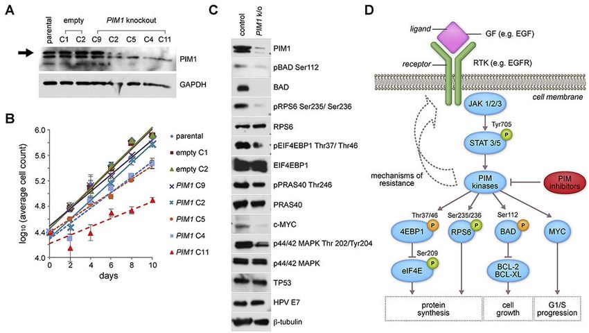
Broutian et al.                                                                                              Page 2

                             Pharmacological inhibition of PIM family kinases decreased growth of UPCI:SCC090 and
Author Manuscript

                             additional HNSCC cell lines in vitro and a xenograft UPCI:SCC090 model in vivo. Based on
                             established interactions between intracellular signaling pathways and relatively high levels of gene
                             expression in almost all HNSCC, we also evaluated combinations of PIM kinase and epidermal
                             growth factor receptor (EGFR) inhibitors. Dual inhibition of these pathways resulted in supra-
                             additive cell death. These data support clinical testing of PIM inhibitors alone or in combination in
                             HNSCC.

                       Keywords
                             head and neck squamous cell carcinoma; human papillomavirus (HPV); PIM serine/threonine
                             kinases; molecular therapeutics; virus insertional mutagenesis
Author Manuscript

                       1.      INTRODUCTION
                                       HNSCC ranks eighth in cancer incidence worldwide [1]. While most cases are attributable to
                                       tobacco and alcohol use, a subset of oropharyngeal cancers caused by HPV infection is
                                       increasing markedly in incidence in numerous developed countries [2]. The transforming
                                       ability of HPV partly has been linked to myriad functions of viral oncoproteins E6 and E7,
                                       which functionally inactivate tumor suppressor proteins p53 and pRb, respectively. While E6
                                       and E7 expression is sufficient for keratinocyte immortalization, secondary genetic events
                                       also are necessary for cellular transformation and formation of cancer [3].

                                       We previously reported a direct association between HPV insertions and various forms of
                                       flanking host genomic instability, including amplifications, rearrangements, deletions and
                                       translocations in human cancer cell lines and primary tumors [4]. Viral insertions frequently
Author Manuscript

                                       were accompanied by disruption in expression of neighboring host genes. We hypothesized
                                       that HPV-mediated alterations in gene expression or structure could serve as critical
                                       secondary genetic events in the pathogenesis of HPV-associated cancers [4]. For example,
                                       HPV insertions with flanking genomic rearrangements altered MYC expression in HeLa
                                       cells [5], GUMC395, a cell line derived from a cervical neuroendocrine cancer [6], and other
                                       cancer cell lines [7]. Marked alterations of MYC expression in these tumors may have
                                       contributed profoundly to their formation.

                                       In studying the HNSCC cell line UPCI:SCC090 using comprehensive genomics methods
                                       including whole genome sequencing (WGS) and RNA sequencing (RNA-Seq), we
                                       discovered HPV insertions flanking a 16-fold somatic amplification of PIM1 (Proviral
                                       insertion site for Moloney murine leukemia virus MuLV), a serine/threonine kinase proto-
Author Manuscript

                                       oncogene located on chromosome 6p21 (Fig. 1) [4]. This HPV insertion-mediated increase
                                       in PIM1 genomic copy number was accompanied by marked increases of PIM1 transcripts,
                                       which were not associated with expression of chimeric HPV-PIM1 fusion transcripts.

                                       The PIM kinase family (PIM1, PIM2, and PIM3) encodes constitutively active serine/
                                       threonine kinases that regulate cell cycle progression and apoptosis [8]. Pim1 originally was
                                       identified as a recurrent provirus integration site for the Moloney murine leukemia virus
                                       (Mo-MLV), resulting in mouse T cell lymphomas [9]. These viral insertions resulted in

                                              Cancer Lett. Author manuscript; available in PMC 2020 May 21.
Broutian et al.                                                                                           Page 3

                                      transcriptional upregulation of the gene, identifying it as a targetable cancer-causing driver
Author Manuscript

                                      gene. Subsequently, Pim2 was identified as another common viral insertion site in
                                      transplanted mouse T cell lymphomas, suggesting its important role in cancer progression
                                      [10]. The orthologous human gene, PIM1, also has been reported to be overexpressed in
                                      HNSCC [11]. Therefore, to confirm that HPV insertion-mediated alterations at PIM1 were
                                      required for the HNSCC cancer phenotype in UPCI:SCC090 cells, we investigated the
                                      antiproliferative and biochemical effects of genetic knockdown and small molecule
                                      inhibition of PIM kinases. To test the hypothesis that PIM kinases also contribute to HNSCC
                                      cancer formation more broadly, as candidate cancer-causing driver genes, we extended these
                                      experiments in additional HNSCC cell lines.

                       2.      MATERIALS AND METHODS
                       2.1.     Human HNSCC cancer cell lines and primary HNSCC tumor /normal pairs
Author Manuscript

                                      Human HNSCC cell lines FaDu (HPV-negative), SCC-4 (HPV-negative), SCC-9 (HPV-
                                      negative), SCC-15 (HPV-negative), CAL 27 (HPV-negative), Detroit 562 (hereafter called
                                      D562, HPV-negative), and SCC-25 (HPV-negative) were purchased from American Type
                                      Culture Collection (ATCC) [12–15]; UM-SCC-47 (HPV-positive) and UM-SCC-104 (HPV-
                                      positive) [15], kindly provided by Dr. Thomas Carey, University of Michigan;
                                      UPCI:SCC090 (HPV-positive) [16], Dr. Susanne M. Gollin, University of Pittsburgh; UD-
                                      SCC-2 (HPV-positive) [17], Dr. Henning Bier, University of Dusseldorf; and HMS001
                                      (HPV-positive) [4], Dr. James Rocco, Ohio State University. Cell lines were cultured
                                      according to instructions from ATCC, and as described previously [4] and in Supplementary
                                      Methods.

                                      Patients with newly diagnosed oral cavity or oropharyngeal squamous cell carcinoma
Author Manuscript

                                      presenting at Ohio State University from 2011-2016 provided written, informed consent to
                                      participate in genomics studies [3], approved by Institutional Review Boards at Ohio State
                                      University and at University of Texas MD Anderson Cancer Center. WGS and RNA-Seq
                                      were performed on 101 HPV-positive HNSCC tumor/normal (T/N) pairs, including 84 in the
                                      Ohio cohort and 17 downloaded from the TCGA website at https://gdc.cancer.gov/, and 50
                                      HPV-negative OSCC T/N pairs including 26 from our Ohio cohort and 24 from TCGA [3].

                       2.2.     Quantitative realtime PCR assays for PIM1 and PIM3 expression
                                      TaqMan assays to quantify PIM1 and PIM3 transcript levels were performed as described in
                                      Supplementary Methods.

                       2.3.     Lentiviral shRNA
Author Manuscript

                                      Knockdown of PIM1, PIM3 or FOXE1 was conducted using recombinant lentivirus to
                                      express specific or control scrambled shRNA sequences in the HNSCC cell lines UD-
                                      SCC-2, UM-SCC-47, UPCI:SCC090, CAL 27, and D562, as described in Supplementary
                                      Methods.

                                            Cancer Lett. Author manuscript; available in PMC 2020 May 21.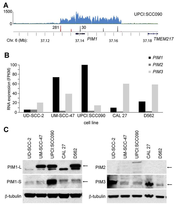
Broutian et al.                                                                                            Page 4

                       2.4.     Generation of PIM1 knockout clones using CRISPR/Cas9 gene editing
Author Manuscript

                                      CRISPR/Cas9-mediated genome editing was used to construct PIM1 knockout clones
                                      derived from UPCI:SCC090 parental cells. A lentiviral construct, LentiCRISPR-PIM1, was
                                      engineered to express a custom PIM1-specific guide RNA sequence, 5’-
                                      GTGGCGTGCAGGTCGTTGCA-3’, and functional effects of this genetic knockdown were
                                      assessed as described in Supplementary Methods.

                       2.5.     Analysis of protein expression and phosphorylation by Western blotting
                                      Expression and phosphorylation levels of proteins including PIM family kinases,
                                      downstream PIM kinase substrates and proteins upstream of the PIM signaling pathway
                                      were evaluated in HNSCC cell lines and CRISPR knockout clones by Western blot analysis,
                                      as described in Supplementary Methods.
Author Manuscript

                       2.6.     Cell cycle and apoptosis analysis
                                      Effects of PIM inhibition on the cell cycle and in inducing apoptosis were quantified using
                                      propidium iodide (Abcam) in flow cytometry and with the AlexaFluor 488 Annexin V dead
                                      cell apoptosis kit (Invitrogen Life Technologies), respectively, as described in
                                      Supplementary Methods.

                       2.7.     Mouse xenograft model
                                      Xenograft models in nude mice were generated by injection of UPCI:SCC090 cells. Female
                                      nude mice (Charles River) were maintained in compliance with a protocol approved by the
                                      Institutional Animal Care and Use Committee (Ohio State University). Mice were injected
                                      in the flank with 1.5x10⁶ UPCI:SCC090 cells in a 1:1 ratio of Matrigel (Corning). Twenty
                                      days after inoculation of cells, when tumor volumes reached ~150 to 200 mm3, mice (n = 7)
Author Manuscript

                                      were arbitrarily assigned to treatment twice daily via oral gavage either with 100mg/kg
                                      INCB053914 in 5% dimethylacetamide and 0.5% methylcellulose, or vehicle. Tumor
                                      volumes were measured every 2 days until euthanasia due to tumor growth and/or toxicity.

                       2.8.     Dual inhibition of PIM and EGFR pathways
                                      To assay effects of PIM pathway inhibition on cell viability, cell lines were treated with pan-
                                      PIM inhibitor INCB053914 (Incyte Corp.) [18], or the PIM1 inhibitors Quercetagetin (Santa
                                      Cruz Technologies) or PIM1 Inhibitor 2 (Santa Cruz Technologies), or a negative control,
                                      DMSO, as described in Supplementary Methods. For dual inhibitor studies of the PIM and
                                      EGFR pathways, cells were incubated with INCB053914 [18] and either erlotinib or afatinib
                                      (Selleckchem), individually or in combination, as described in Supplementary Methods.
Author Manuscript

                       2.9.     Informatics analysis of HNSCC samples
                                      Informatics analysis of PIM gene copy numbers, RNA-Seq data and associations between
                                      copy numbers and gene expression levels in primary HNSCC tumors was performed as
                                      described in Supplementary Methods.

                                            Cancer Lett. Author manuscript; available in PMC 2020 May 21.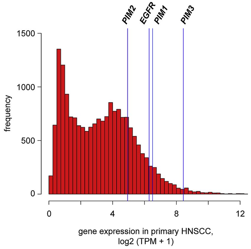
Broutian et al.                                                                                          Page 5

                       3.      RESULTS
Author Manuscript

                       3.1.     HPV integration upregulates PIM1 expression in UPCI:SCC090 cells
                                      We conducted WGS and RNA-Seq of UPCI:SCC090 cells and other HPV-positive and
                                      negative HNSCC cell lines, and of primary HNSCC tumor/ normal (T/N) pairs [3, 4]. In the
                                      HNSCC cell line UPCI:SCC090, we identified ~200 copies of HPV16 genomic DNA per
                                      cell, and found 33 HPV-host insertional breakpoints located on Chromosomes 3p12, 6p21,
                                      and 9q22 [4]. HPV integrants on Chr. 6p21 flanked an ~16-fold amplification of PIM1, a
                                      serine/threonine kinase gene (Fig. 1A). On Chr. 9q22, HPV integrants flanked an ~7 fold
                                      amplification of FOXE1, a member of the forkhead family of transcription factors [4].
                                      Amplification of PIM1 was associated with high levels of PIM1 transcript and protein
                                      expression, as assessed by RNA-Seq and Western blot (Fig. 1B, C). We detected only
                                      minimal numbers of chimeric HPV-PIM1 fusion transcripts, insufficient to account for the
Author Manuscript

                                      strong upregulation of PIM1 transcripts (Supp. Fig. S1). Both isoforms of PIM1 protein (i.e.
                                      PIM1-L [large], 44kDa; and PIM1-S [small], 33kDa) were highly expressed.

                       3.2.     PIM gene family expression varies widely in HNSCC cell lines
                                      We measured expression of all 3 members of the PIM family of kinases in UPCI:SCC090
                                      and additional HNSCC cell lines, including HPV-positive (i.e. UD-SCC-2 and UM-SCC-47)
                                      and HPV-negative (i.e. CAL 27 and D562) lines. Considerable variation was detected in
                                      PIM1 mRNA expression (i.e. ranging from ~10 to 120 fragments per thousand transcripts
                                      per million mapped reads, FPKM) and in PIM3 expression (i.e. 25-60 FPKM) based on
                                      RNA-seq data. By contrast, PIM2 expression was consistently low across these cell lines
                                      (Fig. 1B). Quantitation of PIM1, PIM2 and PIM3 expression by quantitative reverse
                                      transcriptase mediated polymerase chain reaction (qRT-PCR) assays confirmed high
Author Manuscript

                                      expression of PIM1 (in UPCI:SCC090, UM-SCC-47 and D562 cells) and PIM3 (in UD-
                                      SCC-2 and CAL 27 cells) (Supp. Fig. S2).

                                      Consistent with the known regulation of PIM1 expression at a transcriptional level [8, 19],
                                      PIM1 protein levels measured by Western blotting correlated closely with transcript levels as
                                      measured by RNA-Seq (Fig. 1C). By contrast, encoded PIM3 protein expression levels were
                                      not as closely associated with PIM3 transcript levels. For example, based on PIM3 transcript
                                      levels, PIM3 protein levels were higher than predicted in UD-SCC-2 cells, while they were
                                      lower than predicted in D562 cells (Fig. 1B, C). We surveyed expression of PIM1 and PIM3
                                      proteins in additional HNSCC cell lines, again highlighting wide-ranging variability in
                                      expression (Supp. Fig. S3).

                       3.3.     PIM knockdown reduces cell viability
Author Manuscript

                                      We hypothesized that HPV-mediated amplification of PIM1 and FOXE1 could contribute to
                                      cancer formation of UPCI:SCC090 cells. Therefore, we examined a potential relationship
                                      between expression levels of these genes and UPCI:SCC090 cell viability. To knockdown
                                      these genes, we obtained lentivirus vectors to express shRNA directed against PIM1,
                                      FOXE1, or a scrambled shRNA sequence as a control. After anti-PIM1 shRNA was
                                      introduced, UPCI:SCC090 cell viability was reduced by 82% as quantified by cell counting
                                      with Trypan blue exclusion. By comparison, treatment with anti-FOXE1 shRNA reduced

                                            Cancer Lett. Author manuscript; available in PMC 2020 May 21.
Broutian et al. Page 6
cell viability by 38%. As a control, treatment with scrambled shRNA reduced viability by
Author Manuscript
21% (Fig. 2A).
To evaluate impacts of PIM1 or PIM3 knockdown on viability of additional cell lines, we
transduced UD-SCC-2, UM-SCC-47, UPCI:SCC090, CAL 27, and D562 cells each with
recombinant lentiviruses to express the respective knockdown shRNAs or scrambled control.
Knockdown of PIM1 in D562 cells, which express high levels of PIM1 mRNA and protein,
resulted in ~60% reduction in cell viability. By contrast, D562 cell viability was unaffected
by PIM3 knockdown (Fig. 2B). The converse was true in UD-SCC-2 cells, which express
high levels of PIM3 but low PIM1. By contrast, viability of UM-SCC-47 and CAL 27 cells
was unaffected by PIM1 or PIM3 shRNA knockdown, despite high expression levels of
PIM1 and PIM3, respectively (Fig. 2B), suggesting potential compensatory driver genes or
pathways in them.
Author Manuscript
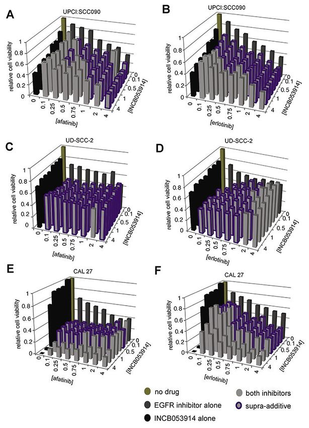
3.4. Genetic knockout of PIM1 reduces UPCI:SCC090 proliferation
Because genetic knockdown experiments involving shRNAs can produce off-target and
transient effects on phenotypes[20], we also generated stable PIM1 genetic knockout
subclones using CRISPR/Cas9 gene editing. This facilitated a comparison of their
proliferation vs. that of wildtype cells at various timepoints in culture. We designed guide
RNAs to target the first and fourth coding exons of PIM1 (Supp. Fig. S4). Five PIM1
knockout subclones (i.e. C9, C2, C5, C4, C11) were generated from parental UPCI:SCC090
cells by introduction of insertion-deletion (indel) and frameshift mutations in PIM1. Each
was confirmed by PCR amplification and Sanger sequencing of the targeted region, which
demonstrated that each subclonal line harbored both mutated and wildtype PIM1 sequences
(Supp. Fig. S5). A possible explanation for the remaining wildtype allele is that CRISPR/
Cas9 did not mutagenize all genomic copies of PIM1 per cell. Since the gene is amplified
Author Manuscript
16-fold in a concatenated array together with integrated HPV16 in parental UPCI:SCC090
cells (Supp. Fig. S1) [4], some wildtype alleles may have survived CRISPR-mediated gene
editing in resulting cell clones. An alternative explanation is that subclonal cells harboring
wildtype PIM1 persisted in mosaicism within each culture population, although each
subclone was grown up from isolated cells that were considered to be singletons. Despite
persistence of the wildtype allele in cloned lines, a 70 to 100% reduction in PIM1 protein
expression was confirmed by Western blot (Fig. 3A and Supp. Fig. S6).
These reductions in PIM1 protein expression, due to CRISPR-mediated genetic knockout in
UPCI:SCC090 subclones, correlated with reductions in their proliferation rates (Fig. 3B). A
nearly complete reduction of PIM1 expression in clone C11 was associated with a ~58%
reduction in proliferation rate when compared to control cells (Fig. 3B). By contrast,
Author Manuscript
minimal or more modest reductions of PIM1 expression in PIM clones C9 and C2 (Fig. 3A)
were associated with proliferation rates that were essentially unchanged when compared
with parental control cells (Fig. 3B). Upon serial passage of the CRISPR knockout clones,
we observed a gradual recovery in PIM1 protein expression accompanied by an accelerated
proliferation rate, again revealing an association between PIM1 expression levels and the
cells’ proliferation rates (data not shown).
Cancer Lett. Author manuscript; available in PMC 2020 May 21.Broutian et al. Page 7
Despite genetic knockdown of PIM1, no change in HPV E7 oncoprotein was detected (Fig.
Author Manuscript
3). Since only minimal numbers of RNA-Seq reads were identified that documented
expression of chimeric HPV-host transcripts (Supp. Fig. S1), this result confirmed our
prediction that PIM1 disruption would not affect the autonomously expressed, independent
E7 oncogene.
3.5. Downstream consequences of PIM1 genetic knockout
To explore potential mechanisms for reduced cell proliferation of PIM1 knockout cells, we
examined phosphorylation levels of PIM1 kinase substrates in knockout clone C11. PIM1
has been shown to inhibit apoptosis through phosphorylation of Ser112 of the pro-apoptotic
BCL2-associated agonist of death promoter (BAD) protein, thereby inducing anti-apoptotic
activity of BCL2/BCL-xL and increasing cell survival [21]. Western blotting confirmed
marked decreases in PIM1, pBAD Ser112 phosphorylation and total BAD levels in the
Author Manuscript
knockout cells (Fig. 3 and Supp. Fig. S6).
PIM1 also has been shown to regulate protein synthesis downstream of the
phosphatidylinositol-4,5-bisphosphonate 3-kinase catalytic subunit alpha, AKT serine/
threonine kinase, mammalian target of rapamycin (PIK3CA/AKT/mTOR) signaling
pathway. It activates ribosomal protein S6 (RPS6, also termed S6) via phosphorylation of
Ser235/236, and inhibits eukaryotic translation initiation factor 4E-binding protein
(EIF4EBP1) via phosphorylation of Thr37 and Thr46, thereby promoting cap-dependent
translation and cell growth [22, 23]. Upon knockdown of PIM1 in UPCI:SCC090 cells, we
observed decreased phosphorylation of pRPS6 and of EIF4EBP1 (i.e. pEIF4EBP1),
respectively, while overall RPS6 and EIF4EBP1 protein levels remained unchanged (Figs.
3C and 3D). However, upon knockdown of PIM1, we did not observe decreases in the PIM1
phosphorylation target AKT1 substrate 1 (AKT1S1) at Thr246 or in total AKT1S1 levels
Author Manuscript
(not shown), in contrast to treated leukemia cells [24].
PIM1 protein reportedly stabilizes MYC protein and promotes prostate cancer tumorigenesis
through activation of mitogen-activated protein kinase 1 (MAPK1, also termed ERK) [25].
Decreased levels of MYC protein and phosphorylated MAPK1 (termed pMAPK1,
phosphorylated at Thr 202/Tyr204) were observed in the CRISPR-generated PIM1 knockout
clone C11 when compared with control (Figs. 3C and 3D). Similarly, reduced levels of
MYC and phosphorylated MYC were observed in another CRISPR knockout clone (Supp.
Fig. S6). By contrast, both HPV16 E7 and p53 protein expression levels were unaffected by
PIM1 knockdown, arguing against an explanation for reduced cellular proliferation that
involves off-target effects on HPV oncoproteins (Fig. 3C). We conclude that reduced
proliferation and increased cell death upon PIM1 knockdown may be attributed to a
Author Manuscript
combination of factors including inhibition of protein synthesis, generation of pro-apoptotic
signals, and cell cycle arrest due to reduction in MYC function.
3.6. Pan-PIM Inhibitors inhibit HNSCC cell line growth
Genetic knockdown or knockout of PIM1 reduced UPCI-SCC090 cells’ viability and
proliferation. These observations prompted us to investigate the effect of small molecule
inhibitors of PIM kinase activity upon cell viability in our panel of HNSCC cell lines.
Cancer Lett. Author manuscript; available in PMC 2020 May 21.Broutian et al. Page 8
Several PIM kinase inhibitors have been developed for potential therapeutic uses. We
Author Manuscript
evaluated the anti-proliferative effects of INCB053914 [18], PIM1 Inhibitor 2 [26], and
quercetagetin [27], using neutral red and WST-1 cell viability assays (Fig. 4A, Supp. Fig.
S7). INCB053914 is a pan-PIM inhibitor, while PIM1 inhibitor 2 and quercetagetin are
competitive inhibitors of ATP binding to the PIM1 active site.
Consistent with shRNA knockdown experiments, UPCI:SCC090, UD-SCC-2 and D562 cells
each were sensitive to treatments with the pan-PIM inhibitor INCB053914 at low doses. The
doses at which growth was half-maximally inhibited (EC50) ranged from approximately
0.045 to 8 μM (Fig. 4A). By contrast, UM-SCC-47 and CAL 27 cells were more resistant to
this drug (Fig. 4A). Similar findings were obtained upon fitting drug inhibition curves
according to a non-linear model (not shown). Treatment of seven additional HNSCC cell
lines (i.e. SCC-9, SCC15, SCC-25, HMS-001, UM-SCC-104, SCC4, and FaDu cells) with
INCB053914 revealed EC50 < 8 μM in SCC-9 and SCC15 cells (Supp. Fig. S8, Supp. Table
Author Manuscript
S1). In total, 5 of 12 HNSCC cell lines were at least modestly sensitive to the pan-PIM
inhibitor INCB053914, as defined by EC50Broutian et al. Page 9
To elucidate possible mechanisms of sensitivity or resistance to INCB053914, we evaluated
Author Manuscript
alterations in phosphorylation of PIM1 substrates by Western blot. In sensitive cell lines
such as UPCI:SCC090, UD-SCC-2, and D562, we observed a consistent, dose-dependent
decrease in pRPS6 phosphorylation at serine residues 235/236 (Supp. Fig. S10). By contrast,
levels of pRPS6 phosphorylation were essentially unchanged in resistant cells such as UM-
SCC-47 treated with a wide range of doses of INCB053914 (Supp. Fig. S11).
A functional deficiency of phosphorylated pRPS6 has been linked to reduced cell size [22,
28]. Consistent with this report, UPCI:SCC090 and UD-SCC-2 cells displayed 44% and
29% reductions in mean cell sizes, respectively, following 48 hours of exposure to 1.25μM
INCB053914. The cell sizes were measured by flow cytometry as mean forward scatter
height (FSC-H) of the gated G1 cell population. By contrast, UM-SCC-47 cells, resistant to
INCB053914, displayed only a 7% decrease in cell size upon such treatment (Supp. Fig.
S9).
Author Manuscript
When assessing phosphorylation of other PIM substrates after treatment with INCB053914,
we observed reductions in phosphorylation of BAD serine 112 in drug-sensitive
UPCI:SCC090 and D562 cells (Supp. Fig. S11). In the former cells, reductions in BAD
phosphorylation were modest, whereas they were more robust in D562 cells. By contrast, no
such inhibition was observed in drug-sensitive UD-SCC-2 cells. This indicated that
modulation of phosphorylated pBAD is not required for growth inhibition. We also did not
observe altered phosphorylation of AKT1S1 at Thr246 as previously reported [24]. Protein
levels of MYC varied with INCB053914 treatment for each cell line (Supp. Fig. S11).
Western blot analysis of UM-SCC-47 cell lysates showed an increase in inactivating
phosphorylation of EIF4EBP1 at Thr37 and Thr46, which negatively regulates cap-
dependent translation. In addition, increased activating phosphorylation of eIF4E protein at
Author Manuscript
Ser209 was observed (Supp. Fig. S11). These modifications promote protein synthesis and
could contribute to modest reductions in size of UM-SCC-47 cells upon treatment with
INCB053914 (Supp. Fig. S9).
CAL 27 cells display high levels of PIM3 expression, but their viability was not affected
either by pan-PIM inhibitor treatment or by genetic knockdown (Fig. 4A). Nevertheless, we
observed modest, dose-dependent reductions in phosphorylation of pRPS6 at Ser235/236
(Supp. Fig. S12) and more robust decreases in phosphorylation of BAD at Ser112,
phosphorylation of EIF4EBP1 at Thr37 and Thr46, and phosphorylation of eIF4E at Ser209,
in response to treatment by pan-PIM inhibitor INCB053914 (Supp. Fig. S12). However,
expression levels of activated, phosphorylated signal transducer and activator of transcription
3 (STAT3) at Tyr705 and of PIM3 were increased upon treatment with INCB053914 (Supp.
Author Manuscript
Fig. S12). Since PIM expression has been reported to be regulated at the level of
transcription by STAT proteins, we hypothesized that CAL 27 resistance to PIM inhibition
could involve up-regulation of PIM3 by interacting signaling feedback loops involving
STAT3 activation (Supp. Fig. S12).
3.9. Supra-additive inhibition of growth by pan-PIM and EGFR inhibitors
The oncogenic pathways driven by EGFR are interconnected with the complex network of
PIM signaling pathways. A recent study showed that EGF and TGF-alpha signaling through
Cancer Lett. Author manuscript; available in PMC 2020 May 21.Broutian et al. Page 10
EGFR can induce increased expression and nuclear translocation of PIM1 in 5 HNSCC cell
Author Manuscript
lines, contributing to radiation resistance [29]. We noted that cell lines resistant to pan-PIM
inhibitors (i.e. UM-SCC-47 and CAL 27) harbored ~10-fold amplification of EGFR copy
number as shown by WGS [4], but sensitive cell lines had no such amplification (Supp.
Table S4).
To test the hypothesis that combined inhibition of both upstream EGFR and downstream
PIM1 could demonstrate supra-additivity, we assayed viability of the HNSCC cell lines
UPCI:SCC090, UD-SCC-2, UM-SCC-47, CAL 27 and D562 after 72 h exposure to various
concentrations of both INCB053914 and a small molecule EGFR inhibitor, i.e. afatinib or
erlotinib. As indicated by a combination index (CI) 2.5) was identified in 22 (14.6%) of
151 primary HNSCC tumors (Supp. Table S5). These were identified more frequently in 101
HPV-positive HNSCC (PIM1, n=6; PIM2, n=11; PIM3, n=1; total 17.8%) than in 50 HPV-
negative tumors (all PIM family members, n=4; total 8%). We also observed EGFR
amplification in 2 (2.0%) of 101 HPV-positive tumors and in 14 (28%) of 50 HPV-negative
tumors (Supp. Table S5).
Mean PIM1 mRNA expression levels were significantly higher in tumors with PIM1 copy
number amplification (expressed as log2(transcript fragments per kilobase million), TPM),
Supp. Fig. S14). However, similar associations were not observed for PIM2 or PIM3 (Supp.
Fig. S15). Mean EGFR expression levels were significantly higher in tumors with EGFR
Author Manuscript
amplification (Supp. Fig. S14), as previously reported [30, 31]. However, EGFR copy
number amplification was not associated with PIM gene expression in our panel of HNSCC
cell lines (Supp. Fig. S16).
RNA-seq analysis of the 151 primary HNSCC tumors revealed high levels of expression
(defined as TPM ≥ 30) of PIM1, PIM2, and PIM3 in 146 (96.7%), 75 (49.7%) and 151
(100%) of the samples, respectively (Fig. 6, Supp. Table S6). When compared with
expression of all annotated genes across these primary HNSCC tumors, median PIM1
Cancer Lett. Author manuscript; available in PMC 2020 May 21.Broutian et al. Page 11
transcript levels ranked in the top 6.5th percentile, PIM2 ranked in the 21.4th percentile,
Author Manuscript
PIM3 ranked in the top 1.3rd percentile and EGFR ranked in the 7.7th percentile (Fig. 6). By
comparison, based on the same definition, only ~23% of 18,640 RefSeq genes were
expressed at these high median levels across the 151 HNSCC samples (Fig. 6). While high
levels of PIM1 and PIM3 expression were detected in almost all of the HPV-positive and
HPV-negative HNSCC tumors, high levels of PIM2 expression were identified in a majority
of only the HPV-positive tumors (Supp. Table S6). EGFR was highly expressed (TPM ≥ 30)
in 134 (88.7%) of the HNSCC tumors studied.
4. DISCUSSION
The transforming ability of HPV has been attributed primarily to inactivation of host p53
and pRb family members by viral E6 and E7 oncoproteins, respectively [32]. While
expression of E6 and E7 is essential to the viral life cycle and sufficient for cellular
Author Manuscript
immortalization, secondary genetic events are necessary for development of cancer.
Analyses of genomic landscapes of HNSCC [3, 33, 34] and cervical cancers [35] by next
generation DNA sequencing have identified numerous recurrent somatic mutations and
genomic structural rearrangements. We and others have reported that HPV can act as an
insertional mutagen, wherein HPV integration is directly associated with alterations in host
genome structure and expression [4, 5, 7, 36]. We hypothesize that resulting alterations can
drive clonal selection and are necessary for the malignant phenotype, and the data presented
here support this hypothesis. As reported here, the study of HPV integration has led to the
identification of a candidate driver gene (i.e. PIM1 kinase) that is a rational target for
therapeutic agent development in HNSCC. Notably, groundbreaking studies of Mo-MuLV
insertions in mouse lymphomas led to the initial identification of members of the same
family of serine/threonine kinase genes [9, 10], activated by genomic insertions of a distinct
Author Manuscript
virus in a different species and tissue type, again driving cancer formation or progression.
We previously showed that HPV insertion at PIM1 in the HNSCC cell line UPCI:SCC090
was associated with flanking genomic amplification and markedly increased expression of
that serine/threonine kinase gene [4]. The 16-fold amplification of the PIM1 gene, its
marked overexpression, and its well-documented role as a candidate oncogene in other
cancers [37, 38] prompted us to investigate the hypothesis that the large-scale upregulation
of PIM1 in UPCI:SCC090 cells, attributable to virus integration, could have contributed to
the malignant phenotype and formation of this particular HNSCC [39]. We detected very
low expression levels of chimeric HPV-PIM1 fusion transcripts, which we concluded were
insufficient to account for the strong upregulation of PIM1. We experimentally inhibited
PIM1 in UPCI:SCC090 cells at the DNA, RNA, and protein levels, using CRISPR-mediated
Author Manuscript
genome editing, shRNA inhibition, and several pharmacological inhibitors. Resulting
reductions of PIM1 serine/threonine kinase activity consistently were associated with
decreased UPCI:SCC090 cell viability and proliferation, and also resulted in well-
documented changes in phosphorylation of PIM substrates. Treatment with the pan-PIM
inhibitor INCB053914 significantly decreased both UPCI:SCC090 cell growth in vitro and
tumor volume in a mouse xenograft model in vivo. The growth of additional HNSCC cell
lines also was inhibited by similar experimental treatments.
Cancer Lett. Author manuscript; available in PMC 2020 May 21.Broutian et al. Page 12
Subclonal cells derived from UPCI:SCC090 that underwent CRISPR/Cas9-mediated genetic
Author Manuscript
mutagenesis of PIM1 displayed no changes in HPV E7 expression (Fig. 3). This result was
expected, since minimal chimeric HPV-PIM1 fusion transcripts were detected in parental
cells (Supp. Fig. S1). These proof-of-principle data support the hypothesis that HPV-
mediated genetic rearrangements could alter expression, structure and function of candidate
cancer-causing driver genes, and thereby contribute directly to cancer formation independent
of viral oncogene expression. Comparable evidence was obtained in HPV-positive cervical
neuroendocrine cancer GUMC395 cells, where experimental knockdown of MYC adjacent
to sites of HPV integration resulted in decreased cell viability. These effects again were
independent of the viral E6 and E7 transcripts [6].
PIM1 overexpression has been identified in HNSCC compared with matched normal
controls, and has been associated with poor survival [11, 29, 40]. Here, we identified high
levels of expression of at least one member of the PIM kinase family in almost all primary
Author Manuscript
HNSCC (Fig. 6, Supp. Table S6). We attributed this finding in part to genomic copy number
gains at these genes, found in ~13% of HNSCC. Overexpression of PIM family kinases also
has been reported in numerous hematological malignancies and other solid tumors,
including Hodgkin lymphoma [41], multiple myeloma [42], prostate [43], pancreatic [44]
and triple negative breast cancers [45]. PIM kinase overexpression may contribute to
chemotherapy resistance [46] and cellular survival by blocking apoptotic cell death in the
context of oncogene-induced stress [47] and hypoxia [48].
In the case of the UPCI:SCC090 cell line, PIM1 overexpression was attributable to HPV-
integrant mediated amplification, but we observed PIM family kinases to be overexpressed
in other cell lines. This is likely because PIM1 kinase is downstream of several cellular
signaling pathways frequently activated by host genetic alterations in HNSCC, including the
Author Manuscript
PIK3CA/AKT/mTOR, EGFR and NFKB signaling pathways [3, 49]. PIM1 also increases
the stability and transcriptional activity of MYC [50], and the PIM kinases and MYC are
recognized oncogenic collaborators in cellular transformation [8]. The substrates of PIM
kinase phosphorylation in turn regulate several critical cellular processes, including cell
cycle progression (e.g. p21, p27), cell growth (e.g. eIF4E, 4EPB2), and cell death (e.g. BAD,
BCL2; Fig. 3D) [8]. As a regulator of cell metabolism and growth, PIM1 phosphorylates
several substrates in the PIK3CA/AKT/mTOR pathway. PIM1 inactivates negative
regulators of this pathway (i.e. TSC2 and AKT1S1) and promotes pathway activation
through eIF4B phosphorylation. As shown here, sensitivity to INCB053914 was linked to
reduced levels of phosphorylated pRPS6 protein.
In several experimental models of other solid tumors, overexpression of PIM kinases has
Author Manuscript
been implicated in resistance to PIK3CA/AKT/mTOR inhibitors. For example, in breast
cancer cells treated with PI3K inhibitors, upregulation of PIM1 serves as a secondary
resistance mechanism [51]. Similarly, resistance to PI3K-AKT inhibition in a prostate cancer
model is mediated by PIM1 kinase [52]. In this latter model, PIM1 increased translation of
the transcription factor NFR2 and downstream regulators of ROS scavengers and
metabolism, promoting cell survival [53]. Treatment with a combination of PI3K/AKT and
PIM kinase inhibitors overcame this resistance [52].
Cancer Lett. Author manuscript; available in PMC 2020 May 21.Broutian et al. Page 13
Several HNSCC cell lines with high PIM expression demonstrated sensitivity to PIM
Author Manuscript
inhibitors. However, we also found that a few cell lines with high levels of PIM1 and PIM3
expression, respectively, were nevertheless resistant to PIM inhibition (i.e. UM-SCC-47 and
CAL 27), in agreement with a previous report about PIM in other cancers [46]. Further
analysis of factors upstream and downstream of PIM suggested potential mechanisms of
resistance. Treatment of CAL 27 cells with INCB053914 induced compensatory STAT3
activation and increased PIM3 expression. By contrast, treatment of UM-SCC-47 cells
resulted in increased phosphorylation of EIF4EBP1 and eIF4E proteins, suggesting
alternative mechanisms of mTOR pathway activation. Both cell lines harbor EGFR gene
amplification [4], raising the possibility that increased signaling through the activated EGFR
pathway might promote PIM inhibitor resistance.
Based on these findings, we inferred that frequent, aberrant activation of signaling pathways
upstream of PIM1 (including the EGFR and PIK3CA/AKT/mTOR pathways) in HNSCC
Author Manuscript
could result in decreased sensitivity to PIM inhibition (Fig. 3D). Therefore, we tested effects
of dual PIM and EGFR inhibition. We found strong supra-additivity in cell lines treated with
various combined doses of inhibitors, regardless of their sensitivity to PIM inhibition alone.
These data demonstrating supra-additivity in HNSCC are consistent with recently
demonstrated synergy between PIM inhibitors and PI3K inhibitors in prostate cancer [54],
MET inhibitors in lung cancer[55], mTOR inhibitors in leukemia [56] and Janus kinase
(JAK) inhibitors in myeloproliferative neoplasms [57, 58].
We note that multiple mechanisms of resistance to EGFR inhibitors may limit their efficacy
in the clinic, including acquisition of secondary EGFR mutations, synchronous activation of
redundant receptor tyrosine kinases (e.g. c-Met or G-protein-coupled receptors), and
aberrant activation of bypass pathways including the MAPK, RAS and PI3K/AKT pathways
Author Manuscript
which maintain mitogenic signaling [59, 60]. Several proteins in these pathways activate
PIM family kinases.
In summary, HPV insertion-mediated amplification of PIM1 in an HNSCC cell line
prompted us to investigate that kinase gene’s functional roles in cancer cell viability and
growth. The preclinical data shown here strongly suggest that the use of PIM inhibitors in
HNSCC could provide a promising new therapeutic approach. In addition, since PIM
kinases are involved in several signaling pathways dysregulated in cancer, including the
EGFR pathway, the future clinical development of PIM inhibitors could be enhanced by
studies of synergistic combinations of therapeutic agents that inhibit multiple targets in such
pathways.
Author Manuscript
Supplementary Material
Refer to Web version on PubMed Central for supplementary material.
ACKNOWLEDGMENTS
We thank the patients with oropharyngeal and oral cavity cancers at Ohio State University who enrolled in our
genomics study. We thank Dr. Holly Koblish at Incyte Corporation; Dr. Faye M. Johnson at MD Anderson Cancer
Center; members of Dr. Broutian’s doctoral thesis advisory committee including Drs. Gillison, Quintin Pan, Dawn
Chandler and Nyla Heerema; and members of the Gillison and Symer laboratories for insightful comments at
Cancer Lett. Author manuscript; available in PMC 2020 May 21.Broutian et al. Page 14
various stages of this study; TCGA for access to WGS and RNA-seq data; and Jordan Pietz and Jeff Flasik (MDA)
for help preparing graphical figures. We gratefully acknowledge Incyte Corporation for providing the pan-PIM
Author Manuscript
inhibitor INCB053914.
Financial support: This study was funded by the Cancer Prevention Research Institute of Texas (RR170005;
MLG), University of Texas MD Anderson Cancer Center (DES, MLG), the Ohio State University Comprehensive
Cancer Center (DES, MLG), the Ohio Supercomputer Center (PAS0425; DES), the Oral Cancer Foundation
(MLG), and National Cancer Institute grant R50CA211533 (K.A.). Dr. Gillison is a CPRIT Scholar. We
acknowledge the Analytical Cytometry Shared Resource and Genomics Shared Resource at OSUCCC, supported
by NCI CCSG P30CA016058. We gratefully acknowledge Incyte Corporation for providing the pan-PIM inhibitor
INCB053914.
REFERENCES
[1]. Bray F, Ferlay J, Soerjomataram I, Siegel RL, Torre LA, Jemal A, Global cancer statistics 2018:
GLOBOCAN estimates of incidence and mortality worldwide for 36 cancers in 185 countries,
CA Cancer J Clin, 68 (2018) 394–424. [PubMed: 30207593]
[2]. Chaturvedi AK, Anderson WF, Lortet-Tieulent J, Curado MP, Ferlay J, Franceschi S, Rosenberg
Author Manuscript
PS, Bray F, Gillison ML, Worldwide trends in incidence rates for oral cavity and oropharyngeal
cancers, J Clin Oncol, 31 (2013) 4550–4559. [PubMed: 24248688]
[3]. Gillison ML, Akagi K, Xiao W, Jiang B, Pickard RKL, Li J, Swanson BJ, Agrawal AD, Zucker M,
Stache-Crain B, Emde AK, Geiger HM, Robine N, Coombes KR, Symer DE, Human
papillomavirus and the landscape of secondary genetic alterations in oral cancers, Genome Res,
29 (2019) 1–17.
[4]. Akagi K, Li J, Broutian TR, Padilla-Nash H, Xiao W, Jiang B, Rocco JW, Teknos TN, Kumar B,
Wangsa D, He D, Ried T, Symer DE, Gillison ML, Genome-wide analysis of HPV integration in
human cancers reveals recurrent, focal genomic instability, Genome Res, 24 (2014) 185–199.
[PubMed: 24201445]
[5]. Adey A, Burton JN, Kitzman JO, Hiatt JB, Lewis AP, Martin BK, Qiu R, Lee C, Shendure J, The
haplotype-resolved genome and epigenome of the aneuploid HeLa cancer cell line, Nature, 500
(2013) 207–211. [PubMed: 23925245]
[6]. Yuan H, Krawczyk E, Blancato J, Albanese C, Zhou D, Wang N, Paul S, Alkhilaiwi F, Palechor-
Ceron N, Dakic A, Fang S, Choudhary S, Hou TW, Zheng YL, Haddad BR, Usuda Y, Hartmann
Author Manuscript
D, Symer D, Gillison M, Agarwal S, Wangsa D, Ried T, Liu X, Schlegel R, HPV positive
neuroendocrine cervical cancer cells are dependent on Myc but not E6/E7 viral oncogenes, Sci
Rep, 7 (2017) 45617. [PubMed: 28378747]
[7]. Durst M, Croce CM, Gissmann L, Schwarz E, Huebner K, Papillomavirus sequences integrate near
cellular oncogenes in some cervical carcinomas, Proc Natl Acad Sci U S A, 84 (1987) 1070–
1074. [PubMed: 3029760]
[8]. Nawijn MC, Alendar A, Berns A, For better or for worse: the role of Pim oncogenes in
tumorigenesis, Nat Rev Cancer, 11 (2011) 23–34. [PubMed: 21150935]
[9]. Cuypers HT, Selten G, Quint W, Zijlstra M, Maandag ER, Boelens W, van Wezenbeek P, Melief C,
Berns A, Murine leukemia virus-induced T-cell lymphomagenesis: integration of proviruses in a
distinct chromosomal region, Cell, 37 (1984) 141–150. [PubMed: 6327049]
[10]. Breuer ML, Cuypers HT, Berns A, Evidence for the involvement of pim-2, a new common
proviral insertion site, in progression of lymphomas, EMBO J, 8 (1989) 743–748. [PubMed:
2721500]
Author Manuscript
[11]. Beier UH, Weise JB, Laudien M, Sauerwein H, Gorogh T, Overexpression of Pim-1 in head and
neck squamous cell carcinomas, Int J Oncol, 30 (2007) 1381–1387. [PubMed: 17487358]
[12]. Rheinwald JG, Beckett MA, Tumorigenic keratinocyte lines requiring anchorage and fibroblast
support cultured from human squamous cell carcinomas, Cancer Res, 41 (1981) 1657–1663.
[PubMed: 7214336]
[13]. Gioanni J, Fischel JL, Lambert JC, Demard F, Mazeau C, Zanghellini E, Ettore F, Formento P,
Chauvel P, Lalanne CM, et al., Two new human tumor cell lines derived from squamous cell
carcinomas of the tongue: establishment, characterization and response to cytotoxic treatment,
Eur J Cancer Clin Oncol, 24 (1988) 1445–1455. [PubMed: 3181269]
Cancer Lett. Author manuscript; available in PMC 2020 May 21.Broutian et al. Page 15
[14]. Peterson WD Jr., Stulberg CS, Simpson WF, A permanent heteroploid human cell line with type
B glucose-6-phosphate dehydrogenase, Proc Soc Exp Biol Med, 136 (1971) 1187–1191.
Author Manuscript
[PubMed: 5554463]
[15]. Cooper T, Biron VL, Fast D, Tam R, Carey T, Shmulevitz M, Seikaly H, Oncolytic activity of
reovirus in HPV positive and negative head and neck squamous cell carcinoma, J Otolaryngol
Head Neck Surg, 44 (2015) 8. [PubMed: 25890191]
[16]. White JS, Weissfeld JL, Ragin CC, Rossie KM, Martin CL, Shuster M, Ishwad CS, Law JC,
Myers EN, Johnson JT, Gollin SM, The influence of clinical and demographic risk factors on the
establishment of head and neck squamous cell carcinoma cell lines, Oral Oncol, 43 (2007) 701–
712. [PubMed: 17112776]
[17]. Lin CJ, Grandis JR, Carey TE, Gollin SM, Whiteside TL, Koch WM, Ferris RL, Lai SY, Head
and neck squamous cell carcinoma cell lines: established models and rationale for selection,
Head Neck, 29 (2007) 163–188. [PubMed: 17312569]
[18]. Koblish H, Li YL, Shin N, Hall L, Wang Q, Wang K, Covington M, Marando C, Bowman K,
Boer J, Burke K, Wynn R, Margulis A, Reuther GW, Lambert QT, Dostalik Roman V, Zhang K,
Feng H, Xue CB, Diamond S, Hollis G, Yeleswaram S, Yao W, Huber R, Vaddi K, Scherle P,
Author Manuscript
Preclinical characterization of INCB053914, a novel pan-PIM kinase inhibitor, alone and in
combination with anticancer agents, in models of hematologic malignancies, PLoS One, 13
(2018) e0199108. [PubMed: 29927999]
[19]. Saurabh K, Scherzer MT, Shah PP, Mims AS, Lockwood WW, Kraft AS, Beverly LJ, The PIM
family of oncoproteins: small kinases with huge implications in myeloid leukemogenesis and as
therapeutic targets, Oncotarget, 5 (2014) 8503–8514. [PubMed: 25238262]
[20]. Lin A, Giuliano CJ, Palladino A, John KM, Abramowicz C, Yuan ML, Sausville EL, Lukow DA,
Liu L, Chait AR, Galluzzo ZC, Tucker C, Sheltzer JM, Off-target toxicity is a common
mechanism of action of cancer drugs undergoing clinical trials, Sci Transl Med, 11 (2019).
[21]. Macdonald A, Campbell DG, Toth R, McLauchlan H, Hastie CJ, Arthur JS, Pim kinases
phosphorylate multiple sites on Bad and promote 14–3-3 binding and dissociation from Bcl-XL,
BMC Cell Biol, 7 (2006) 1. [PubMed: 16403219]
[22]. Ruvinsky I, Sharon N, Lerer T, Cohen H, Stolovich-Rain M, Nir T, Dor Y, Zisman P, Meyuhas O,
Ribosomal protein S6 phosphorylation is a determinant of cell size and glucose homeostasis,
Genes Dev, 19 (2005) 2199–2211. [PubMed: 16166381]
Author Manuscript
[23]. Schatz JH, Oricchio E, Wolfe AL, Jiang M, Linkov I, Maragulia J, Shi W, Zhang Z, Rajasekhar
VK, Pagano NC, Porco JA Jr., Teruya-Feldstein J, Rosen N, Zelenetz AD, Pelletier J, Wendel
HG, Targeting cap-dependent translation blocks converging survival signals by AKT and PIM
kinases in lymphoma, J Exp Med, 208 (2011) 1799–1807. [PubMed: 21859846]
[24]. Zhang F, Beharry ZM, Harris TE, Lilly MB, Smith CD, Mahajan S, Kraft AS, PIM1 protein
kinase regulates PRAS40 phosphorylation and mTOR activity in FDCP1 cells, Cancer Biol Ther,
8 (2009) 846–853. [PubMed: 19276681]
[25]. Wang J, Kim J, Roh M, Franco OE, Hayward SW, Wills ML, Abdulkadir SA, Pim1 kinase
synergizes with c-MYC to induce advanced prostate carcinoma, Oncogene, 29 (2010) 2477–
2487. [PubMed: 20140016]
[26]. Pierce AC, Jacobs M, Stuver-Moody C, Docking study yields four novel inhibitors of the
protooncogene Pim-1 kinase, J Med Chem, 51 (2008) 1972–1975. [PubMed: 18290603]
[27]. Holder S, Zemskova M, Zhang C, Tabrizizad M, Bremer R, Neidigh JW, Lilly MB,
Characterization of a potent and selective small-molecule inhibitor of the PIM1 kinase, Mol
Author Manuscript
Cancer Ther, 6 (2007) 163–172. [PubMed: 17218638]
[28]. Fingar DC, Salama S, Tsou C, Harlow E, Blenis J, Mammalian cell size is controlled by mTOR
and its downstream targets S6K1 and 4EBP1/eIF4E, Genes Dev, 16 (2002) 1472–1487.
[PubMed: 12080086]
[29]. Peltola K, Hollmen M, Maula SM, Rainio E, Ristamaki R, Luukkaa M, Sandholm J, Sundvall M,
Elenius K, Koskinen PJ, Grenman R, Jalkanen S, Pim-1 kinase expression predicts radiation
response in squamocellular carcinoma of head and neck and is under the control of epidermal
growth factor receptor, Neoplasia, 11 (2009) 629–636. [PubMed: 19568408]
Cancer Lett. Author manuscript; available in PMC 2020 May 21.Broutian et al. Page 16
[30]. Sheu JJ, Hua CH, Wan L, Lin YJ, Lai MT, Tseng HC, Jinawath N, Tsai MH, Chang NW, Lin CF,
Lin CC, Hsieh LJ, Wang TL, Shih Ie M, Tsai FJ, Functional genomic analysis identified
Author Manuscript
epidermal growth factor receptor activation as the most common genetic event in oral squamous
cell carcinoma, Cancer Res, 69 (2009) 2568–2576. [PubMed: 19276369]
[31]. Leemans CR, Braakhuis BJ, Brakenhoff RH, The molecular biology of head and neck cancer, Nat
Rev Cancer, 11 (2011) 9–22. [PubMed: 21160525]
[32]. Munger K, Baldwin A, Edwards KM, Hayakawa H, Nguyen CL, Owens M, Grace M, Huh K,
Mechanisms of human papillomavirus-induced oncogenesis, J Virol, 78 (2004) 11451–11460.
[PubMed: 15479788]
[33]. Cancer Genome Atlas Network, Comprehensive genomic characterization of head and neck
squamous cell carcinomas, Nature, 517 (2015) 576–582. [PubMed: 25631445]
[34]. Stransky N, Egloff AM, Tward AD, Kostic AD, Cibulskis K, Sivachenko A, Kryukov GV,
Lawrence MS, Sougnez C, McKenna A, Shefler E, Ramos AH, Stojanov P, Carter SL, Voet D,
Cortes ML, Auclair D, Berger MF, Saksena G, Guiducci C, Onofrio RC, Parkin M, Romkes M,
Weissfeld JL, Seethala RR, Wang L, Rangel-Escareno C, Fernandez-Lopez JC, Hidalgo-Miranda
A, Melendez-Zajgla J, Winckler W, Ardlie K, Gabriel SB, Meyerson M, Lander ES, Getz G,
Author Manuscript
Golub TR, Garraway LA, Grandis JR, The mutational landscape of head and neck squamous cell
carcinoma, Science, 333 (2011) 1157–1160. [PubMed: 21798893]
[35]. Ojesina AI, Lichtenstein L, Freeman SS, Pedamallu CS, Imaz-Rosshandler I, Pugh TJ, Cherniack
AD, Ambrogio L, Cibulskis K, Bertelsen B, Romero-Cordoba S, Trevino V, Vazquez-Santillan K,
Guadarrama AS, Wright AA, Rosenberg MW, Duke F, Kaplan B, Wang R, Nickerson E, Walline
HM, Lawrence MS, Stewart C, Carter SL, McKenna A, Rodriguez-Sanchez IP, Espinosa-Castilla
M, Woie K, Bjorge L, Wik E, Halle MK, Hoivik EA, Krakstad C, Gabino NB, Gomez-Macias
GS, Valdez-Chapa LD, Garza-Rodriguez ML, Maytorena G, Vazquez J, Rodea C, Cravioto A,
Cortes ML, Greulich H, Crum CP, Neuberg DS, Hidalgo-Miranda A, Escareno CR, Akslen LA,
Carey TE, Vintermyr OK, Gabriel SB, Barrera-Saldana HA, Melendez-Zajgla J, Getz G,
Salvesen HB, Meyerson M, Landscape of genomic alterations in cervical carcinomas, Nature,
506 (2014) 371–375. [PubMed: 24390348]
[36]. Koneva LA, Zhang Y, Virani S, Hall PB, McHugh JB, Chepeha DB, Wolf GT, Carey TE, Rozek
LS, Sartor MA, HPV Integration in HNSCC Correlates with Survival Outcomes, Immune
Response Signatures, and Candidate Drivers, Mol Cancer Res, 16 (2018) 90–102. [PubMed:
Author Manuscript
28928286]
[37]. Mondello P, Cuzzocrea S, Mian M, Pim kinases in hematological malignancies: where are we
now and where are we going?, J Hematol Oncol, 7 (2014) 95. [PubMed: 25491234]
[38]. Horiuchi D, Camarda R, Zhou AY, Yau C, Momcilovic O, Balakrishnan S, Corella AN, Eyob H,
Kessenbrock K, Lawson DA, Marsh LA, Anderton BN, Rohrberg J, Kunder R, Bazarov AV,
Yaswen P, McManus MT, Rugo HS, Werb Z, Goga A, PIM1 kinase inhibition as a targeted
therapy against triple-negative breast tumors with elevated MYC expression, Nat Med, 22 (2016)
1321–1329. [PubMed: 27775705]
[39]. Ferris RL, Martinez I, Sirianni N, Wang J, Lopez-Albaitero A, Gollin SM, Johnson JT, Khan S,
Human papillomavirus-16 associated squamous cell carcinoma of the head and neck (SCCHN): a
natural disease model provides insights into viral carcinogenesis, Eur J Cancer, 41 (2005) 807–
815. [PubMed: 15763658]
[40]. Chiang WF, Yen CY, Lin CN, Liaw GA, Chiu CT, Hsia YJ, Liu SY, Up-regulation of a serine-
threonine kinase proto-oncogene Pim-1 in oral squamous cell carcinoma, Int J Oral Maxillofac
Surg, 35 (2006) 740–745. [PubMed: 16546353]
Author Manuscript
[41]. Szydlowski M, Prochorec-Sobieszek M, Szumera-Cieckiewicz A, Derezinska E, Hoser G,
Wasilewska D, Szymanska-Giemza O, Jablonska E, Bialopiotrowicz E, Sewastianik T, Polak A,
Czardybon W, Galezowski M, Windak R, Zaucha JM, Warzocha K, Brzozka K, Juszczynski P,
Expression of PIM kinases in Reed-Sternberg cells fosters immune privilege and tumor cell
survival in Hodgkin lymphoma, Blood, 130 (2017) 1418–1429. [PubMed: 28698206]
[42]. Asano J, Nakano A, Oda A, Amou H, Hiasa M, Takeuchi K, Miki H, Nakamura S, Harada T,
Fujii S, Kagawa K, Endo I, Yata K, Sakai A, Ozaki S, Matsumoto T, Abe M, The serine/
threonine kinase Pim-2 is a novel anti-apoptotic mediator in myeloma cells, Leukemia, 25 (2011)
1182–1188. [PubMed: 21475253]
Cancer Lett. Author manuscript; available in PMC 2020 May 21.Broutian et al. Page 17
[43]. Jimenez-Garcia MP, Lucena-Cacace A, Robles-Frias MJ, Narlik-Grassow M, Blanco-Aparicio C,
Carnero A, The role of PIM1/PIM2 kinases in tumors of the male reproductive system, Sci Rep,
Author Manuscript
6 (2016) 38079. [PubMed: 27901106]
[44]. Xu J, Zhang T, Wang T, You L, Zhao Y, PIM kinases: an overview in tumors and recent advances
in pancreatic cancer, Future Oncol, 10 (2014) 865–876. [PubMed: 24799066]
[45]. Braso-Maristany F, Filosto S, Catchpole S, Marlow R, Quist J, Francesch-Domenech E, Plumb
DA, Zakka L, Gazinska P, Liccardi G, Meier P, Gris-Oliver A, Cheang MC, Perdrix-Rosell A,
Shafat M, Noel E, Patel N, McEachern K, Scaltriti M, Castel P, Noor F, Buus R, Mathew S,
Watkins J, Serra V, Marra P, Grigoriadis A, Tutt AN, PIM1 kinase regulates cell death, tumor
growth and chemotherapy response in triple-negative breast cancer, Nat Med, 22 (2016) 1303–
1313. [PubMed: 27775704]
[46]. Rebello RJ, Huglo AV, Furic L, PIM activity in tumours: A key node of therapy resistance, Adv
Biol Regul, 67 (2018) 163–169. [PubMed: 29111105]
[47]. Jinesh GG, Mokkapati S, Zhu K, Morales EE, Pim kinase isoforms: devils defending cancer cells
from therapeutic and immune attacks, Apoptosis, 21 (2016) 1203–1213. [PubMed: 27651368]
[48]. Chen J, Kobayashi M, Darmanin S, Qiao Y, Gully C, Zhao R, Yeung SC, Lee MH, Pim-1 plays a
Author Manuscript
pivotal role in hypoxia-induced chemoresistance, Oncogene, 28 (2009) 2581–2592. [PubMed:
19483729]
[49]. Puram SV, Rocco JW, Molecular Aspects of Head and Neck Cancer Therapy, Hematol Oncol
Clin North Am, 29 (2015) 971–992. [PubMed: 26568543]
[50]. Zhang Y, Wang Z, Li X, Magnuson NS, Pim kinase-dependent inhibition of c-Myc degradation,
Oncogene, 27 (2008) 4809–4819. [PubMed: 18438430]
[51]. Le X, Antony R, Razavi P, Treacy DJ, Luo F, Ghandi M, Castel P, Scaltriti M, Baselga J,
Garraway LA, Systematic Functional Characterization of Resistance to PI3K Inhibition in Breast
Cancer, Cancer Discov, 6 (2016) 1134–1147. [PubMed: 27604488]
[52]. Song JH, Singh N, Luevano LA, Padi SKR, Okumura K, Olive V, Black SM, Warfel NA,
Goodrich DW, Kraft AS, Mechanisms Behind Resistance to PI3K Inhibitor Treatment Induced
by the PIM Kinase, Mol Cancer Ther, 17 (2018) 2710–2721. [PubMed: 30190422]
[53]. Mitsuishi Y, Taguchi K, Kawatani Y, Shibata T, Nukiwa T, Aburatani H, Yamamoto M,
Motohashi H, Nrf2 redirects glucose and glutamine into anabolic pathways in metabolic
reprogramming, Cancer Cell, 22 (2012) 66–79. [PubMed: 22789539]
Author Manuscript
[54]. Mologni L, Magistroni V, Casuscelli F, Montemartini M, Gambacorti-Passerini C, The Novel
PIM1 Inhibitor NMS-P645 Reverses PIM1-Dependent Effects on TMPRSS2/ERG Positive
Prostate Cancer Cells And Shows Anti-Proliferative Activity in Combination with PI3K
Inhibition, J Cancer, 8 (2017) 140–145. [PubMed: 28123608]
[55]. An N, Xiong Y, LaRue AC, Kraft AS, Cen B, Activation of Pim Kinases Is Sufficient to Promote
Resistance to MET Small-Molecule Inhibitors, Cancer Res, 75 (2015) 5318–5328. [PubMed:
26670562]
[56]. Harada M, Benito J, Yamamoto S, Kaur S, Arslan D, Ramirez S, Jacamo R, Platanias L,
Matsushita H, Fujimura T, Kazuno S, Kojima K, Tabe Y, Konopleva M, The novel combination
of dual mTOR inhibitor AZD2014 and pan-PIM inhibitor AZD1208 inhibits growth in acute
myeloid leukemia via HSF pathway suppression, Oncotarget, 6 (2015) 37930–37947. [PubMed:
26473447]
[57]. Mazzacurati L, Lambert QT, Pradhan A, Griner LN, Huszar D, Reuther GW, The PIM inhibitor
AZD1208 synergizes with ruxolitinib to induce apoptosis of ruxolitinib sensitive and resistant
Author Manuscript
JAK2-V617F-driven cells and inhibit colony formation of primary MPN cells, Oncotarget, 6
(2015) 40141–40157. [PubMed: 26472029]
[58]. Mazzacurati L, Collins RJ, Pandey G, Lambert-Showers QT, Amin NE, Zhang L, Stubbs MC,
Epling-Burnette PK, Koblish HK, Reuther GW, The pan-PIM inhibitor INCB053914 displays
potent synergy in combination with ruxolitinib in models of MPN, Blood Adv, 3 (2019) 3503–
3514. [PubMed: 31725895]
[59]. Liu Q, Yu S, Zhao W, Qin S, Chu Q, Wu K, EGFR-TKIs resistance via EGFRindependent
signaling pathways, Mol Cancer, 17 (2018) 53. [PubMed: 29455669]
Cancer Lett. Author manuscript; available in PMC 2020 May 21.You can also read