Regulation of Candida albicans Hyphal Morphogenesis by Endogenous Signals - MDPI
←
→
Page content transcription
If your browser does not render page correctly, please read the page content below
Journal of
           Fungi
Review
Regulation of Candida albicans Hyphal
Morphogenesis by Endogenous Signals
Daniel Kornitzer
 Department of Molecular Microbiology, B. Rappaport Faculty of Medicine, Technion-Israel Institute of
 Technology, Haifa, Israel; danielk@technion.ac.il

 Received: 10 January 2019; Accepted: 26 February 2019; Published: 28 February 2019

 Abstract: Candida albicans is a human commensal fungus that is able to assume several morphologies,
 including yeast, hyphal, and pseudohyphal. Under a range of conditions, C. albicans performs a
 regulated switch to the filamentous morphology, characterized by the emergence of a germ tube
 from the yeast cell, followed by a mold-like growth of branching hyphae. This transition from
 yeast to hyphal growth has attracted particular attention, as it has been linked to the virulence of
 C. albicans as an opportunistic human pathogen. Signal transduction pathways that mediate the
 induction of the hyphal transcription program upon the imposition of external stimuli have been
 extensively investigated. However, the hyphal morphogenesis transcription program can also be
 induced by internal cellular signals, such as inhibition of cell cycle progression, and conversely,
 the inhibition of hyphal extension can repress hyphal-specific gene expression, suggesting that
 endogenous cellular signals are able to modulate hyphal gene expression as well. Here we review
 recent developments in the regulation of the hyphal morphogenesis of C. albicans, with emphasis on
 endogenous morphogenetic signals.

 Keywords: Candida albicans; hyphae; morphogenesis; cell cycle; transcription

1. Introduction
     Candida albicans is a human commensal organism found in the gastrointestinal tract and other
mucosal surfaces of a majority of the population [1–3]. It can cause superficial mucosal infections in
immunocompetent individuals, but among immunocompromised or debilitated patients, it can be
responsible for life-threatening systemic disease [4]. In the United States, 9% of nosocomial bloodstream
infections are caused by Candida spp. [5,6], of which 40%–70% are caused by C. albicans, and the rest
by other Candida species [7,8]. Bloodstream infection, or candidemia, develops into deep-seated
candidiasis when the fungus invades internal organs [9]. The mortality rate for invasive candidiasis
has remained stubbornly high at 30%–40% over the last several decades in spite of the introduction of
new classes of antifungals such as the echinocandins [6,8,10–14]. Candidiasis is currently responsible
for an estimated 700,000 annual deaths worldwide [15].
     C. albicans has been historically distinguished from other Candida species by its ability to
switch between a yeast form of growth, with rounded cells that disperse after septation; a
pseudohyphal form, characterized by chains of elongated yeast cells; and a hyphal, or mold form,
characterized by branching chains of tubular cells without constrictions at the sites of septation [16].
While most other Candida species are only able to form yeast and pseudohyphae (reviewed in
Reference [17]), C. dubliniensis and C. tropicalis, the Candida species most closely related to C. albicans,
are nonetheless also able to form true hyphae, albeit less efficiently and under a more restricted range
of conditions [18,19]. Recently, the emerging pathogen C. auris was found to form hyphae as well,
but only after passage in an animal host [20].

J. Fungi 2019, 5, 21; doi:10.3390/jof5010021                                          www.mdpi.com/journal/jof
J. Fungi 2019, 5, 21 2 of 15
Several environmental stimuli can promote the switch from yeast to hyphal growth in C. albicans.
Incubation in serum at 37 ◦ C is a potent stimulus and provides the basis for a diagnostic test for
C. albicans in the clinical laboratory. Other environmental stimuli that are known to promote the
switch to hyphal growth in C. albicans include neutral or alkaline pH, carbon starvation, nitrogen
starvation, cell density via quorum sensing molecules, low oxygen and elevated CO2 , and the presence
of N-acetylglucosamine (GlcNAc) (reviewed in References [21,22]).
2. The Role of Hyphal Morphogenesis in the Virulence of C. albicans
The observation that C. albicans is found predominantly in the hyphal form in tissue samples
of candidiasis patients [23] suggests that the yeast-to-hyphal morphogenetic switch plays a role in
the transition from candidemia to the subsequent tissue invasion. The establishment of candidemia
itself might also be aided by the enhanced ability of the hyphal morphology to penetrate the mucous
membranes and underlying tissues, and to enter the bloodstream [24]. Furthermore, the hyphae
formation in the phagosome was shown to contribute to the ability of C. albicans cells to escape
phagocytosis and kill the macrophage [25–27]. Finally, optimal biofilm formation on synthetic
substrates, an ability that enables C. albicans to colonize indwelling devices and cause iatrogenic
candidemia, is highly correlated with the capacity of the strain to form hyphae [28]. Animal models of
infection provide support for the role of the yeast to hyphal transition in pathogenesis. In a mouse
model of systemic infection, C. albicans mutants that are unable to switch from the yeast form to the
hyphal form demonstrate significantly reduced virulence [29,30]. Studies of strains engineered so
that the yeast-to-hyphal switch can be regulated in vivo suggested that hyphal morphogenesis after
injection into the bloodstream is essential for virulence [29,31] while inhibiting hyphal morphogenesis
early in the infection significantly increased the survival of the host [32]. Whereas a genomic screen to
identify determinants of hyphal growth and/or virulence in mice revealed only a partial overlap [33],
a more recent global analysis reaffirmed the link between hyphal morphogenesis and virulence by
showing that among 177 mutant strains tested for virulence in mice, attenuation of virulence was
significantly correlated with decreased hyphal morphogenesis [34]. On the other hand, most studies
and observations appear to support a strong link between hyphal morphogenesis and C. albicans
pathogenicity [35,36].
Mucosal infections that affect both healthy and immunosuppressed individuals include oral
candidiasis and vulvovaginal candidiasis, whereas esophageal candidiasis occurs in patients with
chronic diseases [37]. These types are much more prevalent than systemic candidiasis and, although
usually less threatening than invasive disease, can impose a significant burden on patients [38].
These superficial infections appear to involve the yeast-to-hyphal transition as well [39–42].
3. Mechanism of Hyphal Morphogenesis
The two main fungal morphologies, yeast and hyphae, are distinguished by a different
polarization of cellular growth. In both cases, the growth is differentially distributed: in hyphae,
the growth is concentrated at the tip of the extending filament, and in yeast, the growth occurs mainly
in the bud and the daughter cell, and very little in the mother cell. However, in contrast to hyphal
growth which occurs permanently at the apex, the yeast bud only maintains apical growth in the
initial stages after emergence from the mother cell, after which it switches to isotropic growth, lending
the cell its final oval morphology. The transition from apical to isotropic growth in the yeast bud is
best understood in the baker’s yeast S. cerevisiae, where it depends on the sequential activation of the
different cyclin–cyclin-dependent kinase (CDK) complexes involved in the cell cycle progression [43].
In the early bud, the activity of the G1 cyclins Cln1 and Cln2 promotes focused apical growth by
concentrating the activity of the small GTPase Cdc42 at the bud tip, which induces a polarization of
the actin cytoskeleton towards the tip, whereas in G2, activation of the mitotic cyclins causes a switch
from apical to diffuse isotropic growth and the delocalization of Cdc42 from the bud tip (reviewed
in Reference [44]). It was, therefore, suggested that the difference between the yeast and hyphalJ. Fungi 2019, 5, 21 3 of 15
modes of growth in polymorphic fungi such as C. albicans can be reduced largely to a difference in the
polarization of the actin cytoskeleton [45].
In filamentous fungi, the localization of cellular growth into a small area of the cell surface
at the hyphal apex requires strong polarization of the cellular biosynthetic machinery, involving a
large-scale movement of vesicles containing membranes and cell wall precursors towards the hyphal
tip (recently reviewed in References [46,47]). This movement depends on both the microtubule and the
actin cytoskeleton, and is coordinated by a vesicle organizing center located just behind the hyphal
tip: the Spitzenkörper. The rapid exocytosis of the transported vesicles drives hyphal tip elongation.
This exocytosis must be counterbalanced by endocytosis in order to recuperate excess membranes and
membrane-anchored enzymes that participate in cell wall biosynthesis [48,49]. A collar of endocytic
actin patches surrounds the hypha subapically, suggesting a tight coupling between apical exocytosis
and subapical endocytosis [50]. Although the mechanics of hyphal elongation are thought to be broadly
similar in C. albicans hyphal cells and in filamentous fungi, there are, nonetheless, some significant
differences: for example, the C. albicans hyphae extension is relatively slower and does not appear to
require microtubules, although it does still require the actin cytoskeleton [51,52]. Furthermore, while C.
albicans hyphae also have a Spitzenkörper near the apex [53], a recent report concludes that the travel
of most secretory vesicles is short-range, compared to that in typical filamentous fungi [54].
The establishment and maintenance of polarization of the hyphal cytoskeleton require polarity
markers at the hyphal apex. As in other fungi, a protein complex called polarisome forms a cap at
the site of growth in C. albicans hyphae, as well as in yeast and pseudohyphal cells [53]. Compared
to the Spitzenkörper, the hyphal polarisome proteins show much less turnover [55]. The polarisome
recruits the formin Bni1, which, in turn, may stimulate actin polymerization at the hyphal tip [56,57].
Polarized growth in C. albicans, like in many cell types, also requires several small GTPases such
as Cdc42 [58,59] and other Ras-like proteins (reviewed in Reference [60]). The regulation of Cdc42
activity and localization by the activity of its GTP exchange factor Cdc24 and by its GTPase-activating
proteins Rga2 and Bem3 are thought to be central to the regulation of hyphal morphogenesis [61,62].
Another small GTPase, Rsr1, is also required for the maintenance of the polar localization of Cdc42 [63].
In particular, Rsr1 is required for the contact-dependent (thigmotropic) directionality of hyphal growth:
in its absence, hyphae lose their ability to follow the contour of solid growth surfaces [64,65].
4. Induction of the Hyphal Morphogenesis of C. albicans by External Stimuli
Several signal transduction regulators, notably components of the MAPK- [66] and cAMP/PKA-
dependent pathways [67,68], can mediate the yeast-to-hyphal switch. Hyphal morphogenesis is
accompanied by the increased expression of a large number of genes [69,70], and many transcription
factors (TFs) have been identified that can influence filamentous growth, including Cph1 [71],
Efg1 [30,72], Cph2 [73], Czf1 [74], Tec1 [75], Rim101 [76], Hms1 [77], Tup1 [78], Nrg1 [79], Flo8 [80],
Brg1/Gat2 [81], Mcm1 [82], Fkh2 [83] and Ume6 [84,85]. Deletions of the genes encoding these
TFs exhibited reduced filamentous growth (or, in the case of the transcriptional repressors Nrg1
and Tup1, enhanced filamentous growth). While mutants of some TFs such as Efg1 are defective
in hyphal morphogenesis induced by many conditions, the effect of the deletion of many of these
TFs is only detectable under specific induction conditions. This suggests that specific TFs can be
activated by specific signaling pathways and, indeed, several of these TFs are direct targets of
hyphal-inducing signal transduction pathways [66,86,87]. The transcription program that accompanies
the yeast-to-hyphal switch also varies according to the mode of hyphal induction [69,70,88]. However,
a “core” set of induced genes common to many hyphal induction modes can be defined, which includes
genes encoding hyphal cell surface components such as the Hwp1 and Als3 proteins, and the cytolytic
toxin Ece1 [89].
Although most of the transcription factors above are necessary for hyphal growth at least under
some conditions, their ectopic expression is not sufficient to induce authentic hyphal morphogenesis
or the full hyphal gene expression profile. One exception is Ume6, high artificial expression of whichJ. Fungi 2019, 5, 21 4 of 15
can induce hyphae whilst intermediate expression causes pseudohyphal chain formation [85,90].
Conversely, ume6−/− mutants, while capable of initiating germ tube formation when transferred
to hyphal-inducing conditions, are profoundly defective in hyphal extension under most tested
J. Fungi 2019, 5[84,85].
conditions FOR PEER REVIEW
The UME6 gene is regulated by many of the transcription factors required for4
hyphal morphogenesis, including Cph1, Tec1, Flo8, Rfg1 and Nrg1 [84,85].
The environmental inputs that activate the hyphal growth pathways are varied, as mentioned
The environmental inputs that activate the hyphal growth pathways are varied, as mentioned
above, and can activate distinct signal transduction pathways. Among the best characterized are the
above, and can activate distinct signal transduction pathways. Among the best characterized are the
signal transduction pathways that involve the ammonium permease Mep2 [91] and the G-coupled
signal transduction pathways that involve the ammonium permease Mep2 [91] and the G-coupled
receptor Gpr1 [92]. On the one hand, via Ras (in part at least), these activate both the AMP cyclase
receptor Gpr1 [92]. On the one hand, via Ras (in part at least), these activate both the AMP cyclase
Cyr1/Cdc35 and the protein kinase A pathway, thus activating the transcription factor Efg1 [86,93],
Cyr1/Cdc35 and the protein kinase A pathway, thus activating the transcription factor Efg1 [86,93],
and, on the other hand, they activate Cph1 via the MAP kinase pathway [94,95]. The temperature
and, on the other hand, they activate Cph1 via the MAP kinase pathway [94,95]. The temperature signal
signal was shown to depend on the chaperone Hsp90 and the transcription factor Hms1, as well as
was shown to depend on the chaperone Hsp90 and the transcription factor Hms1, as well as the CDK
the CDK Pho85 with its cyclin Pcl1 [77,96]. The Efg1 and Cph1 transcription factors activate various
Pho85 with its cyclin Pcl1 [77,96]. The Efg1 and Cph1 transcription factors activate various genes, but a
genes, but a central target is the transcription factor gene UME6 [85,90]. Ume6 functions in a large
central target is the transcription factor gene UME6 [85,90]. Ume6 functions in a large measure via the
measure via the induction of a hyphal-specific cyclin gene, HGC1 [97]. Hgc1, a cyclin of the cell-cycle
induction of a hyphal-specific cyclin gene, HGC1 [97]. Hgc1, a cyclin of the cell-cycle CDK Cdc28 that
CDK Cdc28 that is essential for hyphal morphogenesis, is a homolog of the S. cerevisiae G1 cyclins
is essential for hyphal morphogenesis, is a homolog of the S. cerevisiae G1 cyclins Cln1 and Cln2 [29].
Cln1 and Cln2 [29]. Hgc1 in conjunction with Cdc28 phosphorylates several effectors of
Hgc1 in conjunction with Cdc28 phosphorylates several effectors of morphogenesis, including the
morphogenesis, including the Cdc42 GAP Rga2, a central regulator of polar growth [98]; the GEF
Cdc42 GAP Rga2, a central regulator of polar growth [98]; the GEF Sec2, a regulator of polarized
Sec2, a regulator of polarized secretion at the tip of the hyphae [99]; the exocyst component Exo84
secretion at the tip of the hyphae [99]; the exocyst component Exo84 [100]; the septin Cdc11 [101];
[100]; the septin Cdc11 [101]; and the Spa2 polarisome scaffold protein, maintaining it at the hyphal
and the Spa2 polarisome scaffold protein, maintaining it at the hyphal tip [102]. A simplified hyphal
tip [102]. A simplified hyphal induction pathway, activated by a single stimulus—low nitrogen—is
induction pathway, activated by a single stimulus—low nitrogen—is depicted in Figure 1.
depicted in Figure 1.
A simplified
Figure1.1.The
Figure simplifiedoutline
outlineof oneC.
ofone C.albicans
albicanshyphal
hyphalmorphogenesis
morphogenesissignaling
signalingpathway.
pathway.
The pathway shown in Figure 1 is linear, leading from nitrogen starvation via cellular signal
The pathway shown in Figure 1 is linear, leading from nitrogen starvation via cellular signal
transduction pathways to the activation of transcription factors, which in turn leads to the expression
transduction pathways to the activation of transcription factors, in turn, leading to the expression of
of direct effectors of hyphal morphogenesis. This scheme is highly simplified: for example, the Mep2
direct effectors of hyphal morphogenesis. This scheme is highly simplified: for example, the Mep2
receptor is involved in nitrogen starvation sensing [91], while other stimuli such as alkaline pH or
receptor is involved in nitrogen starvation sensing [91], while other stimuli such as alkaline pH or
GlcNAc, activate other receptors [103,104]; HGC1 is transcribed by Ume6 during hyphal extension, but
GlcNAc, activate other receptors [103,104]; HGC1 is transcribed by Ume6 during hyphal extension,
initial activation relies on other TFs [97]. More comprehensive descriptions of the various pathways
but initial activation relies on other TFs [97]. More comprehensive descriptions of the various
involved in hyphal induction under different conditions and by alternative stimuli can be found in
pathways involved in hyphal induction under different conditions and by alternative stimuli can be
recent reviews [22,105]. In common with the scheme in Figure 1, these parallel pathways (examples
found in recent reviews [22,105]. In common with the scheme in Figure 1, these parallel pathways
of which are shown in red in Figure 2 below) all involve linear transduction of signals from the
(examples of which are shown in red in Figure 2 below) all involve the linear transduction of signals
environment via gene expression regulation to the activation of hyphal effector proteins. These signals
from the environment via gene expression regulation to the activation of hyphal effector proteins.
also include quorum sensing factors [106], of which the best studied is farnesol [107], a molecule
These signals also include quorum sensing factors [106], of which the best studied is farnesol [107], a
that inhibits hyphal morphogenesis by affecting many hyphal-inducing pathways via the direct
molecule that inhibits hyphal morphogenesis by affecting many hyphal-inducing pathways via the
inactivation of Cyr1 [108], via the indirect stabilization of the transcriptional repressor Nrg1 [109],
direct inactivation of Cyr1 [108], via the indirect stabilization of the transcriptional repressor Nrg1
and via an unknown mechanism involving the Eed1 protein [110]. Thus, the individual fungal
[109], and via an unknown mechanism involving the Eed1 protein [110]. Thus, the individual fungal
cell integrates the inputs of many external stimuli in order to determine whether to initiate hyphal
cell integrates the inputs of many external stimuli in order to determine whether to initiate hyphal
morphogenesis. However additional internal cellular signals also feed into the hypha-specific gene
morphogenesis. However additional internal cellular signals also feed into the hypha-specific-genes
(HSG) expression mechanism. These are discussed below.
(HSG) expression mechanism. These are discussed below.
While many genes were shown to be involved in hyphal development under several or under
While many genes were shown to be involved in hyphal development under several or under
specific conditions, only some of these can induce hyphal development in the absence of external
specific conditions, only some of these can induce hyphal development in the absence of external
stimuli when ectopically expressed. These include the genes for Ras1G13V [56,111,112], Ste11∆N [113],
stimuli when ectopically expressed. These include the genes for Ras1 G13V [56,111,112], Ste11ΔN [113],
Ume6 [90] and Hgc1 [113], which can all induce the switch from yeast to hyphae and hyphal-specific
Ume6 [90] and Hgc1 [113], which can all induce the switch from yeast to hyphae and hyphal-specific
gene expression under yeast growth conditions, particularly when expressed in the activated form.
gene expression under yeast growth conditions, particularly when expressed in the activated form.
A large number of genes are typically induced following the stimulation of hyphal growth
A large number of genes are typically induced following the stimulation of hyphal growth from
from a yeast culture (the hypha-specific genes (HSG)) [69,70]. However, only a few of these genes
a yeast culture (the hypha-specific genes (HSG)) [69,70]. However, only a few of these genes are
are actually essential for hyphal morphogenesis. Chief among them are the UME6 and HGC1 genes,
actually essential for hyphal morphogenesis. Chief among them are the UME6 and HGC1 genes,
described above. Conversely, according to one report at least, it appears that under some
circumstances, hyphal morphogenesis can be induced without any detectable induction of HSGs
[114]. Furthermore, some HSGs such as RBT5 can also be expressed in yeast-form cells under certain
conditions such as iron starvation [115,116]. Thus, it was suggested that the hypha-specific genespromote, at sub-toxic levels, polarized cellular growth [145]. Interestingly, however, a recent report
indicates that the C. albicans hyphal cells can themselves generate H2O2 via disproportionation
through the superoxide dismutase Sod5 of the superoxide ion generated by the NADPH oxidase Fre8
at the hyphal tip and that this H2O2 contributes to hyphal morphogenesis [146]. FRE8 is itself strongly
induced
J. under
Fungi 2019, 5, 21 hyphal growth conditions, potentially providing a reinforcing loop that sustains
5 of 15
morphogenesis using this "autocrine" mechanism.
In another recent study, endogenous NO was found to be associated with hyphal
described above. Conversely, according to one report at least, it appears that under some circumstances,
morphogenesis: the pharmacological inhibition of NO generation prevented hyphal morphogenesis
hyphal morphogenesis can be induced without any detectable induction of HSGs [114]. Furthermore,
via the prevention of the normal degradation of the HSG suppressor Nrg1 under hyphal induction
some HSGs such as RBT5 can also be expressed in yeast-form cells under certain conditions such
conditions [147]. Consistent with the notion that elevated NO promotes hyphal morphogenesis, the
as iron starvation [115,116]. Thus, it was suggested that the hypha-specific genes (HSGs) might be
deletion of the main NO detoxifying enzyme, Yhb1, caused hyper-filamentation [148]. YHB1
more correctly referred to as hypha-associated genes (HAGs) [105]. Nonetheless, we will retain the
expression is suppressed under hyphal induction conditions [149], providing another potential
customary nomenclature here and call these genes HSGs.
reinforcing feedback regulation on hyphal morphogenesis.
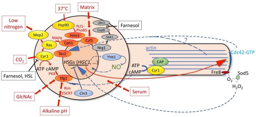
Figure 2. Schematic depiction of some of the positive stimuli on hyphal induction (boxed in red and red
Figure 2. The schematic depiction of some of the positive stimuli on hyphal induction (boxed in red
arrows) and inhibitory signals (black). The cellular mediators of the hyphal morphogenetic pathways
and red arrows) and inhibitory signals (black). The cellular mediators of the hyphal morphogenetic
are depicted in yellow ovals and the transcription activators that induce hyphal growth are in orange
pathways are depicted in yellow ovals and the transcription activators that induce hyphal growth are
ovals. The internal signals feeding back from the morphogenetic apparatus (dashed lines) and the cell
in orange
cycle ovals. (solid
apparatus The internal
lines) tosignals feeding backgene
the hypha-specific from(HSG)
the morphogenetic apparatus
expression program (dashedinlines)
are shown blue.
See text for details of the individual interactions. The question mark indicates that it isprogram
and the cell cycle apparatus (solid lines) to the hypha-specific-genes (HSG) expression are
still unclear
whether Cdc42 affects the HSG expression directly or indirectly via its effect on the cytoskeleton. it
shown in blue. See text for details of the individual interactions. The question mark indicates that
is still unclear whether Cdc42 affects the HSG expression directly or indirectly via its effect on the
5. Regulation of Hyphal Morphogenesis and HSG Expression by Internal Signals
cytoskeleton.
5.1. Interference with Yeast Proliferation Induces Hyphal Morphogenesis and HSG Expression
7. Conclusions
The
The established role of the S.
hyphal development cerevisiaeonce
program, cell-cycle CDKlikely
initiated, in therequires
coordination of cell cycle
commitment progression
mechanisms in
with
orderbud morphogenesis
to be [43], as wellcould
sustained. Commitment as itsbe
role in S. cerevisiae
achieved pseudohyphal
by positive growth [117,118],
feedback regulatory led as
loops such to
the
the exploration of the
transcriptional role of cell cycle
feed-forward regulators
loops, in C.ofalbicans
the effects morphogenesis
actin cytoskeleton [119]. Interference
polarization with
on the HSG
cell cycle progression
expression program, and in the
theabsence
ROS- and of external
NO-mediatedstimuliregulation
was foundofto inducemorphogenesis
hyphal hyphal-like growth; this
described
hyphal
above. The internal signals that can affect hyphal gene expression are schematically indicatedcell
morphogenesis is often accompanied by an induction of HSG expression. The types of in
cycle
Figureinterferences that induce hyphal growth and HSG expression include mitotic inhibition by the
2 with blue arrows.
depletion of the
Besides, in essential mitotichyphae-inducing
the external polo kinase Cdc5stimuli[120,121],
thatS-phase inhibition by
are transmitted viathewell-studied
addition of
the DNA synthesis inhibitor hydroxyurea, or through the depletion
transduction pathways to the nucleus to induce the HSG expression program and hyphalof the ribonucleotide reductase
growth
Rnr1, other genotoxic stresses [121,122], or cell cycle arrest in G1 through the depletion of the essential
G1 cyclin Cln3 [123,124]. Interference with the activity of the SCF ubiquitin ligase complex, which
in S. cerevisiae is essential for the G1-to-S transition [125], by mutating its essential cullin component
Cdc53 or by interfering with its Rub1 modification, also induced a filamentous morphology, albeit
pseudohyphal rather than hyphal in most cases [126,127]. In contrast, the deletion of CDC4, which
encodes one of the alternative substrate recognition components of the SCF complex, led to a strong
constitutive hyphal phenotype with aerial hyphae protruding from colonies [128]. The constitutive
hyphal phenotype of the cdc4−/− mutant is a consequence of the combined stabilization of two SCFCDC4
substrates, the CDK inhibitor Sol1, which delays cell cycle progression at the G1-to-S transition, and the
TF Ume6, which promotes hyphal extension [129].J. Fungi 2019, 5, 21 6 of 15
Diminished expression of the essential cell cycle kinases Cak1, an activator of the cell cycle CDK
Cdk1/Cdc28, or of Kin28, involved in RNA polymerase II activity, also causes filamentous growth and
HSG expression, and so do the reduced activity of mutants in a number of additional essential genes,
a phenomenon that has been called "essential process impairment-induced filamentation" [130,131].
Conversely, the acceleration of the cell cycle either by deletion of the cell cycle inhibitor Nrm1 [132] or by
the overexpression of Cln3 [129] inhibits hyphal morphogenesis and HSG expression. The pathways
for hyphal induction upon interference with the cell cycle progression include the transcription
factor Ume6 and the Cdc28 cyclin Hgc1 in the case of the depletion of Cdc5 or Cln3, but not for
hydroxyurea-induced filamentous growth. The latter, however, still required the Ras-cAMP pathway
activity [133,134]. HSG expression induced by diminished CAK1 expression was surprisingly resistant
to the deletion of TFs that are normally essential for hyphal growth under many conditions, in
particular, Ume6 and Brg1. Deletion of both UME6 and BRG1 together did, nonetheless, greatly reduce
HSG expression in the diminished CAK1 expression background, suggesting that several parallel
HSG expression pathways are induced in this background [130]. The signals that induce hyphal
morphogenesis upon cell cycle inhibition or essential process impairment, in general, are yet unknown.
5.2. Actin Depolymerization Blocks Hyphal Morphogenesis and Inhibits HSG Expression
The linear pathway shown in Figure 1 would suggest that after induction of hyphal morphogenesis
by external stimuli, interference with the actin cytoskeleton using actin depolymerizing drugs such as
cytochalasin A and latrunculin A should inhibit cellular morphogenesis, but not HSG expression. This
is, however, not what was observed. These two drugs inhibited hyphal morphogenesis, but also caused
a strong suppression of HSG expression [58,135]. Similar suppression was noted with mutants that are
defective in actin nucleation but, conversely, the drug jasplakinolide that inhibits hyphal elongation by
stabilizing filamentous actin was not defective in HSG expression [135]. These observations suggest the
existence of a mechanism linking the filamentous actin cytoskeleton, possibly via cAMP levels [135], to
the regulation of HSG expression. This mechanism may involve the adenylyl cyclase Cyr1, which is
essential for the hyphal morphogenesis gene expression program and which, via the Cap1 protein,
binds to actin [136]. However, the details of this mechanism are unknown.
5.3. Inhibition of Endocytosis Inhibits Hyphal Morphogenesis and HSG Expression
As mentioned in Section 2, hyphal morphogenesis requires that exocytosis be balanced by
endocytosis, and indeed, a collar of endocytic actin patches is located sub-apically in growing hyphae.
Thus, not surprisingly, when general endocytosis is inhibited either pharmacologically [113] using
Trifluoperazine or related compounds, or genetically through the deletion of the BAR proteins Rvs161
or Rvs167 [137], or by overexpressing the inhibitory Akl1 kinase [138], hyphal extension is inhibited.
However, in addition, HSG expression is also strongly inhibited under these conditions [113,138].
Furthermore, the activation of endocytosis through deletion of the inhibitory kinase Akl1 or through
the overexpression of the endocytic scaffold protein Pan1 both increased the rate of hyphal elongation
and the expression of HSGs [138]. Since there is no known connection between the endocytic proteins
Akl1 and Pan1 and the signal transduction pathways of hyphal induction, it appears that interference
with the mechanism of hyphal extension can indirectly affect the HSG expression.
6. Feedback Regulations in Hyphal Morphogenesis
6.1. Positive Feedback Regulations of Hyphal Transcription
To achieve sustained hyphal extension may require positive feedback (or "feed-forward")
regulations in order to ensure the continuation of the HSG expression program beyond its initial
activation. One instance of a feed-forward regulation is that of the transcription factor Ume6, whose
gene is activated under hyphal induction conditions. Ume6 was shown to activate its own gene’s
promoter under hyphal induction conditions and this reinforcing feedback loop was essential for theJ. Fungi 2019, 5, 21 7 of 15
maintenance of hyphal elongation [139]. Another mechanism that could contribute to the persistence of
the HSG expression program relies on chromatin modifiers that perpetuate an active chromatin state by
histone deacetylation. The initiation of HSG expression induces the down-regulation of the expression
of a transcriptional repressor, Nrg1 [140], as well as the degradation of the Nrg1 protein [109], causing
inter alia the activation of the TF gene BRG1 [87]. Brg1 then recruits the deacetylase Hda1 to HSG
promoters, which causes histone deacetylation indirectly through the inactivation of the NuA4 histone
acetyltransferase complex [87,141]. Upon Nrg1 removal, Brg1 can itself also activate its own promoter
in another feed-forward loop that can perpetuate the expression of HSGs [141].
6.2. Cdc42
The small GTPase Cdc42 is an essential protein for yeast growth and proliferation and it is also
specifically involved in hyphal elongation. Misregulation of Cdc42 activity or localization either
through mutation, by increasing or decreasing its expression, or by manipulating its associated GTP
exchange factor (Cdc24) or its associated proteins Bem1 and Rsr1, causes a disruption in hyphal
morphogenesis [58,59,61,63,142,143]. Interestingly, in the absence of proper hyphal morphogenetic
Cdc42 activity, including properly focused localization at the hyphal tip, the initial HSG expression
cannot be sustained [61,63,143]. This could be due to the role of the Cdc42 activity in actin cytoskeleton
polarization which, in turn, can affect the HSG expression program, as discussed above. On the
other hand, the toxicity of the activated CDC42G12V allele could be suppressed by the deletion of the
MAP kinase signaling pathway protein Cst20, suggesting that in addition to its cytoskeletal role in
hyphal morphogenesis, Cdc42 might be directly involved in the transduction of the hyphal induction
signals via the MAPK pathway [59]. Nonetheless, additional observations indicate that Cdc42 plays
a role in maintenance, rather than in the initial induction, of the HSG expression during hyphal
morphogenesis [61]. Furthermore, the focused localization of the Cdc42 protein at the hyphal apex is
necessary for its effect on HSG expression [63]. Thus, whereas a direct role for Cdc42 in the MAPK
signal transduction pathway cannot be excluded, it is likely that the Cdc42 morphogenetic complex
impacts HSG expression indirectly via its effect on hyphal morphogenesis.
6.3. Reactive Oxygen and Nitrogen Species
Nitric oxide (NO) and reactive oxygen species (ROS) such as the superoxide radical and
hydrogen peroxide are toxic compounds generated by host phagocytic cells in response to invading
microorganisms. Conversely, the microorganisms have evolved mechanisms to detoxify these reactive
species (recently reviewed in Reference [144]). In addition, exogenous H2 O2 was shown to promote,
at sub-toxic levels, polarized cellular growth [145]. Interestingly, however, a recent report indicates
that the C. albicans hyphal cells can themselves generate H2 O2 via disproportionation through the
superoxide dismutase Sod5 of the superoxide ion generated by the NADPH oxidase Fre8 at the hyphal
tip, and that this H2 O2 contributes to hyphal morphogenesis [146]. FRE8 is itself strongly induced
under hyphal growth conditions, potentially providing a reinforcing loop that sustains morphogenesis
using this "autocrine" mechanism.
In another recent study, endogenous NO was found to be associated with hyphal morphogenesis:
the pharmacological inhibition of NO generation prevented hyphal morphogenesis via the prevention
of the normal degradation of the HSG suppressor Nrg1 under hyphal induction conditions [147].
Consistent with the notion that elevated NO promotes hyphal morphogenesis, the deletion of the
main NO detoxifying enzyme, Yhb1, caused hyper-filamentation [148]. YHB1 expression is suppressed
under hyphal induction conditions [149], providing another potential reinforcing feedback regulation
on hyphal morphogenesis.
7. Conclusions
The hyphal development program, once initiated, likely requires commitment mechanisms in
order to be sustained. Commitment could be achieved by positive feedback regulatory loops suchJ. Fungi 2019, 5, 21 8 of 15
as the transcriptional feed-forward loops, the effects of actin cytoskeleton polarization on the HSG
expression program, and the ROS- and NO-mediated regulation of hyphal morphogenesis described
above. The internal signals that can affect hyphal gene expression are schematically indicated in
Figure 2 with blue arrows.
Besides the external hyphae-inducing stimuli that are transmitted via well-studied transduction
pathways to the nucleus to induce the HSG expression program and hyphal growth (red arrows in
Figure 2), a significant number of cellular conditions have been identified that can also induce hyphal
morphogenesis. These include mutations or treatments that inhibit cell cycle progression or otherwise
inhibit essential cellular processes. Given that the abilities to form hyphae and to generate biofilms
are correlated, it was suggested that hyphal induction upon the inhibition of proliferation represents
an evolved cellular response, which maximizes cell attachment and substrate penetration upon
encountering adverse conditions, thereby enabling, e.g., biofilm formation as a coping mechanism [131].
This possibility implies the existence of sensors of cellular growth capacity that transmit information
on the internal state of the cell to the HSG expression system. One candidate for such a sensor
is the essential G1 cyclin Cln3: high levels of Cln3 promote yeast proliferation and inhibit hyphal
morphogenesis [129,138], whereas reduced Cln3 levels promote hyphal morphogenesis [123,124,130].
It is not known how C. albicans Cln3 levels are regulated, but one possibility is that by analogy
with S. cerevisiae, where Cln3 protein levels are strongly associated with cell proliferation rates [150],
the C. albicans Cln3 cyclin likewise serves as a sensor of the cellular biosynthetic capacity.
It is, however, also possible that rather than being an evolved function, the cell cycle inhibition-
mediated hyphae formation is a consequence of mechanisms that normally ensure commitment
to hyphal morphogenesis. For example, cell cycle inhibition could induce an initial cytoskeletal
polarization due to, e.g., an imbalance between the actions of the different Cdc28 cyclins. This initial
polarization could then be further amplified to the extent that cytoskeletal polarization can affect the
HSG expression, leading to hyphal morphogenesis in the absence of regular stimuli. The cell cycle
inhibition-induced mode of hyphal morphogenesis could thus represent a spurious consequence of the
existence of the positively reinforcing cellular mechanisms that ensure commitment to morphogenesis
after germ tube initiation.
The possibilities that cell cycle inhibition-induced hyphal morphogenesis represents an evolved
response or that it is a consequence of reinforcing feedback loops in the morphogenetic pathway
that are activated under certain artificial conditions are not mutually exclusive. In either case, future
challenges will include the elucidation of the molecular mechanisms that connect the hyphal extension
apparatus to the HSG expression program. Cell cycle inhibition-induced hyphal morphogenesis,
regardless of its evolutionary basis, provides a useful experimental window into the mechanisms that
sustain hyphal growth.
Acknowledgments: Research in the author’s laboratory is funded by the Israel Science Foundation and by the
U.S.-Israel Binational Science Foundation.
Conflicts of Interest: The author declares no conflict of interest.
References
1. Nash, A.K.; Auchtung, T.A.; Wong, M.C.; Smith, D.P.; Gesell, J.R.; Ross, M.C.; Stewart, C.J.; Metcalf, G.A.;
Muzny, D.M.; Gibbs, R.A.; et al. The gut mycobiome of the Human Microbiome Project healthy cohort.
Microbiome 2017, 5, 153. [CrossRef] [PubMed]
2. Ghannoum, M.A.; Jurevic, R.J.; Mukherjee, P.K.; Cui, F.; Sikaroodi, M.; Naqvi, A.; Gillevet, P.M.
Characterization of the Oral Fungal Microbiome (Mycobiome) in Healthy Individuals. PLoS Pathog. 2010, 6,
e1000713. [CrossRef] [PubMed]
3. Kam, A.P.; Xu, J. Diversity of commensal yeasts within and among healthy hosts. Diagn. Microbiol. Infect.
Dis. 2002, 43, 19–28. [CrossRef]
4. Calderone, R.A.; Clancy, C.J. Candida and Candidiasis, 2nd ed.; Calderone, R.A., Clancy, C.J., Eds.; ASM Press:
Washington, DC, USA, 2012; ISBN 9781555815394.J. Fungi 2019, 5, 21 9 of 15
5. Pfaller, M.A.; Diekema, D.J. Epidemiology of invasive candidiasis: A persistent public health problem.
Clin. Microbiol. Rev. 2007, 20, 133–163. [CrossRef] [PubMed]
6. Pfaller, M.A.; Diekema, D.J. Epidemiology of invasive mycoses in North America. Crit. Rev. Microbiol. 2010,
36, 1–53. [CrossRef] [PubMed]
7. Falagas, M.E.; Roussos, N.; Vardakas, K.Z. Relative frequency of albicans and the various non-albicans Candida
spp among candidemia isolates from inpatients in various parts of the world: A systematic review. Int. J.
Infect. Dis. 2010, 14, e954–e966. [CrossRef] [PubMed]
8. Pfaller, M.; Neofytos, D.; Diekema, D.; Azie, N.; Meier-Kriesche, H.U.; Quan, S.P.; Horn, D. Epidemiology and
outcomes of candidemia in 3648 patients: Data from the Prospective Antifungal Therapy (PATH Alliance(R))
registry, 2004-2008. Diagn. Microbiol. Infect. Dis. 2012, 74, 323–331. [CrossRef] [PubMed]
9. Kullberg, B.J.; Arendrup, M.C. Invasive Candidiasis. N. Engl. J. Med. 2015, 373, 1445–1456. [CrossRef] [PubMed]
10. Gudlaugsson, O.; Gillespie, S.; Lee, K.; Vande Berg, J.; Hu, J.; Messer, S.; Herwaldt, L.; Pfaller, M.; Diekema, D.
Attributable mortality of nosocomial candidemia, revisited. Clin. Infect. Dis. 2003, 37, 1172–1177. [CrossRef]
[PubMed]
11. Hassan, I.; Powell, G.; Sidhu, M.; Hart, W.M.; Denning, D.W. Excess mortality, length of stay and cost
attributable to Candidaemia. J. Infect. 2009, 59, 360–365. [CrossRef] [PubMed]
12. Mora-Duarte, J.; Betts, R.; Rotstein, C.; Colombo, A.L.; Thompson-Moya, L.; Smietana, J.; Lupinacci, R.;
Sable, C.; Kartsonis, N.; Perfect, J. Comparison of caspofungin and amphotericin B for invasive candidiasis.
N. Engl. J. Med. 2002, 347, 2020–2029. [CrossRef] [PubMed]
13. Cuervo, G.; Garcia-Vidal, C.; Nucci, M.; Puchades, F.; Fernández-Ruiz, M.; Obed, M.; Manzur, A.; Gudiol, C.;
Pemán, J.; Aguado, J.M.; et al. Breakthrough Candidaemia in the era of broad-spectrum antifungal therapies.
Clin. Microbiol. Infect. 2016, 22, 181–188. [CrossRef] [PubMed]
14. Pappas, P.G.; Kauffman, C.A.; Andes, D.R.; Clancy, C.J.; Marr, K.A.; Ostrosky-Zeichner, L.; Reboli, A.C.;
Schuster, M.G.; Vazquez, J.A.; Walsh, T.J.; et al. Clinical Practice Guideline for the Management of Candidiasis:
2016 Update by the Infectious Diseases Society of America. Clin. Infect. Dis. 2015, 62, civ933. [CrossRef]
[PubMed]
15. Bongomin, F.; Gago, S.; Oladele, R.; Denning, D. Global and Multi-National Prevalence of Fungal
Diseases—Estimate Precision. J. Fungi 2017, 3, 57. [CrossRef] [PubMed]
16. Sudbery, P.; Gow, N.; Berman, J. The distinct morphogenic states of Candida albicans. Trends Microbiol. 2004,
12, 317–324. [CrossRef] [PubMed]
17. Sharma, J.; Rosiana, S.; Razzaq, I.; Shapiro, R. Linking Cellular Morphogenesis with Antifungal Treatment
and Susceptibility in Candida Pathogens. J. Fungi 2019, 5, 17. [CrossRef] [PubMed]
18. Stokes, C.; Moran, G.P.; Spiering, M.J.; Cole, G.T.; Coleman, D.C.; Sullivan, D.J. Lower filamentation rates of
Candida dubliniensis contribute to its lower virulence in comparison with Candida albicans. Fungal Genet. Biol.
2007, 44, 920–931. [CrossRef] [PubMed]
19. Lackey, E.; Vipulanandan, G.; Childers, D.S.; Kadosh, D. Comparative Evolution of Morphological Regulatory
Functions in Candida Species. Eukaryot. Cell 2013, 12, 1356–1368. [CrossRef] [PubMed]
20. Yue, H.; Bing, J.; Zheng, Q.; Zhang, Y.; Hu, T.; Du, H.; Wang, H.; Huang, G. Filamentation in Candida auris, an
emerging fungal pathogen of humans: Passage through the mammalian body induces a heritable phenotypic
switch. Emerg. Microbes Infect. 2018, 7, 188. [CrossRef] [PubMed]
21. Hall, R.A.; Cottier, F.; Muhlschlegel, F.A. Molecular networks in the fungal pathogen Candida albicans.
Adv. Appl. Microbiol. 2009, 67, 191–212. [CrossRef] [PubMed]
22. Sudbery, P.E. Growth of Candida albicans hyphae. Nat. Rev. Microbiol. 2011, 9, 737–748. [CrossRef] [PubMed]
23. Richardson, M.D.; Warnock, D.W. Fungal Infection–Diagnosis and Management, 2nd ed.; Blackwell Sciences
Ltd.: Oxford, UK, 1997.
24. Koh, A.Y.; Kohler, J.R.; Coggshall, K.T.; Van Rooijen, N.; Pier, G.B. Mucosal damage and neutropenia are
required for Candida albicans dissemination. PLoS Pathog. 2008, 4, e35. [CrossRef] [PubMed]
25. Marcil, A.; Harcus, D.; Thomas, D.Y.; Whiteway, M. Candida albicans killing by RAW 264.7 mouse macrophage
cells: Effects of Candida genotype, infection ratios, and gamma interferon treatment. Infect. Immun.
Infect. Immun. 2002, 70, 6319–6329. [CrossRef]
26. Ghosh, S.; Navarathna, D.H.M.L.P.; Roberts, D.D.; Cooper, J.T.; Atkin, A.L.; Petro, T.M.; Nickerson, K.W.
Arginine-Induced germ tube formation in Candida albicans is essential for escape from murine macrophage
line RAW 264.7. Infect. Immun. 2009, 77, 1596–1605. [CrossRef] [PubMed]J. Fungi 2019, 5, 21 10 of 15
27. McKenzie, C.G.; Koser, U.; Lewis, L.E.; Bain, J.M.; Mora-Montes, H.M.; Barker, R.N.; Gow, N.A.; Erwig, L.P.
Contribution of Candida albicans cell wall components to recognition by and escape from murine macrophages.
Infect. Immun. 2010, 78, 1650–1658. [CrossRef] [PubMed]
28. Nobile, C.J.; Johnson, A.D. Candida albicans Biofilms and Human Disease. Annu. Rev. Microbiol. 2015, 69,
71–92. [CrossRef] [PubMed]
29. Zheng, X.; Wang, Y. Hgc1, a novel hypha-specific G1 cyclin-related protein regulates Candida albicans hyphal
morphogenesis. EMBO J. 2004, 23, 1845–1856. [CrossRef] [PubMed]
30. Lo, H.-J.; Kohler, J.; DiDomenico, B.; Loebenberg, D.; Cacciapuoti, A.; Fink, G.R. Nonfilamentous C. albicans
mutants are avirulent. Cell 1997, 90, 939–950. [CrossRef]
31. Saville, S.P.; Lazzell, A.L.; Monteagudo, C.; Lopez-Ribot, J.L. Engineered control of cell morphology in vivo
reveals distinct roles for yeast and filamentous forms of Candida albicans during infection. Eukaryot. Cell 2003,
2, 1053–1060. [CrossRef] [PubMed]
32. Saville, S.P.; Lazzell, A.L.; Bryant, A.P.; Fretzen, A.; Monreal, A.; Solberg, E.O.; Monteagudo, C.;
Lopez-Ribot, J.L.; Milne, G.T. Inhibition of filamentation can be used to treat disseminated candidiasis.
Antimicrob. Agents Chemother. 2006, 50, 3312–3316. [CrossRef] [PubMed]
33. Noble, S.M.; French, S.; Kohn, L.A.; Chen, V.; Johnson, A.D. Systematic screens of a Candida albicans
homozygous deletion library decouple morphogenetic switching and pathogenicity. Nat. Genet. 2010, 42,
590–598. [CrossRef] [PubMed]
34. O’Meara, T.R.; Veri, A.O.; Ketela, T.; Jiang, B.; Roemer, T.; Cowen, L.E. Global analysis of fungal morphology
exposes mechanisms of host cell escape. Nat. Commun. 2015, 6, 6741. [CrossRef] [PubMed]
35. Jacobsen, I.D.; Wilson, D.; Wachtler, B.; Brunke, S.; Naglik, J.R.; Hube, B. Candida albicans dimorphism as a
therapeutic target. Expert Rev. Anti-Infect. Ther. 2012, 10, 85–93. [CrossRef] [PubMed]
36. Vila, T.; Romo, J.A.; Pierce, C.G.; McHardy, S.F.; Saville, S.P.; Lopez-Ribot, J.L. Targeting Candida albicans
filamentation for antifungal drug development. Virulence 2017, 8, 150–158. [CrossRef] [PubMed]
37. Ruhnke, M. Skin and mucous membrane infections. In Candida and candidiasis; Calderone, R.A., Ed.; ASM
Press: Washington, DC, USA, 2002; pp. 307–325.
38. Blostein, F.; Levin-Sparenberg, E.; Wagner, J.; Foxman, B. Recurrent vulvovaginal candidiasis. Ann. Epidemiol.
2017, 27, 575–582.e3. [CrossRef] [PubMed]
39. Sobel, J.D.; Muller, G.; Buckley, H.R. Critical role of germ tube formation in the pathogenesis of Candidal
vaginitis. Infect. Immun. 1984, 44, 576–580. [PubMed]
40. Martin, M.V.; Craig, G.T.; Lamb, D.J. An investigation of the role of true hypha production in the pathogenesis
of experimental oral candidosis. Sabouraudia 1984, 22, 471–476. [CrossRef] [PubMed]
41. Hisajima, T.; Ishibashi, H.; Yamada, T.; Nishiyama, Y.; Yamaguchi, H.; Funakoshi, K.; Abe, S. Invasion process
of Candida albicans to tongue surface in early stages of experimental murine oral candidiasis. Med. Mycol.
2008, 46, 697–704. [CrossRef] [PubMed]
42. Peters, B.M.; Palmer, G.E.; Nash, A.K.; Lilly, E.A.; Fidel, P.L., Jr.; Noverr, M.C. Fungal morphogenetic
pathways are required for the hallmark inflammatory response during Candida albicans vaginitis. Infect.
Immun. 2014, 82, 532–543. [CrossRef] [PubMed]
43. Lew, D.J.; Reed, S.I. Morphogenesis in the yeast cell cycle: Regulation by Cdc28 and cyclins. J. Cell Biol. 1993,
120, 1305–1320. [CrossRef] [PubMed]
44. Chiou, J.; Balasubramanian, M.K.; Lew, D.J. Cell Polarity in Yeast. Annu. Rev. Cell Dev. Biol. 2017, 33, 77–101.
[CrossRef] [PubMed]
45. Liu, H. Transcriptional control of dimorphism in Candida albicans. Curr. Opin. Microbiol. 2001, 4, 728–735.
[CrossRef]
46. Steinberg, G.; Peñalva, M.A.; Riquelme, M.; Wösten, H.A.; Harris, S.D. Cell Biology of Hyphal Growth.
Microbiol. Spectr. 2017, 5, 1–34. [CrossRef]
47. Riquelme, M.; Aguirre, J.; Bartnicki-García, S.; Braus, G.H.; Feldbrügge, M.; Fleig, U.; Hansberg, W.;
Herrera-Estrella, A.; Kämper, J.; Kück, U.; et al. Fungal Morphogenesis, from the Polarized Growth of
Hyphae to Complex Reproduction and Infection Structures. Microbiol. Mol. Biol. Rev. 2018, 82, e00068-17.
[CrossRef] [PubMed]
48. Bartnicki-Garcia, S.; Garduño-Rosales, M.; Delgado-Alvarez, D.L.; Mouriño-Pérez, R.R. Experimental
measurement of endocytosis in fungal hyphae. Fungal Genet. Biol. 2018, 118, 32–36. [CrossRef] [PubMed]J. Fungi 2019, 5, 21 11 of 15
49. Hernández-González, M.; Bravo-Plaza, I.; Pinar, M.; de Los Ríos, V.; Arst, H.N.; Peñalva, M.A. Endocytic recycling
via the TGN underlies the polarized hyphal mode of life. PLoS Genet. 2018, 14, e1007291. [CrossRef] [PubMed]
50. Araujo-Bazán, L.; Peñalva, M.A.; Espeso, E.A. Preferential localization of the endocytic internalization
machinery to hyphal tips underlies polarization of the actin cytoskeleton in Aspergillus nidulans.
Mol. Microbiol. 2008, 67, 891–905. [CrossRef] [PubMed]
51. Yokoyama, K.; Kaji, H.; Nishimura, K.; Miyaji, M. The role of microfilaments and microtubules in apical
growth and dimorphism of Candida albicans. J. Gen. Microbiol. 1990, 136, 1067–1075. [CrossRef] [PubMed]
52. Rida, P.C.G.; Nishikawa, A.; Won, G.Y.; Dean, N. Yeast-to-Hyphal Transition Triggers Formin-dependent Golgi
Localization to the Growing Tip in Candida albicans. Mol. Biol. Cell 2006, 17, 4364–4378. [CrossRef] [PubMed]
53. Crampin, H.; Finley, K.; Gerami-Nejad, M.; Court, H.; Gale, C.; Berman, J.; Sudbery, P. Candida albicans
hyphae have a Spitzenkörper that is distinct from the polarisome found in yeast and pseudohyphae. J. Cell
Sci. 2005, 118, 2935–2947. [CrossRef] [PubMed]
54. Weiner, A.; Orange, F.; Lacas-Gervais, S.; Rechav, K.; Ghugtyal, V.; Bassilana, M.; Arkowitz, R.A. On-site
secretory vesicle delivery drives filamentous growth in the fungal pathogen Candida albicans. Cell. Microbiol.
2018, e12963. [CrossRef] [PubMed]
55. Jones, L.A.; Sudbery, P.E. Spitzenkörper, exocyst, and polarisome components in Candida albicans hyphae
show different patterns of localization and have distinct dynamic properties. Eukaryot. Cell 2010, 9, 1455–1465.
[CrossRef] [PubMed]
56. Martin, R.; Walther, A.; Wendland, J. Ras1-induced hyphal development in Candida albicans requires the
formin Bni1. Eukaryot. Cell 2005, 4, 1712–1724. [CrossRef] [PubMed]
57. Li, C.R.; Wang, Y.M.; De Zheng, X.; Liang, H.Y.; Tang, J.C.W.; Wang, Y. The formin family protein CaBni1p
has a role in cell polarity control during both yeast and hyphal growth in Candida albicans. J. Cell Sci. 2005,
118, 2637–2648. [CrossRef] [PubMed]
58. Hazan, I.; Liu, H. Hyphal tip-associated localization of Cdc42 is F-actin dependent in Candida albicans.
Eukaryot Cell 2002, 1, 856–864. [CrossRef] [PubMed]
59. Ushinsky, S.C.; Harcus, D.; Ash, J.; Dignard, D.; Marcil, A.; Morchhauser, J.; Thomas, D.Y.; Whiteway, M.;
Leberer, E. CDC42 is required for polarized growth in human pathogen Candida albicans. Eukaryot. Cell 2002,
1, 95–104. [CrossRef] [PubMed]
60. Arkowitz, R.A.; Bassilana, M. Regulation of hyphal morphogenesis by Ras and Rho small GTPases.
Fungal Biol. Rev. 2015, 29, 7–19. [CrossRef]
61. Bassilana, M.; Hopkins, J.; Arkowitz, R.A. Regulation of the Cdc42/Cdc24 GTPase module during Candida
albicans hyphal growth. Eukaryot Cell 2005, 4, 588–603. [CrossRef] [PubMed]
62. Court, H.; Sudbery, P. Regulation of Cdc42 GTPase Activity in the Formation of Hyphae in Candida albicans.
Mol. Biol. Cell 2007, 18, 265–281. [CrossRef] [PubMed]
63. Pulver, R.; Heisel, T.; Gonia, S.; Robins, R.; Norton, J.; Haynes, P.; Gale, C.A. Rsr1 focuses CDC42 activity at
hyphal tips and promotes maintenance of hyphal development in Candida albicans. Eukaryot. Cell 2013, 12,
482–495. [CrossRef] [PubMed]
64. Brand, A.; Vacharaksa, A.; Bendel, C.; Norton, J.; Haynes, P.; Henry-Stanley, M.; Wells, C.; Ross, K.;
Gow, N.A.R.; Gale, C.A. An internal polarity landmark is important for externally induced hyphal behaviors
in Candida albicans. Eukaryot. Cell 2008, 7, 712–720. [CrossRef] [PubMed]
65. Thomson, D.D.; Wehmeier, S.; Byfield, F.J.; Janmey, P.A.; Caballero-Lima, D.; Crossley, A.; Brand, A.C.
Contact-induced apical asymmetry drives the thigmotropic responses of Candida albicans hyphae.
Cell. Microbiol. 2015, 17, 342–354. [CrossRef] [PubMed]
66. Csank, C.; Schroppel, K.; Leberer, E.; Harcus, D.; Mohamed, O.; Meloche, S.; Thomas, D.Y.; Whiteway, M.
Roles of the Candida albicans mitogen-activated protein kinase homolog, Cek1p, in hyphal development and
systemic candidiasis. Infect. Immun. 1998, 66, 2713–2721. [PubMed]
67. Rocha, C.R.; Schroppel, K.; Harcus, D.; Marcil, A.; Dignard, D.; Taylor, B.N.; Thomas, D.Y.; Whiteway, M.;
Leberer, E. Signaling through adenylyl cyclase is essential for hyphal growth and virulence in the pathogenic
fungus Candida albicans. Mol. Biol. Cell 2001, 12, 3631–3643. [CrossRef] [PubMed]
68. Cloutier, M.; Castilla, R.; Bolduc, N.; Zelada, A.; Martineau, P.; Bouillon, M.; Magee, B.B.; Passeron, S.;
Giasson, L.; Cantore, M.L. The two isoforms of the cAMP-dependent protein kinase catalytic subunit are
involved in the control of dimorphism in the human fungal pathogen Candida albicans. Fungal Genet. Biol.
2003, 38, 133–141. [CrossRef]J. Fungi 2019, 5, 21 12 of 15
69. Nantel, A.; Dignard, D.; Bachewich, C.; Harcus, D.; Marcil, A.; Bouin, A.P.; Sensen, C.W.; Hogues, H.; van het
Hoog, M.; Gordon, P.; et al. Transcription profiling of Candida albicans cells undergoing the yeast-to-hyphal
transition. Mol. Biol. Cell 2002, 13, 3452–3465. [CrossRef] [PubMed]
70. Kadosh, D.; Johnson, A.D. Induction of the Candida albicans filamentous growth program by relief of
transcriptional repression: A genome-wide analysis. Mol. Biol. Cell 2005, 16, 2903–2912. [CrossRef] [PubMed]
71. Liu, H.; Kohler, J.; Fink, G.R. Suppression of hyphal formation in Candida albicans by mutation of a STE12
homolog. Science 1994, 266, 1723–1726. [CrossRef] [PubMed]
72. Stoldt, V.R.; Sonneborn, A.; Leuker, C.E.; Ernst, J.F. Efg1p, an essential regulator of morphogenesis of
the human pathogen Candida albicans, is a member of a conserved class of bHLH proteins regulating
morphogenetic processes in fungi. EMBO J. 1997, 16, 1982–1991. [CrossRef] [PubMed]
73. Lane, S.; Zhou, S.; Pan, T.; Dai, Q.; Liu, H. The basic helix-loop-helix transcription factor Cph2 regulates
hyphal development in Candida albicans partly via TEC1. Mol Cell Biol 2001, 21, 6418–6428. [CrossRef]
[PubMed]
74. Brown, D.H., Jr.; Giusani, A.D.; Chen, X.; Kumamoto, C.A. Filamentous growth of Candida albicans in
response to physical environmental cues and its regulation by the unique CZF1 gene. Mol. Microbiol. 1999,
34, 651–662. [CrossRef] [PubMed]
75. Schweizer, A.; Rupp, S.; Taylor, B.N.; Rollinghoff, M.; Schroppel, K. The TEA/ATTS transcription factor
CaTec1p regulates hyphal development and virulence in Candida albicans. Mol. Microbiol. 2000, 38, 435–445.
[CrossRef] [PubMed]
76. Davis, D.; Edwards, J.E., Jr.; Mitchell, A.P.; Ibrahim, A.S. Candida albicans RIM101 pH response pathway is
required for host-pathogen interactions. Infect Immun. 2000, 68, 5953–5959. [PubMed]
77. Shapiro, R.S.; Sellam, A.; Tebbji, F.; Whiteway, M.; Nantel, A.; Cowen, L.E. Pho85, Pcl1, and Hms1 Signaling
Governs Candida albicans Morphogenesis Induced by High Temperature or Hsp90 Compromise. Curr. Biol.
2012, 22, 461–470. [CrossRef] [PubMed]
78. Braun, B.R.; Johnson, A.D. Control of filament formation in Candida albicans by the transcriptional repressor
TUP1. Science 1997, 277, 105–109. [CrossRef] [PubMed]
79. Murad, A.M.; Leng, P.; Straffon, M.; Wishart, J.; Macaskill, S.; MacCallum, D.; Schnell, N.; Talibi, D.;
Marechal, D.; Tekaia, F.; et al. NRG1 represses yeast-hypha morphogenesis and hypha-specific gene
expression in Candida albicans. EMBO J. 2001, 20, 4742–4752. [CrossRef] [PubMed]
80. Cao, F.; Lane, S.; Raniga, P.P.; Lu, Y.; Zhou, Z.; Ramon, K.; Chen, J.; Liu, H. The Flo8 Transcription Factor
Is Essential for Hyphal Development and Virulence in Candida albicans. Mol. Biol. Cell 2006, 17, 295–307.
[CrossRef] [PubMed]
81. Du, H.; Guan, G.; Xie, J.; Sun, Y.; Tong, Y.; Zhang, L.; Huang, G. Roles of Candida albicans Gat2, a GATA-Type
Zinc Finger Transcription Factor, in Biofilm Formation, Filamentous Growth and Virulence. PLoS ONE 2012,
7, e29707. [CrossRef] [PubMed]
82. Rottmann, M.; Dieter, S.; Brunner, H.; Rupp, S. A screen in Saccharomyces cerevisiae identified CaMCM1, an
essential gene in Candida albicans crucial for morphogenesis. Mol. Microbiol. 2003, 47, 943–959. [CrossRef]
[PubMed]
83. Bensen, E.S.; Filler, S.G.; Berman, J. A forkhead transcription factor is important for true hyphal as well as
yeast morphogenesis in Candida albicans. Eukaryot. Cell 2002, 1, 787–798. [CrossRef] [PubMed]
84. Banerjee, M.; Thompson, D.S.; Lazzell, A.; Carlisle, P.L.; Pierce, C.; Monteagudo, C.; Lopez-Ribot, J.L.;
Kadosh, D. UME6, a novel filament-specific regulator of Candida albicans hyphal extension and virulence.
Mol. Biol. Cell 2008, 19, 1354–1365. [CrossRef] [PubMed]
85. Zeidler, U.; Lettner, T.; Lassnig, C.; Muller, M.; Lajko, R.; Hintner, H.; Breitenbach, M.; Bito, A. UME6 is a
crucial downstream target of other transcriptional regulators of true hyphal development in Candida albicans.
FEMS Yeast Res 2009, 9, 126–142. [CrossRef] [PubMed]
86. Bockmuhl, D.P.; Ernst, J.F. A potential phosphorylation site for an A-type kinase in the Efg1 regulator protein
contributes to hyphal morphogenesis of Candida albicans. Genetics 2001, 157, 1523–1530. [PubMed]
87. Lu, Y.; Su, C.; Wang, A.; Liu, H. Hyphal development in Candida albicans requires two temporally linked
changes in promoter chromatin for initiation and maintenance. PLoS Biol 2011, 9, e1001105. [CrossRef]
88. Lane, S.; Birse, C.; Zhou, S.; Matson, R.; Liu, H. DNA array studies demonstrate convergent regulation
of virulence factors by Cph1, Cph2, and Efg1 in Candida albicans. J. Biol. Chem. 2001, 276, 48988–48996.
[CrossRef] [PubMed]You can also read