Mesenchymal Stem Cell-Based Therapy for Stroke: Current Understanding and Challenges - Frontiers
←
→
Page content transcription
If your browser does not render page correctly, please read the page content below
REVIEW
                                                                                                                                       published: 09 February 2021
                                                                                                                                   doi: 10.3389/fncel.2021.628940

                                             Mesenchymal Stem Cell-Based
                                             Therapy for Stroke: Current
                                             Understanding and Challenges
                                             Weifeng Li 1† , Linli Shi 1,2† , Bei Hu 1 , Yimei Hong 1 , Hao Zhang 3 , Xin Li 1* and Yuelin Zhang 1,2*
                                             1
                                              Department of Emergency Medicine, Guangdong Provincial People’s Hospital, Guangdong Academy of Medical Sciences,
                                             Guangzhou, China, 2 The Second School of Clinical Medicine, Southern Medical University, Guangzhou, China, 3 Faculty
                                             of Pharmacy, Bengbu Medical College, Bengbu, China

                                             Stroke, the most prevalent cerebrovascular disease, causes serious loss of neurological
                                             function and is the leading cause of morbidity and mortality worldwide. Despite
                                             advances in pharmacological and surgical therapy, treatment for functional rehabilitation
                                             following stroke is limited with a consequent serious impact on quality of life. Over
                                             the past decades, mesenchymal stem cell (MSCs)-based therapy has emerged as a
                                             novel strategy for various diseases including stroke due to their unique properties that
                         Edited by:          include easy isolation, multipotent differentiation potential and strong paracrine capacity.
                         Yujie Chen,
       Army Medical University, China
                                             Although MSCs have shown promising results in the treatment of stroke, there remain
                       Reviewed by:
                                             many challenges to overcome prior to their therapeutic application. In this review, we
                           Shengli Hu,       focus on the following issues: the scientific data from preclinical studies and clinical
      Army Medical University, China
                                             trials of MSCs in the treatment of stroke; the potential mechanisms underlying MSC-
          María Gutiérrez Fernández,
     University Hospital La Paz, Spain       based therapy for stroke; the challenges related to the timing and delivery of MSCs and
                *Correspondence:             MSC senescence.
                                 Xin Li
                                             Keywords: mesenchymal stem cell, stroke, cell therapy, mechanisms, challenges
                     xlidoct@qq.com
                         Yuelin Zhang
        orzhangyuelin1999@163.com
     † These   authors have contributed
                                            INTRODUCTION
                    equally to this work
                                            Stroke, one of the major diseases of the central nervous system, is a global health problem with
                   Specialty section:       limited treatment options. It is classified as hemorrhagic (13%), caused by rupture of blood vessels,
        This article was submitted to       or ischemic (87%), caused by disruption of blood supply (Kalladka and Muir, 2014). With an
             Cellular Neurophysiology,      increasing elderly population, the mortality and morbidity of stroke are increasing. Approximately
                a section of the journal    15 million individuals worldwide are affected by stroke each year, of whom 5 million will ultimately
    Frontiers in Cellular Neuroscience      die and 5 million will suffer long-term disability (Roy-O’Reilly and McCullough, 2014). Ischemic
       Received: 13 November 2020           stroke is caused by occlusion of a supply artery due to embolus or thrombus. As a result of
        Accepted: 14 January 2021           cerebral ischemia, excitatory amino acids react with tissues and generate a large number of calcium
       Published: 09 February 2021
                                            ions and free radicals. This produces carbon monoxide with consequent irreversible necrosis
                              Citation:     of brain cells (Rastogi et al., 2006). The necrotic portion, also known as the ischemic core, is
             Li W, Shi L, Hu B, Hong Y,     surrounded by the peri-infarct region or penumbra that represents the functionally impaired
    Zhang H, Li X and Zhang Y (2021)
                                            but potentially salvageable tissue and is the primary target for the developing neuroprotective
       Mesenchymal Stem Cell-Based
            Therapy for Stroke: Current
                                            strategies (Candelario-Jalil and Paul, 2021). Rapid restoration of cerebral blood flow is the focus
       Understanding and Challenges.        of the treatment for acute stroke. Currently, there are no proven options for stroke patients aside
     Front. Cell. Neurosci. 15:628940.      from dissolution of thrombus via tissue plasminogen activator (e.g., alteplase), or mechanical
      doi: 10.3389/fncel.2021.628940        thrombectomy (Hacke et al., 2008; Powers et al., 2015; Saver et al., 2016). However, thrombolysis

Frontiers in Cellular Neuroscience | www.frontiersin.org                         1                                      February 2021 | Volume 15 | Article 628940
Li et al. Mesenchymal Stem Cell for Stroke Treatment
has a narrow therapeutic window, being clinically effective only (SD) rats or Wistar rats were used to establish a model of
within 4.5 h after stroke and losing its effect when the thrombus cerebral ischemia, induced by middle cerebral artery occlusion
is large or the stroke is extensive (Bhaskar et al., 2018). Fewer (MCAO). It has been shown that transplantation of MSCs
than 5% of ischemic stroke patients receive such treatment and following ischemic stroke promotes improvement of cerebral
still suffer post-treatment neurological deficits with no therapy function (Toyoshima et al., 2015; Moisan et al., 2016; Hu
available to promote recovery (Lyden et al., 2019). Mechanical et al., 2019) effectively protects ischemic neurons and restores
thrombectomy exhibits the significant therapeutic efficacy in brain damage (Son et al., 2019). However, several studies
acute ischemic stroke caused by intracranial proximal artery used young adult and healthy animals, without taking into
occlusion. However, this technique is not yet fully developed, account the fact that a large proportion of ischemic stroke
and the efficacy and safety of endovascular reperfusion beyond patients are elderly and the presence of comorbidities such as
6 h remains controversial (Berkhemer et al., 2015; Smith, 2019). hypertension and diabetes (Howells et al., 2010; Laso-Garcia
Limited numbers of stroke patients can benefit from these et al., 2019). Herein, these animal models create the barriers to
approaches and achieve good outcomes (Detante et al., 2017). the translation of the findings to clinical trials. Therefore, we here
Over the past decades, stem cell-based therapy has attracted focused on studies that incorporated comorbidities into animal
great interest as an emerging treatment in stroke in the hope models of stroke.
that it can repair the damaged central neural networks (Wan It has been estimated that about 75% of strokes occur
Safwani et al., 2017; Choi et al., 2018). Stem cell therapy displays in the elderly (Yousufuddin and Young, 2019). Shen et al.
significant effects of functional improvement for ischemic stroke, (2007a) selected 10–12 month-old female retired breeder rats
offering hope for the preservation of neural tissue in the acute to establish an ischemic stroke model, and confirmed the long-
phase of stroke and the replacement of lost tissue in the chronic term neurological protective effects of MSC on ischemic stroke.
stage (Wei et al., 2017). In addition, Saraf et al. demonstrated that stroke induced CaN
MSCs are pluripotent, non-hematopoietic stem cells with hyperactivation, triggering an apoptotic pathway in neurons that
the ability to differentiate into a diverse number of cell lineages, further led to neuronal death in middle-aged ovariectomized
including chondrocytes, osteoblasts, and neuron-like cells female rats. MSCs treatment rescued neurons and promoted
(Uccelli et al., 2006; Williams and Keating, 2008). They can neuronal survival via reducing CaN expression (Saraf et al., 2019).
be isolated from almost all tissues in mammals including Hypertension is the major risk factor for all types of stroke
bone marrow (BM), adipose tissue or other tissues (Pinho (Hong, 2017; Cipolla et al., 2018), Hypertensive ischemic stroke
et al., 2020) and are easy to culture and effectively expand. models mostly use stroke-prone spontaneously hypertensive rats
BM-MSCs are the most common, while in recent years (SHRSP), an animal model that develops 100% hypertension
adipose-derived MSCs have become increasingly popular without genetic modification, has cerebrovascular pathology
due to their easy availability and high yield (Faghih et al., and physiology very similar to that of human hypertension,
2017; Perteghella et al., 2017). MSCs from bone marrow, and induces spontaneous strokes at a rate of more than
adipose tissue have full trilineage (adipogenic, osteogenic, 60% (Liao et al., 2013). Calio et al. (2014) demonstrated
and chondrogenic) differentiation capacity and excellent that MSCs induced an increase in the anti-apoptotic gene
immunomodulatory properties compared to other sources, Bcl-2 and protected brain tissue through anti-apoptosis and
and therefore represent the optimal stem cell sources for antioxidation, suggesting that MSCs have a protective effect
tissue engineering and regenerative medicine (Heo et al., on neuronal cells in SHRSP rats. In another study, placental
2016). Even in the acute stage of stroke, MSCs are suitable derived MSCs treatment greatly improved functional recovery
for transplantation and have substantial neurotrophic effects and reduced infarct size in mice with hypertensive ischemic
(Walczak et al., 2008). In addition, MSCs derived from adult (Kranz et al., 2010). Diabetes is a definite risk factor for
tissues pose no risk of tumorigenesis and their low expression stroke. Patients with diabetes have an increased probability
of major histocompatibility complex (MHC)-I and MHC-II of developing ischemic stroke, and hyperglycemia exacerbates
antigens eliminates the need for immunosuppression following microvascular and macrovascular damage in ischemic strokes
allogeneic administration (Bhatia and Hare, 2005; Williams and (Rehni et al., 2017; Lau et al., 2019). Therefore, studying
Hare, 2011). The therapeutic effects of MSCs are mediated by diabetic stroke models is of great importance. It has been
many mechanisms including anti-inflammation, anti-apoptosis, reported that 6 weeks after permanent MCAO, lesions were
angiogenesis, and neurogenesis. They have become the focus of more severe in the hyperglycemic group than in the non-
many preclinical and clinical studies (Li et al., 2016; Moniche hyperglycemic group. Although human adipose tissue-derived
et al., 2016). This review will focus on the application of MSCs in MSCs treatment for hyperglycemic stroke rats did not reduce
the treatment of stroke. lesion size, it significantly improved neurological function
(Gomez-de Frutos et al., 2019). Cui et al. (2016) demonstrated
the beneficial effects of BM- MSCs in type 1 diabetic rats
PRECLINICAL STUDIES with stroke via mediating miR-145. In type 2 diabetic Wistar
rats with stroke, treatment with exosomes harvested from BM-
The application of MSCs in the treatment of stroke has been MSCs significantly improved blood-brain barrier (BBB) integrity,
studied for nearly two decades. Several recent animal studies increased white matter remodeling, and promoted neural repair
are summarized in Table 1. In most studies, Sprague Dawley (Venkat et al., 2020).
Frontiers in Cellular Neuroscience | www.frontiersin.org 2 February 2021 | Volume 15 | Article 628940Li et al. Mesenchymal Stem Cell for Stroke Treatment
TABLE 1 | Overview of animal studies of MSC-based therapy for stroke.
Animal Stroke Comorbidity Cell Cell number Delivery route Timing Results References
species type source
SD MCAO – BM 1 × 10⁵ IA (carotid artery) 10 days Neuronal regeneration Hu et al., 2019
SD MCAO – BM 3 × 10⁶ IV (tail vein) 8 days Angiogenesis Moisan et al., 2016
Wistar MCAO – BM 1 × 10⁶ IA 1, 6, 24, Reduce infarction volume Toyoshima et al.,
and 48 h 2015
SD MCAO – BM 2 × 10⁵ IC (brain tissue) 1 days Protect ischemic neurons Son et al., 2019
Wistar MCAO Aging BM 2 × 10⁶ IA (carotid artery) 1 days Long-term improvement in Shen et al., 2007a
functional outcome
SD MCAO Aging BM 1 × 10⁵ IA 6h Improve the functional Saraf et al., 2019
outcome
SHR Stroke Hypertension BM 1 × 10⁶ IC (atlanto-occipital – Neuroprotective and Calio et al., 2014
prone membrane) antioxidant potential
SHR MCAO Hypertension Placenta 1 × 10⁶ IV (tail vein) 8 and 24 h Functional recovery Kranz et al., 2010
SD MCAO Hyperglycemia Adipose 1 × 10⁶ IV (tail vein) 48 h Neurological recovery Gomez-de Frutos
tissue et al., 2019
Wistar MCAO Diabetes BM 5 × 10⁶ IV (tail vein) 24 h Neurorepair effects Cui et al., 2016
Wistar MCAO Diabetes BM 3 × 10¹¹ IV (tail vein) 3 days Improve the functional Venkat et al., 2020
outcome
MCAO, middle cerebral artery occlusion; BM, bone marrow; IA, intra-arterial; IC, intracerebral; IV, intravenous.
Comorbidities in humans can profoundly affect stroke of autologous intravenous BMSCs transplantation and got
pathophysiology, lesion development, and recovery (Cho and similar results (Lee et al., 2010). In 2011, the reduction in
Yang, 2018). Despite of the beneficial effects of MSCs, more infarct lesion volume and recovery of neurological function
preclinical studies are warranted to exploring MSC therapy were obtained following administration of serum-expanded
for stroke due to the limited number of relevant studies in autologous BMSCs to chronic stroke patients (Honmou et al.,
stroke comorbidity models. Therefore, the importance of using 2011). Afterwards, a phase I/II study of intracerebral cell
preclinical comorbidity model should be emphasized when transplantation in patients with chronic stroke has reported
establishing guidelines on how to improve the validity of animal that intracerebral transplantation of genetically modified MSCs
models of stroke. significantly improved neurological function (Steinberg et al.,
2016, 2018). A single-center, open-label Randomized Controlled
Trial study showed that intravenous injection of autologous
CLINICAL TRIALS BMSCs also improved the motor function, suggesting that
MSCs treatment is feasible therapeutic strategy for stroke
Although preclinical studies have shown that MSCs displays (Jaillard et al., 2020).
beneficial effects on stroke (Moisan et al., 2016; Cunningham Recently, Savitz et al. (2019) conducted a randomized, sham
et al., 2018), the safety problems inflammation, tumor controlled, phase II trial in which autologous BM-derived
development, metastasis in clinical trials have been reported aldehyde dehydrogenase (ALDH)-bright cells were transplanted
(Gazdic et al., 2015; Dhere et al., 2016; Volarevic et al., 2018). intra-arterially to patients with disabling middle cerebral artery
Over the past few decades, the safety, feasibility and effectiveness stroke and showed no adverse events in the treatment patients
of MSCs in the treatment of stroke have been widely studied group, although there was a higher incidence of small lesions on
in clinical trials (Table 2). Previous clinical trials have shown MRI. In addition, superparamagnetic iron oxide-labeled BMSCs
that MSCs isolated from different tissues have shown the high were also used to treat stroke patients and proved to be safe and
efficiency for stroke treatment (Detante et al., 2017; Wechsler tolerated (Shichinohe et al., 2017). Compared to BMSCs, adipose
et al., 2018; Chrostek et al., 2019; Cui et al., 2019; Suda et al., tissue-derived mesenchymal stem cells (AD-MSCs) are easier and
2020). Several routes of delivery have been proposed including safer to prepare without adverse side effects and ethical concerns
intracerebral (IC), intra-arterial (IA), and intravenous (IV) (Ra et al., 2011; Gutierrez-Fernandez et al., 2013a,b). In 2014,
(Toyoshima et al., 2017). Among them, the intracerebral allogeneic intravenous AD-MSCs transplantation was carried out
pathway is the most effective and invasive route. In contrast, in patients with subacute stroke. The results demonstrated that
the intravenous pathway is the least invasive, but the number allogeneic AD-MSCs had no association with the development
of MSC cells reaching the injured brain is the most limited. The of tumors. The study concluded that AD-MSCs-based cell
intra-arterial pathway is relatively neutral. In 2005, autologous therapy was safe and could promote rehabilitation of stroke
BMSCs transplantation was performed intravenously for the (Diez-Tejedor et al., 2014).
first time in five patients with acute ischemic stroke and no Human umbilical cord-derived MSCs (hUC-MSCs) have great
adverse reactions were reported (Bang et al., 2005). In 2010, advantages for stroke treatment due to low immunogenicity
a large long-term study evaluated the safety and efficacy and no substantial ethical problems (Yin et al., 2019). A Phase
Frontiers in Cellular Neuroscience | www.frontiersin.org 3 February 2021 | Volume 15 | Article 628940Li et al. Mesenchymal Stem Cell for Stroke Treatment
TABLE 2 | Clinical trials of MSC-based therapy for stroke.
Phase Patients Delivery Cell source Cell number Timing Results References
number route
I 5 IV Autologous BM-MSCs 1 × 10⁸ 7 days Improve in BI Bang et al., 2005
II 16 IV Autologous BM-MSCs 5 × 10⁷ 5–7 weeks Improve in mRS Lee et al., 2010
I 12 IV Autologous BM-MSCs 1 × 10⁸ 36–133 days Improve in NIHSS Honmou et al.,
2011
I 8 IV Autologous BM-MSCs 5–6 × 10⁷ 3 months–1 year Improve in Fugle-Meyer Bhasin et al., 2011
and mRS, increase in
number of cluster activation
of Brodmann areas BA 4
and BA 6
II 20 IV Allogeneic AD-MSCs 1 × 10⁶ cells/kg 2 week Safe and effective Diez-Tejedor et al.,
2014
I/IIa 18 IC Modified MSCs dose-escalation: 6–60 months improve in Steinberg et al.,
(SB623) 2.5 × 10⁶ , 5.0 × 10⁶ , ESS,NIHSS,Fugle-Meyer 2016, 2018
or 10 × 106
II 48 IA BM-ALDHbr cells 0.5 × 10⁵ –2.5 × 10⁷ 9–15 days Safe Savitz et al., 2019
I 10 IV Allogeneic UC-MSCs 5 × 10⁶ –5 × 10⁷ /Kg 7–10 days Safe and feasible Laskowitz et al.,
2018
II 16 IV Autologous BM-MSCs 10 × 107 –30 × 107 14 days Improve in Jaillard et al., 2020
motor-NIHSS,Fugle-Meyer,
task-related fMRI activity
BI, Barthel Index; mRS, modified Rankin Scale; NIHSS, National Institute of Health Stroke Scale; ESS, European Stroke Scale; BM, bone marrow; MSC, mesenchymal
stem cell; UC-MSC, umbilical cord mesenchymal stem cell; ALDHbr , aldehyde dehydrogenase bright; IA, intra-arterial; IC, intracerebral; IV, intravenous.
I clinical trial using hUC-MSCs for acute stroke treatment 2016). In the presence of injury and inflammation, MSCs are
demonstrated a significant functional recovery, indicating that directly transplanted or homed to the damaged site (Shi et al.,
hUC-MSCs treatment is safe and feasible option for acute stroke 2012). In a specific microenvironment of a tissue or organ, MSCs
(Laskowitz et al., 2018). have the ability to divide and proliferate, differentiate and develop
Although MSC transplantation has been proven to be safe into the same cell type as the tissue or organ, including neuronal
and feasible in small phase I/II trials, no significant improvement cells, and effect repair (Clevers et al., 2014; Shi et al., 2016). It
was observed in a randomized controlled intravenous phase has been reported that MSCs isolated from human umbilical
II trial (Hess et al., 2017). Their efficacy in the treatment of cord can differentiate into neuron-like cells and maintain their
ischemic stroke therefore remains controversial (Hess et al., immunomodulatory and antioxidant activities (Li et al., 2012).
2017). Several reasons are proposed for the inconsistent results
of MSC transplantation in ischemic stroke. First, trials differ in Paracrine Effects
patient numbers, dose and type of cell delivery, timing of cell There is accumulating evidence that the ability of MSCs to
therapy, and treatment modalities. Second, the location/extent differentiate into neuron cells cannot on its own explain the
and severity of the lesions are different. The studies also applied predominant benefits of MSC-based therapy (Ghittoni et al.,
different evaluation criteria when assessing neurological function 2005; Wang et al., 2012; Bang and Kim, 2019; Brown et al.,
and adverse reactions. More optimized and well-designed large 2020). Compared with BM-MSC transplantation, infusion of
sample multicenter studies are needed to explore the therapeutic MSC-derived conditioned medium has been shown to equally
efficacy of MSCs in ischemic stroke. improve injured brain function (Ghittoni et al., 2005; Gnecchi
et al., 2006; Volarevic et al., 2011). A paracrine effect of
MSCs is thus concluded to be a major mechanism underlying
POTENTIAL MECHANISMS OF MSC the benefits of MSC-based therapy for stroke (Liang et al.,
THERAPY FOR STROKE 2014). There is a close interaction between the soluble factors
derived from MSCs and immune cells (such as dendritic cells,
Currently, the underlying mechanisms of MSC-based therapy lymphocytes, natural killer cells, and macrophages) (Gao et al.,
for stroke have not been fully elucidated. Many experimental 2014). A paracrine role is reflected in immune regulation: MSCs
studies have revealed that MSCs protect against stroke through secrete soluble factors through direct cell-cell interaction, are
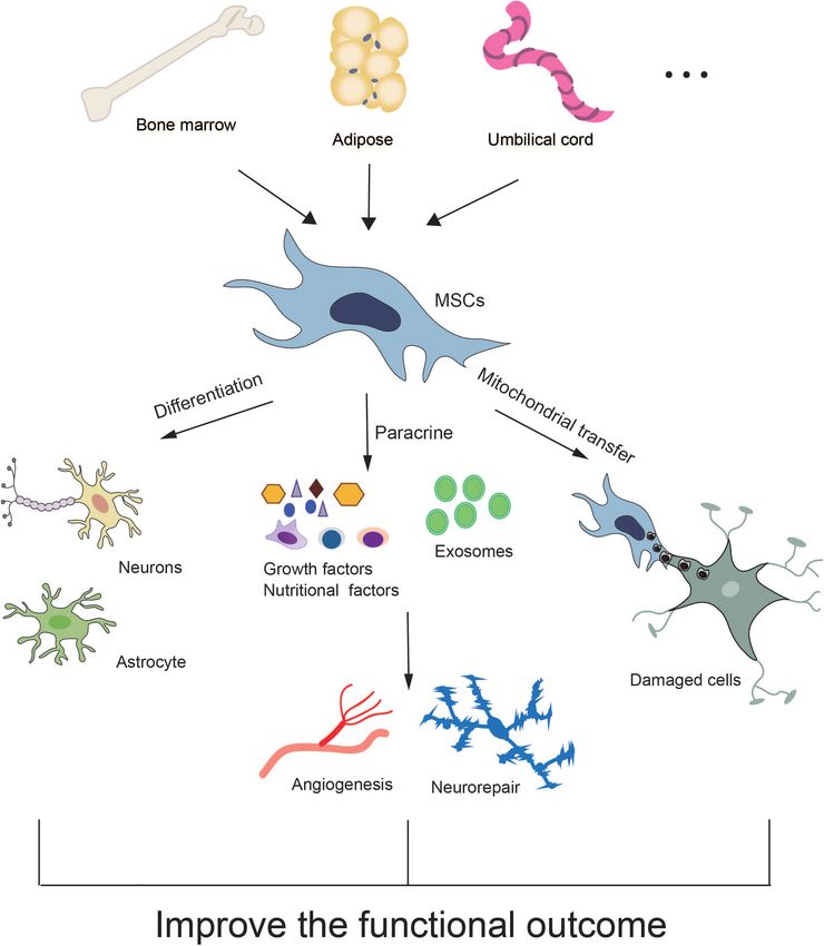
multiple mechanisms including direct differentiation, paracrine involved in immune regulation and induce immune tolerance,
effects and mitochondrial transfer (Figure 1). and can improve and regulate the destructive inflammatory
response (Wu et al., 2017; de Witte et al., 2018). Numerous
MSC Differentiation paracrine components form a complex exocrine factor network
MSCs are pluripotent adult mesenchymal cells with the ability of to ensure the stability of cells and enhance the regeneration
self-renewal and multi-differentiation (Klimczak and Kozlowska, response. Many MSC-based tissue repair models are largely
Frontiers in Cellular Neuroscience | www.frontiersin.org 4 February 2021 | Volume 15 | Article 628940Li et al. Mesenchymal Stem Cell for Stroke Treatment FIGURE 1 | Potential mechanisms of MSC therapy for stroke. dependent on the paracrine action of MSCs (Weiss and basic fibroblast growth factor and vascular endothelial growth Dahlke, 2019; Wu et al., 2020). Another manifestation of their factor induce endothelial cell proliferation and migration to paracrine effect is in the promotion of angiogenesis (De Luca form new vascular branches from existing vascular branches et al., 2011; Gnecchi et al., 2016; Chen et al., 2020). Both (Gnecchi et al., 2006). The various nutritional factors secreted Frontiers in Cellular Neuroscience | www.frontiersin.org 5 February 2021 | Volume 15 | Article 628940
Li et al.                                                                                                Mesenchymal Stem Cell for Stroke Treatment

by MSCs, including enzymes, growth factors, chemokines,                    dysfunction has been considered a sign of ischemia/reperfusion
matrix metalloproteinases, and adhesion molecules, all have                injury in the complex cell process, so mitochondrial transfer may
an effect on several key steps of angiogenesis. They can                   be one of the mechanisms by which MSC treatment is of benefit
induce the proliferation, migration and tubular formation                  for stroke (Han et al., 2020). Co-cultured BM-MSCs can transfer
of vascular endothelial cells, as well as inhibit apoptosis of             intact mitochondria through transient tunneling nanotubes
endothelial cells (Kinnaird et al., 2004; Rehman et al., 2004).            (TNT) to damaged cells, restoring their mitochondrial function
Transplantation of adipose tissue-derived MSCs has also been               (Han et al., 2016). Yang et al. demonstrated that iPSC-MSCs
shown to promote angiogenesis and improve behavioral recovery              could protect damaged PC12 cells by restoring mitochondrial
in SD rats after MCAO operation (Mu et al., 2019). BM-MSCs                 function. This was not just due to the paracrine effect of
can increase the expression of astrocyte-derived VEGF and                  MSCs, but also attributed to the mitochondria transferred from
BDNF in the ischemic boundary zone after stroke and promote                MSCs to the injured PC12 cells (Yang et al., 2020). There
angiogenesis, as well as the recruitment and proliferation                 is accumulating evidence that mitochondrial transfer between
of reactive astrocytes, leading to nerve injury repair (Guo                MSCs and damaged cells is mainly mediated through tunneled
et al., 2012; Zhang et al., 2017). Moreover, Human BM-MSCs                 nanotubes and microvesicles (Spees et al., 2006; Islam et al.,
have been shown to increase cerebral vascular generation in                2012; Li et al., 2014; Murray and Krasnodembskaya, 2019). In
stroke lesions by releasing endogenous angiogenic factors that             addition, Babenko et al. (2015) reported that BM-MSCs can
enhance the stability of new blood vessels (Moisan et al., 2016).          donate mitochondria to injured astrocytes and restore their
Therefore, the paracrine effect of stem cells is likely to play an         mitochondrial function, demonstrating the protective function of
important role in increasing capillary density and angiogenesis in         MSCs on nerves. Tseng et al. (2020) demonstrated that transfer
the damaged brain.                                                         of mitochondria from MSCs to damaged neurons induced by
    Notably, exosomes are the most important agents in the                 oxidative stress in vivo and in vitro resulted in metabolic benefits.
process of information transmission and inducing repair for                The researchers tagged BM-MSCs and tracked the transplanted
many secreted cytokines (Elahi et al., 2020). The paracrine                mitochondria. They observed the mitochondria transfer and a
effect produced by their external secretion plays a critical role          protective effect on the damaged cerebral microvascular system
in stroke recovery (Xin et al., 2013a; Hao et al., 2014; Zhang             in rats with cerebral ischemia (Liu et al., 2019; Yang et al., 2020).
Y. et al., 2015; Zhang et al., 2019). Exosomes from MSCs are               Thus, mitochondrial transfer from MSCs to damaged cells may
30–100 nm diameter lipid particles with a double membrane                  offer a new avenue in the treatment of stroke.
structure containing micro RNAs, mRNAs, DNAs, and bioactive
substances such as protein and lipids. They display similar
properties and functions to MSCs including low immunogenicity              CHALLENGES OF MSC THERAPY FOR
and the ability to stimulate nerve vascular repair with no                 STROKE
risk of tumor formation (Yaghoubi et al., 2019). Increasing
lines of evidence have confirmed that exosomes contribute                  Although many animal studies and clinical trials of MSC-based
significantly to the benefits of cell-based therapies, including the       therapy for stroke have obtained promising results,there remain
treatment of stroke, traumatic brain injury and other neurological         many challenges to overcome before MSCs can be widely applied
diseases (Xin et al., 2013b; Zhang Y. et al., 2015; Stonesifer             in clinical practice.
et al., 2017; Zhang et al., 2019). Dabrowska et al. (2019)                    First, the optimum time for MSC administration remains
demonstrated that intra-arterial delivery of exosomes derived              controversial. Currently, most preclinical studies recommend
from BM-MSCs reduced neuroinflammation induced by focal                    transplanting MSCs during the acute stroke stage (
Li et al. Mesenchymal Stem Cell for Stroke Treatment
Another challenge is to determine the best treatment. methodological rigorism is a great challenge for preclinical and
Although MSCs have shown general immune evasion and clinical studies.
tolerance in clinical studies of stroke, an increasing number Sixth, senescence of MSCs has attracted extensive attention in
of preclinical studies have proved the therapeutic efficacy of recent years. The passage times of MSCs are limited. Extension
conditioned medium (CM) and extracellular vesicles (EVs) of expansion time will inevitably lead to replicative senescence.
derived from MSCs that reduces the dependence on and need Moreover, MSCs isolated from the elderly exhibit an aging
for cells (Xin et al., 2013b; Cunningham et al., 2018). These cell- phenotype with a decline in function, leading to decreased
free substitutes can be cryopreserved with no concerns about cell therapeutic efficacy (Wagner et al., 2008). Therefore, developing
survival. Cells can be preserved for a long time and transported strategies to deal with MSC senescence is another future challenge
around the world. Nevertheless, there is no clear consensus on (Kim and Park, 2017).
the optimal culture conditions and pretreatment strategies to Finally,the comorbidities of patients are also the challenges
maximize the regenerative potential of MSC-derived CM or EVs for MSC therapy (Cui et al., 2019). Many stroke patients have
(Reiner et al., 2017). Further clinical studies are needed to clarify comorbidities such as hypertension, diabetes and heart disease
their therapeutic value for stroke. that may exert an impact on therapy efficacy (Chen et al., 2011).
Third, the route of MSC administration is another major The medications such as antidiabetics and antiplatelet drugs often
challenge. Previous studies have used both systemic and direct influence MSC function, limiting the therapeutic effects (Ortega
approaches such as IV, IA, and IC. Compared with more invasive et al., 2013). Unfortunately, most of preclinical studies haven’t
routes (e.g., intrathecal and IC approaches), minimally invasive investigated influence factors, leading to a big knowledge gap in
routes (e.g., IV and IA approaches) may cause less damage at translate stroke research to clinic.
the injection site although each route has its own advantages and
disadvantages. How to choose a simpler and safer delivery route
for MSCs is a major hurdle to their clinical application with much
greater care required by the clinician. CONCLUSION
Fourth, the best source of MSCs for stroke treatment has
MSCs have many advantages: they are immune evasive, easy to
not been determined. Although most preclinical studies (>90%)
harvest, expand and store for a long time, and convenient to
use fresh MSCs from healthy, young donors, half of the clinical
manage in various ways. Additionally, their clinical use does
studies used autologous MSC products. Autologous MSCs may
not raise many ethical issues. Increasing evidence supports the
circumvent the logistical ethical problems and have been proven
potential of MSCs to treat stroke, but there are challenges
to be more effective than those obtained from healthy donors
to overcome. We have systematically reviewed the safety and
(Block et al., 2017; van de Vyver, 2017). Nonetheless, expanding
efficacy of MSCs in the treatment of ischemic stroke and
enough stem cells for transplantation requires a long time,
hemorrhagic stroke. In preclinical studies, MSC treatment has
so it is impossible to use autologous MSC cells in the acute
shown considerable efficacy in some neurological function tests,
stage of stroke, especially from elderly patients or those with
but there remains no large-scale randomized, double-blind,
serious diseases. Genetic engineering or reprogramming to
multicenter clinical study to prove their effectiveness. The
amplify MSCs can lead to uncontrolled proliferation and genetic
heterogeneity of MSCs is the main barrier to their clinical
abnormality, limiting their viability and therapeutic potential
application and therapeutic effect. Key parameters such as the
(Zhang J. et al., 2015). Furthermore, whether modified MSCs
source of MSCs, dosage, route of administration, administration
can successfully differentiate into fully functional neural cells
time and other key parameters directly affect the application
in patients remains elusive (Strioga et al., 2012). One large
effect. More importantly, many clinical trials have similar
randomized controlled clinical trial reported that cryopreserved
limitations in detecting the role of MSCs, including small size,
allogeneic MSCs from healthy donors had poor viability and poor
lack of a control arm, and inconsistent methods for use of
clinical efficacy (Matthay et al., 2019). Therefore, when using
MSCs. Homogenization and quality control are key issues in their
allogeneic cells from healthy donors care should be taken to assess
clinical application. Future preclinical and clinical studies should
their viability in order to match preclinical conditions.
consider adoption of a well-designed randomized controlled
Fifth, another challenge is the heterogeneity in study designs.
study design, method rigor and intervention measures, so as to
The poor methodological rigorism of both preclinical and
determine the effect of MSC therapy in the treatment of stroke
clinical studies may have contributed to the current conflicting
(Napoli and Borlongan, 2016; Squillaro et al., 2016). Nonetheless,
results. Preclinical studies rarely adopted randomized or blinding
despite these issues, MSCs have exciting potential as a means to
designs, or carried out confirmatory studies, a pre-requisite of
protect neurons and improve the outcome for stroke patients.
clinical trials (Macleod et al., 2008; Hirst et al., 2014). Similar
problems also existed in clinical studies. Studies included RCT
(Lee et al., 2010; Hess et al., 2017), single-arm trial (Steinberg
et al., 2016), or case series (Honmou et al., 2011), and could AUTHOR CONTRIBUTIONS
not be compared. Previous clinical measures of efficacy reported
included NIHSS, mRS, BI, Fugle-Meyer scale, and ESS. A unified WL and LS searched the literature and wrote the manuscript. BH,
method for evaluation of neurological function is lacking so it YH, and HZ searched the literature and provided comments. XL
is difficult to reach a consistent conclusion about the safety and and YZ designed the study and wrote the manuscript. All authors
effectiveness of MSCs in clinical application. How to increase contributed to the article and approved the submitted version.
Frontiers in Cellular Neuroscience | www.frontiersin.org 7 February 2021 | Volume 15 | Article 628940Li et al. Mesenchymal Stem Cell for Stroke Treatment
FUNDING (DFJH201918 to YZ, DFJH2020020 to WL), and a grant from
the Science and Technology Planning Project of Guangzhou
This research was supported in part by the High-level Hospital (201804010335 to BH) and Tibet Autonomous Region Research
Construction Project of Guangdong Provincial People’s Hospital Projects (XZ2018-01-GB-09 to XL).
REFERENCES circulation. J. Cereb. Blood Flow Metab. 38, 2129–2149. doi: 10.1177/
0271678X18800589
Babenko, V. A., Silachev, D. N., Zorova, L. D., Pevzner, I. B., Khutornenko, A. A., Clevers, H., Loh, K. M., and Nusse, R. (2014). Stem cell signaling. An
Plotnikov, E. Y., et al. (2015). Improving the post-stroke therapeutic potency of integral program for tissue renewal and regeneration: Wnt signaling
mesenchymal multipotent stromal cells by cocultivation with cortical neurons: and stem cell control. Science 346:1248012. doi: 10.1126/science.12
the role of crosstalk between cells. Stem Cells Transl. Med. 4, 1011–1020. doi: 48012
10.5966/sctm.2015-0010 Cui, C., Ye, X., Chopp, M., Venkat, P., Zacharek, A., Yan, T., et al. (2016). miR-145
Bang, O. Y., and Kim, E. H. (2019). Mesenchymal stem cell-derived extracellular regulates diabetes-bone marrow stromal cell-induced neurorestorative effects
vesicle therapy for stroke: challenges and progress. Front. Neurol. 10:211. doi: in diabetes stroke rats. Stem Cells Transl. Med. 5, 1656–1667. doi: 10.5966/sctm.
10.3389/fneur.2019.00211 2015-0349
Bang, O. Y., Lee, J. S., Lee, P. H., and Lee, G. (2005). Autologous mesenchymal Cui, L. L., Golubczyk, D., Tolppanen, A. M., Boltze, J., and Jolkkonen, J. (2019).
stem cell transplantation in stroke patients. Ann. Neurol. 57, 874–882. doi: Cell therapy for ischemic stroke: are differences in preclinical and clinical study
10.1002/ana.20501 design responsible for the translational loss of efficacy? Ann. Neurol. 86, 5–16.
Berkhemer, O. A., Fransen, P. S., Beumer, D., van den Berg, L. A., Lingsma, doi: 10.1002/ana.25493
H. F., Yoo, A. J., et al. (2015). A randomized trial of intraarterial treatment Cunningham, C. J., Redondo-Castro, E., and Allan, S. M. (2018). The therapeutic
for acute ischemic stroke. N. Engl. J. Med. 372, 11–20. doi: 10.1056/NEJMoa potential of the mesenchymal stem cell secretome in ischaemic stroke.
1411587 J. Cereb. Blood Flow Metab. 38, 1276–1292. doi: 10.1177/0271678X18
Bhasin, A., Srivastava, M. V. P., Kumaran, S. S., Mohanty, S., Bhatia, R., Bose, 776802
S., et al. (2011). Autologous mesenchymal stem cells in chronic stroke. Dabrowska, S., Andrzejewska, A., Strzemecki, D., Muraca, M., Janowski, M.,
Cerebrovasc. Dis. Extra 1, 93–104. doi: 10.1159/000333381 and Lukomska, B. (2019). Human bone marrow mesenchymal stem cell-
Bhaskar, S., Stanwell, P., Cordato, D., Attia, J., and Levi, C. (2018). Reperfusion derived extracellular vesicles attenuate neuroinflammation evoked by focal
therapy in acute ischemic stroke: dawn of a new era? BMC Neurol. 18:8. doi: brain injury in rats. J. Neuroinflammation 16:216. doi: 10.1186/s12974-01
10.1186/s12883-017-1007-y 9-1602-5
Bhatia, R., and Hare, J. M. (2005). Mesenchymal stem cells: future source for De Luca, A., Gallo, M., Aldinucci, D., Ribatti, D., Lamura, L., D’Alessio, A., et al.
reparative medicine. Congest. Heart Fail. 11, 87–91. doi: 10.1111/j.1527-5299. (2011). Role of the EGFR ligand/receptor system in the secretion of angiogenic
2005.03618.x quiz 92-83, factors in mesenchymal stem cells. J. Cell Physiol. 226, 2131–2138. doi: 10.1002/
Block, T. J., Marinkovic, M., Tran, O. N., Gonzalez, A. O., Marshall, A., Dean, D. D., jcp.22548
et al. (2017). Restoring the quantity and quality of elderly human mesenchymal de Witte, S. F. H., Luk, F., Sierra Parraga, J. M., Gargesha, M., Merino, A., Korevaar,
stem cells for autologous cell-based therapies. Stem Cell Res. Ther. 8:239. doi: S. S., et al. (2018). Immunomodulation by therapeutic mesenchymal stromal
10.1186/s13287-017-0688-x cells (MSC) is triggered through phagocytosis of MSC by monocytic cells. Stem
Brown, J., Park, Y. J., Lee, J. Y., Chase, T. N., Koga, M., and Borlongan, Cells 36, 602–615. doi: 10.1002/stem.2779
C. V. (2020). Bone marrow-derived NCS-01 cells advance a novel cell- Detante, O., Moisan, A., Hommel, M., and Jaillard, A. (2017). Controlled clinical
based therapy for stroke. Int. J. Mol. Sci. 21:2845. doi: 10.3390/ijms210 trials of cell therapy in stroke: meta-analysis at six months after treatment. Int.
82845 J. Stroke 12, 748–751. doi: 10.1177/1747493017696098
Calio, M. L., Marinho, D. S., Ko, G. M., Ribeiro, R. R., Carbonel, A. F., Oyama, Dhere, T., Copland, I., Garcia, M., Chiang, K. Y., Chinnadurai, R., Prasad, M.,
L. M., et al. (2014). Transplantation of bone marrow mesenchymal stem cells et al. (2016). The safety of autologous and metabolically fit bone marrow
decreases oxidative stress, apoptosis, and hippocampal damage in brain of a mesenchymal stromal cells in medically refractory Crohn’s disease – a phase
spontaneous stroke model. Free Radic. Biol. Med. 70, 141–154. doi: 10.1016/j. 1 trial with three doses. Aliment. Pharmacol. Ther. 44, 471–481. doi: 10.1111/
freeradbiomed.2014.01.024 apt.13717
Candelario-Jalil, E., and Paul, S. (2021). Impact of aging and comorbidities Diez-Tejedor, E., Gutierrez-Fernandez, M., Martinez-Sanchez, P., Rodriguez-
on ischemic stroke outcomes in preclinical animal models: a translational Frutos, B., Ruiz-Ares, G., Lara, M. L., et al. (2014). Reparative therapy
perspective. Exp. Neurol. 335:113494. doi: 10.1016/j.expneurol.2020.113494 for acute ischemic stroke with allogeneic mesenchymal stem cells from
Chen, J., Ye, X., Yan, T., Zhang, C., Yang, X. P., Cui, X., et al. (2011). Adverse adipose tissue: a safety assessment: a phase II randomized, double-
effects of bone marrow stromal cell treatment of stroke in diabetic rats. Stroke blind, placebo-controlled, single-center, pilot clinical trial. J. Stroke
42, 3551–3558. doi: 10.1161/STROKEAHA.111.627174 Cerebrovasc. Dis. 23, 2694–2700. doi: 10.1016/j.jstrokecerebrovasdis.2014.
Chen, L., Wang, Y., Li, S., Zuo, B., Zhang, X., Wang, F., et al. (2020). 06.011
Exosomes derived from GDNF-modified human adipose mesenchymal stem Doeppner, T. R., Herz, J., Gorgens, A., Schlechter, J., Ludwig, A. K.,
cells ameliorate peritubular capillary loss in tubulointerstitial fibrosis by Radtke, S., et al. (2015). Extracellular vesicles improve post-stroke
activating the SIRT1/eNOS signaling pathway. Theranostics 10, 9425–9442. doi: neuroregeneration and prevent postischemic immunosuppression.
10.7150/thno.43315 Stem Cells Transl. Med. 4, 1131–1143. doi: 10.5966/sctm.20
Cho, S., and Yang, J. (2018). What do experimental models teach us about 15-0078
comorbidities in stroke? Stroke 49, 501–507. doi: 10.1161/STROKEAHA.117. Elahi, F. M., Farwell, D. G., Nolta, J. A., and Anderson, J. D. (2020). Preclinical
017793 translation of exosomes derived from mesenchymal stem/stromal cells. Stem
Choi, J. R., Yong, K. W., and Choi, J. Y. (2018). Effects of mechanical loading on Cells 38, 15–21. doi: 10.1002/stem.3061
human mesenchymal stem cells for cartilage tissue engineering. J. Cell. Physiol. Faghih, H., Javeri, A., and Taha, M. F. (2017). Impact of early subcultures on
233, 1913–1928. doi: 10.1002/jcp.26018 stemness, migration and angiogenic potential of adipose tissue-derived stem
Chrostek, M. R., Fellows, E. G., Crane, A. T., Grande, A. W., and Low, W. C. cells and their resistance to in vitro ischemic condition. Cytotechnology 69,
(2019). Efficacy of stem cell-based therapies for stroke. Brain Res. 1722:146362. 885–900. doi: 10.1007/s10616-017-0104-5
doi: 10.1016/j.brainres.2019.146362 Gao, S., Mao, F., Zhang, B., Zhang, L., Zhang, X., Wang, M., et al.
Cipolla, M. J., Liebeskind, D. S., and Chan, S. L. (2018). The importance of (2014). Mouse bone marrow-derived mesenchymal stem cells induce
comorbidities in ischemic stroke: impact of hypertension on the cerebral macrophage M2 polarization through the nuclear factor-kappaB
Frontiers in Cellular Neuroscience | www.frontiersin.org 8 February 2021 | Volume 15 | Article 628940Li et al. Mesenchymal Stem Cell for Stroke Treatment and signal transducer and activator of transcription 3 pathways. mesenchymal stem cells in stroke. Brain 134, 1790–1807. doi: 10.1093/brain/ Exp. Biol. Med. (Maywood) 239, 366–375. doi: 10.1177/15353702135 awr063 18169 Howells, D. W., Porritt, M. J., Rewell, S. S., O’Collins, V., Sena, E. S., Gazdic, M., Volarevic, V., Arsenijevic, N., and Stojkovic, M. (2015). Mesenchymal van der Worp, H. B., et al. (2010). Different strokes for different stem cells: a friend or foe in immune-mediated diseases. Stem Cell Rev. Rep. 11, folks: the rich diversity of animal models of focal cerebral ischemia. 280–287. doi: 10.1007/s12015-014-9583-3 J. Cereb. Blood Flow Metab. 30, 1412–1431. doi: 10.1038/jcbfm. Ghittoni, R., Patrussi, L., Pirozzi, K., Pellegrini, M., Lazzerini, P. E., Capecchi, P. L., 2010.66 et al. (2005). Simvastatin inhibits T-cell activation by selectively impairing the Hu, B., Chen, S., Zou, M., He, Z., Shao, S., and Liu, B. (2016). Effect of extracellular function of Ras superfamily GTPases. FASEB J. 19, 605–607. doi: 10.1096/fj.04- vesicles on neural functional recovery and immunologic suppression after 2702fje rat cerebral apoplexy. Cell. Physiol. Biochem. 40, 155–162. doi: 10.1159/0004 Gnecchi, M., Danieli, P., Malpasso, G., and Ciuffreda, M. C. (2016). Paracrine 52533 mechanisms of mesenchymal stem cells in tissue repair. Methods Mol. Biol. Hu, Y., Chen, W., Wu, L., Jiang, L., Qin, H., and Tang, N. (2019). Hypoxic 1416, 123–146. doi: 10.1007/978-1-4939-3584-0_7 preconditioning improves the survival and neural effects of transplanted Gnecchi, M., He, H., Noiseux, N., Liang, O. D., Zhang, L., Morello, F., et al. (2006). mesenchymal stem cells via CXCL12/CXCR4 signalling in a rat model Evidence supporting paracrine hypothesis for Akt-modified mesenchymal stem of cerebral infarction. Cell Biochem. Funct. 37, 504–515. doi: 10.1002/ cell-mediated cardiac protection and functional improvement. FASEB J. 20, cbf.3423 661–669. doi: 10.1096/fj.05-5211com Islam, M. N., Das, S. R., Emin, M. T., Wei, M., Sun, L., Westphalen, K., et al. Gomez-de Frutos, M. C., Laso-Garcia, F., Diekhorst, L., Otero-Ortega, (2012). Mitochondrial transfer from bone-marrow-derived stromal cells to L., Fuentes, B., Jolkkonen, J., et al. (2019). Intravenous delivery of pulmonary alveoli protects against acute lung injury. Nat. Med. 18, 759–765. adipose tissue-derived mesenchymal stem cells improves brain repair in doi: 10.1038/nm.2736 hyperglycemic stroke rats. Stem Cell Res. Ther. 10:212. doi: 10.1186/s13287-01 Jaillard, A., Hommel, M., Moisan, A., Zeffiro, T. A., Favre-Wiki, I. M., Barbieux- 9-1322-x Guillot, M., et al. (2020). Autologous mesenchymal stem cells improve motor Guo, F., Lv, S., Lou, Y., Tu, W., Liao, W., Wang, Y., et al. (2012). Bone recovery in subacute ischemic stroke: a randomized clinical trial. Transl. Stroke marrow stromal cells enhance the angiogenesis in ischaemic cortex after stroke: Res. 11, 910–923. doi: 10.1007/s12975-020-00787-z involvement of notch signalling. Cell Biol. Int. 36, 997–1004. doi: 10.1042/ Kalladka, D., and Muir, K. W. (2014). Brain repair: cell therapy in stroke. Stem Cells CBI20110596 Cloning 7, 31–44. doi: 10.2147/SCCAA.S38003 Gutierrez-Fernandez, M., Rodriguez-Frutos, B., Otero-Ortega, L., Ramos-Cejudo, Kim, H. J., and Park, J. S. (2017). Usage of human mesenchymal stem cells in J., Fuentes, B., and Diez-Tejedor, E. (2013a). Adipose tissue-derived stem cells cell-based therapy: advantages and disadvantages. Dev. Reprod. 21, 1–10. doi: in stroke treatment: from bench to bedside. Discov. Med. 16, 37–43. 10.12717/DR.2017.21.1.001 Gutierrez-Fernandez, M., Rodriguez-Frutos, B., Ramos-Cejudo, J., Teresa Vallejo- Kinnaird, T., Stabile, E., Burnett, M. S., Lee, C. W., Barr, S., Fuchs, S., et al. (2004). Cremades, M., Fuentes, B., Cerdan, S., et al. (2013b). Effects of intravenous Marrow-derived stromal cells express genes encoding a broad spectrum of administration of allogenic bone marrow- and adipose tissue-derived arteriogenic cytokines and promote in vitro and in vivo arteriogenesis through mesenchymal stem cells on functional recovery and brain repair markers paracrine mechanisms. Circ. Res. 94, 678–685. doi: 10.1161/01.RES.0000118601. in experimental ischemic stroke. Stem Cell Res. Ther. 4:11. doi: 10.1186/ 37875.AC scrt159 Klimczak, A., and Kozlowska, U. (2016). Mesenchymal stromal cells and tissue- Hacke, W., Kaste, M., Bluhmki, E., Brozman, M., Davalos, A., Guidetti, D., et al. specific progenitor cells: their role in tissue homeostasis. Stem Cells Int. (2008). Thrombolysis with alteplase 3 to 4.5 hours after acute ischemic stroke. 2016:4285215. doi: 10.1155/2016/4285215 N. Engl. J. Med. 359, 1317–1329. doi: 10.1056/NEJMoa0804656 Kranz, A., Wagner, D. C., Kamprad, M., Scholz, M., Schmidt, U. R., Nitzsche, F., Han, D., Zheng, X., Wang, X., Jin, T., Cui, L., and Chen, Z. (2020). et al. (2010). Transplantation of placenta-derived mesenchymal stromal cells Mesenchymal stem/stromal cell-mediated mitochondrial transfer and the upon experimental stroke in rats. Brain Res. 1315, 128–136. doi: 10.1016/j. therapeutic potential in treatment of neurological diseases. Stem Cells Int. brainres.2009.12.001 2020:8838046. doi: 10.1155/2020/8838046 Lalu, M. M., Montroy, J., Dowlatshahi, D., Hutton, B., Juneau, P., Wesch, N., Han, H., Hu, J., Yan, Q., Zhu, J., Zhu, Z., Chen, Y., et al. (2016). Bone marrow- et al. (2020). From the lab to patients: a systematic review and meta-analysis derived mesenchymal stem cells rescue injured H9c2 cells via transferring of mesenchymal stem cell therapy for stroke. Transl. Stroke Res. 11, 345–364. intact mitochondria through tunneling nanotubes in an in vitro simulated doi: 10.1007/s12975-019-00736-5 ischemia/reperfusion model. Mol. Med. Rep. 13, 1517–1524. doi: 10.3892/mmr. Laskowitz, D. T., Bennett, E. R., Durham, R. J., Volpi, J. J., Wiese, J. R., Frankel, M., 2015.4726 et al. (2018). Allogeneic umbilical cord blood infusion for adults with ischemic Hao, L., Zou, Z., Tian, H., Zhang, Y., Zhou, H., and Liu, L. (2014). Stem cell-based stroke: clinical outcomes from a phase I safety study. Stem Cells Transl. Med. 7, therapies for ischemic stroke. Biomed. Res. Int. 2014:468748. doi: 10.1155/2014/ 521–529. doi: 10.1002/sctm.18-0008 468748 Laso-Garcia, F., Diekhorst, L., Gomez-de Frutos, M. C., Otero-Ortega, L., Heo, J. S., Choi, Y., Kim, H. S., and Kim, H. O. (2016). Comparison of molecular Fuentes, B., Ruiz-Ares, G., et al. (2019). Cell-based therapies for stroke: profiles of human mesenchymal stem cells derived from bone marrow, promising solution or dead end? Mesenchymal stem cells and comorbidities umbilical cord blood, placenta and adipose tissue. Int. J. Mol. Med. 37, 115–125. in preclinical stroke research. Front. Neurol. 10:332. doi: 10.3389/fneur.2019. doi: 10.3892/ijmm.2015.2413 00332 Hess, D. C., Wechsler, L. R., Clark, W. M., Savitz, S. I., Ford, G. A., Chiu, D., Lau, L. H., Lew, J., Borschmann, K., Thijs, V., and Ekinci, E. I. (2019). et al. (2017). Safety and efficacy of multipotent adult progenitor cells in acute Prevalence of diabetes and its effects on stroke outcomes: a meta-analysis ischaemic stroke (MASTERS): a randomised, double-blind, placebo-controlled, and literature review. J. Diabetes Investig. 10, 780–792. doi: 10.1111/jdi. phase 2 trial. Lancet Neurol. 16, 360–368. doi: 10.1016/S1474-4422(17) 12932 30046-7 Lee, J. S., Hong, J. M., Moon, G. J., Lee, P. H., Ahn, Y. H., Bang, O. Y., et al. (2010). Hirst, J. A., Howick, J., Aronson, J. K., Roberts, N., Perera, R., Koshiaris, C., A long-term follow-up study of intravenous autologous mesenchymal stem et al. (2014). The need for randomization in animal trials: an overview cell transplantation in patients with ischemic stroke. Stem Cells 28, 1099–1106. of systematic reviews. PLoS One 9:e98856. doi: 10.1371/journal.pone.00 doi: 10.1002/stem.430 98856 Li, G., Yu, F., Lei, T., Gao, H., Li, P., Sun, Y., et al. (2016). Bone marrow Hong, K. S. (2017). Blood pressure management for stroke prevention mesenchymal stem cell therapy in ischemic stroke: mechanisms of action and and in acute stroke. J. Stroke 19, 152–165. doi: 10.5853/jos.2017. treatment optimization strategies. Neural Regen Res. 11, 1015–1024. doi: 10. 00164 4103/1673-5374.184506 Honmou, O., Houkin, K., Matsunaga, T., Niitsu, Y., Ishiai, S., Onodera, R., Li, J., Li, D., Ju, X., Shi, Q., Wang, D., and Wei, F. (2012). Umbilical cord-derived et al. (2011). Intravenous administration of auto serum-expanded autologous mesenchymal stem cells retain immunomodulatory and anti-oxidative activities Frontiers in Cellular Neuroscience | www.frontiersin.org 9 February 2021 | Volume 15 | Article 628940
Li et al.                                                                                                                                   Mesenchymal Stem Cell for Stroke Treatment

    after neural induction. Neural Regen. Res. 7, 2663–2672. doi: 10.3969/j.issn.                       stroke association. Stroke 46, 3020–3035. doi: 10.1161/STR.0000000000
    1673-5374.2012.34.003                                                                               000074
Li, X., Zhang, Y., Yeung, S. C., Liang, Y., Liang, X., Ding, Y., et al. (2014).                      Ra, J. C., Shin, I. S., Kim, S. H., Kang, S. K., Kang, B. C., Lee, H. Y., et al. (2011).
    Mitochondrial transfer of induced pluripotent stem cell-derived mesenchymal                         Safety of intravenous infusion of human adipose tissue-derived mesenchymal
    stem cells to airway epithelial cells attenuates cigarette smoke-induced                            stem cells in animals and humans. Stem Cells Dev. 20, 1297–1308. doi: 10.1089/
    damage. Am. J. Respir. Cell Mol. Biol. 51, 455–465. doi: 10.1165/rcmb.2013-0                        scd.2010.0466
    529OC                                                                                            Rastogi, L., Godbole, M. M., Ray, M., Rathore, P., Rathore, P., Pradhan, S., et al.
Liang, X., Ding, Y., Zhang, Y., Tse, H. F., and Lian, Q. (2014). Paracrine                              (2006). Reduction in oxidative stress and cell death explains hypothyroidism
    mechanisms of mesenchymal stem cell-based therapy: current status and                               induced neuroprotection subsequent to ischemia/reperfusion insult. Exp.
    perspectives. Cell Transplant. 23, 1045–1059. doi: 10.3727/096368913X                               Neurol. 200, 290–300. doi: 10.1016/j.expneurol.2006.02.013
    667709                                                                                           Rehman, J., Traktuev, D., Li, J., Merfeld-Clauss, S., Temm-Grove, C. J., Bovenkerk,
Liao, S. J., Huang, R. X., Su, Z. P., Zeng, J. S., Mo, J. W., Pei, Z., et al. (2013). Stroke-           J. E., et al. (2004). Secretion of angiogenic and antiapoptotic factors by
    prone renovascular hypertensive rat as an animal model for stroke studies: from                     human adipose stromal cells. Circulation 109, 1292–1298. doi: 10.1161/01.CIR.
    artery to brain. J. Neurol. Sci. 334, 1–5. doi: 10.1016/j.jns.2013.07.2517                          0000121425.42966.F1
Liu, K., Guo, L., Zhou, Z., Pan, M., and Yan, C. (2019). Mesenchymal stem cells                      Rehni, A. K., Liu, A., Perez-Pinzon, M. A., and Dave, K. R. (2017). Diabetic
    transfer mitochondria into cerebral microvasculature and promote recovery                           aggravation of stroke and animal models. Exp. Neurol. 292, 63–79. doi: 10.1016/
    from ischemic stroke. Microvasc. Res. 123, 74–80. doi: 10.1016/j.mvr.2019.01.                       j.expneurol.2017.03.004
    001                                                                                              Reiner, A. T., Witwer, K. W., van Balkom, B. W. M., de Beer, J., Brodie, C.,
Lyden, J., Grant, S., and Ma, T. (2019). Altered metabolism for neuroprotection                         Corteling, R. L., et al. (2017). Concise review: developing best-practice models
    provided by mesenchymal stem cells. Brain Circ. 5, 140–144. doi: 10.4103/bc.                        for the therapeutic use of extracellular vesicles. Stem Cells Transl. Med. 6,
    bc_36_19                                                                                            1730–1739. doi: 10.1002/sctm.17-0055
Macleod, M. R., van der Worp, H. B., Sena, E. S., Howells, D. W.,                                    Roy-O’Reilly, M., and McCullough, L. D. (2014). Sex differences in stroke: the
    Dirnagl, U., and Donnan, G. A. (2008). Evidence for the efficacy of                                 contribution of coagulation. Exp. Neurol. 259, 16–27. doi: 10.1016/j.expneurol.
    NXY-059 in experimental focal cerebral ischaemia is confounded by                                   2014.02.011
    study quality. Stroke 39, 2824–2829. doi: 10.1161/STROKEAHA.108.                                 Saraf, J., Sarmah, D., Vats, K., Kaur, H., Pravalika, K., Wanve, M., et al. (2019).
    515957                                                                                              Intra-arterial stem cell therapy modulates neuronal calcineurin and confers
Matthay, M. A., Calfee, C. S., Zhuo, H., Thompson, B. T., Wilson, J. G., Levitt, J. E.,                 neuroprotection after ischemic stroke. Int. J. Neurosci. 129, 1039–1044. doi:
    et al. (2019). Treatment with allogeneic mesenchymal stromal cells for moderate                     10.1080/00207454.2019.1633315
    to severe acute respiratory distress syndrome (START study): a randomised                        Saver, J. L., Goyal, M., van der Lugt, A., Menon, B. K., Majoie, C. B., Dippel,
    phase 2a safety trial. Lancet Respir. Med. 7, 154–162. doi: 10.1016/S2213-                          D. W., et al. (2016). Time to treatment with endovascular thrombectomy and
    2600(18)30418-1                                                                                     outcomes from ischemic stroke: a meta-analysis. JAMA 316, 1279–1288. doi:
Moisan, A., Favre, I., Rome, C., De Fraipont, F., Grillon, E., Coquery, N., et al.                      10.1001/jama.2016.13647
    (2016). Intravenous injection of clinical grade human MSCs after experimental                    Savitz, S. I., Yavagal, D., Rappard, G., Likosky, W., Rutledge, N., Graffagnino,
    stroke: functional benefit and microvascular effect. Cell Transplant. 25, 2157–                     C., et al. (2019). A phase 2 randomized, sham-controlled trial of
    2171. doi: 10.3727/096368916X691132                                                                 internal carotid artery infusion of autologous bone marrow-derived
Moniche, F., Rosado-de-Castro, P. H., Escudero, I., Zapata, E., de la Torre Laviana,                    ALD-401 cells in patients with recent stable ischemic stroke (RECOVER-
    F. J., Mendez-Otero, R., et al. (2016). Increasing dose of autologous bone                          stroke). Circulation 139, 192–205. doi: 10.1161/CIRCULATIONAHA.117.
    marrow mononuclear cells transplantation is related to stroke outcome: results                      030659
    from a pooled analysis of two clinical trials. Stem Cells Int. 2016:8657173.                     Shen, L. H., Li, Y., Chen, J., Cui, Y., Zhang, C., Kapke, A., et al. (2007a).
    doi: 10.1155/2016/8657173                                                                           One-year follow-up after bone marrow stromal cell treatment in middle-aged
Mu, J., Bakreen, A., Juntunen, M., Korhonen, P., Oinonen, E., Cui, L., et al.                           female rats with stroke. Stroke 38, 2150–2156. doi: 10.1161/STROKEAHA.106.
    (2019). Combined adipose tissue-derived mesenchymal stem cell therapy and                           481218
    rehabilitation in experimental stroke. Front. Neurol. 10:235. doi: 10.3389/fneur.                Shen, L. H., Li, Y., Chen, J., Zacharek, A., Gao, Q., Kapke, A., et al. (2007b).
    2019.00235                                                                                          Therapeutic benefit of bone marrow stromal cells administered 1 month
Murray, L. M. A., and Krasnodembskaya, A. D. (2019). Concise review:                                    after stroke. J. Cereb. Blood Flow Metab. 27, 6–13. doi: 10.1038/sj.jcbfm.96
    intercellular communication via organelle transfer in the biology and                               00311
    therapeutic applications of stem cells. Stem Cells 37, 14–25. doi: 10.1002/stem.                 Shi, W., Huang, C. J., Xu, X. D., Jin, G. H., Huang, R. Q., Huang, J. F., et al. (2016).
    2922                                                                                                Transplantation of RADA16-BDNF peptide scaffold with human umbilical
Napoli, E., and Borlongan, C. V. (2016). Recent advances in stem cell-based                             cord mesenchymal stem cells forced with CXCR4 and activated astrocytes for
    therapeutics for stroke. Transl. Stroke Res. 7, 452–457. doi: 10.1007/s12975-016-                   repair of traumatic brain injury. Acta Biomater. 45, 247–261. doi: 10.1016/j.
    0490-6                                                                                              actbio.2016.09.001
Ortega, F. J., Jolkkonen, J., Mahy, N., and Rodriguez, M. J. (2013). Glibenclamide                   Shi, Y., Su, J., Roberts, A. I., Shou, P., Rabson, A. B., and Ren, G. (2012).
    enhances neurogenesis and improves long-term functional recovery after                              How mesenchymal stem cells interact with tissue immune responses. Trends
    transient focal cerebral ischemia. J. Cereb. Blood Flow Metab. 33, 356–364.                         Immunol. 33, 136–143. doi: 10.1016/j.it.2011.11.004
    doi: 10.1038/jcbfm.2012.166                                                                      Shichinohe, H., Kawabori, M., Iijima, H., Teramoto, T., Abumiya, T., Nakayama,
Perteghella, S., Martella, E., de Girolamo, L., Perucca Orfei, C., Pierini, M.,                         N., et al. (2017). Research on advanced intervention using novel bone marrOW
    Fumagalli, V., et al. (2017). Fabrication of innovative silk/alginate microcarriers                 stem cell (RAINBOW): a study protocol for a phase I, open-label, uncontrolled,
    for mesenchymal stem cell delivery and tissue regeneration. Int. J. Mol. Sci.                       dose-response trial of autologous bone marrow stromal cell transplantation in
    18:1829. doi: 10.3390/ijms18091829                                                                  patients with acute ischemic stroke. BMC Neurol. 17:179. doi: 10.1186/s12883-
Pinho, A. G., Cibrao, J. R., Silva, N. A., Monteiro, S., and Salgado, A. J. (2020).                     017-0955-6
    Cell secretome: basic insights and therapeutic opportunities for CNS disorders.                  Sinden, J. D., Vishnubhatla, I., and Muir, K. W. (2012). Prospects for stem cell-
    Pharmaceuticals (Basel) 13:31. doi: 10.3390/ph13020031                                              derived therapy in stroke. Prog. Brain Res. 201, 119–167. doi: 10.1016/B978-0-
Powers, W. J., Derdeyn, C. P., Biller, J., Coffey, C. S., Hoh, B. L., Jauch, E. C.,                     444-59544-7.00007-X
    et al. (2015). 2015 American heart association/American stroke association                       Smith, W. S. (2019). Endovascular stroke therapy. Neurotherapeutics 16, 360–368.
    focused update of the 2013 guidelines for the early management of patients                          doi: 10.1007/s13311-019-00724-5
    with acute ischemic stroke regarding endovascular treatment: a guideline                         Son, J. W., Park, J., Kim, Y. E., Ha, J., Park, D. W., Chang, M. S., et al. (2019).
    for healthcare professionals from the American heart association/American                           Glia-like cells from late-passage human MSCs protect against ischemic stroke

Frontiers in Cellular Neuroscience | www.frontiersin.org                                        10                                            February 2021 | Volume 15 | Article 628940
You can also read