Crosstalk between nitric oxide and retinoic acid pathways is essential for amphioxus pharynx development
←
→
Page content transcription
If your browser does not render page correctly, please read the page content below
RESEARCH ARTICLE

                                     Crosstalk between nitric oxide and
                                     retinoic acid pathways is essential for
                                     amphioxus pharynx development
                                     Filomena Caccavale1, Giovanni Annona1, Lucie Subirana2, Hector Escriva2,
                                     Stephanie Bertrand2, Salvatore D’Aniello1*
                                     1
                                      Department of Biology and Evolution of Marine Organisms (BEOM), Stazione
                                     Zoologica Anton Dohrn Napoli, Napoli, Italy; 2Sorbonne Université CNRS, Biologie
                                     Intégrative des Organismes Marins (BIOM), Observatoire Océanologique, Banyuls-
                                     sur-Mer, France

                                     Abstract During animal ontogenesis, body axis patterning is finely regulated by complex
                                     interactions among several signaling pathways. Nitric oxide (NO) and retinoic acid (RA) are potent
                                     morphogens that play a pivotal role in vertebrate development. Their involvement in axial
                                     patterning of the head and pharynx shows conserved features in the chordate phylum. Indeed, in
                                     the cephalochordate amphioxus, NO and RA are crucial for the correct development of pharyngeal
                                     structures. Here, we demonstrate the functional cooperation between NO and RA that occurs
                                     during amphioxus embryogenesis. During neurulation, NO modulates RA production through the
                                     transcriptional regulation of Aldh1a.2 that irreversibly converts retinaldehyde into RA. On the other
                                     hand, RA directly or indirectly regulates the transcription of Nos genes. This reciprocal regulation
                                     of NO and RA pathways is essential for the normal pharyngeal development in amphioxus and it
                                     could be conserved in vertebrates.

*For correspondence:
salvatore.daniello@szn.it
                                     Introduction
                                     The ontogenesis of the vertebrate head is a complex developmental process in which both neural
Competing interests: The             crest and non-neural crest cells participate. The craniofacial development and the correct antero-
authors declare that no
                                     posterior patterning of head structures are driven by complex interactions among several signaling
competing interests exist.
                                     pathways and epigenetic mechanisms (Haworth et al., 2007; Jacox et al., 2014; Kong et al., 2014;
Funding: See page 13                 Francis-West and Crespo-Enriquez, 2016). In this context nitric oxide (NO) is a potent morphogen
Received: 26 April 2020              playing crucial roles in head structures development. Loss-of-function of neuronal nitric oxide syn-
Preprinted: 23 June 2020             thase (Nos1) in Xenopus and zebrafish induces mouth opening arrest, smaller eyes, and substantial
Accepted: 31 July 2021               aberrations in cartilage and bone formation (Jacox et al., 2014). Moreover, inhibition of NO produc-
Published: 24 August 2021            tion is responsible for severe defects in pharyngeal arch patterning, consistent with the observation
                                     of alterations in the Hox code (Kong et al., 2014).
Reviewing editor: Kristin
Tessmar-Raible, University of
                                        The development, as well as the antero-posterior and dorso-ventral patterning, of the head and
Vienna, Austria                      pharynx shows conserved features within the chordate phylum. In amphioxus, which belongs to the
                                     cephalochordate subphylum, the pharynx is characterized by a marked left-right asymmetry which is
   Copyright Caccavale et al. This
                                     controlled by the Nodal signaling pathway, namely by the Cerberus-Nodal-Lefty-Pitx cascade
article is distributed under the
                                     (Bertrand et al., 2015; Soukup et al., 2015; Li et al., 2017). The antero-posterior patterning and
terms of the Creative Commons
Attribution License, which           development of amphioxus pharyngeal slits are driven by a conserved set of transcription factor
permits unrestricted use and         genes, among which Hox1, Pax2/5/8, Pitx, and Tbx1/10, that are also involved in vertebrate pharyn-
redistribution provided that the     geal arches formation (Schubert et al., 2005; Bertrand et al., 2015; Wang et al., 2019).
original author and source are          In amphioxus, NO is enzymatically produced by synthases encoded by three distinct genes –
credited.                            NosA, NosB, and NosC – that are derived from cephalochordate-specific gene duplications and

Caccavale et al. eLife 2021;10:e58295. DOI: https://doi.org/10.7554/eLife.58295                                                     1 of 16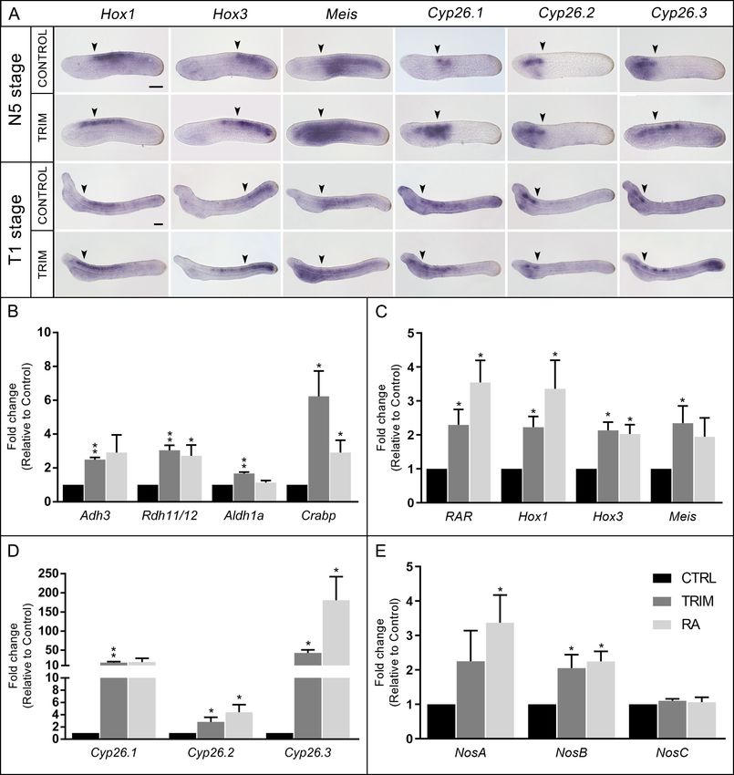
Research article                                                                                          Developmental Biology

                                   show a complementary expression pattern during development (Annona et al., 2017;
                                   Marlétaz et al., 2018). During amphioxus embryogenesis, as a consequence of pharmacological
                                   inhibition of endogenous NO production, the opening of the mouth is prevented as well as the cor-
                                   rect development of other important pharyngeal structures, such as the endostyle and the club-
                                   shaped gland (Annona et al., 2017). Moreover, the treated larvae show a posteriorized phenotype,
                                   resembling the well-described phenotype induced by exogenous retinoic acid (RA) administration
                                   during amphioxus embryogenesis (Escriva et al., 2002; Schubert et al., 2005; Koop et al., 2014).
                                   This experimental evidence prompted us to analyze the molecular effects of NO synthesis inhibition
                                   in detail by using a differential transcriptomic approach in the cephalochordate Branchiostoma lan-
                                   ceolatum. Our results show that the pharyngeal phenotype observed after reduction of endogenous
                                   NO production derives from an alteration of the RA signaling pathway. Moreover, we highlight the
                                   existence of a crosstalk between these pathways and propose that it is crucial to fine-tune the NO/
                                   RA balance that is required for proper pharyngeal development.

                                   Results
                                   NO controls pharyngeal development during early neurulation in
                                   amphioxus
                                   Previous studies, using pharmacological inhibition approaches, have highlighted the involvement of
                                   NO in the specification of amphioxus pharyngeal structures during neurulation (Annona et al.,
                                   2017). To better characterize the key role of NO during embryonic development, we narrowed
                                   down the time window of pharmacological treatment by defining the exact timing during which an
                                   inhibition of endogenous NO production affects the development of the pharynx. Therefore, we per-
                                   formed short-term in vivo treatments with the Nos activity inhibitor 1-[2-(trifluoromethyl)phenyl] 1H-
                                   imidazole (TRIM) during B. lanceolatum development testing different drug exposure times between
                                   the early neurula stage (N2 stage, 24 hr post fertilization [hpf] at 18˚C) and the pre-mouth larva stage
                                   (transition stage T1, 48 hpf at 18˚C) (Figure 1A). During the selected time window, the endogenous
                                   NO mainly derives from the activity of NosC, whose gene expression is observed from the N2 stage
                                   until the larval stage (L1, 72 hpf at 18˚C) (Figure 1—figure supplement 1; Annona et al., 2017). We
                                   cannot exclude a contribution to the NO production by NosB, whose gene is expressed from gas-
                                   trula (G3 stage, 10 hpf at 18˚C) to neurula (N2) (Annona et al., 2017), although we think it should be
                                   minimal because its mRNA synthesis runs out at 24 hpf. On the other hand, NosA is expressed in the
                                   adult and not during embryonic development.
                                       The phenotype resulting from the different treatments was scored at the open-mouth L1 stage.
                                   The morphological alterations included: (i) a mean reduction of 31% of the pharynx length being
                                   overall unchanged the body length (Figure 1B and Figure 1—figure supplement 2A,B), (ii) the
                                   complete or partial absence of mouth opening on the left side of the pharynx (Figure 1B, panels I,
                                   III and V, VII), and (iii) the incomplete formation of the club-shaped gland and of the endostyle
                                   (Figure 1B, panels I, III and V, VII), the latter being positioned more ventrally than in controls
                                   (Figure 1B, panels II, IV and VI, VIII).
                                       The inhibition of NO production during the 24–30 hpf time window at 18˚C was the shortest
                                   treatment inducing a significant effect by producing 100% of abnormal larvae. Delayed treatments
                                   starting at 30 or 36 hpf, for 6, 12, or 18 hr, resulted in approximately 70% of affected larvae
                                   (Figure 1A). Moreover, the endogenous NO concentration was measured at the endpoints of TRIM
                                   incubation intervals (24–30, 24–36, and 24–42 hpf) and in comparison to controls it decreased by
                                   66%, 57%, and 55%, respectively, demonstrating the efficiency of the treatments (Figure 1—figure
                                   supplement 2C). Conversely, when TRIM treatment was performed later, between 42 and 48 hpf,
                                   the larvae were not affected (Figure 1A). These results suggest that pharyngeal development is, at
                                   least in part, under the control of NO during neurulation (24–42 hpf) and we observed that the mini-
                                   mal time window of TRIM treatment that induces pharynx malformations corresponds to the first 6
                                   hr (24–30 hpf).
                                       The results presented here are slightly different from those previously published by our group
                                   (Annona et al., 2017). In our preceding study, we suggested that the developmental time window
                                   of Nos action was between 36 and 48 hpf because the treatment between 24 and 36 hpf was ineffi-
                                   cient in inducing a phenotype. However, in the present work we show that a treatment between 24

Caccavale et al. eLife 2021;10:e58295. DOI: https://doi.org/10.7554/eLife.58295                                                     2 of 16
Research article                                                                                                          Developmental Biology

Figure 1. Characterization of in vivo 1-[2-(trifluoromethyl)phenyl] 1H-imidazole (TRIM) treatment during early amphioxus embryogenesis. (A) Schematic
representation of time intervals during which embryos were grown in presence of TRIM and the resulting phenotype. (B) 3D reconstruction of control
and TRIM-treated larvae showing anatomical alterations in pharyngeal region (panels I, II, V, VI). Only internal anatomical structures are highlighted in
panels III, IV, VII, VIII. Larvae orientation: anterior to the left, dorsal to the top. Scale bar: 50 mm. Color code: green = pre-oral pit, violet = endostyle,
yellow = mouth, blue = club-shaped gland, orange = gill slit. (C) Gene expression heatmap, for selected genes, of the differential transcriptomic
analysis (control versus TRIM).
The online version of this article includes the following source data and figure supplement(s) for figure 1:
Source data 1. DESeq2 output for TRIM treated versus control condition.
Figure supplement 1. NosC expression pattern by whole-mount in situ hybridization.
Figure supplement 2. Pharynx and body lenght measurement and Nitric oxide quantification.
Figure supplement 3. RNA-seq data quality.
Figure supplement 4. Phylogenetic analysis for two retinoic acid (RA) pathway genes.
Figure supplement 5. Validation of the RNA-seq data by gene expression analyses.

                                       and 30 hpf is fully penetrant. In Annona et al., 2017, the inhibition of NO synthesis was achieved by
                                       using the L-NAME (Nw-nitro-L-arginine methyl ester), which is an L-arginine analog and is able to
                                       slow down the Nos activity. On the other hand, here we used TRIM, a molecule that is able to inter-
                                       fere with the binding of Nos enzymes with its substrate L-arginine and co-factor tetrahydrobiopterin.
                                       Thus, the dissimilarity in the time window of inhibition between the two treatments can be explained
                                       by the significant difference in Nos inhibition efficiency between the two molecules. Indeed, TRIM is
                                       effective at much lower concentrations than L-NAME (Annona et al., 2017), which probably justifies
                                       why a shorter treatment duration leads to the pharyngeal defect phenotype when we used TRIM,
                                       and hence the apparent discrepancy with previous data.

Caccavale et al. eLife 2021;10:e58295. DOI: https://doi.org/10.7554/eLife.58295                                                                         3 of 16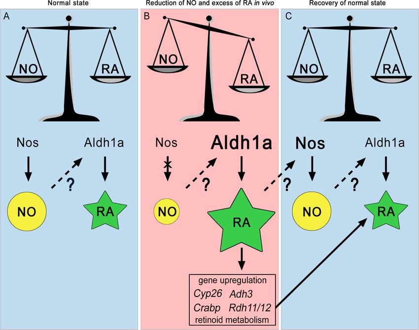
Research article Developmental Biology
Based on the experimental evidence obtained in the present work, we performed a differential
transcriptomic analysis comparing TRIM-treated N4 embryos (continuous treatment from 24 to 30
hpf) with wild-type embryos in order to define the genes acting downstream of NO signaling during
pharyngeal development in amphioxus (Figure 1C, and Figure 1—figure supplement 3).
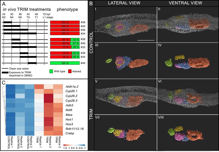
Inhibition of NO synthesis in vivo induces up-regulation and ectopic
expression of RA pathway genes
The differential RNA-seq analysis revealed the up-regulation of 392 genes and the down-regulation
of 50 genes upon TRIM treatment (Figure 1C, and Figure 1—figure supplement 3). Interestingly,
several differentially up-regulated genes are implicated in RA metabolism and signaling pathways
(synthesis and storage, catabolism and known direct RA target genes): Adh3, Rdh11/12.18,
Aldh1a.2, Crabp, Cyp26.1, Cyp26.2, Cyp26.3, RAR, Hox1, Hox3, Meis (Figure 1C and Figure 1—fig-
ure supplement 4). In order to confirm this finding, we additionally validated RNA-seq data by
quantitative RT-PCR (qRT-PCR) analyses of up-regulated, down-regulated, and unaffected genes.
The results showed a consistent expression trend with the RNA-seq data (Figure 1—figure supple-
ment 5A–C’). Moreover, the expression pattern of RA target genes Hox1, Hox3, Meis, and that of
Cyp26 genes was further investigated by whole-mount in situ hybridization in both control and
TRIM-treated embryos at the neurula N5 (36 hpf at 18˚C) and pre-mouth T1 (48 hpf at 18˚C) develop-
mental stages. Such analyses showed that endogenous NO reduction produced an effect not only
on the expression level of RA metabolism and signaling pathway genes, but also on the expression
territories of most of them. The Hox1, Hox3, and Meis anterior limit of expression was shifted anteri-
orly in TRIM-treated embryos in comparison to controls, indicative of the embryo’s body posteriori-
zation (Figure 2A). The RA catabolism enzyme genes that are duplicated in amphioxus, Cyp26.1,
Cyp26.2, and Cyp26.3, showed a heterogeneous behavior: Cyp26.2 was slightly up-regulated and its
expression pattern did not change after TRIM treatment, whereas Cyp26.1 and Cyp26.3 were
strongly up-regulated and showed an ectopic expression (Figure 2A,D). In particular, after inhibition
of NO production, Cyp26.1 expression was shifted anteriorly, while Cyp26.3 expression was shifted
posteriorly. Moreover, Cyp26.3 showed an additional domain of expression in the tailbud, mainly in
T1 stage embryos (Figure 2A). As a negative control we tested the expression pattern of Pitx, Six1/
2, IrxC, and Cdx, whose expression levels were not affected after TRIM treatment in the RNA-seq
data. Likewise, we observed no modification of their expression pattern after treatment (Figure 1—
figure supplement 5D).
Aldh1a.2 expression is specifically regulated by NO
The above-mentioned gene expression results (Figure 2A,C,D) suggested that the abnormal pharyn-
geal development of TRIM-treated embryos could be the result of an overactivation of the RA sig-
naling pathway based on previous studies (Escriva et al., 2002; Schubert et al., 2006; Koop et al.,
2014; Carvalho et al., 2017b). In order to test this hypothesis, we performed two in vivo experi-
ments in parallel in which neurula stage embryos were incubated for 6 hr, from 24 to 30 hpf, in the
presence of either TRIM or RA. Then, we analyzed the relative expression of three groups of genes
by qRT-PCR, which we previously found to be up-regulated in the RNA-seq analysis: (i) genes
involved in the synthesis and storage of RA (Adh3, Rdh11/12.18, Aldh1a.2, and Crabp); (ii) genes
that mediate RA effects (RAR, Hox1, Hox3, and Meis); and (iii) genes involved in RA degradation
(Cyp26.1, Cyp26.2, and Cyp26.3). All analyzed genes were up-regulated after both TRIM and RA
treatment, with the exception of Aldh1a.2 which was exclusively up-regulated after TRIM treatment
(Figure 2B,C,D).
NosA and NosB respond to exogenous RA during development
The expression analysis of the three amphioxus Nos genes after TRIM treatment revealed transcrip-
tional up-regulation for two of them, NosA and NosB, while NosC, remained insensitive to the phar-
macological treatment (Figure 2E).
In order to check if NosA and NosB up-regulation could be due to an indirect effect of the intra-
cellular increase of RA caused by the TRIM treatment, we tested their expression levels after the
addition of exogenous RA (Figure 2E). Similarly to the TRIM treatment, we observed that NosA and
NosB expression were significantly up-regulated as a consequence of RA administration (Figure 2E).
Caccavale et al. eLife 2021;10:e58295. DOI: https://doi.org/10.7554/eLife.58295 4 of 16Research article Developmental Biology Figure 2. Analysis of gene expression. (A) Gene expression pattern by in situ hybridization of Hox1, Hox3, Meis, Cyp26.1, Cyp26.2, and Cyp26.3 in 1-[2- (trifluoromethyl)phenyl] 1H-imidazole (TRIM)-treated and control embryos at N5 and T1 stage. The anterior (Hox1, Hox3, Meis, Cyp26.1) and posterior (Cyp26.2 and Cyp26.3) limits of gene expression territories in wild-type embryos are indicated with arrowheads in both control and TRIM-treated conditions. Fifteen embryos were used for each probe and all showed the pattern presented here. Embryos orientation: anterior to the left, dorsal to the top. Scale bar: 50 mm. (B–E) Quantitative RT-PCR (qRT-PCR) on N4 stage embryos showing expression level changes after 6 hr of pharmacological TRIM or retinoic acid (RA) treatments (24–30 hpf) of: (B) genes encoding enzymes for RA synthesis: Adh3, Rdh11/12.18, Aldh1a.2, and binding protein for storage: Crabp; (C) RA direct target genes: RAR, Hox1, Hox3, Meis; (D) genes encoding RA degradation enzymes: Cyp26.1, Cyp26.2, Cyp26.3; (E) Nos genes: NosA, NosB, NosC. The statistical significance indicated is: * = p-value < 0.05; ** = p-value < 0.01. Figure 2 continued on next page Caccavale et al. eLife 2021;10:e58295. DOI: https://doi.org/10.7554/eLife.58295 5 of 16
Research article                                                                                             Developmental Biology

Figure 2 continued
The online version of this article includes the following source data and figure supplement(s) for figure 2:
Source data 1. Source raw data of qRT-PCR in Figure 2B-E.
Figure supplement 1. Nos genes expression pattern by quantitative RT-PCR (qRT-PCR) after retinoic acid (RA) treatment.

                                      Moreover, exogenous RA induced up-regulation of NosA expression up to 36 hpf (N5) and of NosB
                                      expression up to 48 hpf (T1) (Figure 2—figure supplement 1).
                                         These results suggest a transcriptional control of RA on Nos genes expression during embryogen-
                                      esis in amphioxus, but whether it is direct or indirect remains to be investigated.

                                      A RALDH inhibitor and a RAR antagonist are able to rescue the normal
                                      phenotype after inhibition of NO synthesis
                                      To confirm that the up-regulation of Aldh1a.2, which could result in an endogenous RA increase,
                                      was the key event underlying pharyngeal alterations in TRIM-treated larvae, we performed two inde-
                                      pendent phenotypic rescue experiments using the retinaldehyde dehydrogenases (RALDH) inhibitor
                                      DEAB (N,N-diethylaminobenzaldehyde) and the RA antagonist BMS009. Both DEAB and BMS009
                                      were applied in combination with TRIM to embryos at 24 hpf and removed at 30 hpf. As a control,
                                      the TRIM treatment was performed in parallel on another batch of embryos. The combined treat-
                                      ment with TRIM and DEAB resulted in a wild-type phenotype in 76% of the total observed larvae
                                      (‘rescue’ in Figure 3A,B panels I and II). On the other hand, 14% of the larvae showed an intermedi-
                                      ate phenotype with normal pharynx length and organization in comparison to wild-type larvae,
                                      although the mouth was smaller and the club-shaped gland had an abnormal morphology (‘partial
                                      rescue’ in Figure 3A,B panels III and IV). The remaining larvae showed an affected phenotype as
                                      described above for the TRIM treatment (‘altered’ in Figure 3A,B panels V and VI). Similarly, the
                                      phenotype rescue experiment performed using the combination of TRIM and BMS009 led to 54% of
                                      wild-type larval morphology and 21% of larvae with a smaller mouth (Figure 3A). Moreover, the
                                      morphological rescue obtained by TRIM+DEAB treatment was associated with the rescued expres-
                                      sion pattern of RA catabolism (Cyp26.1 and Cyp26.3) and RA target genes (Hox1, Hox3, and Meis)
                                      (Figure 3C) at the N5 neurula stage. Therefore, by using two independent experiments, we demon-
                                      strated that the reduction of RA pathway activity in TRIM-treated embryos rescued the wild-type
                                      phenotype, suggesting that the observed effects of the inhibition of NO synthesis are produced by
                                      an increase in RA signaling.

                                      Discussion
                                      NO controls the RA concentration
                                      The inhibition of NO production during amphioxus neurulation affects the normal formation and pat-
                                      terning of pharyngeal structures at the larva stage, including the length of the pharynx. From a
                                      molecular point of view, the differential RNA-seq approach revealed a clear up-regulation of differ-
                                      ent RA pathway players after Nos activity inhibition, suggesting that such de-regulation is responsi-
                                      ble for the observed phenotype. The role of RA in pharyngeal morphogenesis has been extensively
                                      described in the literature; RA acts through Hox1 in establishing the posterior limit of amphioxus
                                      pharynx. Hox1 is co-expressed with the RA receptor (RAR) in the midgut endoderm and, in turn,
                                      represses the expression of pharyngeal endoderm markers, such as Pax1/9 and Otx (Schubert et al.,
                                      2005). Nevertheless, the formation of pharyngeal slits requires low levels of RA. This condition is
                                      guaranteed: (i) by the activity of RA degradation enzymes (Cyp26), (ii) by the expression of TR2/4, a
                                      transcriptional repressor which binds to Retinoic Acid Response Elements (RARE) and decreases RA
                                      signaling in the anterior part of the animal, and (iii) by the fact that the central region of the embryo
                                      producing RA moves posteriorly as the embryo elongates (Escriva et al., 2002; Koop et al., 2014).
                                      In the present work, as a result of the inhibition of Nos activity, we observed the up-regulation of RA
                                      target genes, Hox1, Hox3 and Meis, and that their anterior limit of expression shifted anteriorly. Fur-
                                      thermore, the RA degrading enzyme genes, Cyp26.1 and Cyp26.3, were also sensitive to the inhibi-
                                      tion of endogenous NO production showing an increased and ectopic expression. Cyp26 are
                                      required for RA degradation in the endoderm and ectoderm and have a key role in the

Caccavale et al. eLife 2021;10:e58295. DOI: https://doi.org/10.7554/eLife.58295                                                         6 of 16
Research article Developmental Biology
Figure 3. Phenotypic rescue effect of N,N-diethylaminobenzaldehyde (DEAB) and BMS009 on 1-[2-(trifluoromethyl)
phenyl] 1H-imidazole (TRIM)-treated embryos. (A) Pie charts of the phenotypes observed after TRIM treatment
and the combinatorial pharmacological treatments TRIM (100 mM) + DEAB (25 mM) or TRIM (100 mM) + BMS009
(10 6 M). The percentages of each observed phenotype are reported in the respective portions of the graphs. For
each treatment, the total number of observed larvae is indicated below the chart. (B) Pictures of the pharyngeal
region of larvae presenting the three different classes of phenotype observed in the rescue experiments: rescue,
partial rescue, and altered. The mouth is highlighted in yellow and the club-shaped gland in blue. Larvae
orientation: anterior to the left, dorsal to the top. Scale bar: 50 mm. (C) Expression pattern by in situ hybridization
of Hox1, Hox3, Meis, Cyp26.1, and Cyp26.3 after rescue assay with DEAB showing the restoration of wild-type
expression territories. Numbers indicate the ratio between embryos showing a restored expression pattern and
the total number of embryos analyzed. Embryo orientation: anterior to the left, dorsal to the top. Scale bar: 50 mm.
establishment and maintenance of the antero-posterior RA concentration gradient in amphioxus
(Carvalho et al., 2017a). The up-regulation of Cyp26 genes is a known consequence of RA excess,
which is responsible for the posteriorization of larval body structures and for the pharynx loss
(Escriva et al., 2002; Schubert et al., 2005; Schubert et al., 2006; Minoux and Rijli, 2010;
Koop et al., 2010; Koop et al., 2014; Bertrand et al., 2015; Carvalho et al., 2017b). Altogether,
these results suggest that the observed phenotype in TRIM-treated amphioxus embryos could be
due to an increase in RA production. In previous studies, it has been shown that exogenous RA
administration disrupts its endogenous gradient and causes the whole body posteriorization
(Escriva et al., 2002; Koop et al., 2014; Osborne et al., 2009), highlighted for example by the
anterior shift of Cdx expression. Here, instead, we show that the TRIM treatment only partially phe-
nocopies exogenous RA application, resulting in a local posteriorization mainly restricted to the
anterior part of the body including the pharynx area, while the posterior region is unaffected as
demonstrated by the unaltered Cdx expression (Figure 1—figure supplement 5D). Therefore, this
suggests the occurrence of an NO-mediated regulation of the RA pathway in anterior tissues versus
an NO-independent mechanism in the posterior tissues.
Caccavale et al. eLife 2021;10:e58295. DOI: https://doi.org/10.7554/eLife.58295 7 of 16Research article Developmental Biology
Intracellular RA is synthesized by the reversible oxidation of retinol into retinaldehyde by either
alcohol dehydrogenases (ADH) or retinol dehydrogenases (RDH). Subsequently, retinaldehyde is irre-
versibly oxidized to RA by RALDH, mainly by ALDH1A (Gallego et al., 2006; Duester, 2008). In our
experiments we observed a transcriptional up-regulation of Adh3, Rdh11/12.18, and Aldh1a.2 after
TRIM treatment. While a unique ortholog of vertebrate Adh genes, Adh3, has been identified in
amphioxus (Cañestro et al., 2002); 22 Rdh11/12 genes, derived from a lineage-specific expansion,
were identified. These Rdh11/12 genes are related to human Rdh11, Rdh12, Rdh13, and Rdh14, that
together with Rdh10 correspond to retinaldehyde reductases predominantly involved in retinoid
metabolism and homeostasis (Albalat et al., 2011; D’Aniello et al., 2015; Figure 1—figure supple-
ment 4B). For Aldh1, a total of six genes were identified in amphioxus, orthologs of human
Aldh1A1-3, which are major players in the oxidation of RA (Cañestro et al., 2006; Figure 1—figure
supplement 4A). In our study, Adh3 and Rdh11/12.18 genes were also up-regulated after adminis-
tration of exogenous RA, suggesting a feedback regulation of RA synthesis, at least on reversible
enzymatic steps. On the other hand, Aldh1a.2 was insensitive to exogenous RA administration.
Based on these results, we hypothesize that, under physiological conditions, NO transcriptionally
regulates Aldh1a.2 and, as a consequence, controls the production of endogenous RA. Further evi-
dence to support our hypothesis is provided by the two independent rescue experiments. Techni-
cally, in association with TRIM, we used two drugs that specifically act on the most crucial steps of
RA signaling pathway: an inhibitor of the RALDH enzymes activity that would compensate the excess
of Aldh1a.2 protein, and an RA antagonist, which is able to compensate the RA over-production
through its binding to RAR. In both experiments we observed a wild-type phenotype at the larva
stage as a consequence of the recovery of the normal development, further supported by the resto-
ration of the proper expression pattern of both RA-target and RA-degrading enzyme genes.
Based on our results, we propose that NO plays a key role in the regulation of RA level during
neurulation in amphioxus by fine-tuning the expression of Aldh1a.2, keeping RA concentration within
the optimal range. This precise balance between intracellular concentrations of NO and RA guaran-
tees the correct expression level and localization of all RA downstream target genes. When NO is
removed from the system, the RA metabolism machinery malfunctions, resulting in a cascade of
events leading to the up-regulation of the entire RA signaling pathway.
The missing piece of the puzzle, therefore, seems to be an unknown molecular link, which is able
to explain the control of Aldh1a.2 transcription by NO. In other chordates, it has been demonstrated
that the mechanisms by which NO regulates transcription of target genes are: (i) the control of the
extracellular-regulated kinase (ERK) and the Mitogen-activated protein kinase (MAPK) phosphatases
activity and, as a result, the modulation of phosphorylation or dephosphorylation of target transcrip-
tion factors (Castellano et al., 2014), and (ii) the direct modulation of target proteins, like transcrip-
tion factors, histone acetyltransferases, and deacetylases or DNA methyltranserases, through S-
nitrosylation of specific cysteine residues (Bogdan, 2001; Nott et al., 2008; Sha and Marshall,
2012).
Thus, NO could control Aldh1a.2 expression by modulating the phosphorylation or S-nitrosylation
of a specific transcription factor/chromatin remodeling protein, the nature of which still requires fur-
ther research to be discovered.
It would be important to improve this knowledge since very little information, restricted to verte-
brates, is reported on the control of RA metabolism by NO. Some cytochrome P450 enzymes,
involved in RA metabolism, were identified as putative NO-regulated proteins, but no evidence
about putative transcriptional regulation has been reported so far (Lee et al., 2014; Lee et al.,
2017).
Crosstalk between NO and RA signaling pathways
The exogenous administration of RA induces the expression of amphioxus NosA and NosB that are
normally not expressed during the developmental time window investigated in this study
(Annona et al., 2017). Moreover, such transcriptional regulation is maintained throughout the critical
time period during which NO is necessary for pharyngeal development (i.e. 24–42 hpf, Figure 2—
figure supplement 1). Conversely, NosC was not affected by the increase of RA level. A possible
explanation could be that while in normal conditions RA does not regulate NosA and NosB expres-
sion, it directly or indirectly regulates such gene expression in the case of an NO/RA imbalance as a
Caccavale et al. eLife 2021;10:e58295. DOI: https://doi.org/10.7554/eLife.58295 8 of 16Research article Developmental Biology
Figure 4. The NO:RA hypothesis. Schematic representation of the possible crosstalk occurring between Nitric Oxide (NO) and Retinoic Acid (RA)
during chordate development. NO is represented by a yellow circle, RA by a green star, the ratio between NO and RA by a scale symbol. The decrease
of NO and the increase of RA, Aldh1a and Nos, are indicated by different sizes of symbols relative to the normal state. Arrows represent enzymatic
processes, while discontinuous arrows correspond to transcriptional regulation. Question marks indicate that it is not known exactly how NO regulates
Aldh1a.2 transcription and RA regulates Nos transcription. The cross over the arrow between Nos and NO illustrates the inactivation of Nos.
way to restore the correct NO/RA ratio. Additionally, it could be that RA is able to force the regula-
tory mechanism normally used at another developmental stage or in the adult.
In vertebrates, the role of NO and RA in the correct development of the pharynx and craniofacial
structures has already been described (Abe et al., 2008; Liu et al., 2013; Jacox et al., 2014;
Chawla et al., 2018). However, the existence of a possible crosstalk between NO and RA pathways
has not yet been revealed. As mentioned above, there are few published data on the regulatory
effect of NO on RA pathways, while the control of RA on NO production in vertebrates is more
documented. For instance, the inhibitory or activator effect of RA on NO production through the
activation of inducible and endothelial Nos, at both the protein and transcript levels, has been dem-
onstrated in human cell lines (Sirsjö et al., 2000; Achan et al., 2002; Hattori et al., 2002;
Behairi et al., 2015; Moon, 2019).
Caccavale et al. eLife 2021;10:e58295. DOI: https://doi.org/10.7554/eLife.58295 9 of 16Research article Developmental Biology
To summarize the regulatory loop between NO and RA signaling pathways in amphioxus, we for-
mulated the NO:RA hypothesis, shown in Figure 4, arguing that their in vivo crosstalk is critical to
maintain NO and RA concentrations in a correct ratio for normal pharyngeal development. During
embryogenesis (Figure 4A), the balance of these two signaling molecules guarantees a physiological
RA concentration necessary for a correct pharynx development. A reduction of NO (Figure 4B), as
shown by our results albeit indirectly, induces an excess of RA, through the overexpression of
Aldh1a.2. We propose that in the developing embryo an excess of RA compared to NO level indu-
ces both the expression of genes that contribute to RA level reduction, and to the overexpression of
Nos genes resulting in NO production increase (Figure 4C) which in turn contributes to the decrease
in RA concentration through an unknown mechanism. Thus, this regulatory loop would allow a rees-
tablishment of the normal NO/RA ratio (Figure 4C) and would contribute to a correct development
of the amphioxus pharynx.
Conclusions
Our results show the existence of a functional crosstalk between NO and RA signals in the pharyn-
geal region of the cephalochordate amphioxus during neurulation. This opens new questions about
the evolutionary conservation of this regulatory loop in vertebrates.
The role of RA, as well as that of NO, in amphioxus development and antero-posterior patterning
of the pharynx has been described in several studies. Our results suggest the occurrence of a regula-
tory crosstalk between these two ancient and essential signaling pathways that has previously been
neglected. Our results allow us to propose that, during amphioxus development, a precise NO/RA
balance is necessary for the correct antero-posterior patterning of the pharynx. This endogenous
intracellular balance is preserved by the reciprocal regulation of NO and RA pathways. Taking into
account the role that these two signaling pathways and their crosstalk have for the correct develop-
ment of the amphioxus pharynx, as well as the known role that, separately, has been demonstrated
for these two pathways in vertebrates, we hypothesize that the functional and regulatory crosstalk
between NO and RA pathways could be a conserved feature in vertebrates.
Materials and methods
Key resources table
Reagent type (species) or resource Designation Source or reference Identifiers Additional information
Strain, strain Wild type Collected in Argelès- NCBI Taxon: 7740
background, (Branchiostoma lanceolatum) sur-mer, France
Chemical 2,3-Diaminonaphthalene (DAN) Sigma-Aldrich D2757
compound, drug
Chemical Nitrate reductase Sigma-Aldrich N7265
compound, drug (NAD[P]H) from Aspergillus niger
Chemical Flavin adenine Sigma-Aldrich F6625
compound, drug dinucleotide disodium
salt hydrate (FAD)
Chemical b-Nicotinamide Sigma-Aldrich N7505
compound, drug adenine dinucleotide
20 -phosphate reduced
tetrasodium salt
hydrate (NADPH)
Chemical 1-[2-(Trifluoromethyl) Cayman chemical 81310
compound, drug phenyl]-1H-imidazole (TRIM)
Chemical all-trans-Retinoic Sigma-Aldrich R2625
compound, drug acid (RA)
Chemical N,N-diethylamino Sigma-Aldrich D86256
compound, drug benzaldehyde (DEAB)
Amphioxus embryos collection
Ripe adult European amphioxus (B. lanceolatum) were collected in Argelès-sur-mer (France) with a
specific permission delivered by the Prefect des Pyrénées-Orientales. B. lanceolatum is not a
Caccavale et al. eLife 2021;10:e58295. DOI: https://doi.org/10.7554/eLife.58295 10 of 16Research article Developmental Biology
protected species. Spawning was induced during late spring and beginning of summer by employing
a thermal shock as described by Fuentes et al., 2007. After in vitro fertilization, embryos were cul-
tured in 0.22 mm filtered seawater at 18˚C in plastic Petri dishes. According to the recent amphioxus
ontology and staging (Bertrand et al., 2021; Carvalho et al., 2021) at 18˚C, 24 hpf corresponds to
neurula two stage (N2), 30 hpf to neurula four stage (N4), 36 hpf to neurula five stage (N5), 42 hpf
to transition 0 stage (T0), 48 hpf to transition one stage (T1), and 72 hpf to larva 1 (L1). Embryos at
desired developmental stages were incubated in the presence of specific pharmacological drugs,
frozen in liquid nitrogen and kept at 80˚C for subsequent RNA extraction or fixed with 4% parafor-
maldehyde in MOPS buffer overnight at 4˚C and then stored in 70% ethanol at 20˚C until use.
Pharmacological treatments
Amphioxus embryos were treated at different developmental stages with the Nos inhibitor 1-[2-(tri-
fluoromethyl)phenyl] 1H-imidazole (TRIM), with the RALDH inhibitor N,N-diethylaminobenzalde-
hyde (DEAB), with the RA antagonist BMS009 and with all-trans RA. All the drugs were dissolved in
dimethyl sulfoxide (DMSO), and control embryo groups for each treatment were prepared adding
an equal amount of DMSO. For TRIM treatments, a final concentration of 100 mM was used. For RA
treatments, a final concentration of 10 6 M was used. All the treatments were performed in biologi-
cal triplicates.
For rescue experiments, embryos at 24 hpf were treated simultaneously with a combination of
100 mM TRIM and 25 mM DEAB, or 100 mM TRIM and 10 6 M BMS009. At 30 hpf they were rinsed in
filtered seawater and allowed to develop until the 72 hpf stage when the phenotype was observed.
The experiment was performed in biological duplicates.
Imaging
Control and TRIM-treated larvae at 72 hpf were stained with DAPI. A high-resolution Z-stack was
acquired using a Zeiss confocal microscopy LSM 800, and a medium speed fast interactive deconvo-
lution was applied. The 3D reconstruction was made employing Imaris 9.3.1 software; different larval
body structures have been stained using the following five color blindness-friendly colors: #A6D854
green; #E78AC3 violet; #FFD92F yellow; #8DA0CB blue; #FC8D62 orange. Pharynx and body length
measurements were performed on 72 hpf larvae captured with Axio Imager.Z2 microscope using a
10 objective, and employing the Ruler tool in Photoshop CS5 with a digital zoom of 50%. The
pharynx was measured from the most anterior part of the pre-oral pit to the most posterior part of
the first pharyngeal slit. An unpaired t-test was applied for the statistical analysis.
Fluorimetric determination of endogenous NO concentration
Control and TRIM-treated frozen embryos at different developmental stages (from 24 hpf to 30, 36,
and 42 hpf) were homogenized in PBS, sonicated (3 cycles of 1 min) and centrifuged at 20,000 g for
30 min at 4˚C. The supernatants were collected for NO level analysis; 80 ml of each sample was incu-
bated for 1 hr at room temperature in the presence of the nitrate reductase (0.06 U/ml), 2.5 mM
FAD, and 100 mM NADPH. Then, 10 ml of 2,3-diaminonaphthalene (DAN) (0.05 mg/ml in 0.62 M HCl)
were added and the samples were incubated for 15 min in the dark. The fluorescent product was
stabilized in 1 N sodium hydroxide. Fluorescence was measured using the spectrofluorometer
(Tecan) with excitation and emission at 365 and 425 nm, respectively, adding water up to a final vol-
ume of 200 ml. The results were normalized on the protein content. Total protein concentration was
determined by the Bradford assay using a Bio-Rad Protein Assay Reagent (Bio-Rad) and bovine
serum albumin as a standard.
RNA-seq analysis
Total RNA was extracted from embryos using the RNeasy Plus Mini Kit (Qiagen) after sample
homogenization using the TissueLyser (Qiagen). The RNA integrity number (RIN) was assessed by
using TapeStatio4200 while RNA concentration and purity were estimated using a Nanodrop spec-
trophotometer. Indexed libraries were prepared from 1 mg/ea purified RNA with TruSeq Stranded
Total RNA Library Prep Kit. Libraries were quantified using the Agilent 2100 Bioanalyzer (Agilent
Technologies) and pooled so that each index-tagged sample was present in equimolar amounts,
with a final concentration of 2 nM. The pooled samples at a final concentration of 10 pM were
Caccavale et al. eLife 2021;10:e58295. DOI: https://doi.org/10.7554/eLife.58295 11 of 16Research article Developmental Biology
subjected to cluster generation and sequencing using an Illumina NextSeq500 System in a 175 sin-
gle read format (30 million reads). The raw sequence files generated (fastq files) underwent quality
control analysis using FastQC. Transcriptome sequences were deposited in the NCBI Sequence
Read Archive (SRA) database with the accession number: PRJNA630453.
Reads were mapped on the B. lanceolatum transcriptome (Oulion et al., 2012) using the aligner
Bowtie2 with default parameters (Langmead and Salzberg, 2012). The read counts were obtained
using IdxStats (Li et al., 2009; Cock et al., 2013) and the differential expression analysis between
treated and wild-type embryos was performed using the R package DESeq2 (Love et al., 2014).
Mapping and read counting were performed on the Roscoff ABiMS Galaxy platform.
Phylogenetic analysis
Protein alignments were generated with ClustalX program using the sequence database reported in
Handberg-Thorsager et al., 2018. Phylogenetic trees were reconstructed using maximum likelihood
inferences calculated with PhyML v3.0 (Guindon et al., 2010).
Gene expression analysis by whole-mount in situ hybridization and
immunostaining
Hox1, Hox3, Meis, Cyp26.1, Cyp26.2, Cyp26.3, Pitx, Six1/2, IrxC, Cdx, and NosC were cloned in
pGEM-T vector (Promega) using primers listed in Supplementary file 1. Antisense labeled ribop-
robes were synthesized and in situ hybridizations were performed as previously described
(Annona et al., 2017; Carvalho et al., 2021). Whole-mount immunostaining of acetylated tubulin
using monoclonal antibody produced in mouse (6-11B-1, Sigma) was performed as previously
described in Coppola et al., 2018. Embryos were mounted in 80% glycerol in PBS and photo-
graphed using an Axio Imager.Z2 or a confocal microscope Zeiss LSM700.
Gene expression analysis by qRT-PCR
Total RNA was extracted from embryos at different developmental stages: 30, 36, 42, 48, and 72
hpf using the RNeasy Plus Mini Kit (Qiagen); 350–1000 ng of total RNA were retrotranscribed in
cDNA which was used undiluted (only for Nos genes) or diluted 1:10 for the qRT-PCR. Each reaction
contained a final concentration of 0.7 mM of each primer and Fast SYBR Green Master mix with ROX
(Applied Biosystems) in 10 ml total volume. qRT-PCR were run in a ViiA 7 Real-Time PCR System
(Applied Biosystems). The cycling conditions were: 95˚C for 20 s, 40 cycles with 95˚C for 1 s, 60˚C for
20 s, 95˚C for 15 s, 60˚C 1 min, followed by a dissociation curve analysis using a gradient from 60˚C
to 95˚C with a continuous detection at 0.015˚C/s increment for 15 min. The results were analyzed
using the ViiA 7 Software and exported into Microsoft Excel for further analysis. Each sample was
processed in biological triplicates. The 2 DDCt Rpl32 method was used to calculate the relative gene
expression. Ribosomal protein L32 (Rpl32), expressed at a constant level during development, was
used as a reference gene for the normalization of each gene expression level (Annona et al., 2017).
Primers used are listed in Supplementary file 1. For the statistical analysis, we used the GraphPad
Prism software employing the paired t-test. Statistical significance cut-off criteria was set at p <
0.05.
Acknowledgements
The authors thank Rebecca Adikes and Hannah Rosenblatt, course assistants at the MBL Embryology
Course 2019 in Woods Hole (USA), and Periklis Paganos for their help with imaging. We are also
grateful to Enrico D’Aniello, Ricard Albalat, Marion Picard, and Haley Flom for their critical reading
of the manuscript. We thank Ángel R de Lera for providing the RA antagonist BMS009, and Carola
Murano and Anna Palumbo for their help with the NO quantification (DAN assay). We are grateful to
the Institut Français de Bioinformatique and the Roscoff Bioinformatics platform ABiMS for providing
computing and storage resources for the RNA-seq analysis, and the BIO2MAR platform (EMBRC-
France) supported by ANR grant no. ANR-10-INBS-02 for giving us access to analytical material. SD
and FC acknowledge the Assemble Plus project (contract numbers BA010618 and 360BA0619) and
The Company of Biologists (grant number DEVTF-170211; sponsoring journal: Development) for
supporting research visits to the Observatoire Océanologique of Banyuls-sur-Mer (France). FC was
Caccavale et al. eLife 2021;10:e58295. DOI: https://doi.org/10.7554/eLife.58295 12 of 16Research article Developmental Biology
supported by an OU-SZN PhD fellowship. SB is supported by the Institut Universitaire de France
(IUF). HE is supported by the Centre national de la recherche scientifique and Agence Nationale de
la Recherche (ANR) grants number ANR-16-CE12-0008-01 and ANR-19-CE13-0011. HE is supported
by the European Commission ASSEMBLE Plus network (H2020-INFRAIA-1-2016–2017; grant number
730984).
Additional information
Funding
Funder Grant reference number Author
European Commission BA010618 and 360BA0619 Salvatore D’Aniello
Company of Biologists DEVTF-170211 Filomena Caccavale
Agence Nationale de la Re- ANR-16-CE12-0008-01 and Hector Escriva
cherche ANR-19-CE13-0011
European Commission H2020-INFRAIA-1-2016– Hector Escriva
2017 n. 730984
The funders had no role in study design, data collection and interpretation, or the
decision to submit the work for publication.
Author contributions
Filomena Caccavale, Conceptualization, Data curation, Investigation, Methodology, Writing - original
draft, Writing - review and editing; Giovanni Annona, Data curation, Investigation, Methodology,
Writing - review and editing; Lucie Subirana, Data curation, Formal analysis, Methodology; Hector
Escriva, Conceptualization, Writing - review and editing; Stephanie Bertrand, Conceptualization,
Data curation, Writing - review and editing; Salvatore D’Aniello, Conceptualization, Supervision,
Investigation
Author ORCIDs
Giovanni Annona http://orcid.org/0000-0001-7806-6761
Salvatore D’Aniello https://orcid.org/0000-0001-7294-1465
Decision letter and Author response
Decision letter https://doi.org/10.7554/eLife.58295.sa1
Author response https://doi.org/10.7554/eLife.58295.sa2
Additional files
Supplementary files
. Supplementary file 1. Primers for the preparation of WISH probes cloning and for quantitative RT-
PCR.
. Transparent reporting form
Data availability
Sequencing data have been deposited in NCBI SRA under accession code PRJNA630453. All data
generated or analysed in this study are included in the manuscript and supporting files. Source data
files have been provided for Figures 1 and 2.
The following dataset was generated:
Database and
Author(s) Year Dataset title Dataset URL Identifier
Caccavale F, 2020 Crosstalk between Nitric Oxide and https://www.ncbi.nlm. NCBI Sequence Read
Annona G, Retinoic Acid pathways is essential nih.gov/sra/ Archive, PRJNA630
Subirana L, Escriva for amphioxus pharynx PRJNA630453 453
Caccavale et al. eLife 2021;10:e58295. DOI: https://doi.org/10.7554/eLife.58295 13 of 16Research article Developmental Biology
H, Bertrand S, development
D’Aniello S
References
Abe M, Maeda T, Wakisaka S. 2008. Retinoic acid affects craniofacial patterning by changing Fgf8 expression in
the pharyngeal ectoderm. Development, Growth & Differentiation 50:717–729. DOI: https://doi.org/10.1111/j.
1440-169X.2008.01069.x, PMID: 19046160
Achan V, Tran CTL, Arrigoni F, Whitley GSJ, Leiper JM, Vallance P. 2002. All- trans -Retinoic acid increases nitric
oxide synthesis by endothelial cells. Circulation Research 90:764–769. DOI: https://doi.org/10.1161/01.RES.
0000014450.40853.2B
Albalat R, Brunet F, Laudet V, Schubert M. 2011. Evolution of retinoid and steroid signaling: vertebrate
diversification from an amphioxus perspective. Genome Biology and Evolution 3:985–1005. DOI: https://doi.
org/10.1093/gbe/evr084, PMID: 21856648
Annona G, Caccavale F, Pascual-Anaya J, Kuratani S, De Luca P, Palumbo A, D’Aniello S. 2017. Nitric oxide
regulates mouth development in amphioxus. Scientific Reports 7:8432. DOI: https://doi.org/10.1038/s41598-
017-08157-w, PMID: 28814726
Behairi N, Belkhelfa M, Mesbah-Amroun H, Rafa H, Belarbi S, Tazir M, Touil-Boukoffa C. 2015. All-trans-retinoic
acid modulates nitric oxide and interleukin-17A production by peripheral blood mononuclear cells from
patients with Alzheimer’s disease. Neuroimmunomodulation 22:385–393. DOI: https://doi.org/10.1159/
000435885, PMID: 26278415
Bertrand S, Aldea D, Oulion S, Subirana L, de Lera AR, Somorjai I, Escriva H. 2015. Evolution of the role of RA
and FGF signals in the control of somitogenesis in chordates. PLOS ONE 10:e0136587. DOI: https://doi.org/
10.1371/journal.pone.0136587, PMID: 26371756
Bertrand S, Carvalho JE, Dauga D, Matentzoglu N, Daric V, J-k Y, Schubert M, Escrivá H. 2021. The ontology of
the amphioxus anatomy and life cycle (AMPHX). front. Cell & Developmental Biology 9:668025. DOI: https://
doi.org/10.3389/fcell.2021.668025
Bogdan C. 2001. Nitric oxide and the regulation of gene expression. Trends in Cell Biology 11:66–75.
DOI: https://doi.org/10.1016/S0962-8924(00)01900-0
Cañestro C, Albalat R, Hjelmqvist L, Godoy L, Jörnvall H, Gonzàlez-Duarte R. 2002. Ascidian and amphioxus adh
genes correlate functional and molecular features of the ADH family expansion during vertebrate evolution.
Journal of Molecular Evolution 54:81–89. DOI: https://doi.org/10.1007/s00239-001-0020-2, PMID: 11734901
Cañestro C, Postlethwait JH, Gonzàlez-Duarte R, Albalat R. 2006. Is retinoic acid genetic machinery a chordate
innovation? Evolution Development 8:394–406. DOI: https://doi.org/10.1111/j.1525-142X.2006.00113.x,
PMID: 16925675
Carvalho JE, Lahaye F, Croce JC, Schubert M. 2017a. CYP26 function is required for the tissue-specific
modulation of retinoic acid signaling during amphioxus development. The International Journal of
Developmental Biology 61:733–747. DOI: https://doi.org/10.1387/ijdb.170227ms
Carvalho JE, Theodosiou M, Chen J, Chevret P, Alvarez S, De Lera AR, Laudet V, Croce JC, Schubert M. 2017b.
Lineage-specific duplication of amphioxus retinoic acid degrading enzymes (CYP26) resulted in sub-
functionalization of patterning and homeostatic roles. BMC Evolutionary Biology 17:24. DOI: https://doi.org/
10.1186/s12862-016-0863-1
Carvalho JE, Lahaye F, Yong LW, Croce JC, Escrivá H, Jk Y, Schubert M. 2021. An updated staging system for
cephalochordate development: one table suits them all. Front Cell Dev. Biol 9:668006. DOI: https://doi.org/10.
3389/fcell.2021.668006
Castellano I, Ercolesi E, Palumbo A. 2014. Nitric Oxide Affects ERK Signaling through Down-Regulation of MAP
Kinase Phosphatase Levels during Larval Development of the Ascidian Ciona intestinalis. PLOS ONE 9:e102907.
DOI: https://doi.org/10.1371/journal.pone.0102907
Chawla B, Swain W, Williams AL, Bohnsack BL. 2018. Retinoic acid maintains function of neural Crest–Derived
Ocular and Craniofacial Structures in Adult Zebrafish. Investigative Opthalmology & Visual Science 59:1924–
1935. DOI: https://doi.org/10.1167/iovs.17-22845
Cock PJ, Grüning BA, Paszkiewicz K, Pritchard L. 2013. Galaxy tools and workflows for sequence analysis with
applications in molecular plant pathology. PeerJ 1:e167. DOI: https://doi.org/10.7717/peerj.167, PMID: 2410
9552
Coppola U, Caccavale F, Scelzo M, Holland ND, Ristoratore F, D’Aniello S. 2018. Ran GTPase, an eukaryotic
gene novelty, is involved in amphioxus mitosis. PLOS ONE 13:e0196930. DOI: https://doi.org/10.1371/journal.
pone.0196930, PMID: 30300344
D’Aniello E, Ravisankar P, Waxman JS. 2015. Rdh10a provides a conserved critical step in the synthesis of
retinoic acid during zebrafish embryogenesis. PLOS ONE 10:e0138588. DOI: https://doi.org/10.1371/journal.
pone.0138588, PMID: 26394147
Duester G. 2008. Retinoic acid synthesis and signaling during early organogenesis. Cell 134:921–931.
DOI: https://doi.org/10.1016/j.cell.2008.09.002, PMID: 18805086
Escriva H, Holland ND, Gronemeyer H, Laudet V, Holland LZ. 2002. The retinoic acid signaling pathway regulates
anterior/posterior patterning in the nerve cord and pharynx of amphioxus, a chordate lacking neural crest.
Development 129:2905–2916. DOI: https://doi.org/10.1242/dev.129.12.2905, PMID: 12050138
Caccavale et al. eLife 2021;10:e58295. DOI: https://doi.org/10.7554/eLife.58295 14 of 16Research article Developmental Biology
Francis-West P, Crespo-Enriquez I. 2016. Vertebrate Embryo: Craniofacial Development ELS. Chichester, UK:
John Wiley & Sons, Ltd. DOI: https://doi.org/10.1002/9780470015902.a0026602
Fuentes M, Benito E, Bertrand S, Paris M, Mignardot A, Godoy L, Jimenez-Delgado S, Oliveri D, Candiani S,
Hirsinger E, D’Aniello S, Pascual-Anaya J, Maeso I, Pestarino M, Vernier P, Nicolas J-F, Schubert M, Laudet V,
Geneviere AM, Albalat R, et al. 2007. Insights into spawning behavior and development of the european
amphioxus (Branchiostoma lanceolatum). Journal of Experimental Zoology Part B: Molecular and
Developmental Evolution 308B:484–493. DOI: https://doi.org/10.1002/jez.b.21179
Gallego O, Belyaeva OV, Porté S, Ruiz FX, Stetsenko AV, Shabrova EV, Kostereva NV, Farrés J, Parés X,
Kedishvili NY. 2006. Comparative functional analysis of human medium-chain dehydrogenases, short-chain
dehydrogenases/reductases and aldo-keto reductases with retinoids. Biochemical Journal 399:101–109.
DOI: https://doi.org/10.1042/BJ20051988
Guindon S, Dufayard JF, Lefort V, Anisimova M, Hordijk W, Gascuel O. 2010. New algorithms and methods to
estimate maximum-likelihood phylogenies: assessing the performance of PhyML 3.0. Systematic Biology 59:
307–321. DOI: https://doi.org/10.1093/sysbio/syq010, PMID: 20525638
Handberg-Thorsager M, Gutierrez-Mazariegos J, Arold ST, Kumar Nadendla E, Bertucci PY, Germain P,
Tomançak P, Pierzchalski K, Jones JW, Albalat R, Kane MA, Bourguet W, Laudet V, Arendt D, Schubert M.
2018. The ancestral retinoic acid receptor was a low-affinity sensor triggering neuronal differentiation. Science
Advances 4:eaao1261–16. DOI: https://doi.org/10.1126/sciadv.aao1261, PMID: 29492455
Hattori MA, Kato Y, Fujihara N. 2002. Retinoic acid suppression of endothelial nitric oxide synthase in porcine
oocyte. Canadian Journal of Physiology and Pharmacology 80:777–782. DOI: https://doi.org/10.1139/y02-099,
PMID: 12269787
Haworth KE, Wilson JM, Grevellec A, Cobourne MT, Healy C, Helms JA, Sharpe PT, Tucker AS. 2007. Sonic
hedgehog in the pharyngeal endoderm controls arch pattern via regulation of Fgf8 in head ectoderm.
Developmental Biology 303:244–258. DOI: https://doi.org/10.1016/j.ydbio.2006.11.009, PMID: 17187772
Jacox L, Sindelka R, Chen J, Rothman A, Dickinson A, Sive H. 2014. The extreme anterior domain is an essential
craniofacial organizer acting through Kinin-Kallikrein signaling. Cell Reports 8:596–609. DOI: https://doi.org/10.
1016/j.celrep.2014.06.026, PMID: 25043181
Kong Y, Grimaldi M, Curtin E, Dougherty M, Kaufman C, White RM, Zon LI, Liao EC. 2014. Neural crest
development and craniofacial morphogenesis is coordinated by nitric oxide and histone acetylation. Chemistry
& Biology 21:488–501. DOI: https://doi.org/10.1016/j.chembiol.2014.02.013, PMID: 24684905
Koop D, Holland ND, Sémon M, Alvarez S, de Lera AR, Laudet V, Holland LZ, Schubert M. 2010. Retinoic acid
signaling targets hox genes during the amphioxus gastrula stage: insights into early anterior-posterior
patterning of the chordate body plan. Developmental Biology 338:98–106. DOI: https://doi.org/10.1016/j.
ydbio.2009.11.016, PMID: 19914237
Koop D, Chen J, Theodosiou M, Carvalho JE, Alvarez S, de Lera AR, Holland LZ, Schubert M. 2014. Roles of
retinoic acid and Tbx1/10 in pharyngeal segmentation: amphioxus and the ancestral chordate condition.
EvoDevo 5:36. DOI: https://doi.org/10.1186/2041-9139-5-36, PMID: 25664163
Langmead B, Salzberg SL. 2012. Fast gapped-read alignment with bowtie 2. Nature Methods 9:357–359.
DOI: https://doi.org/10.1038/nmeth.1923, PMID: 22388286
Lee CM, Lee BS, Arnold SL, Isoherranen N, Morgan ET. 2014. Nitric oxide and Interleukin-1 b stimulate the
Proteasome-Independent degradation of the retinoic acid hydroxylase CYP2C22 in primary rat hepatocytes.
Journal of Pharmacology and Experimental Therapeutics 348:141–152. DOI: https://doi.org/10.1124/jpet.113.
209841
Lee CM, Tripathi S, Morgan ET. 2017. Nitric oxide-regulated proteolysis of human CYP2B6 via the ubiquitin-
proteasome system. Free Radical Biology and Medicine 108:478–486. DOI: https://doi.org/10.1016/j.
freeradbiomed.2017.04.015, PMID: 28427998
Li H, Handsaker B, Wysoker A, Fennell T, Ruan J, Homer N, Marth G, Abecasis G, Durbin R, 1000 Genome
Project Data Processing Subgroup. 2009. The sequence alignment/Map format and SAMtools. Bioinformatics
25:2078–2079. DOI: https://doi.org/10.1093/bioinformatics/btp352, PMID: 19505943
Li G, Liu X, Xing C, Zhang H, Shimeld SM, Wang Y. 2017. Cerberus-Nodal-Lefty-Pitx signaling cascade controls
left-right asymmetry in amphioxus. PNAS 114:3684–3689. DOI: https://doi.org/10.1073/pnas.1620519114,
PMID: 28320954
Liu Y, Lu X, Xiang FL, Lu M, Feng Q. 2013. Nitric oxide synthase-3 promotes embryonic development of
atrioventricular valves. PLOS ONE 8:e77611. DOI: https://doi.org/10.1371/journal.pone.0077611, PMID: 24204
893
Love MI, Huber W, Anders S. 2014. Moderated estimation of fold change and dispersion for RNA-seq data with
DESeq2. Genome Biology 15:550. DOI: https://doi.org/10.1186/s13059-014-0550-8, PMID: 25516281
Marlétaz F, Firbas PN, Maeso I, Tena JJ, Bogdanovic O, Perry M, Wyatt CDR, de la Calle-Mustienes E, Bertrand
S, Burguera D, Acemel RD, van Heeringen SJ, Naranjo S, Herrera-Ubeda C, Skvortsova K, Jimenez-Gancedo S,
Aldea D, Marquez Y, Buono L, Kozmikova I, et al. 2018. Amphioxus functional genomics and the origins of
vertebrate gene regulation. Nature 564:64–70. DOI: https://doi.org/10.1038/s41586-018-0734-6
Minoux M, Rijli FM. 2010. Molecular mechanisms of cranial neural crest cell migration and patterning in
craniofacial development. Development 137:2605–2621. DOI: https://doi.org/10.1242/dev.040048,
PMID: 20663816
Moon K-Y. 2019. Upregulation of Nitric Oxide Synthase Activity by All- trans Retinoic Acid and 13- cis Retinoic
Acid in Human Malignant Keratinocytes . Biomedical Science Letters 25:196–200. DOI: https://doi.org/10.
15616/BSL.2019.25.2.196
Caccavale et al. eLife 2021;10:e58295. DOI: https://doi.org/10.7554/eLife.58295 15 of 16You can also read