A candidate gene identified in converting platycoside E to platycodin D from Platycodon grandiflorus by transcriptome and main metabolites ...
←
→
Page content transcription
If your browser does not render page correctly, please read the page content below
www.nature.com/scientificreports
OPEN A candidate gene identified
in converting platycoside E
to platycodin D from Platycodon
grandiflorus by transcriptome
and main metabolites analysis
Xinglong Su1,2, Yingying Liu3, Lu Han1, Zhaojian Wang1,2, Mengyang Cao1,2, Liping Wu1,
Weimin Jiang4, Fei Meng1, Xiaohu Guo1, Nianjun Yu1, Shuangying Gui1,5, Shihai Xing1,2,6* &
Daiyin Peng1,2,7*
Platycodin D and platycoside E are two triterpenoid saponins in Platycodon grandiflorus, differing
only by two glycosyl groups structurally. Studies have shown β-Glucosidase from bacteria can convert
platycoside E to platycodin D, indicating the potential existence of similar enzymes in P. grandiflorus.
An L9(34) orthogonal experiment was performed to establish a protocol for calli induction as
follows: the optimal explant is stems with nodes and the optimum medium formula is MS + NAA
1.0 mg/L + 6-BA 0.5 mg/L to obtain callus for experimental use. The platycodin D, platycoside E and
total polysaccharides content between callus and plant organs varied wildly. Platycodin D and total
polysaccharide content of calli was found higher than that of leaves. While, platycoside E and total
polysaccharide content of calli was found lower than that of leaves. Associating platycodin D and
platycoside E content with the expression level of genes involved in triterpenoid saponin biosynthesis
between calli and leaves, three contigs were screened as putative sequences of β-Glucosidase gene
converting platycoside E to platycodin D. Besides, we inferred that some transcription factors can
regulate the expression of key enzymes involved in triterpernoid saponins and polysaccharides
biosynthesis pathway of P. grandiflorus. Totally, a candidate gene encoding enzyme involved in
converting platycoside E to platycodin D, and putative genes involved in polysaccharide synthesis in P.
grandiflorus had been identified. This study will help uncover the molecular mechanism of triterpenoid
saponins biosynthesis in P. grandiflorus.
Abbreviations
AACT Acetoacetyl-coenzyme A
6-BA 6-Benzylaminopurine
Amylosucrase 1,4-α-D-glucan-4-α-D-glucosyltransferase-glucan
AXS UDP-apiose/xylose synthase
B5 Gamborg B5 Medium
CAS Cycloartenol synthase
CMK 4-Diphosphocytidyl-2-C-methyl-D-erythritol kinase
CMS 2-C-methyl-D-erythritol 4-phosphate cytidylyltransferase (4-diphosphocytidyl-2C-methyl-
D-erythritol synthase)
1
School of Pharmacy, Anhui University of Chinese Medicine, Hefei 230012, China. 2Institute of Traditional
Chinese Medicine Resources Protection and Development, Anhui Academy of Chinese Medicine, Hefei 230012,
China. 3College of Humanities and International Education Exchange, Anhui University of Chinese Medicine,
Hefei 230012, China. 4College of Life Sciences and Environment, Hengyang Normal University, Hengyang 421008,
Hunan, China. 5Anhui Province Key Laboratory of Pharmaceutical Preparation Technology and Application,
Anhui University of Chinese Medicine, Hefei 230012, China. 6Anhui Province Key Laboratory of Research and
Development of Chinese Medicine, Hefei 230012, China. 7Synergetic Innovation Center of Anhui Authentic Chinese
Medicine Quality Improvement, Hefei 230038, China. *email: xshshihai@163.com; pengdy@ahtcm.edu.cn
Scientific Reports | (2021) 11:9810 | https://doi.org/10.1038/s41598-021-89294-1 1
Vol.:(0123456789)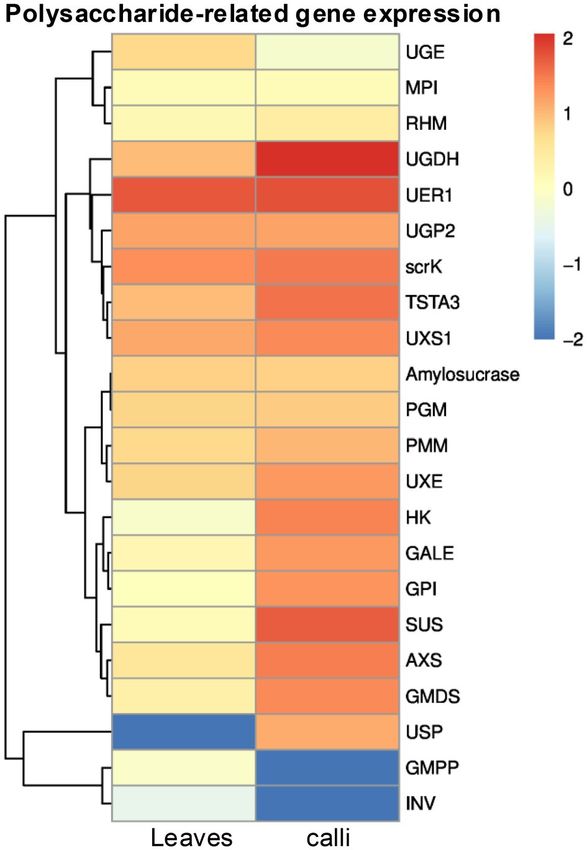
www.nature.com/scientificreports/
DXR 1-Deoxy-D-xylulose 5-phosphate reductoisomerase
DXS 1-Deoxy-D-xylulose 5-phosphate synthase
FPPS Farnesyl-diphosphate synthase
GALE UDP-glucose 4-epimerase
GMDS GDP-mannose-4,6-dehydratase
GMPP Mannose-1-phosphate Guanylyltransferase
GPI Glucose-6-phosphate isomerase
GPPS Geranylgeranyl pyrophosphate synthase
HDR 4-Hydroxy-3-methylbut-2-enyl diphosphate reductase
HDS (E)-4-hydroxy-3-methylbut-2-enyl-diphosphate synthase
HK Hexokinase
HMGR 3-Hydroxy-3-methylglutaryl-coenzyme A reductase
HMGS Hydroxymethyl glutaryl-CoA synthase
IDI Sopentenyl-diphosphate Delta-isomerase
INV Isopentenyl-diphosphate Delta-isomerase
LS Lupeol synthase
MCS 2-C-methyl-D-erythritol 2,4-cyclodiphosphate synthase
MEP/DOXP Non-mevalonate pathway
MPI Mannose-6-phosphate isomerase
MS Murashige & Skoog Medium
MVA Mevalonic acid pathway
MVD Mevalonate diphosphate decarboxylase
MVK Mevalonate kinase
PD Platycodin D
PE Platycoside E
PGM Phosphoglucomutase
PGPS Platycodon grandiflorus Polysaccharides
PMK Phosphomevalonate kinase
PMM Phosphomevalonate kinase
RHM UDP-glucose-4,6-dehydratase
SacA β-Fructofuranosidase
ScrA Sucrose-specific enzyme II
ScrK Fructokinase
SE Squalene epoxidase
SS Squalene synthase
SUS Sucrose synthase
TSTA3 GDP-L-fucose synthase
UER1 3,5-Epimerase-4-reductase
UGDH UDP-glucose-6-dehydrogenase
UGE UDP-glucuronate 4-epimerase
UGE UDP-glucuronate 4-epimerase
UGP2 UTP-glucose-1-phosphate Uridylyltransferase
USP UDP-sugar pyrophosphorylase
UXE UDP-arabinose-4-epimerase
UXS1 UDP-glucuronate decarboxylase
WPM Lloyd & McCown Woody Plant Basal Medium
α-NAA α-Naphthylacetic acid
β-A28O Isolate CYP716A140 beta-amyrin 28-oxidase
β-AS Beta-amyrin synthase
Platycodon grandiflorus (Jacq.) A. DC., a perennial herb, is the sole species in genus Platycodon within the Cam-
panulaceae family. The flowers of P. grandiflorus are blue purple or white, which can be used for ornamental and
horticultural purpose. The root (platycodi radix) of P. grandiflorus has diverse pharmacological activities and can
be used for the treatment of some chronic inflammatory diseases such as asthma, bronchitis and t uberculosis1.
The dried form of the platycodi radix is officially listed as a traditional herbal medicine in the Chinese, Korean
and Japanese Pharmacopoeia2. It is also being pickled in northeast China, and made into kimchi in the Korean
Peninsula. It has medicinal, edible, ornamental value in one, with immeasurable development prospects.
As a traditional Chinese herb, P. grandiflorus is a rich source of natural secondary metabolic products that
have various chemical structural types. More than 100 compounds have been isolated from P. grandiflorus,
including steroidal saponins, flavonoids, phenolic acids, polyacetylenes, and sterols, etc.3. Triterpenoid saponins
are the main active components in P. grandiflorus, including platycodin D (PD), platycoside E (PE), platycodi-
genin and platyconic acid A, etc. PD is one of the major triterpenoid saponins with higher pharmacological
activity than the other platycosides from platycodi radix, and have multiple pharmacological effects, such as
immunostimulation4, anti-inflammation5, anti-obesity6, anti-atherosclerosis1, and anticancer7. The structure of
platycoside E is similar to that of platycodin D, and both of them are oleanane-type triterpenoid saponin. PE
has two additional glucose groups compared to P D8. PE could convert to PD through the hydrolysis action of a
de-glucosidase. P. grandiflorus polysaccharides (PGPs) are another important active component in this medicinal
plant, and studies have confirmed that PGPs are involved in antioxidant a ctivity9, it can activate macrophage
Scientific Reports | (2021) 11:9810 | https://doi.org/10.1038/s41598-021-89294-1 2
Vol:.(1234567890)www.nature.com/scientificreports/
and enhance non-specific immunity f unction10. A research has shown that a selenium polysaccharide from the
platycodi radix may be considered as a potential and useful antioxidant a gent9.
Pentacyclic triterpenoid saponins have been well-known as important secondary metabolites in plants.
2, 3-oxidosqualene, a direct precursor of triterpenoid saponins, is synthesized mainly by the mevalonic acid
(MVA) pathway11. However, the operator of the MVA pathway in regulating the biosynthesis of triterpenoids
even phytosterols in P. grandiflorus has not been clearly d escribed12. Farnesyl pyrophosphate (farnesyl-PP) is
synthesized from isopentenyl diphosphate (IPP) and dimethylallyl diphosphate (DMAPP) under the catalysis
of farnesyl pyrophosphate synthase (FPP)11,13. Under the catalysis of enzymes such as squalene synthase (SS)
and squalene epoxidase (SE), 2,3-oxidosqualene is produced t hereafter14. Subsequently, triterpenoids saponins
synthesized from 2,3-oxidosqualene by three kinds of enzymes which are oxidized squalene cyclase, cytochrome
P450 monooxygenase and uridine diphosphate-dependent glycosyltransferase. In other words, 2,3-oxidosqualene
is converted to polygalacic acid, platycodigenin and platycogenic acid A by successive enzymes such as β-amyrin
synthase (β-AS), β-amyrin 28-oxidase (β-A28O) and a series of cytochrome P450 (CYPs)15. Subsequently, the
conversion of polygalacic acid into polygalacin D, platycodigenin into platycoside E, and platycogenic acid
A into platyconic acid A are all catalyzed by certain kinds of GTs (Glycosyltransferases)16. Platycodin D, an
oleanane-type triterpenoid saponin, is the main bioactive component and has stronger pharmacological activi-
ties, but little is clear on its biosynthesis in P. grandiflorus at present. It has been found that platycoside E is a
precursor of platycodin D, and PE can be converted to PD by enzyme catalysis. Their biological activities can be
increased and their bioavailability and cell permeability would be improved due to their reduced size resulted
by de-glycosylation of s aponins17.
From the chemical structure of platycodin D and platycoside E, it can be predicted that there are enzymes
which can catalyze degradation of glycosyl group from PE to PD. Two extracellular experiments showed that β-D-
glucosidase from Aspergillus usamii and Caldicellulosiruptor bescii can successfully catalyze conversion of PE and
platycodin D3 into PD under optimal reactions c onditions18,19. In this study we attempt to find out appropriate
candidate genes encoding enzymes involving in conversion of PE, platycodin D3 into PD in traditional Chinese
medicinal plant P. grandiflorus. The pathway of triterpenoid saponins in P. grandiflorus is predicted referring
to the terpenoid backbone and saponin biosynthesis in KEGG20 (https://www.kegg.jp/dbget-bin/www_bget?
map00900), as shown in Fig. 1.
Polysaccharides are extremely important bio-macromolecules, and have a wide range of industrial value and
clinical role21. There are enormous types of polysaccharides in P. grandiflorus, named as P. grandiflorus polysac-
charides (PGPs), which have important pharmacological activities. However, few relevant studies and analyses
on its biosynthetic pathways were reported. Sucrose is firstly produced in plants through photosynthesis in
plant chloroplast. Sucrose generates UDP-Glc in the presence of sucrose synthase (SUS), and then UDP-Glc is
converted into GDP-Glc under successive catalytic reactions, including UDP-sugar pyrophosphorylase (USP)22,
UDP-glucose phosphorylase (UGP), GDP-glucose pyrophos-phorylase (GGP)23. Sucrose-6P is biosynthesized
from sucrose under the catalysis of sucrose-specific enzyme II (ScrA)24,25. Subsequently, a series of enzymes such
as β-Fructofuranosidase (SacA), Mannose-6-phosphate isomerase11, Mannose-1-phosphate Guanylyl-transferase
(GMPP) and other enzymes catalyze sucurose-6P to generate GDP-Man. Then, GDP-Fuc is synthesized from
GDP-Man by enzymes such as GDP-Mann 4,6-dehydratase (GMDS) and GDP-L-Fucose (TSTA3)26,27. In addi-
tion, through the catalysis of nucleotide-diphospho-sugar (NDP-sugar) interconversion enzymes (NSEs), many
other NDP sugars were produced from UDP-Glc and GDP-Man28. Another way is that sucrose invertase (INV)
and hexokinase (HK) catalyze the production of D-Glucose-6P from sucrose29. Subsequently, D-Glucose-6P
is converted into UDP-Gal, UDP-GalA, UDP-Rha and other polysaccharide precursors under the catalysis of
enzymes such as Phosphoglucomutase (PGM), UTP-glucose-1-phosphate Uridylyltransferase (UGP2), and UDP-
apiose/xylose synthase (AXS) et al30. Finally, through the catalysis of various glycosyltransferases (GTs), the active
monosaccharide unit NDP-sugar is added to the sugar residues of various polysaccharides and glycoconjugates26.
The biosynthesis pathway of polysaccharides in P. grandiflorus was inferred as shown in Supplementary Figure S1.
RNA-Seq has been widely used to analyze the pathway of secondary metabolite biosynthesis and regulation,
excavate key enzyme genes and transcription factors in medicinal plants31,32, which provides a scientific basis
for the efficient accumulation and effective utilization of active ingredients in medicinal plants. Transcriptome
sequencing have been completed in many medicinal plants such as Taxus chinensis33, Panax quinquefolius34,
Coptis chinensis35, Panax zingiberensis15, and many other medicinal plants. Many functional genes are discovered
and partial specific biosynthetic pathways of secondary metabolite are analyzed.
In this paper, calli of P. grandiflorus were induced by tissue culture techniques. The contents of PE, PD and
PGPs in calli and each organ of original plant of P. grandiflorus were determined by high performance liquid
chromatography (HPLC) and Phenol–sulfuric acid method, respectively. Based on the differences of the sec-
ondary metabolites between calli and organs of original plant P. grandiflorus, RNA-seq was performed between
calli and leaves. Comparative analysis of the transcriptome data provides valuable resources for further studies
of the molecular mechanisms of terpenoids, saponins and polysaccharides biosynthesis in P. grandiflorus. In
this study, the differences between calli and organs of original plants were analyzed, and candidate key genes
and transcription factors were identified to help our knowledge of the metabolism and regulation of secondary
metabolites in P. grandiflorus.
Results
Calli induction of Platycodon grandiflorus. The effects of four explants, different basic media, and plant
growth regulator combinations on calli induction of P. grandiflorus were studied by orthogonal experimental
design of 4 factors and 3 levels (L9(34)) without consideration of interactions among factors. Each treatment has
Scientific Reports | (2021) 11:9810 | https://doi.org/10.1038/s41598-021-89294-1 3
Vol.:(0123456789)www.nature.com/scientificreports/
MEP/DOXP Glycolysis Mevalonate
pathway pathway
D-Glyceraldehyde-3-phosphate Acetyl-Coa
DXS
AACT
1-Deoxy-D-xylulose-5-phosphate
DXR Acetoacetyl-CoA
2-C-Methyl-D-erythritol-4-phosphate HMGS
CMS 3-Hydroxy-3-methyl-glutaryl-CoA
4-(Cytidine-5'-diphospho)-2-C-methyl-D-erythritol HMGR
CMK Mevalonate
2-phospho-4-(Cytidine-5'-diphospho)-2-C- MVK
methyl-D-erythritol
MCS Mevalonate-5P
2-C-Methyl-D-erythritol-2,4-cyclodiphosphate PMK
HDS Mevalonate-5PP
1-Hydroxy-2-Methyl-2-butenyl-4-diphosphate
HDR MVD
Dimethylallyl-PP Isopentenyl-PP
IDI
GPPS
Squalene SS Farnesyl-PP FPPS Geranyl-PP
SE
˟-AS Beta-amycrin
2,3-oxidosqualene
˟-A28O
GTs Polygalacic acid CYPs Oleanolic acid
᧻
Polygalacin D CYP
GTs
Platycodigenin Platycoside E
CYP ᧻
Platycogenic acid A Platycodin D3
GTs ᧻
Platyconic acid A Platycodin D ᧻
Figure 1. Triterpenoid saponin biosynthetic pathways predicted in P. grandiflorus. Arrows with solid lines
represent the identified enzymatic reactions, and arrows with dashed lines represent multiple enzymatic
reactions through multiple steps and putative enzymatic reactions.
60 independent duplicates (20 bottles with 3 pieces). The calli induced are shown in Supplementary Figure S2,
and the results are shown in Table 1.
The induction rates of the calli were calculated with the number of the calli divided by the number of uncon-
taminated explants. The K value and R value were obtained by statistical analysis of orthogonal experiment
data, as the K value was the average of the rates from each level, and the R value called extreme difference is the
difference between the maximum and minimum average values of each factor.
By analysis of variance (ANOVA), a significant difference among these factors and their levels (F = 8.67 > F0.01(3,
4) = 6.59) was identified. It can be concluded from the R value that the category of explants had a great influ-
ence on the calli induction, and stem with nodes is the optimal explants. It also can be concluded that the MS
Scientific Reports | (2021) 11:9810 | https://doi.org/10.1038/s41598-021-89294-1 4
Vol:.(1234567890)www.nature.com/scientificreports/
Factors
Explants NAA mg/L 6-BA mg/L Inoculation Num (non-
Media (A) (B) (C) (D) contaminated) Calli Num Induction rate (%)
1 B5 Leaves 0.1 0.5 56 4 7.14
2 B5 Stems with nods 0.2 1.0 58 22 37.93
3 B5 Stems 1.0 2.0 57 8 14.04
4 MS Leaves 0.2 2.0 53 6 11.32
5 MS Stems with nods 1.0 0.5 57 57 100.00
6 MS stems 0.1 1.0 54 2 3.70
7 WPM leaves 1.0 1.0 55 0 0.00
8 WPM Stems with nods 0.1 2.0 58 23 39.66
9 WPM stems 0.2 0.5 56 4 7.14
k1 59.11 18.46 50.50 114.28
k2 115.02 177.59 56.39 41.63
k3 46.80 24.88 114.04 65.02
K1 19.70 6.15 16.83 38.09
K2 38.34 59.20 18.80 13.88
K3 15.60 8.29 38.01 21.67
R 22.74 53.05 21.18 24.21
Table 1. Orthogonal experiment (L9(34)) analyzing the effects of different factors on callus induction of P.
grandiflorus.
medium is better than the other two basic media and 6-BA could get calli induction more efficiently than NAA,
and the optimal concentrations are 0.5 mg/L and 1.0 mg/L, respectively. Based on the above analysis, the best
combination is A2B2C3D1. In order to verify whether the A2B2C3D1 is the best combination or not, a total of
30 stems with nodes were inoculated in 10 bottles with 3 duplicates per bottle, a total of 25 calli were induced
after 50 days, and the induction rate is up to 83.33%.
It can be concluded from what is stated above that the best protocol for calli induction of P.grandiflorus is
to use stem with nodes as the explants, and take MS + NAA 1.0 mg/L + 6-BA 0.5 mg/L as the optimal formula.
Comparative of PD and PE content in organs and calli. Two kinds of specific platyopsis saponins PD
and PE were identified by comparing the retention t ime34 and maximum uptake of the samples and standards at
a wavelength of 210 nm by HPLC. The results showed that the concentrations of PD and PE are positively cor-
related with the area of the peak in the test range ( R2 > 0.9999, R2 > 0.9946), and the content of each saponins was
calculated from the peak area versus its own standard curve. The retention time and peak profile of PD and PE
in standard and one sample are shown in Fig. 2. The content variance of PD and PE in callus and different organs
in P. grandiflorus was shown in Supplementary Table S1 and Fig. 3.
ANOVA indicated that the distribution of PD content varied wildly among organs in plants of P. grandiflorus
(Fig. 3A). It can be concluded that PD was most enriched in flower buds, followed by the roots, while PD content
in leaves, stems and the whole plants is low. When it comes to the distribution of PD content in stems and roots,
the contents in the bark and phloem are higher than that in the xylem. The results also showed that the content
of PD in callus is higher than that of the whole plants, leaves, stems and roots, and it only lower than that in the
flower buds. It can be inferred that PD could be obtained more efficiently from flowers and calli than from any
other organs and tissues. Since the biomass of flower buds in the whole plants is very low, callus of P. grandiflorus
may become an alternative material for PD production.
As for the distribution of PE content in P. grandiflorus, only the PE content in leaves is higher than that in
the whole plant, while the PE content of flowers, roots and stems is much lower than that of the whole plant
(Fig. 3B). It can be concluded from PE content distribution in each organ that the PE content of phloem and
bark is higher than that of xylem which is consistent with the distribution of PD content distribution described
above. The result also shows that the content of PE of calli is lower than that of all the organs in P. grandiflorus.
We found the content of PD in calli was higher than that in leaves which was sharply contrast to the highest PE
and lower PD content in leaves. Experiments in vitro have shown that through enzymes catalysis, PE could con-
vert to PD with higher pharmacological a ctivities18,19. Therefore, we speculate the existence of putative enzymes
converting PE to PD in P. grandiflorus. Based on the significant differences of PD and PE content between calli
and leaves of P. grandiflorus, an RNA-Seq experiment was designed to identify the candidate genes.
Total polysaccharide content variation among calli and plant organs. A standard curve was
drawn from the absorbance value versus 7 gradient concentrations of standard glucose work solution with
blank at 486 nm following chromogenic reaction (Fig. 4A). The regression equation of the standard curve is
Y = 0.026X − 0.0226, R2 = 0.9991, showing that the quantification responds to linearity within the tested con-
centrations. The total polysaccharide content of callus and organs from the plant was detected by phenol-con-
centrated sulfuric acid method mentioned above (Fig. 4B) (Supplementary Table S1).
Scientific Reports | (2021) 11:9810 | https://doi.org/10.1038/s41598-021-89294-1 5
Vol.:(0123456789)www.nature.com/scientificreports/
Platycoside E Platycodin D
568.76
426.57
A
284.37
142.18
B
-0.02
0.00 3.62 7.25 10.87 14.49 18.12 21.74 25.36
Figure 2. HPLC peak profiles of (A) standard products of PD and PE, and of (B) the P. grandiflorus extract.
The X axis represents the retention time (min) of peaks, and the Y axis represents the height of the peak (mAU).
Figure 3. The contents of main triterpenoid saponins in callus and plant organs of P. grandiflorus. (A)
Platycodin D (PD) content in different samples. (B) Platycoside E (PE) content in different samples. The X axis
is the sample name and the Y axis is the content (mg/g DW), DW is dried weight. Bars with different letters
within same histogram, represent significant difference at p ≤ 0.01.
As for the distribution of polysaccharides in the P. grandiflorus also showed significant difference among
samples by ANOVA, the content of polysaccharides of root is much higher than that of the whole plant which is
consistent with the use of platycodi radix as medicinal part, which indicates that the roots of P. grandiflorus are
an important place for the storage of polysaccharides. The results showed that the content of polysaccharides
of the calli induced by this optimal medium is higher by comparing to the whole plants and organs in P. gran-
diflorus, implying callus might be a high-efficiency material and an important alternative for polysaccharides
production than P. grandiflorus plants. At the same time, we also found that the polysaccharides content of root
phloem was higher than that of the root xylem. This indicates that the root phloem is a more important place
for storing polysaccharides. Moreover, we found that the content of polysaccharides of calli is much higher than
that of leaves, which is in stark contrast to the PE content of callus and leaves.
Scientific Reports | (2021) 11:9810 | https://doi.org/10.1038/s41598-021-89294-1 6
Vol:.(1234567890)www.nature.com/scientificreports/
Figure 4. The contents of total polysaccharide among callus and different organ samples. (A) Polysaccharides
content in different samples. The X axis is the sample name and the Y axis is the content (mg/g DW), and
DW means dried weight. Bars with different letters within same histogram, represent significant difference at
p ≤ 0.01. (B) Standard curve, R2 = 0.9991. The X axis is the standard concentration (μg/mL), and the Y axis is the
absorbance value.
Sequencing and De novo assembly. A total of 39.44 Gb data were generated using the BGISEQ-500
platform by sequencing the established six libraries (leaf and callus each has three duplicates). The raw data were
filtered by trimmomatic software (version 0.36, parameters are illuminaclip:2:30:10, leading:3, trailing:3, sliding-
window:4:15 minlen:50) to obtain clean reads by removing the reads containing connector, or unknown bases
content more than 5%, or reads with low quality. The clean read numbers of each library were counted by SOAP-
nuke (version 1.4.0, parameters are -l 5 -q 0.5 -n 0.1). Trinity software was used to assemble clean reads, and then
Tgicl software was used to cluster the transcripts to remove redundancy and obtain unigenes. Finally, 152,777
unigenes were obtained, which is higher than those (34,053 unigenes) obtained by Chunhua Ma et al16. The total
length, average length, N50, and GC content were 228,154,936 bp, 1,493 bp, 2,514 bp, and 40.58%, respectively.
A single-copy ortholog database, BUSCO (https://busco.ezlab.org/) (Supplementary Figure S3), was used to
evaluate the quality of the assembled transcripts. By comparing with conserved genes, the results showed the
good integrity of the transcriptome assembly. A total of 80,826 coding sequences (N50 = 1,380) were identified,
with a maximum length of 16,398 and a minimum length of 297 by TransDecoder software.
Unigenes functional annotation and expression analysis of unigenes. Unigenes were compared
to the seven major functional databases to annotate. There were 97,878 (NR: 64.07%), 82,833 (NT: 54.22%),
73,371 (SwissProt: 48.02%), 77,858 (KOG: 50.96%), 78,197 (KEGG: 51.18%), 73,988 (GO: 48.43%), and 73,302
(Pfam: 47.98%) unigenes received functional annotations. 80,826 CDS were detected using Transdecoder soft-
ware. At the same time, 77,478 SSRs were distributed among 50,171 unigenes, and 3,153 unigenes encoding tran-
scription factors were predicted (Supplementary Table S2: X1, X2 and X3 are the three independent replicates
of leaves, X4, X5 and X6 are the three independent replicates of callus). The sequencing and analyzing data had
been stored in website: https://www.ncbi.nlm.nih.gov/geo/query/acc.cgi?acc=GSE153777.
Identification of candidate genes involved in triterpenoid saponin biosynthesis by expression
level analysis. KEGG analysis was applied to gain insight into pathways of unigenes. A total of 22,842 uni-
genes are annotated in the KEGG database20. In order to discover the most important biological pathways, the
KEGG metabolic pathways involved in genes are divided into 5 branches: cellular processes, environmental
information processing, genetic information processing, metabolism, and organismal systems (Supplemen-
tary Table S2), including 19 subcategories (135 routes). Eight pathways (ko00906, ko00908, ko00909, ko00904,
ko00903, ko00902, ko00900 and ko00905) of "Metabolism of terpenoids and polyketides" containing 864 uni-
genes were analyzed. These genes encoding enzymes for terpenoid synthesis that are mainly distributed in
upstream of MEP and MVP, while some are distributed in downstream (Fig. 1) (Table 2).
Expression level of putative genes encoding enzymes for triterpenoid saponins biosynthesis in P. grandiflorus
between callus and leaf was measured by value of log2 (FC) of similar unigene from RNA-Seq data, and the
similar unigene was obtained by aligning the amino acid sequences between putative genes and the unigene.
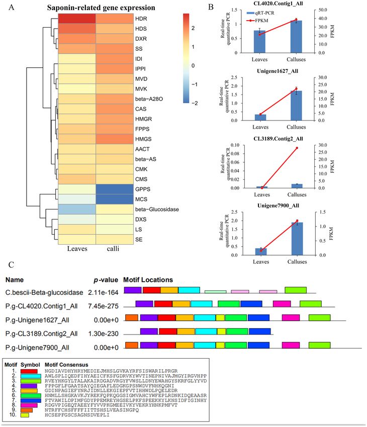
Expression of genes encoding enzymes in the saponin biosynthetic pathway such as FPPS, HMGR, HMGS, IDI,
MVD, MVK, SS, beta-A28O, beta-AS and beta-Glucosidase are up-regulated significantly (Fig. 5A). The expres-
sion level of a putative gene predicted to encode a de-glucosidase (ACM59590, β-Glucosidase) is consistent
with the variance of PD and PE content, and two extracellular experiments indicate this enzyme may function
Scientific Reports | (2021) 11:9810 | https://doi.org/10.1038/s41598-021-89294-1 7
Vol.:(0123456789)www.nature.com/scientificreports/
Enzyme name EC number
AACT (acetoacetyl-coenzyme A) 2.3.1.9
HMGS (hydroxymethylglutaryl-CoA synthase) 2.3.3.10
HMGR (3-hydroxy-3-methylglutaryl-coenzyme A reductase) 1.1.1.34
MVK (mevalonate kinase) 2.1.7.36
PMK (phosphomevalonate kinase) 2.7.4.2
MVD (mevalonate diphosphate decarboxylase) 4.1.1.33
GPPS (geranylgeranyl pyrophosphate synthase) 2.5.1.29
FPPS (Farnesyl-diphosphate synthase) 2.5.1.1, 2.5.1.10
SS (squalene synthase) 2.5.1.21
SE (squalene epoxidase) 1.14.14.17
β-AS (beta-amyrin synthase) 5.4.99.39
β-A28O (isolate CYP716A140 beta-amyrin 28-oxidase) 1.14.13.-
LS (lupeol synthase) 5.4.99.41
CAS (cycloartenol synthase) 5.4.99.8
IDI (isopentenyl-diphosphate Delta-isomerase) 5.3.3.2
DXS (1-deoxy-D-xylulose 5-phosphate synthase) 2.2.1.7
DXR (1-deoxy-D-xylulose 5-phosphate reductoisomerase) 1.1.1.267
CMS (2-C-methyl-D-erythritol 4-phosphate cytidylyltransferase
2.7.7.60
(4-diphosphocytidyl-2C-methyl-D-erythritol synthase))
CMK (4-diphosphocytidyl-2-C-methyl-D-erythritol kinase) 2.7.1.148
MCS (2-C-methyl-D-erythritol 2,4-cyclodiphosphate synthase) 4.6.1.12
HDS ((E)-4-hydroxy-3-methylbut-2-enyl-diphosphate synthase) 1.17.7.1, 1.17.1.3
HDR (4-hydroxy-3-methylbut-2-enyl diphosphate reductase) 1.17.1.4
ACM59590 (β-Glucosidase) 3.2.1.21
Table 2. Putative key enzymes involved in the triterpenoid saponins biosynthesis pathway in P. grandiflorus.
as the β-Glucosidase, which showed that exogenous β-Glucosidase can catalyze the conversion of PE into PD
through removing two glycosyl groups from PE in vitro18,19. From expression level analysis, 4 putative unigenes
(CL4020.Contig1_All, Unigene 1627_All, CL3189.Contig2_All and Unigene7900_All) have high identity with
the β-glucosidase from Caldicellulosiruptor bescii with amino acid s equence18. The result of real-time quantitative
PCR indicated that the expression patterns of putative gene sequences encoding enzyme involved in converting
PE to PD is consistent with the result from transcriptome analysis (Fig. 5B). Furthermore, to better select the
putative sequences which might encode enzyme involved in converting PE to PD, the conserved motifs were
analyzed using MEME (Fig. 5C). The result showed 3 unigenes (CL4020.Contig1_All, Unigene 1627_All and
Unigene7900_All) were screened as candidate fragments of the target gene.
Gene expression variance involved in polysaccharide biosynthesis. Furthermore, fifteen path-
ways (ko00010, ko00500, ko00620, ko00020, ko00051, ko00562, ko00052, ko00630, ko00030, ko00040, ko00053,
ko00520, ko00640, ko00650 and ko00660) of “Carbohydrate metabolism” were analyzed, including 4,441 uni-
genes. There are 1,148 unigenes involved in starch and sucrose metabolism, and 765 unigenes involved in amino
sugar and nucleotide sugar metabolism. Based on KEGG pathway analysis, we developed a model to infer the
biosynthetic pathway of polysaccharides in P. grandiflorus (Supplementary Figure S4) (Table 3).
The value of log2 (FC) was also used to analyze the expression of genes encoding enzymes for polysaccharide
biosynthesis, and it showed that the expression levels of putative genes encoding AXS, GALE, GMDS, GPI, HK,
PMM, SUS, TSTA3, UGDH, USP and UXE in the polysaccharide biosynthetic pathway were also upregulated
significantly (Fig. 6). The result of gene expression quantity is consistent with the fact that the content of total
polysaccharides in calli is higher than that of leaves.
Detection of transcription factors. Transcription factors (TFs) can temporarily and spatially regulate the
activity of target genes, and play a key role in plant development and response to the external environment36–38.
A total of 3,153 candidate TFs were identified in the P. grandiflorus transcriptome database and classified into 57
different TF families (Fig. 7A), among these families that MYB family (326 unigenes) accounted for the largest
proportion of TF families, followed by mTERF (286 unigenes), bHLH (200 unigenes), AP2-EREBP (185 uni-
genes), C3H (160 unigenes), ABI3VP1 (152 unigenes), NAC (132 unigenes), WRKY (130 unigenes), and FAR1
(118 unigenes). In these target TFs 1,542 were up-regulated and 1,444 were down-regulated in the calli versus
leaves (Fig. 7B). We found that 54 candidate genes among transcription factors are expressed only in leaves, and
23 are expressed only in calli (Fig. 7C) (Supplementary Table S3).
By KEGG pathway analysis it can be concluded that total 11 (4 Trihelix and 7 FHA) and 15 (3 bHLH, 6 C2H2,
3 Trihelix, 1 G2-like, 1 FHA and 1 VARL) TFs may relate to the biosynthesis of saponins and polysaccharides,
Scientific Reports | (2021) 11:9810 | https://doi.org/10.1038/s41598-021-89294-1 8
Vol:.(1234567890)www.nature.com/scientificreports/
Figure 5. Expression level analysis of genes from triterpenoid saponins biosynthetic pathway in P. grandiflorus
and conserve motif analysis of predictive β-glucosidase. (A) The expression levels of a single gene encoding an
enzyme from each step of triterpenoid saponins biosynthetic pathway are shown. Red and green represent high
and low expression levels, respectively. (B) Real-time quantitative PCR analysis of CL4020.Contig1_All, Unigene
1627_All, CL3189.Contig2_All and Unigene7900_All in leaves and calli. 18S rRNA as internal reference gene.
Error bars indicate SD (n = 3). The blue bars represent the real-time quantitative PCR data and the red line
represents the FPKM value from RNA-Seq. (C) Comparison of the conserve motifs among β-glucosidase from
C. bescii and the amino acid sequences of putative orthologous unigenes from P. grandiflorus. Each block shows
the position and strength of a motif site. The height of a block gives an indication of the significance of the site
as taller blocks are more significant. The height is calculated to be proportional to the negative logarithm of the
p-value of the site, truncated at the height for a p-value of 1e−10.
respectively (Supplementary Table S4). It also found a Trihelix TF gene (Unigene26781_All) is co-expressed with
the putative de-glucosylation enzyme gene (β-Glucosidase).
Scientific Reports | (2021) 11:9810 | https://doi.org/10.1038/s41598-021-89294-1 9
Vol.:(0123456789)www.nature.com/scientificreports/
Enzyme name EC number
SUS (sucrose synthase) 2.4.1.13
INV (sucrose invertase) 3.2.1.26
HK (Hexokinase) 2.7.1.1
PGM (Phosphoglucomutase) 5.4.2.2
USP (UDP-sugar pyrophosphorylase) 2.7.7.64
UGP2 (UTP-glucose-1-phosphate Uridylyltransferase) 2.7.7.9
scrK (Fructokinase) 2.7.1.4
MPI (Mannose-6-phosphate isomerase) 5.3.1.8
UGDH (UDP-glucose 6-dehydrogenase) 1.1.1.22
UXS1 (UDP- glucuronate decarboxylase) 4.1.1.35
UXE (UDP-arabinose 4-epimerase) 5.1.3.5
RHM (UDP-glucose 4,6-dehydratase) 4.2.1.76
UER1(3,5-Epimerase-4-reductase) 5.1.3.-, 1.1.1.-
PMM (Phosphomannomutase) 5.4.2.8
GMPP (Mannose-1-phosphate Guanylyltransferase) 2.7.7.13
GMDS (GDP-mannose 4,6-dehydratase) 4.2.1.47
TSTA3 (GDP-L-fucose synthase) 1.1.1.271
GPI (Glucose-6-phosphate isomerase) 5.3.1.9
GALE (UDP-glucose 4-epimerase) 5.1.3.2
UGE (UDP-glucuronate 4-epimerase) 5.1.3.6
AXS (UDP-apiose/xylose synthase) AXS
Amylosucrase (1,4-α-D-glucan 4-α-D-glucosyltransferase-glucan) 2.4.1.14
Table 3. Putative key enzymes involved in polysaccharide biosynthesis in P. grandiflorus.
Discussion
Plant tissue culture is not only a method for plant rapid propagation, but also an ideal way for plant improve-
ment, germplasm preservation and production of useful compounds. Studies have shown that compounds can
be quickly obtained from callus of Citrus junos Siebold ex. T
anaka39 and Ocimum basilicum L.18,19. It was reported
that there was high content of pentacyclic triterpenoids with anti-inflammatory and antinociceptive activities
in callus of Chaenomeles japonica (Thunb.) Lindl. ex Spach40. So far, there’s no specific protocol for the callus
induction of P. grandiflorus. The best callus induction plan of P. grandiflorus was screened out through experi-
ments in this article, which filled the research gap, and provided a basis for the development and utilization of
P. grandiflorus callus.
The contents of PE and PD in the phloem of P. grandiflorus is higher than that in the xylem like some other
secondary compounds41,42, indicating that the phloem of P. grandiflorus may have the function to transport
and store PD and PE. In addition, we found that the content of PE is the highest in leaves, while that in callus
was lower than that in other organs. So, obtaining PE from leaves of P. grandiflorus is probably the best choice.
Studies have shown that PE converted to PD by removing two molecules of glycosyl group under the action
of deglycosylated enzymes18,19. Since callus has much higher content of polysaccharides and PD, and lower
content of PE than leaves, then we highly speculate that the active glycosyltransferase in the callus will catalyze
the conversion of PE into PD, and the glucose groups were released to participate in the biosynthesis of polysac-
charides. This process has a positive effect on the accumulation of polysaccharides in callus.
The number of unigenes is 152,777 of us versus 34,053 of Ma’s16. The average length and N50 values of
genes in this study are also higher than those in Ma’s results (with the total average length of 936 bp and N50
of 1,661 bp)16, and the differences are mainly due to the different materials and sequencing platform used for
RNA-Seq, and different sequencing depth. It was reported that endophytic bacteria are involved in secondary
metabolite biosynthesis, which could be isolated from the interior of P. grandiflorum43, which means there might
be genes in the endophytic bacteria encoding key enzymes catalyzing the formation of secondary m etabolites44.
So, the calli and leaves of P. grandiflorum are used as materials for RNA-Seq to avoid the residual RNA from
endophytes. Transcriptome analysis were quite important methods to identify new genes in triterpenoid saponin
biosynthetic pathway in P. grandiflorum. Besides, the difference in plant materials between M a16 et al. and us.
Many key enzymes involved in triterpenoid saponin biosynthesis were discovered in both studies, including
HMGS, HMGR, FPPS, SS, SE, et al. However, UGTs cloned in Ma’s study catalyze the glucosylation; in our
work, 3 unigenes or orthologous sequences (CL4020.Contig1_All, Unigene 1627_All and Unigene7900_All)
were screened as candidate gene which can catalyze degradation of glycosyl group and convert PE to PD. Our
research could supply more transcriptome data, and it is the first time to identify the candidate genes which that
converts PE to PD in P. grandifloras.
Previous studies have shown TFs, such as the bHLH transcription factor AabHLH1 and AaMYC2 in Arte-
misia annua L., have effects on the primary and secondary metabolites of plants, that can effectively regulate the
biosynthesis of a rtemisinin45,46, and the study has shown that the Trihelix family transcription factor BdTHX1
Scientific Reports | (2021) 11:9810 | https://doi.org/10.1038/s41598-021-89294-1 10
Vol:.(1234567890)www.nature.com/scientificreports/
Figure 6. The expression levels of a single gene encoding an enzyme from each step of polysaccharides
biosynthetic pathway in P. grandiflorus are shown. Red and green represent high and low expression levels,
respectively.
likely plays an important role in mixed-linkage glucan biosynthesis and restructuring by regulating the expression
polysaccharide synthase related g enes47. It was reported that Trihelix TF, responding to light signals, regulates the
expression of downstream gene by calcium-dependent phosphorylation and dephosphorylation48. The expression
level of a Trihelix TF gene (Unigene26781_All) is co-expressed with the putative deglucosylation enzyme gene
(β-Glucosidase) in this study, which demonstrated that this Trihelix TF might sense the light signal, and influence
the expression of the putative gene encoding β-Glucosidase, then modify and regulate the conversion of PE to
PD. The discoveries of these TFs may have great potential value and broad application prospects in studying the
regulation of terpenoid saponin and polysaccharide bio-synthesis in P. grandiflorus.
Conclusions
A best protocol for calli induction of P. grandiflorus is that use the stem with nodes as explants, and take
MS + NAA 1.0 mg/L + 6-BA 0.5 mg/L as the formula. We found that the content of PD and polysaccharide in
callus is higher than that in the plant of P. grandiflorus, as well higher than that in the root which is traditional
medicinal parts. Study also showed that PD content of calli is higher than that of leaves, which is in sharp con-
trast to PE content.
We performed comprehensive RNA-Seq analysis on the leaves and calli of P. grandiflorus, and were able to find
the expression of many genes involved in triterpenoid saponins and polysaccharides which were co-expression
with the content of corresponding metabolites. Three putative unigenes with high amino acid sequence identity
were screened as orthologous sequences of candidate β-Glucosidase gene converting PE to PD, which is help-
ful to deeply understand the biosynthesis mechanism of triterpene saponins in P. grandiflorus at the molecular
level. A total of 11 TFs were involved in regulating the biosynthesis of saponins, and 15 TFs were involved in
carbohydrate metabolism were obtained.
In summary, our work may greatly help to promote the molecular biology research and improve the large-
scale production of triterpene saponins and polysaccharides in P. grandiflorus.
Scientific Reports | (2021) 11:9810 | https://doi.org/10.1038/s41598-021-89294-1 11
Vol.:(0123456789)www.nature.com/scientificreports/
A B
zf-HD 19 77
mTERF 286 Up
bZIP 27
bHLH 200 Down
WRKY 130
VOZ 4 NA
VARL 4
ULT 7
Trihelix 76
Tify 57
TUB 26
TIG 23
TCP
TAZ 18
43
1444 1542
Sigma70-like 21
SRS 3
SBP 67
SAP 2
S1Fa-like 3
RWP-RK 63
PLATZ 17
PBF-2-like 6
OFP 14
NAC 132
MYB 326
MADS 75 calli vs leaves
LOB 35
LIM 30
LFY 1
HSF 63
HRT 3
HB
GeBP
12
14 C
GRF 14
GRAS 87
G2-like 96
FHA 49
FAR1 118
EIL 19
E2F-DP 21
DBP 3
CSD 6
CPP 27
C3H 160
C2H2 79
C2C2-YABBY
C2C2-GATA
15
72 23 3077 54
C2C2-Dof 35
C2C2-CO-like 21
BSD 31
BES1 14
BBR/BPC 22
Alfin-like 39
ARR-B 24
ARF 57
AP2-EREBP 185
ABI3VP1 152
0 50 100 150 200 250 300 350
calli vs leaves
Number of genes
Figure 7. Transcription factors (TFs) expression analysis. (A) Classification of gene transcription factors’
family. (B) The expression level of TFs gene is different in calli vs. leaves, NA stands for expression only in calli
or leaves. (C) Venn diagram of TFs gene expression in leaves and calli.
Methods
Plant materials. Plants used in this experiment were identified as Platycodon grandiflora (Jacq.) A. DC.by
expert who is major in plant identification, which were grown at 25 °C during day and 23 °C at night in the green
house of Anhui University of Chinese Medicine, Anhui, China. The seedlings of P. grandiflorus were purchased
from Bozhou Market of Traditional Chinese Medicine, Anhui Province, China. Specimen of P. grandiflorus in
this study was deposited in the Herbarium of Anhui University of Chinese Medicine and the depository No. is
20,200,705. The explants used for callus induction and the materials used for metabolites determination and
RNA-Seq are all from the same plantlets. Each sample in this study has three independent biological duplicates.
Chemicals. Standard compounds of platycodin D (PD, > 98% purity) and platycodin E (PE, > 98% purity)
were purchased from Chengdu Desite Biotech Co., Ltd. and Chengdu Push Bio-technology CO., Ltd., respec-
tively. CTAB-PBIOZOL reagent was purchased from Bioflux. Acetonitrile and Methanol (HPLC grade) were
purchased from Oceanpak. HPLC-grade water was prepared using laboratory water purification system from
Scientific Reports | (2021) 11:9810 | https://doi.org/10.1038/s41598-021-89294-1 12
Vol:.(1234567890)www.nature.com/scientificreports/
Pall Filter Co., Ltd. (Beijing, China). Plant growth regulators of 6-BA (6-Benzylaminopurine) and α-NAA
(α-Naphthylacetic acid) were purchased from Beijing Solarbio Technology Co., Ltd. Chemicals for plant tissue
culture were bought from Sinopharm group, and all other chemicals were of analytical grade.
Calli induction of Platycodon grandifloras. In order to screen the optimal processes of inducing calli
of P. grandiflorus included optimum explants, basic medium and plant growth regulators combination, a L 9(34)
orthogonal experiment was carried out based on the documents and our previous study regardless of the inter-
actions among factors (Supplementary Table S5). The best one was selected from basic media (Factor A) of B5,
MS and WPM for P. grandiflorus calli induction. Leaves, stems with nodes and stems without nodes were used
as explants (B), which are in the same batch with the materials used for content detection and RNA-Seq analysis.
In order to select the optimal combination for calli induction of P. grandiflorus, plant growth regulator combina-
tions with different concentrations of auxin and cytokinin were designed according to relevant literature. NAA
(C) and 6-BA (D) were divided into three concentrations of 0.1 mg/L, 0.2 mg/L and 1.0 mg/L and 0.5 mg/L,
1.0 mg/L and 2.0 mg/L, respectively.
Basic medium B5 (Gamborg B5 Medium), MS (Murashige and Skoog Medium), WPM (Lloyd & McCown
Woody Plant Basal Medium)49 stock solutions were prepared. The full media formulas were prepared according
to the different proportions of components, and different concentrations of plant growth regulators. Finally,
adjusted the pH of media to 5.6 ~ 6.0, followed by the addition of sucrose (30 g/L) and agar (7 g/L), then the
media was sterilized by autoclaving at 121 °C for 30 min.
Explants of P. grandiflorus were rinsed and surface-sterilized, and rinsed 3–5 times with sterile distilled water.
The sterile explants were dried, leaves were cut into 0.5 cm2 in size, and the stems were cut into pieces about 1 cm
in length, then they were inoculated onto the media. Each group of explants were inoculated in 20 bottles with
3 pieces per bottle. The culture conditions were as follows: the photosynthetic photon flux density was 30 μmol/
m2/s for 12 h/d, the culture temperature was (25 ± 1) °C, and the culture lasted for 50 d.
Extraction of triterpenoid saponin D and E. Jaeyoung Kwon et al.2 found that PD degraded in the
minimum at 40 °C in drying process, and suitable for detection. In this paper, all the organs of wild plants and
tissue culture materials of P. grandiflorus were dried at 40 °C to constant weight and pulverized by a ball mill.
Then accurately weighed the sample powders (each sample 0.5 g) and transferred them to a 50 mL of centrifuge
tubes. After adding of 25 mL methanol, each sample solution was adjusted to the same weight and recorded
the final weight followed by ultrasonic extraction lasting for 50 min. And the loss of solution occurring during
ultrasonic extraction was compensated for by adding a certain amount of 100% methanol to the same weight.
Finally, 20 mL of continuous filtrated to 2 mL for further use.
HPLC quantitative analysis of saponin D and E from calli and organs of plants. All analyses of
PD and PE contents were performed on an Agilent Series 1260 system (Agilent Technologies, Germany). Topsil
C18 HPLC column (4.6 mm × 250 mm, 5 µm particle size) was used for chromatography. Elution was carried
out using (A) water and (B) acetonitrile as a mobile phase. The ratio of A to B is 71: 29. The flow rate was 1.0 mL/
min, the sample injection volume was 5 µL, and the column temperature was 30 °C. The Detection wavelength
was 210 nm.
Extraction and determination of total polysaccharide content. Polysaccharides extraction from P.
grandiflorus and determination were performed by the methods reported previously with a few modifications50.
All the samples were dried at 40 °C to constant weight and milled to powder. Accurately weighed samples (0.5 g
each sample), and added 50 mL distilled water to mix evenly. Subsequently, weighed the mixture solution and
allowed it to stand for 30 min, and then refluxed and extracted in a boiling water bath for 1 h. When the solution
was cold, the loss of the solutions was compensated by adding distilled water, then shook and filtered it. Precisely
took 10 mL of the filtrate and evaporated it, added 2 mL distilled water to dissolve the evaporated sample and
then mixed the solution with 10 mL of absolute ethanol to dissolve for 24 h. After centrifugation at 4000 g for
10 min, removed the supernatant and washed the precipitate 3 times with 85% ethanol. Then the precipitate
was dissolved in 50 mL of water and shook intermittently and solution for test was obtained. 1 mL of each raw
material was used to determine the total polysaccharide content by the improved sulfuric acid phenol method.
The standard glucose solution (0.3 mg/mL) was prepared as follows: glucose of 7.5 mg in chromatographic
grade was dissolved in distilled water and diluted to a constant volume of 25 mL. Gradient solutions were pre-
pared by transferring 0.1, 0.2, 0.3, 0.4, 0.5, 0.6, 0.7 mL of standard glucose solution to the test tubes and added
distilled water to a constant volume of 1 mL. Phenol sulfate method was used to determine and draw the standard
curve, and the absorbance were measured at 486 nm using spectrophotometer.
RNA extraction. Total RNA was purified by CTAB-PBIOZOL reagent as extraction solution from leaves
and calli of P. grandiflorus. Around 80 mg samples were ground into powder in liquid nitrogen and transferred
to tubes containing 1.5 mL of preheated 65 °C CTAB-pBIOZOL reagents. The samples were incubated by Solar-
bio mixer for 15 min at 65 °C to make the nucleoprotein complexes in the samples completely dissolved. After
centrifugation at 12,000 g at 4 °C for 5 min, the supernatants were mixed with 400 µL of chloroform per 1.5 mL
of extraction solution and centrifuged at 12,000 g for 10 min at 4 °C. After centrifuge, the supernatants were
transferred to a new 2.0 mL test tubes containing 700 µL acidic phenol and 200 uL chloroform followed by cen-
trifuging at 12,000 g for 10 min at 4 °C. Subsequently, the aqueous phase of each sample was mixed with an equal
volume of chloroform before centrifuging at 12,000 g for 10 min at 4 °C. The supernatants were mixed with an
equal volume of isopropyl alcohol and placed in − 20 °C for 2 h to precipitate, then centrifuged at 12,000 g for
Scientific Reports | (2021) 11:9810 | https://doi.org/10.1038/s41598-021-89294-1 13
Vol.:(0123456789)www.nature.com/scientificreports/
20 min at 4 °C, and the supernatants were removed. After washing twice with 1 mL of 75% ethanol, they were
dried in the bio-safety cabinet, and dissolved with 50 µL of sterilized DEPC-treated water. Finally, total RNA
of each samples was qualified and quantified by a Nano Drop and Agilent 2100 bioanalyzer (Thermo Fisher
Scientific, MA, USA).
Construction of cDNA and RNA‑Seq libraries and sequencing. Oligo(dT) attached magnetic beads
were used to purified mRNA from leaves and calli of P. grandiflorus in three replicates by eliminating rRNA and
tRNA in the total RNA. Purified mRNA was fragmented into small pieces with fragment buffer at appropriate
temperature. Then, First-strand cDNA was generated by random hexamer-primed reverse transcription, fol-
lowed by a second-strand cDNA synthesis. After that, RNA Index Adapters and A-Tailing Mix were added by
incubating to end repair. The fragments of cDNA obtained from previous steps were amplified by PCR, and the
PCR products were purified by Ampure XP Beads, and dissolved in EB solution. The products were validated on
the Agilent Technologies 2100 bioanalyzer for quality control. The double stranded PCR products obtained from
previous steps were denatured by heating and circularized by the splint oligo sequence to get the final library, and
the single stranded circular DNA (ssCir DNA) was formatted as the final library.
The constructional final six libraries were further amplified with phi29 to make DNA nanoball (DNB), each
molecular of which had more than 300 copies, DNBs were loaded into the patterned nano-array and paired-end
of 150 bp base reads were generated on BGIseq 500 platform (BGI-Shenzhen, China).
De novo transcriptome assembly and unigenes annotation. After sequencing, raw data were
obtained, and low-quality, joint contamination and unknown base N were filtered by software SOAPnuke (ver-
sion 1.4.0) to generate clean data. The filtered data which is called clean reads were de novo transcriptome assem-
bled using Trinity software (version 2.0.6). The acquired full-length transcripts for alternatively splicing isoforms
were obtained by splicing transcripts corresponding to paralogous genes, then the redundant transcripts were
removed by TGICL (version 2.06, parameters: − 1 30 –v 35) to acquire non-redundant sequences which called
unigenes. TransDecoder software (Version 3.0.1, parameters: default) was used to identify candidate coding
regions in u nigene51.
The assembled unigenes were subjected to databases as KEGG (Kyoto Encyclopedia of Genes and Genomes),
NT (NCBI non-redundant nucleotide sequence), NR (NCBI non-redundant protein sequences), SwissProt (a
manually annotated and reviewed protein sequence database) and KOG (Clusters of Eukaryotic Orthologous
Groups) by Blast software (version 2.2.23)52 with default parameters (under a threshold E-value ≤ 10–5) to get
the functional annotations. Ultimately, GO (Gene Ontology) annotations and functional classifications were
obtained using Blast2GO program (version 2.5.0, E-value ≤ 10–6)53 based on NR annotations.
Identification of differentially expressed genes (DEGs). All clean reads from all samples were
aligned to unigene sets using Bowtie2 (V2.2.5) with default settings54. The expression level of genes were calcu-
lated by RSEM (v1.2.8) 55 and normalized to fragments per kilobase of transcript per million (FPKM). DEseq2
(v1.4.5)56 was used to detect DEGs (Different expressed genes) with Q value (adjust P value) < 0.001, Unigenes
showing differential expression between two issue types (leaf vs callus) at fold change ≥ 2 or ≤ -2, and a false
discovery rate (FDR) ≤ 0.001 was identified as DEGs using the Posisson distribution method57. The identified
DEGs were subsequently carried into GO and KEGG enrichment with Phyper in R package by Q value ≤ 0.05 as
default. The heatmap was drawn by pheatmap (v1.0.8)56 according to the gene expression in different samples.
Analysis of genes involved in saponin and polysaccharide biosynthesis.. According to the KEGG
annotation results and official classification, we classified the differential genes in biological pathways, and used
the phyper function in R software to perform enrichment analysis, calculated the p-value, and then performed
FDR correction on the p-value. Generally, functions with Q-value ≤ 0.05 are considered significant enrichment
(https://en.wikipedia.org/wiki/Hypergeometric_distribution). The logarithm of normalization of average FPKM
and 0.01 (avoiding the value of FPKM is zero) was used to measure the expression level of each gene in leaf and
callus. The comparison of conserve motif between putative candidate gene and the target gene was performed
based on the results of MEME (http://meme-suite.org/tools/meme) analysis.
Real‑time quantitative PCR analysis. In order to verify RNA-Seq data, real-time quantitative PCR anal-
ysis was performed. Collected leaves and calli samples (each in three independent biological duplicates), which
is same to RNA-Seq, and promptly frozen in liquid nitrogen and stored at − 80 °C. Total RNA was extracted
according to the previous RNA extraction method. 0.5 μg of RNA was used to synthesize the cDNA using FastK-
ing RT Kit (Tiangen, China). Real-time quantitative PCR analysis was conducted by using SuperReal PreMix
Plus (Tiangen, China) on the LightCycle480 machine (Roche, Switzerland). The primer sequences of genes were
designed by Primer Premier (version 5.0) (Supplementary Table S6). Housekeeping gene 18sRNA of P. grandi-
florus was used as internal reference gene. The data from real-time quantitative PCR was statistical analyzed by
the 2−ΔΔCt approach.
Analysis of transcription factors. ORFs (open reading frames) of unigenes were mapped to TF pro-
tein domain in PlnTFDB (plant TF database) based on BlastTX (E-value ≤ 1e−5) using Hmmsearch method53.
High-expressing IFs corresponding to the saponin and polysaccharide were selected by comparing the value of
log2(FC) of each IF in samples.
Scientific Reports | (2021) 11:9810 | https://doi.org/10.1038/s41598-021-89294-1 14
Vol:.(1234567890)You can also read