HUMAN AIRWAY CELLS PREVENT SARS-COV-2 MULTIBASIC CLEAVAGE SITE CELL CULTURE ADAPTATION - ELIFE
←
→
Page content transcription
If your browser does not render page correctly, please read the page content below
RESEARCH ADVANCE

                                   Human airway cells prevent SARS-CoV-2
                                   multibasic cleavage site cell culture
                                   adaptation
                                   Mart M Lamers1, Anna Z Mykytyn1, Tim I Breugem1, Yiquan Wang2,
                                   Douglas C Wu2†, Samra Riesebosch1, Petra B van den Doel1, Debby Schipper1,
                                   Theo Bestebroer1, Nicholas C Wu2,3,4, Bart L Haagmans1*
                                   1
                                    Viroscience Department, Erasmus Medical Center, Rotterdam, Netherlands;
                                   2
                                    Department of Biochemistry, University of Illinois at Urbana-Champaign, Urbana,
                                   United States; 3Center for Biophysics and Quantitative Biology, University of Illinois
                                   at Urbana-Champaign, Urbana, United States; 4Carl R. Woese Institute for Genomic
                                   Biology, University of Illinois at Urbana-Champaign, Urbana, United States

                                   Abstract Virus propagation methods generally use transformed cell lines to grow viruses from
                                   clinical specimens, which may force viruses to rapidly adapt to cell culture conditions, a process
                                   facilitated by high viral mutation rates. Upon propagation in VeroE6 cells, SARS-CoV-2 may mutate
                                   or delete the multibasic cleavage site (MBCS) in the spike protein. Previously, we showed that the
                                   MBCS facilitates serine protease-mediated entry into human airway cells (Mykytyn et al., 2021).
                                   Here, we report that propagating SARS-CoV-2 on the human airway cell line Calu-3 – that
                                   expresses serine proteases – prevents cell culture adaptations in the MBCS and directly adjacent to
                                   the MBCS (S686G). Similar results were obtained using a human airway organoid-based culture
                                   system for SARS-CoV-2 propagation. Thus, in-depth knowledge on the biology of a virus can be
*For correspondence:               used to establish methods to prevent cell culture adaptation.
b.haagmans@erasmusmc.nl

Present address: †Invitae
Corporation, San Francisco,
United States
                                   Introduction
                                   Severe acute respiratory syndrome coronavirus 2 (SARS-CoV-2) is the causative agent of the ongoing
Competing interests: The
                                   coronavirus disease (COVID-19) pandemic. SARS-CoV-2 emerged late 2019 in China and had spread
authors declare that no
                                   globally within a few months (Zhu et al., 2020). An unprecedented rapid vaccine development
competing interests exist.
                                   response has led to approval of the first COVID-19 vaccines at the end of 2020. Conversely, the
Funding: See page 19               quest for efficacious specific antiviral therapies against SARS-CoV-2 was not successful. The lack of
Received: 22 January 2021          antivirals, the high adaptive capacity of the virus, and the emergence of new strains, indicate that
Accepted: 08 April 2021            further research on SARS-CoV-2 biology is necessary.
Published: 09 April 2021               The first step in most SARS-CoV-2 laboratory studies is in vitro virus propagation to obtain highly
                                   concentrated virus stocks. Despite recent advances in physiologically relevant in vitro cell culture sys-
Reviewing editor: Mark Marsh,
University College London,
                                   tems, methods to propagate clinical isolates have not changed since the first cell lines were estab-
United Kingdom                     lished. Traditionally, virus propagation relies on transformed cell lines to produce progeny viruses
                                   after inoculation of these cells with a clinical specimen containing the virus. The most widely used
   Copyright Lamers et al. This
                                   cell line in virology is the Vero cell line, which is derived from the kidney of an African green monkey.
article is distributed under the
                                   This cell line and its derivatives (e.g. VeroE6, Vero118, etc) contain genomic deletions of genes
terms of the Creative Commons
Attribution License, which         involved in the antiviral interferon response (Osada et al., 2014). Such mutations are common in
permits unrestricted use and       transformed cell lines and allow unbridled virus replication, facilitating the production of high titer
redistribution provided that the   virus stocks and allowing research on a wide range of viruses. These isolated viruses are often
original author and source are     adapted to their cell culture systems due to their high mutation rates. The development of first and
credited.                          next generation sequencing methods revealed that these adapted viruses were heavily mutated and

Lamers et al. eLife 2021;10:e66815. DOI: https://doi.org/10.7554/eLife.66815                                                         1 of 22
Research advance                                                                              Microbiology and Infectious Disease

                                   had drifted significantly from their natural counterparts (Alfson et al., 2018; Lau et al., 2020;
                                   Sutter and Moss, 1992; Tamura et al., 2013; Wei et al., 2017). Cell culture adaptive mutations
                                   often affect viruses phenotypically, both in vitro and in vivo.
                                       Coronavirus replication is initiated when the viral spike protein binds to the entry receptor on the
                                   cell and fuses viral and cellular membranes, allowing the viral RNA to enter the cytoplasm
                                   (Hulswit et al., 2016). The spike protein is composed of two domains, the S1 receptor binding
                                   domain and the S2 fusion domain, which are separated by the S1/S2 cleavage site. Proteolytic cleav-
                                   age at the S1/S2 site and the more C-terminal S2’ site is required for coronavirus infectivity as this
                                   turns on the fusogenic activity of the S2 domain (Millet and Whittaker, 2015). A distinctive feature
                                   of SARS-CoV-2 is the presence of a specific S1/S2 cleavage site in the viral spike protein
                                   (Coutard et al., 2020). The SARS-CoV-2 S1/S2 cleavage site contains three basic arginines inter-
                                   rupted by a non-polar alanine (RRAR) and is therefore referred to as a multibasic cleavage site
                                   (MBCS). This feature is remarkable as all other viruses within the clade of SARS-related viruses,
                                   including SARS-CoV, lack a PRRA insertion that creates this multibasic cleavage site, leading to spec-
                                   ulations on whether this site is essential for efficient replication in the human respiratory tract
                                   (Hoffmann et al., 2020). Importantly, SARS-CoV-2 isolates that are cultured in the lab rapidly obtain
                                   mutations or deletions in the MBCS (Davidson et al., 2020; Klimstra et al., 2020; Lau et al., 2020;
                                   Liu et al., 2020; Ogando et al., 2020). On VeroE6 cells, these mutated viruses have a large plaque
                                   phenotype, grow to higher titers and outcompete the wild-type virus within 1–4 passages. These
                                   mutations have rarely been observed in human clinical specimens (Liu et al., 2020; Wong et al.,
                                   2020) and purified MBCS mutants do not efficiently replicate in hamsters (Johnson et al., 2020;
                                   Lau et al., 2020). We have recently shown that the MBCS is not required for entry into VeroE6 cells,
                                   but is essential for entry into human airway organoids (Mykytyn et al., 2021). We also reported that
                                   the MBCS facilitated plasma membrane serine protease-mediated entry, whereas it decreased the
                                   dependency on endosomal cathepsins for entry. The serine protease inhibitor camostat mesylate,
                                   but not a cathepsin inhibitor, effectively inhibited SARS-CoV-2 entry in human airway organoids,
                                   whereas the opposite was observed in VeroE6 cells. These findings demonstrate that SARS-CoV-2
                                   enters relevant airway cells using serine proteases but not cathepsins, and suggest that the multiba-
                                   sic cleavage site is an adaptation to this viral entry strategy. The loss of the MBCS may be an adapta-
                                   tion to the cathepsin-mediated entry pathway present in VeroE6 cells.
                                       In this study, we investigated whether mutations in the SARS-CoV-2 spike MBCS could be pre-
                                   vented in a human airway cell line (Calu-3) and 2D air-liquid interface (ALI) airway organoids in which
                                   SARS-CoV-2 enters using serine proteases.

                                   Results
                                   SARS-CoV-2 isolates that are cultured in the lab rapidly lose their spike MBCS (Davidson et al.,
                                   2020; Klimstra et al., 2020; Lau et al., 2020; Liu et al., 2020; Ogando et al., 2020). To investigate
                                   the extent of cell culture adaptation in our SARS-CoV-2 stocks, we deep-sequenced passage 2, 3,
                                   and 4 stocks (P2, P3, and P4) of the BavPat-1 or Munich-1 strain propagated on VeroE6 cells. These
                                   stocks were produced from a P1 virus stock grown on VeroE6 cells (Figure 1—figure supplement
                                   1A). In the P2 stock, the majority of reads (65.3%) in the MBCS were identical to the WT sequence
                                   (Figure 1A). In the multibasic RxxR motif both the first (R682L) and the last (R685H) arginine were
                                   mutated in 3.5% and 6.1% of reads, respectively. An additional mutation (S686G) directly C-terminal
                                   to the MBCS was detected at 25.1%. As this variant increased during passaging and therefore could
                                   be an adaptation to cell culture, we included it in our analyses. The P3 stock contained 18.8% wild-
                                   type (WT) viruses and the S686G was the major MBCS variant at 45.4% while mutations R685H and
                                   R682L were present at 22.4% and 7.3%, respectively. A deletion (Del679-688) of the entire MBCS
                                   was found as well at 6.1% (Figure 1B). In the P4 virus, the dominant variant contained the R685H
                                   mutation at 33.4%, while the Del679-688 and R682L increased to 13.9% and 10.4%, respectively. In
                                   the P4 virus, only 9% of reads were WT. Despite the strikingly low level of WT viruses in the VeroE6
                                   stocks, the predominant cleavage motif for the P2 and P3 was still RRARS since mutations never co-
                                   occurred (Figure 1A–C). Our results show that a thorough analysis of deep-sequencing data is
                                   required to critically assess culture adaptation. The observations from deep-sequencing data were

Lamers et al. eLife 2021;10:e66815. DOI: https://doi.org/10.7554/eLife.66815                                                         2 of 22
Research advance                                                                                     Microbiology and Infectious Disease

    A        VeroE6 P2              B         VeroE6 P3               C        VeroE6 P4

                                                              R685H
      D                                      R682L
                                                                 S686G

       VeroE6
         P2
                       ACTAATTCTCCTCGGCGGGCACGTAGTGTAGCTAGTCAA
                         T    N S        P R R A R S                      V A S           Q
                                                             R685H
                                             R682L
      E                                                          S686G

       VeroE6
         P3
                       ACTAATTCTCCTCGGCGGGCACGTGGTGTAGCTAGTCAA
                        T     N S        P R R A R G V A S                                Q
                                                             R685H
      F                                     R682L
                                                                S686G

       VeroE6
         P4
                      ACTAATTCTCCTCGGCGGGCACATGGTGTAGCTAGTCAA
                        T     N S       P R R A H G V A S                                Q
       G
                VeroE6 P2                               P3                      P4

                                                 !

Figure 1. SARS-CoV-2 rapidly acquires multibasic cleavage site mutations when propagated on VeroE6 cells. (A–C) Deep-sequencing analysis of
VeroE6 passage 2 (A), passage 3 (B), and passage 4 (C) virus stocks. In each graph, the amino acid sequence logo of the multibasic cleavage site is
shown. (D–F) Sanger sequencing chromatograms of VeroE6 passage 2 (D), passage 3 (E), and passage 4 (F) viruses. Multibasic cleavage site mutations
Figure 1 continued on next page

Lamers et al. eLife 2021;10:e66815. DOI: https://doi.org/10.7554/eLife.66815                                                                 3 of 22
Research advance                                                                                      Microbiology and Infectious Disease

Figure 1 continued
identified by deep-sequencing are indicated with arrows. Translated sequences are indicated below Sanger reads. (G) Plaque size analysis of VeroE6
passage 2–4 virus stocks on VeroE6 cells. Red arrow heads indicate small plaques. Scale bar indicates 1 cm.
The online version of this article includes the following figure supplement(s) for figure 1:
Figure supplement 1. Deep-sequencing analysis of VeroE6 passage 1 virus multibasic cleavage site and full genome deep-sequencing analysis of
passage 1–4 viruses.

                                    consistent with Sanger sequencing analysis (Figure 1D–F). In agreement with the mixed population
                                    of wildtype (WT) and mutant viruses, we observed small (non-adapted) and large (cell culture-
                                    adapted) plaque phenotypes in a plaque assay for the P2 virus, but plaques increased in size during
                                    passaging (Figure 1G).
                                        Whereas the MBCS mutations directly removed arginines (R682L, R685H and the deletion) from
                                    the minimal RxxR furin motif, the most common stock mutation was the S686G. This site lies directly
                                    C-terminal from the MBCS at positions 682–685, indicating that it may also affect the MBCS func-
                                    tionally. To test this, we assessed the infectivity of the 686 mutation using vesicular stomatitis virus
                                    (VSV)-based pseudoviruses expressing a green fluorescent protein (GFP) as described before
                                    (Mykytyn et al., 2021). Western blot analysis of cleaved and uncleaved S1 revealed that proteolytic
                                    cleavage was observed for the WT SARS-CoV-2 pseudovirus and abrogated by all MBCS mutations
                                    tested (del-PRRA, del-RRAR, R682A, R685A, and R685H) (Figure 2A,B). For the S686G
                                    mutation ~10% cleaved S1 was observed, whereas this was ~80% for WT S (Figure 2A,B). The same
                                    difference in cleavage between WT and S686G pseudoviruses was observed for S2 (Figure 2C,D).
                                    As expected based on earlier work (Mykytyn et al., 2021), SARS-CoV-2 pseudoviruses with MBCS
                                    mutations were more infectious on VeroE6 cells and less infectious on Calu-3 cells (Figure 3A–C). A
                                    similar trend was observed for the S686G mutant spike. The infectivity on VeroE6-TMPRSS2 cells
                                    was similar for all spikes tested but the WT spike benefited more from TMPRSS2 expression
                                    (Figure 3D–E). Protease inhibitors camostat and E64D were then used to block serine proteases and
                                    cathepsins, respectively, to assess how spike mutations affect the route of entry. The stable expres-
                                    sion of TMPRSS2 in VeroE6 cells leads to entry of WT pseudoviruses via this protease instead of
                                    cathepsin-mediated entry, but SARS-CoV-2 MBCS mutants and to a lesser extent the S686G mutant
                                    retained partial cathepsin mediated entry (Figure 3F–I). In addition, a GFP-complementation fusion
                                    assay, in which cell-cell fusion occurs at the plasma membrane, showed that MBCS mutations and to
                                    a lesser extent the S686G mutation abrogated fusion in VeroE6, VeroE6-TMPRSS2, and Calu-3 cells
                                    (Figure 4A–C). These data explain why VeroE6-propagated SARS-CoV-2 stocks rapidly accumulate
                                    mutations in the MBCS and at spike position 686. Despite being outside of the MBCS, the S686G
                                    mutation impairs spike cleavage, cell-cell fusion and serine protease usage, but not as dramatically
                                    as the MBCS mutations or deletions. The low infectivity of MBCS mutants and the S686G mutant on
                                    Calu-3 cells indicates that WT viruses could have a selective advantage in these cells.
                                        In order to establish culture conditions in which SARS-CoV-2 is genetically stable, we tested
                                    whether WT viruses would have a selective advantage on Calu-3 cells that possess serine protease
                                    mediated entry and little cathepsin-mediated entry (Mykytyn et al., 2021). For these experiments,
                                    we first produced a Calu-3 P2 virus from the VeroE6 P1 stock (Figure 5A). This stock was 100% WT
                                    in the MBCS and no major variants (>50%) were detected in the rest of the genome (Figure 5—fig-
                                    ure supplement 1C). An additional round of passaging on Calu-3 cells did not lead to any MBCS
                                    mutations, or mutations elsewhere (Figure 5B, Figure 5—figure supplement 1C). A Calu-3 P3 from
                                    a VeroE6 P2 virus did still contain the S686G at low frequency (7.4%) (Figure 5C), but continued pas-
                                    saging to P5 completely removed the S686G (Figure 5D). Again, we did not observe any other
                                    major variant mutations in the rest of the genome (Figure 5—figure supplement 1C). We also pro-
                                    duced Calu-3 P4 virus from a VeroE6 P3 stock and we show that while this Calu-3 P4 virus had lost
                                    all MBCS mutations, the S686G mutation remained at a frequency of 65.7% (Figure 5—figure sup-
                                    plement 1A). The addition of E64D to block any cathepsin-mediated entry decreased the frequency
                                    of S686G by ~11%, to 54.3%, but did not remove S686G entirely (Figure 5—figure supplement
                                    1B). These results support our earlier findings (Figure 2; Figure 3; Figure 4) that the S686G is a less
                                    severe cell culture adaptation compared with MBCS mutations, and more importantly show that
                                    Calu-3 cells can be used to grow genetically stable stocks without MBCS mutations or S686G.

Lamers et al. eLife 2021;10:e66815. DOI: https://doi.org/10.7554/eLife.66815                                                                  4 of 22
Research advance                                                                                     Microbiology and Infectious Disease

    A                                                                B                   100
                                   RA   R   AR
                                            A   A   H
                                   R                     G                                80
                                 -P el-R 682 685 685 686

                                                                        Cleaved S1 (%)
                        T      l
            EV     W        de      d     R  R    R   S
   250 -                                                                                  60
                                                                S0

   100 -                                                                                  40
                                                                S1

                                                                                          20
                                                                VSV-N
     37 -                                                                                  0

                                                                                                  R 2A
                                                                                                  R 5A
                                                                                                 l-R A

                                                                                                  S6 5H
                                                                                                  R R

                                                                                                        G
                                                                                                 l-P T
                                                                                               de W
                                                                                               de RR

                                                                                                      A

                                                                                                     86
                                                                                                    68
                                                                                                    68

                                                                                                    68
                                                                                                    R
                                         T          86G
    C                       EV          W        S6
                                                                     D                   100
                                                          S0
              250 -
                                                                                         80
                                                                        Cleaved S2 (%)

              100 -                                       S1                             60
                                                          S2
                                                                                         40

                                                        VSV-N                            20

                 37 -
                                                                                          0
                                                                                                WT   S686G

Figure 2. Mutations in the multibasic cleavage site and the adjacent serine residue (S686) abrogate S1/S2 cleavage. (A) Analysis of S1/S2 cleavage by
S1 immunoblot of SARS-CoV-2 S (WT), multibasic cleavage site (MBCS) mutant and S686G mutant pseudoviruses. (B) Quantification of S1 cleavage from
four independent pseudovirus productions. (C) Analysis of S1/S2 cleavage by multiplex S1 (red) and S2 (green) immunoblot of SARS-CoV-2 S (WT) and
S686G mutant pseudoviruses. S0 indicates uncleaved spike; S1 indicates the S1 domain of cleaved spike; VSV-N indicates VSV nucleoprotein
(production control). Numbers indicate the molecular weight (kDa) of bands of the protein standard. (D) Quantification of S2 cleavage from four
independent pseudovirus productions. Error bars indicate SD. EV = empty vector. WT = wild type. kDa = kilo dalton.

                                             Additionally, stocks grown on Calu-3 cells reached titers of 1.47  106–2.1  107 TCID50/ml, indicat-
                                             ing that Calu-3 cells support the production of high titer stocks.
                                                To confirm that serine proteases are responsible for the reversal of cell culture adaptation
                                             observed in Calu-3 cells, we passaged the adapted VeroE6 P3 stock (Figure 1B) on regular VeroE6
                                             cells or VeroE6-TMPRSS2 cells. P4 viruses grown on VeroE6 cells were only 9% WT and R685H was
                                             the dominant variant at 33.4% (Figure 6A; redisplay of Figure 1C). R682L and Del679-688 were
                                             present at 10.4% and 13.9%, respectively. Propagation of SARS-CoV-2 in VeroE6-TMPRSS2 cells
                                             resulted in an increase in the frequency of WT viruses at 21.7% and a decrease in the frequency of
                                             MBCS mutations (7.9% R685H; 4.2% R682L; 1.5% Del679-688), but the S686G remained at 64.6%
                                             (Figure 6C). The addition of the serine protease inhibitor camostat (10 mM) to the VeroE6-TMPRSS2
                                             culture, but not the VeroE6 culture (Figure 6B), increased the frequency of MBCS mutations (36.6%
                                             R685H; 13% R682L; 8.8% Del679-688), confirming that serine proteases prevent cell culture adapta-
                                             tion (Figure 6D). As TMPRSS2 expression prevented MBCS mutations, we tested whether the addi-
                                             tion of trypsin (0.7 mg/ml TPCK-Trypsin) would have a similar effect. Surprisingly, the addition of
                                             trypsin to VeroE6 cells, but not VeroE6-TMPRSS2 cells, led to deletion of the entire MBCS (Fig-
                                             ure 6—figure supplement 1A,C). This deletion may arise due to the complete cleavage (S1/S2 and

Lamers et al. eLife 2021;10:e66815. DOI: https://doi.org/10.7554/eLife.66815                                                                  5 of 22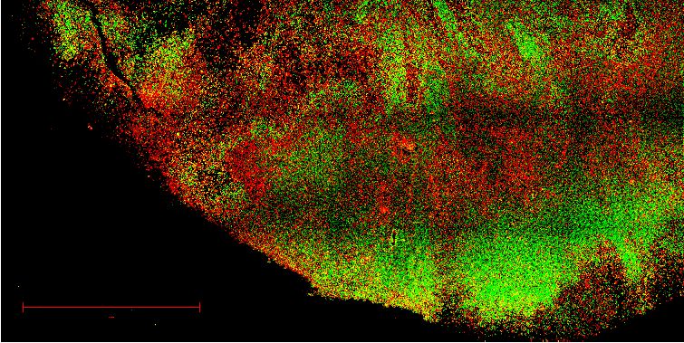
Research advance Microbiology and Infectious Disease
A VeroE6
B Calu-3
C Calu-3 / VeroE6
7 5 0.25
Log10 transduced cells/mL
Log10 transduced cells/mL
Calu-3 titer/ VeroE6 titer
* 0.20
6
* *
* 4
0.15
5 * *
3
* * * 0.10
4 * 0.05
3 2 0.00 * * * * * *
T A R A A H G
T
G
S6 H
R A
5A
R R
l-R RA
l-P T
G
R A
S6 H
R R
5A
l -R A
W RR RA 82 85 85 86
W
2
5
A
de W
86
2
de RR
5
A
86
68
68
68
68
68
R
R
68
R
6 6 6
-P l-R R R R S6
l-P
R
R
l
de de
de
de
D VeroE6-TMPRSS2
EVeroE6-TMPRSS2 / VeroE6
7 25
Log10 transduced cells/mL
VeroE6-TMPRSS2 titer/
20
6
*
VeroE6 titer
* 15
5 * *
10
4
5
3 0
* * * * * *
l-P T
l-P T
G
G
R R
R A
5A
S6 H
R R
R A
R A
5H
l-R A
l-R A
de W
de W
2
de RR
2
5
de RR
A
5
A
86
86
68
68
68
68
68
68
R
R
S6
R
F VeroE6 G VeroE6
150
Entry (% relative to control)
Entry (% relative to control)
150 WT
del-PRRA
del-RRAR
R682A
100 100 R685A
R685H
S686G
50 50
*
0 0
0 0.1 0.5 1 5 10 0 0.1 0.5 1 5 10
Camostat (µM) E64D (µM)
H VeroE6-TMPRSS2 I VeroE6-TMPRSS2
150 150
Entry (% relative to control)
Entry (% relative to control)
100 100
*
50 50
*
0 0
0 0.1 0.5 1 5 10 0 0.1 0.5 1 5 10
Camostat (µM) E64D (µM)
Figure 3. The SARS-CoV-2 multibasic cleavage site and the adjacent serine residue (S686) enhance infectivity and serine protease mediated entry on
Calu-3 and VeroE6-TMPRSS2 cells. (A–B) SARS-CoV-2 (WT), multibasic cleavage site (MBCS) mutant and S686G pseudovirus infectious titers on (A)
VeroE6 and (B) Calu-3 cells. (C) Fold change in SARS-CoV-2, MBCS mutant and S686G pseudovirus infectious titers on Calu-3 cells over infectious titers
on VeroE6 cells. (D) SARS-CoV-2, MBCS mutant and S686G pseudovirus infectious titers on VeroE6-TMPRSS2 cells. (E) Fold change in SARS-CoV-2,
Figure 3 continued on next page
Lamers et al. eLife 2021;10:e66815. DOI: https://doi.org/10.7554/eLife.66815 6 of 22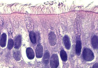
Research advance                                                                                                                                      Microbiology and Infectious Disease

Figure 3 continued
MBCS mutant and S686G pseudovirus infectious titers on VeroE6-TMPRSS2 cells over infectious titers on VeroE6 cells. One-way ANOVA was performed
for statistical analysis comparing all groups with WT. (F–I) SARS-CoV-2, MBCS mutant and S686G pseudovirus entry into (F and G) VeroE6 cells or (H
and I) VeroE6-TMPRSS2 cells pre-treated with a concentration range of either (F and H) camostat mesylate or (G and I) E64D. Two-way ANOVA,
followed by a bonferroni post hoc test was performed for statistical analysis comparing all groups to WT. WT pseudovirus entry into VeroE6 cells
treated with 10 mM E64D was significantly different from del-RRAR, R682A, R685A and S686G pseudovirus entry. * indicates statistical significance
(p
Research advance                                                                                    Microbiology and Infectious Disease

                                                         Calu-3 P2                                            Calu-3 P3
                                       A               (from VeroE6 P1)                   B                   (from Calu-3 P2)

                                                             Calu-3 P3                                           Calu-3 P5
                                       C                   (from VeroE6 P2)                  D              (continued passaging)

                                   Figure 5. SARS-CoV-2 propagation in Calu-3 cells efficiently prevents SARS-CoV-2 cell culture adaptation. (A)
                                   Deep-sequencing analysis of Calu-3 passage 2 virus from a VeroE6 passage 1. (B) Deep-sequencing analysis of
                                   Calu-3 passage 3 virus from the Calu-3 passage 2 in A. (C) Deep-sequencing analysis of Calu-3 passage 3 virus
                                   grown from a VeroE6 passage 2 stock (Figure 1A). Deep-sequencing analysis of Calu-3 passage 5 virus from a
                                   Calu-3 passage 3 stock in C. In each graph, the amino acid sequence logo of the multibasic cleavage site is
                                   shown.
                                   The online version of this article includes the following figure supplement(s) for figure 5:
                                   Figure supplement 1. Multibasic cleavage site deep-sequencing analysis of passage 4 Calu-3 viruses from an
                                   adapted VeroE6 P3 stock and full genome deep-sequencing analysis of Calu-3-propagated viruses.

                                   times to remove unbound particles. On day 2–5 post-infection, apical washes were collected and
                                   stored at 4˚C. During virus collections, bound virus particles were released from cells by pipetting
                                   directly on the cell layer. Virus collections from day 2 and day 3 (d2+3), and day 4 and day 5 (d4+5)
                                   were pooled, centrifuged, and filtered to remove debris, dead cells and mucus. In these cultures, cil-
                                   iated cells were infected, as shown by confocal imaging at day 3 post infection (Figure 7C). At day
                                   5, cultures exhibited widespread infection (Figure 7D) and significant cytopathic effects including
                                   loss of ciliated cells (Figure 7D–E) and syncytium formation (Figure 7E). To remove cytokines that
                                   could interfere in downstream experiments (such as interferons), we exchanged the medium in the
                                   filtered virus collections three times using an Amicon Ultra-15 column (100 kDa cutoff). The resulting

Lamers et al. eLife 2021;10:e66815. DOI: https://doi.org/10.7554/eLife.66815                                                                 8 of 22
Research advance                                                                                   Microbiology and Infectious Disease

                                           Mock                                   Camostat
                         A                                         B

       VeroE6 P4
      (from VeroE6 P3)

                         C                 Mock                      D            Camostat

     VeroE6-
    TMPRSS2
       P4
   (from VeroE6 P3)

Figure 6. Serine protease expression prevents MBCS mutations. (A–B) Deep-sequencing analysis of VeroE6 passage 4 virus from a VeroE6 passage 3 (A
is a redisplay of Figure 1C) mock-treated or treated with 10 mM camostat. (C–D) Deep-sequencing analysis of VeroE6-TMPRSS2 passage 4 virus from a
VeroE6 passage 3 mock-treated or treated with 10 mM camostat. In each graph the amino acid sequence logo of the multibasic cleavage site is shown.
The online version of this article includes the following figure supplement(s) for figure 6:
Figure supplement 1. Multibasic cleavage site and full genome deep-sequencing analysis of passage 4 VeroE6 and VeroE6-TMPRSS2 viruses.

                                   titers from the d2+3 and d4+5 stocks were 5.64  105 and 1.00  107 TCID50/ml, respectively, indi-
                                   cating that high titer virus stocks can be made in human airway organoids. Sequencing demon-
                                   strated that the high titer organoid stock (d4+5) had a 98.9% WT spike sequence, without multibasic
                                   cleavage site mutations and the S686G mutation at only 1.1% (Figure 8A–B). In accordance, the
                                   Organoid P3 virus produced small plaques (Figure 8C). No major variants were detected in the rest
                                   of the genome (Figure 8D). Next, we investigated S1/S2 cleavage of the VeroE6 P2, P3, Calu-3 P3,
                                   and Organoid P3 virus stocks by immunoblot (Figure 8E). The non-adapted Calu-3 and organoid
                                   stocks were >85% cleaved, while the VeroE6 P2 and P3 stocks were 71.2% and 33% cleaved, respec-
                                   tively (Figure 8F). The findings support that the Calu-3 and organoid stocks are non-adapted and
                                   indicate that in vivo the S1/S2 cleavage takes place in the producing cell.

                                   Discussion
                                   The rapid loss of the SARS-CoV-2 MBCS in cell culture has underlined that some in vitro propagation
                                   systems may fail to model key aspects of the viral life cycle. As these mutations directly affect the rel-
                                   evance and translatability of all laboratory SARS-CoV-2 experiments, it is pivotal to sort out exactly
                                   why these occur in order to prevent them. We and others have previously reported that the SARS-
                                   CoV-2 MBCS enhances serine protease-mediated entry, the dominant entry pathway in human

Lamers et al. eLife 2021;10:e66815. DOI: https://doi.org/10.7554/eLife.66815                                                               9 of 22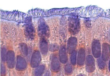
Research advance Microbiology and Infectious Disease
A B B’
B’
C AcTUB NP Hoechst C’ C’’
C’’
’
BC’
D AcTUB NP E AcTUB NP Hoechst E’
E
E’
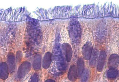
Figure 7. A 2D air-liquid interface human airway organoid model for SARS-CoV-2 propagation. (A) Human airway organoids were dissociated and
plated onto 12 mm transwell inserts. After an 8–12 week differentiation period at air-liquid interface cultures contained ciliated, non-ciliated and basal
cells as shown on a hematoxylin-eosin stain. (B) Air-exposed cells, but not basal cells, expressed the priming protease TMPRSS2 as shown by
immunohistochemistry. (C) Immunofluorescent staining indicated that in these cultures, ciliated cells (acetylated tubulin+ or AcTUB+ cells) were
Figure 7 continued on next page
Lamers et al. eLife 2021;10:e66815. DOI: https://doi.org/10.7554/eLife.66815 10 of 22Research advance Microbiology and Infectious Disease
Figure 7 continued
infected by SARS-CoV-2. (D and E) At 5 days post-infection, whole-well confocal imaging indicated the infection was widespread (D) and cytopathic
effects, including cilia damage (D and E) and syncytial cells (E) were visible. Scale bars indicate 20 mm in A, B, C; 2 mm in D; and 100 mm in E.
airway cells (Hoffmann et al., 2020; Mykytyn et al., 2021). VeroE6 cells, commonly used in the lab
to grow virus stocks, lack this entry pathway, forcing the virus to use endosomal cathepsins for entry.
Cell culture adaptations promoting cathepsin-mediated entry were also observed for the human
coronavirus 229E and adapted viruses showed a reduced ability to replicate in differentiated airway
epithelial cells (Bertram et al., 2013; Shirato et al., 2017). These observations led us to hypothesize
that mutations in the MBCS could be prevented in cells with an active serine protease-mediated
entry pathway. In this study, we show that the ectopic expression of the serine protease TMPRSS2 in
VeroE6 cells prevented MBCS mutations. Virus propagation in Calu-3 cells, which naturally express
serine proteases, also prevented cell culture adaptation. Similar results were obtained using a human
airway organoid-based culture system for SARS-CoV-2 propagation.
Our study shows that SARS-CoV-2 rapidly adapts to VeroE6 cell culture. Therefore, deep-
sequencing of viral stocks, which offers a thorough analysis beyond the consensus sequence, is
essential. As none of the MBCS mutations co-occurred, consensus sequence logos of culture
adapted stocks were often WT, while actually only 10–20% of viral reads contained the WT
sequence. Therefore, besides reporting the consensus sequence SARS-CoV-2 studies should prefera-
bly also report the percentage of WT reads in the MBCS. The first adaptation to occur in our stocks
was the S686G mutation, which lies directly adjacent to the MBCS and decreased Calu-3 infectivity,
fusogenicity and S1/S2 cleavage, but not as severely as MBCS mutations. Interestingly, this mutation
is rapidly positively selected in ferrets (Richard et al., 2020), and also transmitted, suggesting that
there are key differences in transmission between humans and ferrets. Alternatively, it is possible
that S686G optimizes cleavage by a specific ferret protease.
SARS-CoV-2 is generally grown on VeroE6 cells in the presence of 1–10% FBS, as this allows the
production of highly concentrated virus stocks. Here, we show that SARS-CoV-2 rapidly acquired
mutations in the MBCS upon passaging in VeroE6 cells and that the addition of FBS increases the
frequency of MBCS mutations. Currently, we do not know which components of FBS lead to the
increased rate of cell culture adaptation, but we hypothesize that (serine) proteases, naturally pres-
ent in serum (Shimomura et al., 1992) may be responsible as the addition of trypsin dramatically
increased the frequency of MBCS mutations and even led to the deletion of the entire MBCS. The
observation that the MBCS is a disadvantage in the presence of trypsin indicates that S2’ proteolytic
cleavage (performed by trypsin Hoffmann et al., 2020) should not occur in the supernatant where it
would cause all spikes to shed their S1 and adopt their post fusion conformation prior to encounter-
ing the plasma membrane. Alternatively, protease inhibitors in serum (Gstraunthaler, 2003) may
block transmembrane serine proteases. For these reasons, the use of serum should be avoided
when producing virus stocks. The use of defined serum-free media avoids the uncertainty that fac-
tors in serum affect the genetic stability of a virus and increase experimental reproducibility due to
variations in serum sources.
We show that the expression of the serine protease TMPRSS2 decreases the replicative fitness of
MBCS mutant SARS-CoV-2 viruses, which can then be outcompeted by WT viruses. This indicates
that the MBCS is an adaptation to serine proteases and that the serine protease-mediated entry
pathway is used for entry in vivo. This is in agreement with our earlier observations that SARS-CoV-2
enters using serine proteases on airway organoids (Mykytyn et al., 2021) and that MBCS mutant
pseudoviruses could not efficiently infect these cells. Low infectivity of MBCS mutants on the airway
cell line Calu-3 was also noted by Hoffmann et al., 2020. In contrast, two CRISPR-based survival
screens recently identified several endosomal proteins, including cathepsin L, as essential SARS-
CoV-2 genes (Daniloski et al., 2021; Wei et al., 2020). As noted recently by Bailey and Diamond,
2021, the identification of endosomal host factors as proviral in cell-line-based CRISPR screens
requires validation in primary cells. Our observation that WT viruses have a selective advantage in
2D airway organoids confirms that the endosomal entry pathway is of little significance in relevant
cells.
As new SARS-CoV-2 strains are emerging now and will continue to emerge for as long as SARS-
CoV-2 circulates in humans, there is a need to develop propagation systems that will preserve
Lamers et al. eLife 2021;10:e66815. DOI: https://doi.org/10.7554/eLife.66815 11 of 22Research advance Microbiology and Infectious Disease
A Organoid P3
(from VeroE6 P2)
R685H
B R682L
S686G
ACTAATTCTCCTCGGCGGGCACGTAGTGTAGCTAGTCAA
T N S P R R A R S V A S Q
C VeroE6 P2 Organoid P3
D
VeroE6 P2
Organoid P3
(from VeroE6 P2)
E P2 P3 P3 id
P3 F 100
E6 E6 3 no
ro ro lu- rga 80
Ve Ve Ca
Cleaved S1 (%)
O
60
250 - S0
40
S1
100 - 20
50 - 0
NP
P2 P3 P3 P3
6 6 -3 id
ro
E
ro
E
al
u no
Ve Ve C r ga
O
Figure 8. 2D air-liquid interface human airway organoids produce high titer stocks without multibasic cleavage site mutations. (A–B) Deep-sequencing
analysis (A) and Sanger chromatogram (B) of Organoid passage 3 virus from a VeroE6 passage 2 stock (Figure 1A). The amino acid sequence logo of
the multibasic cleavage site is shown. The translated sequence is indicated below the Sanger read. Arrows indicate where cell culture adaptations to
VeroE6 cells occur. (C) Plaque size analysis of VeroE6 passage 2 and Organoid passage 3 virus (the VeroE6 data is a redisplay of Figure 1G). Red arrow
Figure 8 continued on next page
Lamers et al. eLife 2021;10:e66815. DOI: https://doi.org/10.7554/eLife.66815 12 of 22Research advance Microbiology and Infectious Disease
Figure 8 continued
heads indicate large plaques. Scale bar indicates 1 cm. (D) Full genome deep-sequencing analysis of VeroE6 passage 2 and organoid passage three
stocks. In D VeroE6 P2 is a redisplay of VeroE6 P2 in Figure 1—figure supplement 1B. (E) Immunoblot analysis of VeroE6 passage 2 and 3, Calu-3
passage 3 and Organoid passage 3 stocks. S0 indicates uncleaved spike; S1 indicates the S1 domain of cleaved spike; NP indicates nucleoprotein.
Numbers indicate the molecular weight (kDa) of bands of the protein standard. (F) Quantification of cleavage from three immunoblots. Error bars
indicate SD.
The online version of this article includes the following figure supplement(s) for figure 8:
Figure supplement 1. Schematic workflow for the production of SARS-CoV-2 stocks on 2D air-liquid interface differentiated airway organoids.
genetic stability for any given SARS-CoV-2 mutant originating from a human respiratory sample. The
human airway organoid model for SARS-CoV-2 propagation established in this study (Figure 8)
allows high titer SARS-CoV-2 production and was most successful in removing MBCS mutations. We
have not observed any major variants associated with virus propagation in this system, but we can-
not exclude that some minor variants could be culture adaptations. The self-renewing capacity of
organoids allows labs to share organoid lines, allowing a level of reproducibility similar to that of
transformed cell lines. In addition, organoids can be grown from a wide range of organs and species
to best model the in vivo environment of a particular virus. As an accurate modeling of viral target
cells is likely to prevent most forms of cell culture adaptation, we expect that in the future organoid-
based systems are likely to replace transformed cell lines for viral stock production.
In conclusion, this study shows that SARS-CoV-2 rapidly adapts to VeroE6 cell culture propaga-
tion and that this can be prevented by using cell lines with an active serine protease-mediated entry
pathway (VeroE6-TMPRSS2 or Calu-3). Alternatively, a 2D airway organoid-based cell culture model
can be used for SARS-CoV-2 propagation if in the future new variants emerge that are not geneti-
cally stable on Calu-3 cells. Our study also shows that deep-sequencing rather than consensus
sequencing of viral stocks is critical for obtaining relevant and reproducible results in SARS-CoV-2
studies.
Materials and methods
Key resources table
Reagent type
(species) or resource Designation Source or reference Identifiers Additional information
Antibody Rabbit-anti-SARS-CoV Sino Biological Cat# 40143-T62 IF (1:1000)
NP (polyclonal)
Antibody Mouse anti-TMPRSS2 Santa Cruz Cat# sc-515727 IHC (1:200)
(monoclonal)
Antibody Goat-anti-mouse Dako Cat# P0260 IF (1:400)
Antibody Goat anti-rabbit IgG (H+L) Invitrogen Cat# A32740 IF (1:400)
Alexa Fluor Plus 594
Antibody Goat anti-mouse IgG (H+L) Invitrogen Cat# A11029 IF (1:2000)
Alexa Fluor 488
Antibody Mouse-anti-AcTub IgG2A Santa Cruz Cat# sc-23950 AF488 IF (1:100)
Alexa Fluor 488 Biotechnology
(monoclonal)
Antibody Mouse anti-nucleocapsid Sinobiological Cat# 40143-MM05 IF (1:1000)
Antibody Rabbit anti-SARS-CoV S1 Sinobiological Cat# 40150-T62 WB (1:1000)
(polyclonal)
Antibody Mouse-anti-SARS-CoV-2 S2 Genetex Cat# GTX632604 WB (1:1000)
(monoclonal)
Antibody Mouse-anti-VSV-N Absolute Antibody Cat# Ab01403-2.0 WB (1:1000)
(monoclonal)
Biological sample Airway organoids Mykytyn et al., 2021
(Homo sapiens)
Continued on next page
Lamers et al. eLife 2021;10:e66815. DOI: https://doi.org/10.7554/eLife.66815 13 of 22Research advance Microbiology and Infectious Disease
Continued
Reagent type
(species) or resource Designation Source or reference Identifiers Additional information
Cell line VeroE6 ATCC CRL 1586TM
(Cercopithecus aethiops)
Cell line VeroE6 TMPRSS2 Mykytyn et al., 2021
(Cercopithecus aethiops)
Cell line VeroE6 GFP1-10 Mykytyn et al., 2021
(Cercopithecus aethiops)
Cell line VeroE6 GFP1-10 TMPRSS2 Mykytyn et al., 2021
(Cercopithecus aethiops)
Cell line Calu-3 ATCC HTB 55
(Homo sapiens)
Cell line Calu-3 GFP1-10 Mykytyn et al., 2021
(Homo sapiens)
Chemical E64D MedChemExpress Cat# HY-100229
compound, drug
Chemical Camostat mesylate Sigma Cat# SML0057
compound, drug
Chemical Polyethylenimine linear Polysciences Cat# 23966
compound, drug
Chemical Hygromycin B Invitrogen Cat# 10843555001
compound, drug
Chemical Geneticin Invitrogen Cat# 10131035
compound, drug
Chemical Avicel FMC biopolymers -
compound, drug
Chemical Laemmli BioRad Cat# 1610747
compound, drug
Commercial SuperScript Invitrogen Cat# 18090200
assay or kit IV Reverse Transcriptase
Commercial Pfu Ultra II Fusion HS Agilent Technologies Cat# 600674
assay or kit DNA Polymerase
Commercial Qiaquick PCR QIAGEN Cat# 28104
assay or kit Purification Kit
Commercial BigDye Applied Biosystems Cat# 4337456
assay or kit Terminator v3.1 Cycle
Sequencing Kit
Commercial ProtoScript II Reverse New England BioLabs Cat# NEB M0368X
assay or kit Transcriptase
Commercial KAPA HyperPlus Roche Cat# 7962428001
assay or kit
Other Amicon Ultra-15 Centrifugal Millipore Cat#
Filter Unit with UFC910024
Ultracel-100 membrane
Other Opti-MEM I (1X) + GlutaMAX Gibco Cat# 51985–042
Other Advanced DMEM/F12 Thermo Fisher scientific Cat# 12634–010
Other AO medium Sachs et al., 2019 N/A
Other Pneumacult ALI medium Stemcell Cat # 05001
Other TryplE Thermo Fisher scientific Cat# 12605010
Other Basement R and D Systems Cat# 3533-005-02
membrane extract
Other Transwell inserts Corning Cat# 3460
Other Collagen Type I, High Corning Cat# 354249
concentration Rat tail
Continued on next page
Lamers et al. eLife 2021;10:e66815. DOI: https://doi.org/10.7554/eLife.66815 14 of 22Research advance Microbiology and Infectious Disease
Continued
Reagent type
(species) or resource Designation Source or reference Identifiers Additional information
Other 0.45 mm low protein Millipore Cat# SLHV033RS
binding filter
Other Hoechst Thermo Fisher Cat# H1399
Other Ampure XP Beads Beckman Coulter Cat# A63882
Other Illumina sequencer Illumina
V3 MiSeq flowcell
Other ABI PRISM 3100 Applied Biosystems
Genetic Analyzer
Other Odyssey CLx Licor
Other Amersham Typhoon GE Healthcare
Biomolecular Image
Other Amersham Imager 600 GE Healthcare
Other LSM700 confocal microscope Zeiss
Other Carl ZEISS Vert.A1 Zeiss
Software, algorithm ZEN Zeiss
Software, algorithm ImageQuant TL 8.2 GE Healthcare
Software, algorithm Studio Lite Ver 5.2 Licor
Software, algorithm GraphPad PRISM 8, 9 GraphPad
Software, algorithm Illustrator Adobe inc
Strain, strain SARS-CoV-2 BavPat-1 Dr. Christian Drosten European Virus
background Archive Global
(SARS-CoV-2) #026 V-03883
Cell lines
VeroE6 wildtype and retrovirally transduced cell lines were maintained in Dulbecco’s modified
Eagle’s medium (DMEM, Lonza) supplemented with 10% fetal bovine serum (FBS, Sigma, F7524,
heat inactivated for 30 min at 56˚C), HEPES, sodium bicabonate, penicillin (100 IU/mL), and strepto-
mycin (100 IU/mL). VeroE6-TMPRSS2, VeroE6-GFP1-10, VeroE6-TMPRSS2-GFP1-10, and Calu-3-
GFP1-10 cells were generated as described before (Mykytyn et al., 2021). Calu-3 and Calu-3-GFP1-
10 cells were maintained in Eagle’s modified Eagle’s medium (EMEM) supplemented with 10% FBS,
penicillin (100 IU/mL) and streptomycin (100 IU/mL). All cell lines were grown at 37˚C in a humidified
CO2 incubator, and transduced cell lines were cultured in the presence of selection antibiotics.
VeroE6 and Calu-3 cells were derived from ATCC. They were tested negative for mycoplasma.
SARS-CoV-2 propagation in cell lines
SARS-CoV-2 (isolate BetaCoV/Munich/BavPat1/2020; European Virus Archive Global #026 V-03883;
kindly provided by Dr. C. Drosten) was propagated to the indicated passage on VeroE6, VeroE6-
TMPRSS2 or Calu-3 cells, as indicated, in Advanced DMEM/F12 (Gibco), supplemented with HEPES,
Glutamax, penicillin (100 IU/mL) and streptomycin (100 IU/mL) (AdDF+++) at 37˚C in a humidified
CO2 incubator. Infections were performed at a multiplicity of infection (MOI) of 0.01 and virus was
harvested after 72 hr. The culture supernatant was cleared by centrifugation and stored at 80˚C.
Calu-3 stocks were additionally cleared using a 0.45 mM low protein binding filter (Millipore) to
remove mucus debris produced by these cells and the medium was exchanged three times for Opti-
MEM I (1X) + GlutaMAX (Gibco) using an Amicon Ultra-15 column (100 kDa cutoff). At the end of
each centrifugation step, approximately 2 ml was left in the top compartment. After three
exchanges, the purified virus was transferred to a new 50 ml tube and the Amicon Ultra-15 column
was washed ten times by adding 1 ml Opti-MEM I (1X) + GlutaMAX (Gibco) to the top compart-
ment, pipetting up and down several times on the filter and adding each wash to the tube contain-
ing the purified virus preparation. This step was repeated until the volume in the purified virus stock
Lamers et al. eLife 2021;10:e66815. DOI: https://doi.org/10.7554/eLife.66815 15 of 22Research advance Microbiology and Infectious Disease
was equal to the original volume of culture supernatant. Purified virus was stored at 80˚C in ali-
quots. Stock titers were determined by preparing 10-fold serial dilutions in Opti-MEM I (1X) + Gluta-
MAX (Gibco). One-hundred ml of each dilution was added to monolayers of 2 104 VeroE6 cells in
the same medium in a 96-well plate. Plates were incubated at 37˚C for 5 days and then examined for
cytopathic effect. The TCID50 was calculated according to the method of Spearman and Kärber. All
work with infectious SARS-CoV-2 was performed in a Class II Biosafety Cabinet under BSL-3 condi-
tions at Erasmus Medical Center.
Cloning
Cloning of SARS-CoV-2 S WT, del-PRRA, R685A, and R685H constructs for pseudovirus production
and GFP-complementation fusion assay was performed as described before (Mykytyn et al., 2021).
Del-RRAR, R682A, and S686G plasmids were generated by mutagenesis PCR.
Organoid culture and differentiation
Human airway stem cells were isolated and grown into organoids, and passaged as described before
(Lamers et al., 2020a) using a protocol adapted from Sachs et al., 2019. Adult lung tissue was
obtained from residual, tumor-free, material obtained at lung resection surgery for lung cancer. The
Medical Ethical Committee of the Erasmus MC Rotterdam granted permission for this study (METC
2012–512). Study procedures were performed according to the Declaration of Helsinki, and in com-
pliance with relevant Dutch laws and institutional guidelines. The tissues obtained were anonymized
and non-traceable to the donor. In this study we used organoids from one donor, from which bron-
chial and bronchiolar organoids were grown. Differentiation of human airway organoids at air-liquid
interface was performed as described before (Lamers et al., 2020a). Cultures were differentiated
for 8–12 weeks at air-liquid interface.
SARS-CoV-2 stock production on 2D air-liquid interface human airway
organoids
To produce stocks in human airway organoids, we differentiated the bronchial organoids in transwell
inserts at air-liquid interface for twelve weeks. A total of 12 12 mm transwell inserts were washed
three times in AdDF+++ before inoculation at the apical side at a MOI of 0.05. After a 2-hr incuba-
tion, cells were washed three times with AdDF+++ to remove unbound particles. Twenty-four hours
post-infection, cells were washed by adding 500 ul AdDF+++ to the apical side of the cells and incu-
bating at 37˚C 5% CO2 for 30 min to disperse the newly produced virus particles, facilitating the
next round of infection. Next, the medium was removed and discarded, as generally little virus is
produced in the first 24 hr (Lamers et al., 2020a). At days 2–5 post-infection, washes were collected
and stored at 4˚C. During collections, bound virus particles were removed from the cells by pipetting
three times directly on the cell layer after the 30 min incubation step at 37˚C 5% CO2. Virus collec-
tions from day 2 and day 3 (d2+3) and day 4 and day 5 (d4+5) were pooled, mixed by pipetting,
centrifuged at 4000 x g for 4 min, and filtered through a 0.45 um low protein binding filter (Milli-
pore) to remove debris, dead cells, and mucus. To remove cytokines that could interfere in down-
stream experiments (such as interferons), we exchanged the medium in the filtered virus collections
three times with Opti-MEM I (1X) + GlutaMAX (Gibco) using an Amicon Ultra-15 column (100 kDa
cutoff). At the end of each centrifugation step, approximately 2 ml was left in the top compartment.
After three exchanges, the purified virus was transferred to a new 50 ml tube and the Amicon Ultra-
15 column was washed ten times by adding 1 ml Opti-MEM I (1X) + GlutaMAX (Gibco) to the top
compartment, pipetting up and down several times on the filter and adding each wash to the tube
containing the purified virus preparation, resulting in a total volume of ~12 ml. Next, virus prepara-
tions were aliquoted in 500 ml aliquots, stored at 80˚C and thawed for titrations on VeroE6 cells.
Pseudovirus assay
Pseudovirus production, infectivity, and entry assays were performed as described before
(Mykytyn et al., 2021). Briefly, pseudoviruses expressing WT, MBCS mutant, and S686G S were
titrated by preparing 10-fold serial dilutions in Opti-MEM I (1X) + GlutaMAX (Gibco). Thirty ml of
each dilution was added to monolayers of 2 104 VeroE6, VeroE6-TMPRSS2 or 8 104 Calu-3 cells
in the same medium in a 96-well plate. Titrations were performed in triplicate. Plates were incubated
Lamers et al. eLife 2021;10:e66815. DOI: https://doi.org/10.7554/eLife.66815 16 of 22Research advance Microbiology and Infectious Disease
at 37˚C overnight and then scanned on the Amersham Typhoon Biomolecular Imager (channel Cy2;
resolution 10 mm; GE Healthcare). Entry routes were determined by pre-treating monolayers of
VeroE6 or VeroE6-TMPRSS2 cells with a concentration range of camostat mesylate (Sigma) or E64D
(MedChemExpress) diluted in Opti-MEM I (1X) + GlutaMAX (Gibco) for 2 hr prior to infection with 1
103 pseudovirus. Plates were incubated at 37˚C overnight and then scanned on the Amersham
Typhoon Biomolecular Imager (channel Cy2; resolution 10 mm; GE Healthcare). All pseudovirus
experiments were quantified using ImageQuant TL 8.2 image analysis software (GE Healthcare).
Pseudovirus concentration
Pseudoviruses were concentrated as described before (Mykytyn et al., 2021) on a 10% sucrose
cushion (10% sucrose, 15 mM Tris–HCl, 100 mM NaCl, 0.5 mM EDTA) for 1.5 hr at 20,000 x g at 4˚C.
Supernatant was decanted and pseudoviruses resuspended in Opti-MEM I (1X) + GlutaMAX (Gibco)
to achieve 100-fold concentration.
Immunoblotting
Concentrated pseudovirus stocks were diluted to a final concentration of 1x Laemmli loading buffer
(Bio-Rad) containing 5% 2-mercaptoethanol. Authentic viruses were diluted to a final concentration
of 2x Laemmli loading buffer containing 5% 2-mercaptoethanol. All samples were boiled for 30 min
at 95˚C. Samples were used for SDS-PAGE analysis using precast 10% TGX gels (Bio-Rad). Gels were
run in tris-glycine SDS (TGS) buffer at 50V for 30 min and subsequently at 120V for 90 min. Transfer
was performed at 300mA for 55 min onto 0.45 mm Immobilon-FL PVDF membranes in TGS contain-
ing 20% methanol. Spike was stained using polyclonal rabbit-anti-SARS-CoV S1 (1:1000, Sino Biolog-
ical), mouse-anti-SARS-CoV-2 S2 (1:1000, Genetex), SARS-CoV-2 nucleoprotein was stained using
rabbit-anti-SARS-CoV NP (1:1000, Sino Biological) and VSV nucleoprotein was stained using mouse-
anti-VSV-N (1:1000, Absolute Antibody) followed by infrared-labelled secondary antibodies
(1:20,000; Licor). Western blots were scanned on an Odyssey CLx and analyzed using Image Studio
Lite Ver 5.2 software.
GFP-complementation fusion assay
Fusion assays were performed as described before (Mykytyn et al., 2021). Briefly, HEK-293T cells
were transfected with 1.5 mg pGAGGS-spike (all coronavirus S variants described above) DNA and
pGAGGS-b-Actin-P2A-7xGFP11-BFP DNA or empty vector DNA with PEI in a ratio of 1:3 (DNA:
PEI). Transfected HEK-293T cells were incubated overnight at 37˚C 5% CO2, resuspended in PBS
and added to GFP1-10 expressing VeroE6, VeroE6-TMPRSS2 and Calu-3 cells in Opti-MEM I (1X) +
GlutaMAX at a ratio of 1:80 (HEK-293T cells: GFP1-10 expressing cells). Fusion events were quanti-
fied by detecting GFP+ pixels after 18 hr incubation at 37˚C 5% CO2 using Amersham Typhoon Bio-
molecular Imager (channel Cy2; resolution 10 mm; GE Healthcare). Data was analyzed using the
ImageQuant TL 8.2 image analysis software (GE Healthcare) by calculating the sum of all GFP+ pix-
els per well.
Plaque assay
Virus stock were diluted in 10-fold serial dilutions in 2 ml Opti-MEM I (1X) + GlutaMAX (Gibco). One
ml of each dilution was added to monolayers of 2 106 VeroE6 cells in the same medium in a six-
well plate. Cells were incubated at 37˚C for 1 hr and then overlaid with 1.2% Avicel (FMC biopoly-
mers) in Opti-MEM I (1X) + GlutaMAX (Gibco) for 72 hr. Next, they were washed once in PBS, fixed
in formalin, permeabilized in 70% ethanol and washed in PBS again. Cells were blocked in 3% BSA
(bovine serum albumin; Sigma) in PBS, stained with mouse anti-nucleocapsid (Sino biological;
1:1000) in PBS containing 0.1% BSA, washed three times in PBS, then stained with goat anti-mouse
Alexa Fluor 488 (Invitrogen; 1:2000) in PBS containing 0.1% BSA and then washed three times in
PBS. All staining steps were performed at room temperature for one hour. Plates were scanned on
the Amersham Typhoon Biomolecular Imager (channel Cy2; resolution 10 mm; GE Healthcare).
Sanger sequencing
To sequence spike gene fragments, RNA was extracted as described above and used for cDNA syn-
thesis using Superscript IV (Invitrogen), according to the manufacturer’s instructions. PCR was
Lamers et al. eLife 2021;10:e66815. DOI: https://doi.org/10.7554/eLife.66815 17 of 22Research advance Microbiology and Infectious Disease
performed using PfuUltra II Fusion HS DNA Polymerase (Agilent Technologies) and primers 50 -TGA-
CACTACTGATGCTGTCCGTG-30 and 50 -GATGGATCTGGTAATATTTGTG-30 under the following
conditions: initial denaturation at 95˚C for 3 min, followed by 25 cycles of (95˚C for 20 s, 52˚C for 20
s, and 72˚C for 60 s), and a final extension at 72˚C for 10 min. The amplicons were purified (Qiagen
PCR purification kit, according to manufacturer) and sequenced with the forward primer using the
BigDye Terminator v3.1 Cycle Sequencing Kit and an ABI PRISM 3100 genetic analyzer (Applied Bio-
systems). The obtained sequences were assembled and aligned using Benchling (MAFFT algorithm).
Fixed immunofluorescence microscopy and immunohistochemistry
Transwell inserts were fixed in formalin, permeabilized in 0.1% Triton X-100, and blocked for 60 min
in 10% normal goat serum in PBS (blocking buffer). Cells were incubated with primary antibodies
overnight at 4˚C in blocking buffer, washed twice with PBS, incubated with corresponding secondary
antibodies Alexa488-, 594-conjugated secondary antibodies (1:400; Invitrogen) in blocking buffer for
2 hr at room temperature, washed two times with PBS, incubated for 10 min with Hoechst, washed
twice with PBS, and mounted in Prolong Antifade (Invitrogen) mounting medium. SARS-CoV-2 was
stained with rabbit-anti-SARS-CoV nucleoprotein (40143-T62, 1:1000, Sino biological). Ciliated cells
were stained with mouse-anti-AcTub (sc-23950 AF488, 1:100, Santa Cruz Biotechnology). For
TMPRSS2 stainings, formalin-fixed inserts were paraffin-embedded, sectioned, and deparaffinized as
described before prior to staining (Rockx et al., 2020). Samples were imaged on a LSM700 confocal
microscope using ZEN software (Zeiss). Immunohistochemistry was performed as described previ-
ously (Rockx et al., 2020) on formalin fixed, paraffin-embedded Transwell inserts. TMPRSS2 was
stained using mouse-anti-TMPRSS2 (sc-515727, 1:200, Santa Cruz Biotechnology), and visualized
with goat-anti-mouse (PO260, 1:100, Dako) horseradish peroxidase labeled secondary antibody,
respectively. Samples were counterstained using haematoxylin.
Illumina sequencing
For deep- sequencing, RNA was extracted as described above and subsequently cDNA was gener-
ated using ProtoscriptII reverse transcriptase enzyme (New England BiotechnologieBioLabs) accord-
ing to the manufacturer’s protocol. A SARS-CoV-2 specific multiplex PCR was performed as recently
described (Oude Munnink et al., 2020). In short, primers for 86 overlapping amplicons spanning the
entire genome were designed using primal scheme (http://primal.zibraproject.org/). The amplicon
length was set to 500 bp with 75 bp overlap between the different amplicons. Amplicons were puri-
fied with 0.8x AMPure XP beads (Beckman Coulter) and 100 ng of DNA was converted into paired-
end Illumina sequencing libraries using KAPA HyperPlus library preparation kit (Roche) with the
KAPA unique dual-indexed adapters (Roche), following the manufacturer’s recommendations. The
barcode-labeled samples were pooled and analyzed on an Illumina sequencer V3 MiSeq flowcell
(2 300 cycles).
Sequencing data analysis
Adapters from the paired-end sequencing reads were trimmed using cutadapt (https://doi.org/10.
14806/ej.17.1.200) via: cutadapt -B AGATCGGAAGAGCGTCGTGTAGGGAAAGAGTG -b AGA
TCGGAAGAGCACACGTCTGAACTCCAGTCAC –interleaved –minimum-lengthX50. The
trimmed reads were aligned to the genome of Bavpat-1 with Bowtie2 (Langmead and Salzberg,
2012) using parameters: –no-discordantX–dovetailX–no-mixedX–maxins 2000. Primer
sequences were trimmed off from the alignments by soft-clipping the leftmost 33 bases from each
sequencing reads using BamUtil (Jun, Wing, Jun et al., 2015) via: trimbam {bam_file} - -L 30 R 0 –
clip. Variants calling was done using VarScan2 (Koboldt et al., 2012) and SAMtools (Li et al.,
2009) via: samtools mpileup –excl-flagsX2048X–excl-flagsX256X–fasta-refX{REFEREN-
CE_FAASTA}X–max-depthX50000X–min-MQX30X–min-BQX30X{BAM_FILE}X|XvarscanXpi-
leup2cnsX–min-coverageX10X–min-reads2X2X–min-var-freqX0.01X–min-freq-for-
homX0.75X–p-valueX0.05X–variantsX1X>X{snp_file}. Sequence logo were generated with
logomaker (Tareen and Kinney, 2020) using a custom python script. Plotting of mutation frequen-
cies was done using R and ggplot2 (Hadley, 2016). All scripts used for data processing are depos-
ited in GitHub: https://github.com/nicwulab/SARS-CoV-2_in_vitro_adaptation (Wu et al., 2021).
Lamers et al. eLife 2021;10:e66815. DOI: https://doi.org/10.7554/eLife.66815 18 of 22Research advance Microbiology and Infectious Disease
Raw sequencing data has been submitted to the NIH Short Read Archive under accession number:
BioProject PRJNA694097.
Statistics
Statistical analysis was performed with the GraphPad Prism 8 and 9 software using an ANOVA or
two-way ANOVA followed by a Bonferroni multiple-comparison test.
Acknowledgements
This work was supported by the Netherlands Organization for Health Research and Development
(ZONMW) grant agreement 10150062010008 to BLH and co-funded by the PPP Allowance (grant
agreement LSHM19136) made available by Health Holland, Top Sector Life Sciences and Health, to
stimulate public-private partnerships. The present manuscript was part of the research program of
the Netherlands Centre for One Health. The funders had no role in study design, data collection and
interpretation, or the decision to submit the work for publication.
Additional information
Funding
Funder Grant reference number Author
ZonMw 10150062010008 Bart L Haagmans
PPP allowance LSHM19136 Bart L Haagmans
The funders had no role in study design, data collection and interpretation, or the
decision to submit the work for publication.
Author contributions
Mart M Lamers, Anna Z Mykytyn, Tim I Breugem, Conceptualization, Validation, Investigation, Visual-
ization, Methodology, Writing - original draft, Writing - review and editing; Yiquan Wang, Douglas C
Wu, Data curation, Software, Formal analysis, Visualization, Methodology; Samra Riesebosch, Valida-
tion, Investigation, Visualization, Methodology; Petra B van den Doel, Investigation, Writing - original
draft; Debby Schipper, Investigation; Theo Bestebroer, Investigation, Methodology, Writing - origi-
nal draft; Nicholas C Wu, Data curation, Software, Formal analysis, Supervision, Visualization, Writing
- review and editing; Bart L Haagmans, Conceptualization, Supervision, Funding acquisition, Writing
- review and editing
Author ORCIDs
Mart M Lamers https://orcid.org/0000-0002-1431-4022
Anna Z Mykytyn http://orcid.org/0000-0001-7188-6871
Tim I Breugem http://orcid.org/0000-0002-5558-7043
Yiquan Wang http://orcid.org/0000-0002-1954-9808
Douglas C Wu https://orcid.org/0000-0001-6179-3110
Bart L Haagmans https://orcid.org/0000-0001-6221-2015
Decision letter and Author response
Decision letter https://doi.org/10.7554/eLife.66815.sa1
Author response https://doi.org/10.7554/eLife.66815.sa2
Additional files
Supplementary files
. Transparent reporting form
Lamers et al. eLife 2021;10:e66815. DOI: https://doi.org/10.7554/eLife.66815 19 of 22You can also read