Doppler Echocardiography Assessment of Coronary Microvascular Function in Patients With Angina and No Obstructive Coronary Artery Disease
←
→
Page content transcription
If your browser does not render page correctly, please read the page content below
REVIEW
                                                                                                                                       published: 29 October 2021
                                                                                                                                  doi: 10.3389/fcvm.2021.723542

                                            Doppler Echocardiography
                                            Assessment of Coronary
                                            Microvascular Function in Patients
                                            With Angina and No Obstructive
                                            Coronary Artery Disease
                                            Jakob Schroder* and Eva Prescott

                                            Department of Cardiology, Bispebjerg Frederiksberg Hospital, University of Copenhagen, Copenhagen, Denmark

                            Edited by:      Echocardiographic evaluation is an essential part of the diagnostic work-up in
                     Tim van de Hoef,
                    Academic Medical        patients with known or suspected cardiovascular disease. Transthoracic Doppler
                   Center, Netherlands      echocardiography (TTDE) enables straightforward and reliable visualization of flow in the
                       Reviewed by:         left anterior descending artery. In the absence of obstructive coronary artery disease,
                        Andreas Seitz,
     Robert Bosch Hospital, Germany
                                            low TTDE-derived coronary flow velocity reserve (CFVR) is considered a marker of
              Gaetano Antonio Lanza,        coronary microvascular dysfunction (CMD). TTDE CFVR is free from ionizing radiation
     Catholic University of the Sacred      and widely available, utilizing high-frequency transducers, pharmacologic vasodilator
                            Heart, Italy
                                            stress, and pulsed-wave Doppler quantification of diastolic peak flow velocities. European
                  *Correspondence:
                      Jakob Schroder        Society of Cardiology guidelines recommend TTDE CFVR evaluation only following
         jakob.arnborg.schroeder.01@        preceding anatomic invasive or non-invasive coronary imaging excluding obstructive
                           regionh.dk
                                            CAD. Accordingly, clinical use of TTDE CFVR is limited and CMD frequently goes
                    Specialty section:
                                            undiagnosed. An evolving body of evidence underlines that low CFVR is an important and
          This article was submitted to     robust predictor of adverse prognosis and continuing symptoms in angina patients both
    Sex and Gender in Cardiovascular
                                            with and without obstructive CAD. The majority of angina patients have no obstructive
                              Medicine,
                a section of the journal    CAD, particularly among women. This has led to the suggestion that there may be
  Frontiers in Cardiovascular Medicine      a gender-specific female atherosclerotic phenotype with less epicardial obstruction,
           Received: 10 June 2021           and a low CFVR signifying CMD instead. Nevertheless, available evidence indicates
      Accepted: 22 September 2021
        Published: 29 October 2021
                                            low CFVR is an equally important prognostic marker in both men and women. In
                             Citation:
                                            this review, TTDE CFVR was evaluated regarding indication, practical and technical
     Schroder J and Prescott E (2021)       aspects, and interpretation of results. Association with symptoms and prognosis,
            Doppler Echocardiography
                                            comparison with alternative invasive and non-invasive imaging modalities, and possible
              Assessment of Coronary
    Microvascular Function in Patients      interventions in angina patients with low CFVR were discussed, and key research
      With Angina and No Obstructive        questions were proposed.
              Coronary Artery Disease.
   Front. Cardiovasc. Med. 8:723542.        Keywords: coronary flow velocity reserve, stress echocardiography, coronary microvascular dysfunction,
      doi: 10.3389/fcvm.2021.723542         prognosis, sex

Frontiers in Cardiovascular Medicine | www.frontiersin.org                       1                                        October 2021 | Volume 8 | Article 723542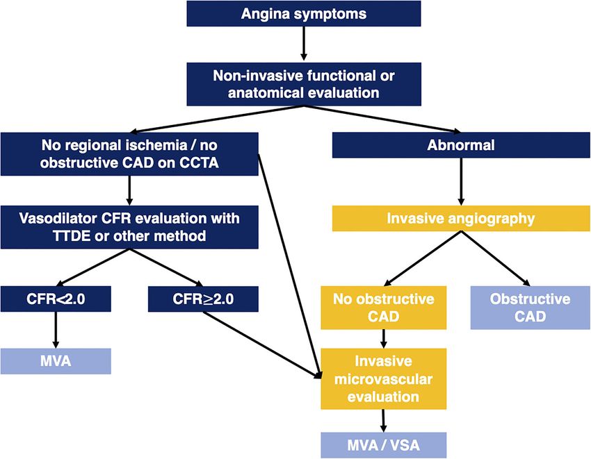
Schroder and Prescott                                                                                                        CFVR in Non-obstructive Angina

INTRODUCTION                                                                        and prognosis, comparison with alternative invasive and non-
                                                                                    invasive imaging modalities, and possible interventions in angina
Coronary artery disease remains one of the leading causes of                        patients with low CFVR were discussed. Finally, we proposed a
morbidity and mortality in Western countries in both men                            possible translation of TTDE CFVR to the clinic and questions in
and women, with male sex as a risk factor for the early                             relation to angina patients and CFVR that remain unanswered.
development of coronary artery disease (CAD) (1, 2). The present
diagnostic paradigm in patients with angina pectoris is focused
on likelihood and subsequent identification of obstructive CAD                      PATHOPHYSIOLOGIC BASIS
(3, 4), but most patients referred for assessment do not fulfill
criteria for invasive coronary angiography (ICA), and in the                        Under resting conditions, the coronary blood flow is kept
subset of patients ultimately examined with ICA, many patients,                     constant at varying coronary pressures. During exertion,
especially women, have no obstructive CAD. Ultimately, only a                       coronary blood flow is increased due to a simultaneous increase
fraction of angina patients is treated with revascularization (5–                   in coronary perfusion pressure and a decrease in coronary
7). In effect, a treatable etiological explanation remains absent in                vascular resistance. Vasodilation is primarily induced at the level
many patients with angina, particularly in women.                                   of small arteries and precapillary arterioles via several regulatory
    More than 30 years ago, efforts to measure coronary                             mechanisms acting in conjunction and interdependent mutual
flow velocities in the coronary epicardial arteries with                            stimulation (19–21). Vasodilatory mechanisms include the flow-
Doppler echocardiography began (8, 9). Early proof of                               mediated endothelium-dependent release of nitric oxide, a
concept studies established a high degree of correlation                            myogenic response to increased transmural arteriolar pressure,
between invasively derived flow measurements and values                             metabolic regulation triggered by local alterations in the levels
obtained via transesophageal, and later transthoracic Doppler                       of vasoactive metabolites, e.g., carbon dioxide and adenosine
echocardiography (TTDE) (10–12). TTDE was considered a                              inducing non-endothelium dependent vasodilation, and neural
promising imaging modality given that echocardiography was                          sympathetic stimulation of β-receptors also contributes to
already a principal part of the diagnostic work-up in nearly                        decreased vascular resistance under conditions of increased
all patients suspected of cardiac disease. Initial studies aimed                    oxygen demand (17, 22–24). In healthy subjects coronary blood
at determining the potential presence and degree of coronary                        flow may be increased 4- to 6-fold during maximal exertion.
epicardial stenosis (13–15). Later on, the focus shifted toward                     This capacity for coronary vasodilation may be expressed as the
visualization and quantification of the most accessible coronary                    ratio between peak and baseline perfusion, the coronary flow
branch, i.e., the left anterior descending (LAD) artery, during                     reserve (CFR) (9, 14, 19). In patients with unobstructed epicardial
rest and pharmacologic stress as an indirect measure of the                         coronary arteries, these mainly serve the purpose of conduit
perfusion capacity of the entire coronary circulation (13, 16, 17).                 vessels transporting blood to the smaller vessels, which regulate
Vasodilation in the microvascular compartment is the main                           the vascular resistance as described above. Conversely, in patients
determinant of increased coronary blood flow during exertion.                       with epicardial obstructive CAD, the CFR will be reduced in
In the absence of any significant epicardial stenosis, changes in                   accordance with the degree of stenosis (17, 19).
coronary flow velocity relative to resting flow is considered an                       It is not possible to visualize the microcirculation in vivo, but
indirect measure of the coronary microvascular function, i.e.,                      in patients with no significant epicardial flow-limiting stenosis
coronary flow velocity reserve (CFVR) (17, 18).                                     (0.8 at ICA) a
    The relevant body of evidence related to TTDE CFVR                              reduced CFR is considered a marker of reduced vasodilatory
in angina patients with no obstructive CAD consists of a                            capacity in the small coronary arteries and arterioles, i.e.,
combination of (i) studies in selected patient cohorts with no                      coronary microvascular dysfunction (CMD) (16, 17, 19, 25).
obstructive CAD and (ii) studies performed in mixed patient                         Pathophysiologically, CMD may be due to both structural and
populations both with and without obstructive CAD. We had                           functional alterations including capillary rarefaction, decreased
chosen to include both study types, while clearly highlighting                      arteriolar lumen/wall ratio, and impaired endothelium- and non-
possible interpretation difficulties and limitations of the evidence                endothelium dependent vasodilatation or excessive adrenergic
related to the mixed nature of the patient populations in the                       vasoconstriction, often with several abnormal findings present
latter group.                                                                       in the same patient. Furthermore, CMD may be present both in
    In this review, we evaluated TTDE CFVR regarding                                patients with and without obstructive CAD or myocardial disease
indication, practical and technical aspects, and interpretation                     (21, 26–29).
of results in patients with angina and no obstructive coronary                         Different vasodilatory and adrenergic agents induce a
artery disease (ANOCA). Moreover, association with symptoms                         near-maximal increase in coronary blood flow during CFR
                                                                                    quantification, partly due to indirect concomitant activation
Abbreviations: ANOCA, angina with no obstructive coronary artery disease;           of both hemodynamic, metabolic, and neural mechanisms.
CAD, coronary artery disease; CFR, coronary flow reserve; CFVR, coronary            Direct quantification of coronary flow reserve of the entire
flow velocity reserve; CMD, coronary microvascular dysfunction; cMRI, cardiac       myocardium can be measured by positron emission tomography
magnetic resonance imaging; Cx, circumflex coronary artery; ICA, invasive
coronary angiography; PET, positron emission tomography; RCA, right coronary
                                                                                    (PET). Quantification of flow reserve in any epicardial vessel
artery; RWMA, regional wall motion abnormality; SE, stress echocardiography;        is obtainable during ICA. TTDE visualization of the main
TTDE, transthoracic Doppler echocardiography.                                       coronary arteries allows assessment of coronary flow velocity as

Frontiers in Cardiovascular Medicine | www.frontiersin.org                      2                                    October 2021 | Volume 8 | Article 723542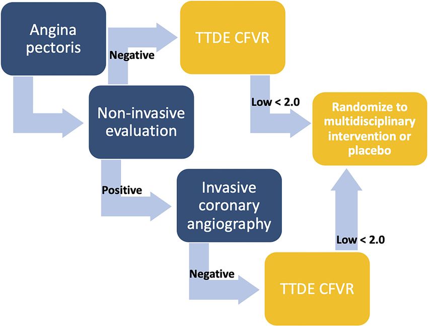
Schroder and Prescott CFVR in Non-obstructive Angina
an alternative to absolute flow (16, 25, 29). Under the assumption these branches may necessitate the use of an echo probe with
that the epicardial vessel lumen is kept relatively constant during a slightly lower frequency range 6 if atrial
TTDE CFVR EXAMINATION fibrillation or other irregular heart rhythms). Stress coronary flow
METHODOLOGY velocity should be the highest value obtained during vasodilator
infusion and three consecutive cardiac cycles should preferably
Coronary flow velocity measurements may be obtained in several be obtained, although this is sometimes challenging due to
of the larger coronary arteries. However, in the context of CMD cardiac motion and hyperventilation during stress. The patient
evaluation, the assessment is most commonly performed via may be instructed to hold their breath to limit hyperventilation
identification of the mid-distal LAD due to its position near the artifacts (44). Readings of coronary flow velocity values have been
chest wall, thus providing reliable and optimal images and flow shown to have good inter- and intra-observer variability and low
curves. Nonetheless, if LAD visualization is difficult in selected coefficient of variation in the range of 5–10% (8, 11, 43, 45–49).
patients, measurements may also be obtained in other larger The pharmacologic stress agents used in CFVR evaluation
coronary branches (8, 16, 25, 30, 32, 33). The TTDE examination are also used to achieve maximal perfusion in other stress
may be performed with commercially available ultrasound imaging modalities, e.g., PET, cardiac MRI (cMRI) perfusion,
machines using a phased array high-frequency ultrasound probe and single-photon emission computed tomography (16, 50). The
usually in the range from >3 to 8 MHz with harmonic imaging first choice agents in CFVR examination are the vasodilators
to obtain high-resolution color Doppler visualization of the mid- adenosine and dipyridamole (16, 51–54). Adenosine acts by
distal LAD (8, 9, 12, 30, 34). The patient is studied in the left directly stimulating the A2A -receptors on vascular smooth
lateral decubitus position. A baseline color scale of ∼1–2.5 kHz muscles cells in the microvascular vessel wall eliciting
(velocity range of ± 10–24 cm/s) may be used as a standard relaxation and vasodilation. Dipyridamole stimulates the
for obtaining the color Doppler. The mid-distal LAD is usually same pathway indirectly by inhibiting adenosine deaminase
located in the interventricular sulcus at the midway between a and phosphodiesterase, effectively increasing the adenosine
foreshortened two- and three-chamber apical view. However, due concentration in the vessel wall (8, 9, 19, 21). Importantly, these
to anatomic variations, the use of modified and apical views is non-endothelium-dependent mechanisms do not cause any
often necessary to obtain optimal LAD visualization. Diastolic direct vasodilation in the larger epicardial arteries (55, 56). It has
maximal coronary flow velocities are measured by pulsed-wave been shown that administration of intracoronary or sublingual
Doppler as a flow signal in the LAD toward the transducer nitroglycerine prior to stress testing dilates the epicardial vessels
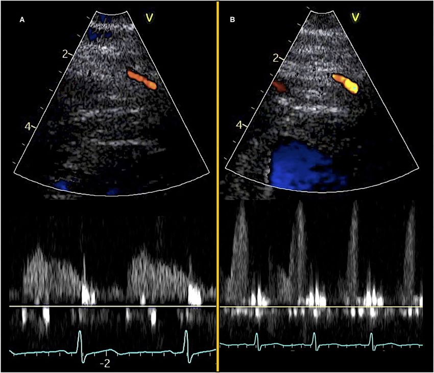
(Figure 1, Supplementary Videos 1, 2). The coronary Doppler resulting in a reduced resting coronary flow velocity without an
flow profile is biphasic with diastolic predominance. To avoid increase in stress coronary flow velocity, ultimately yielding a
systolic motion artifacts and to achieve reproducible maximal higher CFVR ratio (57, 58). However, pre-stress nitroglycerine
velocities the diastolic flow is used for CFVR calculation. The has not been used in any larger clinical studies employing
blood flow direction of the LAD is adjusted to be close to parallel adenosine/dipyridamole stress imaging.
with the direction of the pulsed-wave Doppler ultrasound beam The recommended dose of adenosine stress is 0.14
and a 3–4 mm sample volume is positioned over the LAD color mg/kg/min, and high-dose dipyridamole stress (0.84 mg/kg
flow. Sample volume size is adjusted as needed to balance signal over 6 min) produces comparable values of CFVR although
intensity and noise. In case of unsatisfactory quality of LAD maximal stress coronary flow velocity values are possibly only
color signal or flow velocity profile, it is also possible instead to achieved with adenosine (8, 52). Due to the rapid onset of
visualize and assess flow in either the right coronary artery (RCA) action of adenosine, maximal flow velocity may be recorded
or posterior descending artery in the posterior interventricular quickly after the infusion has started, usually after 1–2 min,
groove (modified two-chamber view) or the circumflex coronary while maximal coronary flow velocity using dipyridamole is
artery (Cx) in the basal part of the lateral left ventricular wall usually obtained after 3–6 min. Most CFVR protocols perform
(apical four-chamber view) (33, 35–38). The deeper position of continuous coronary flow velocity stress measurements during
Frontiers in Cardiovascular Medicine | www.frontiersin.org 3 October 2021 | Volume 8 | Article 723542Schroder and Prescott CFVR in Non-obstructive Angina FIGURE 1 | Transthoracic Doppler echocardiography and pulsed-wave Doppler curves. Color Doppler visualization of mid-distal LAD (top) and diastolic pulsed-wave flow velocity curves (bottom). (A) Images obtained at rest and (B) images obtained during adenosine stress. ∼5–7 min of vasodilator stress. The main contraindication for alternative if adenosine/dipyridamole are contraindicated due to adenosine/dipyridamole is the presence of severe hypotension, a comorbidity (56, 62, 63). reactive pulmonary obstructive disease that may be aggravated Regadenoson is a novel A2A -selective adenosine receptor during adenosine infusion and inadvertent stimulation of A2B agonist which causes vasodilation with a lower risk of adenosine receptors in small airways, and cardiac conduction bronchospasm or atrioventricular conduction delays as A1 and abnormalities, such as higher degree atrioventricular block, A2B adenosine receptors are not stimulated. Regadenoson is which may be aggravated via stimulation of A1 adenosine administered as a weight-unadjusted fixed-dose bolus rapidly receptors (50, 52, 59, 60). Any side effects of adenosine rapidly inducing maximal vasodilation which lasts for ∼2–3 min (64, diminish due to its short half-life, whereas the longer half-life 65). Regadenoson has been suggested as a potential new agent for dipyridamole often necessitates intravenous administration in vasodilator stress testing, but it has not yet been used in of refractory doses of an antagonizing methylxanthine, e.g., larger clinical studies evaluating TTDE CFVR (66–68). Current theophylline and aminophylline (30, 61). limitations that it faces include its price and a lack of overview of The adrenergic agonist dobutamine is often used for SE in all possible adverse effects, and up to now most clinical research the evaluation of wall motion abnormalities due to its positive has been done with non-echocardiography modalities (69, 70). inotropic and chronotropic effects. However, in addition to Abstinence from specific foods, drinks, and medications increasing coronary flow via adrenergic stimulation, it also dilates prior to examination is a prerequisite to gain reliable CFVR the epicardial coronary arteries via β-adrenergic activation, measurements. Methylxanthines are competitive inhibitors resulting in the more difficult interpretation of coronary of adenosine receptors and may significantly attenuate the flow velocity changes. The increase in contractility makes coronary vasodilator effect of all stress agents acting via A2A - consistent measures of coronary flow more difficult. Accordingly, receptors, such as adenosine, dipyridamole, and regadenoson. dobutamine stress in CFVR evaluation is mainly suggested as an Therefore, drinks and foods containing significant amounts of Frontiers in Cardiovascular Medicine | www.frontiersin.org 4 October 2021 | Volume 8 | Article 723542
Schroder and Prescott                                                                                           CFVR in Non-obstructive Angina

methylxanthine, e.g., coffee, tea, cola, chocolate, and banana,        that the use of TTDE CFVR in the United States is limited,
as well as medication used in the treatment of obstructive             and there are no specific recommendations regarding TTDE
pulmonary disease containing theophylline must be withheld             CFVR application (80, 81). Likewise, a recent review of imaging
12–24 h prior to examination. Any medication containing                techniques to assess microvascular dysfunction emphasizes PET
dipyridamole must be withheld >48 h prior to testing to                and possibly cMRI as the most promising candidates for clinical
avoid both an increased risk of side effects during vasodilator        CMD evaluation in the US due to more data and local expertise
stress and an increase in resting flow velocities which would          compared with TTDE CFVR (82).
result in an attenuated CFVR ratio (30, 71–73). Nitrates                  In contrast to the brief coverage of TTDE CFVR in guidelines,
have been shown to cause coronary vasodilation resulting in            an increasing body of White Papers, reviews, and editorials are
decreased resting coronary flow velocity, but the exact effect         proposing a reappraisal of the clinical utilization of coronary
of nitrates on vasodilator stress is not clear. In the context         microvascular assessment (16, 25, 83–86). A role is proposed in
of CFVR evaluation long- and short-acting nitrates should              angina patients in whom obstructive CAD has been ruled out by
be withheld >24 and >1 h, respectively, prior to vasodilator           anatomical imaging, regardless of results from functional tests, as
stress (30, 57, 58). Regarding beta-blockers, dihydropyridine          a part of the diagnostic work-up in angina patients who are never
calcium channel blockers, and angiotensin-converting enzyme            evaluated with ICA (16, 25, 83–86). Furthermore, several large
inhibitors/angiotensin II inhibitors, clinical studies examining       studies performed at centers already utilizing SE for wall motion
the effect on CFVR during vasodilator stress had been small-           scores index as a non-invasive test for risk stratification indicate
scale and to some degree contradictory. In general, it has been        CFVR evaluation adds incremental prognostic value, both in
proposed that these agents may increase CFVR values, thereby           patients with previous or current obstructive CAD and with
lowering the test sensitivity for the detection of reduced CFVR.       no history of obstructive CAD and a normal non-invasive test
However, some studies found no effect or a decrease in CFVR            (67, 68, 87, 88). These studies implying a possibly important role
during treatment (73–78). We recommend withholding anti-               for evaluation of microvascular function have not yet resulted in
ischemic agents and antihypertensive medication 24 h prior to          notable guideline recommendations.
vasodilator stress unless this causes unacceptable symptoms
in the patient, with the aim of reducing uncertainty in the
interpretation of CFVR values.                                         ANGINA PECTORIS SYMPTOMS
                                                                       Symptom characteristics, together with age, gender, and risk
CURRENT INDICATION                                                     factors, play a prominent role when determining the clinical
                                                                       likelihood of obstructive CAD in stable angina patients. The
Contemporary European and North American cardiologic                   rationale for this approach is a relatively strong association
society guidelines suggest clinical utilization of TTDE                between typical angina symptoms and obstructive CAD (3). The
CFVR only in a limited subgroup of angina patients. The                symptom characteristics of MVA are generally not thought to
2019 European Society of Cardiology (ESC) Guideline on                 differ from those observed in patients with obstructive CAD,
Chronic Coronary Syndromes recommended non-invasive                    i.e., symptoms are most often provoked by exertion, cold or
assessment of microcirculatory function with TTDE CFVR as a            emotional stress, but may also occur at rest and appear in the
recommendation IIb and evidence level B. Furthermore, it is only       form of angina equivalents, e.g., shortness of breath (25, 79,
recommended in patients with both (i) clear-cut angina, (ii) an        89). However, few studies have directly compared symptoms
abnormal non-invasive functional test [stress echocardiography         in angina patients with and without CMD (90, 91). In 1684
(SE), single-photon emission computed tomography, PET,                 women with angina and no obstructive CAD at ICA, a low
or stress cMRI], and (iii) epicardial coronary vessels that are        CFVR indicating CMD was not associated with typical angina
normal or have only mild stenosis at coronary computed                 characteristics nor severity. It was additionally found that a
tomography angiography or ICA (3). These criteria are in line          positive non-invasive diagnostic test for regional ischemia did
with the consensus statement from the Coronary Vasomotion              not predict a low CFVR. The lack of concordance between
Disorders International Study Group (COVADIS) for definitive           abnormal functional testing and CMD has also been reported
microvascular angina (MVA) which also necessitates objective           elsewhere (90). These findings challenge the notion that CMD is
demonstration of myocardial ischemia on standard non-invasive          necessarily a condition distinguished by typical angina symptoms
functional testing (79). Accordingly, implementation of TTDE           and a positive non-invasive diagnostic test. Indeed, functional
CFVR in the large group of angina patients with a normal non-          testing is aimed at diagnosing regional ischemia caused by
invasive functional test is not supported in the ESC Guideline         obstruction of an epicardial vessel whereas CMD causes patchy
at present. Furthermore, the Guideline does not mention any            ischemia that does not necessarily lead to abnormal functional
sex-specific differences pertaining to the use of TTDE CFVR,           imaging. A recent study employing 24-h ambulatory ECG
although it is mentioned that the development of CMD often             monitoring in women with CMD found that asymptomatic
precedes the development of epicardial lesions, especially in          electrocardiographic ischemic episodes were frequent, while on
women (3). The American College of Cardiology Guideline on             the other hand, most patients reported symptoms that were not
Stable Ischemic Heart Disease briefly mentions the possibility         accompanied by ischemic ECG changes, further indicating the
of perfusion imaging during SE, but in essence, it is underlined       relation between angina severity, characteristics, CMD presence

Frontiers in Cardiovascular Medicine | www.frontiersin.org         5                                    October 2021 | Volume 8 | Article 723542
Schroder and Prescott CFVR in Non-obstructive Angina
and objective signs of ischemia may be more complex than
optimal cut-off in subgroups
Selected angina population
CAD, coronary artery disease; Cx, circumflex coronary artery; CVD, cardiovascular disease; CFVR, coronary flow velocity reserve; HF, heart failure; n/a, not available; MI, myocardial infarction; RCA, right coronary artery; RWMA, regional
b Eight hundred and nine patients (out of the total 5,577) had a new RWMA, indicating current obstructive CAD. The distribution of these 809 cases between the patient groups with or without prior obstructive CAD was not reported.
100 had significant Cx or
previously assumed (91, 92).
Specific assessment of
RWMA used to assess
Preliminary outcome
Regarding acute angina symptoms, the recently updated ESC
underlying stenosis
Guidelines for the management of acute coronary syndromes
RCA stenosis
in patients presenting without persistent ST-segment elevation
Comment
analysis
emphasize ICA remains the reference standard for any high-
risk patient regardless of sex (4). However, after rule-out
of life-threatening causes of chest pain, the majority of
women/men
these intermediate-risk patients are observed in Emergency
Only women
Not directly
Not directly
Not directly
Not directly
Observation Units and are often discharged with a diagnosis of
reporteda
reporteda
reporteda
reporteda
Effect
non-specific or unexplained chest pain (16, 93). Recent studies
suggest that up to 40% of patients with acute chest pain have
functional signs of CMD. It has been suggested that non-invasive
Effect size
evaluation of possible CMD may be applicable and cost-effective
5.56 (OR)
4.20 (HR)
3.26 (HR)
1.60 (HR)
1.94 (HR)
in this group, especially in the context of prior negative non-
invasive diagnostic tests, no non-cardiac cause of chest pain,
and typical angina symptoms (94, 95). So far, there have not
N, Events Stress agent
been any TTDE-based studies evaluating the prevalence of low
Dipyridamole
Dipyridamole
Dipyridamole
Dipyridamole
Dobutamine
Dobutamine
Adenosine
Adenosine
CFVR in Emergency Department patients. Given that standard
Exercise
transthoracic echocardiography is already indicated in most of
these patients to rule out structural cardiac disease, the potential
add-on of CFVR evaluation may be considered to attain an early
649
218
32
48
96
definitive diagnosis in a significant proportion of these patients,
enabling undelayed initiation of both symptom management
All-cause death, MI, stroke,
and attention to risk factors. Naturally, these potential benefits
CV death, MI, HF, stroke,
CVD death, MI, UAP, HF
must be weighed against the risk of burdening the staff of the
revascularization, HF
All-cause death, MI
Primary outcome
Emergency Departments, and proof of concept has not been
Revascularization
revascularization
effect size was not reported as sex was not a significant predictor of outcome in multivariate-adjusted analysis.
CVD death, MI,
demonstrated in clinical studies.
admission
PROGNOSIS
Larger prognostic studies (n > 200) investigating coronary flow velocity reserve (CFVR) in angina patients.
wall motion abnormality; TTDE, transthoracic Doppler echocardiography, UAP, unstable angina pectoris.
Observational studies have shown a greater risk of major
Follow-up
(median)
4.0 years
2.9 years
1.7 years
1.3 years
4.5 years
adverse cardiovascular events (MACE) in patients with ANOCA
compared with asymptomatic peers, in some studies even similar
to those with obstructive CAD (6, 96). These results have given
cut-offSchroder and Prescott CFVR in Non-obstructive Angina of SE wall motion analysis also included both patients with atherosclerosis on the invasive angiogram (interaction p = 0.91), suspected and previous obstructive CAD but excluded patients but there was significant interaction with BMI. CFVR was if they had any current regional wall motion abnormality not associated with the composite outcome in patients with (RWMA) (87). The composite outcome of CV death, MI, BMI > 30, possibly due to stress CFV acquisition difficulties and revascularization were associated with low CFVR [adjusted and underestimation of CFVR in obese subjects. In short, hazard ratio (HR) 4.2, 95% CI 2.4–7.4] and active smoking, low CFVR was also a significant prognostic marker in this with a further increase in HR with lower CFVR cut-off
Schroder and Prescott                                                                                                CFVR in Non-obstructive Angina

obstructive CAD. However, most of these studies found that                  territory (43, 47). In contrast, a more recent study in women with
sex and obstructive CAD status did not significantly modify the             no obstructive CAD found a more modest agreement between
relation between low CFVR and adverse outcome, suggesting that              the methods with MBFR results consistently higher than CFVR
low CFVR value is a significant and important marker of adverse             results (49). Internal reproducibility of the CFVR results were
prognosis in patients with no obstructive CAD regardless of sex.            good and therefore not the cause of the modest agreement
                                                                            between methods, while PET examinations were not repeated.
                                                                            Interestingly, other studies of MBFR measurement found only
OTHER MODALITIES                                                            modest agreement between repeated PET examinations (107,
                                                                            108). Overall, it was concluded that CFVR and MBFR in
The conception of the relationship between the degree of stenosis           ANOCA patients are not interchangeable, possibly due to the
of the coronary artery lumen and resting and hyperemic flow,                methodological differences.
i.e., coronary flow reserve, was initially established using invasive           Application of cMRI perfusion quantification at rest and
Doppler quantification of coronary flow (9, 13, 14). Accordingly,           during pharmacologic stress for the diagnosis of CMD is a
initial attempts at ultrasound-based quantification of coronary             research field in development with advantages similar to PET
flow, first using transesophageal echocardiographic evaluation              MBFR, e.g., evaluation of all coronary territories and high-quality
of proximal coronary artery segments and later with TTDE of                 assessment in obese individuals (16, 82, 109). The initial semi-
the mid-distal coronary artery segments, were evaluated against             quantitative CFR-surrogate myocardial perfusion reserve index
the gold standard of invasive CFR measurements (10, 11, 13).                (MPRI) is being further supplemented with fully quantitative
Several studies comparing TTDE CFVR and invasive Doppler                    perfusion measurements which may promote cardiac magnetic
CFR have generally shown good agreement, particularly when                  resonance as a beneficial future modality for comprehensive
evaluating presence and degree of stenosis in patients with                 cardiac evaluation. Early studies in patients with ANOCA suggest
obstructive CAD (9, 11, 15, 39). Contemporary comprehensive                 subendocardial hypoperfusion during vasodilator stress (110),
invasive evaluation of coronary microvascular function include              but only a few studies have compared cardiac magnetic resonance
provocation with both adenosines assessing non-endothelium                  imaging and TTDE CFVR. One study measured both TTDE
dependent microvascular dysfunction and acetylcholine                       CFVR, PET MBFR, and cMRI-derived measures of cardiac
assessing endothelium-dependent microvascular dysfunction                   fibrosis in women with ANOCA, and found no associations
and, importantly, macro- or microvascular vasospasm (102, 103).             between fibrosis markers and presence of CMD (111). Directly
Several studies had estimated the prevalence and overlap of                 comparing CMD diagnostic tests, a small study in 18 patients
vasospasm and reduced microvascular dilatation. One study                   with ANOCA compared TTDE CFVR and cMRI perfusion and
in 1,379 patients with stable angina and no obstructive CAD                 found a significant correlation between low CFVR measured in
found acetylcholine infusion resulted in epicardial spasm in                the LAD and stress perfusion defect in the LAD-territory on
26% and microvascular spasm in 33%. However, there were no                  perfusion cMRI (112). Evidently, more studies are needed to
flow measurements nor the assessment of vasodilatory capacity               determine whether CMD as detected by cMRI is correlated with
(CFR) in this study, hence, it remained unknown how many                    low TTDE CFVR.
of the studied patients had a low CFR (104). Another study in                   Cardiac CT perfusion imaging is a further emerging modality
391 patients with angina and no obstructive CAD utilized both               in the assessment of both the functional significance of epicardial
adenosine and acetylcholine and found 52% had isolated CMD                  stenosis and microvascular function, and several studies
(low CFR), 17% had isolated vasospasm, and 21% had a mixed                  demonstrated a decrease in perfusion during pharmacologic
endotype (102). In contrast, a recent study in 111 ANOCA                    stress in patients both with and without obstructive CAD
patients found 63% had isolated vasospasm, only 3% had isolated             (82, 113–115). Comparisons between CT perfusion and other
impaired vasodilation, and 34% had a mixed endotype (105).                  modalities assessing CMD had primarily been made against PET
In sum, a considerable subset of ANOCA patients should be                   MBFR (114, 116), and to our knowledge no studies had compared
expected to suffer from isolated vasospasm which will inherently            TTDE CFVR and cardiac CT perfusion results.
go undetected by TTDE CFVR evaluation.                                          In summary, TTDE CFVR has shown a good correlation with
    Positron emission tomography quantification of mutual and               invasive CFR measurements and ambiguous correlation with
balanced force reduction (MBFR) has long been considered the                PET MBFR, while studies comparing TTDE with cMRI and
non-invasive reference standard for CMD diagnosis (16, 25,                  cardiac CT perfusion were lacking.
106). Since PET assesses global perfusion, while TTDE CFVR
assesses flow velocity only in part of the coronary circulation,
a comparison between positron emission tomography (PET)                     THERAPEUTIC INTERVENTIONS
perfusion and CFVR measurements is of interest. This is partly
because PET MBFR may have more limited availability and                     Given that low CFVR at TTDE is an important predictor
higher associated costs than TTDE. An early study in 10 healthy             of adverse prognosis in angina patients there have been
volunteers comparing CFVR and MBFR found an excellent                       surprisingly few larger interventional studies in this patient group
correlation between the two, while a study in 86 obese patients             targeting CMD (25, 117). Consistently, management guidelines
with prior CAD found acceptable agreement between CFVR                      in ANOCA patients are unclear owing to lack of evidence-based
and MBFR, especially in patients without prior MI in the LAD                data (118). A systematic review of treatment strategies in CMD

Frontiers in Cardiovascular Medicine | www.frontiersin.org              8                                    October 2021 | Volume 8 | Article 723542
Schroder and Prescott CFVR in Non-obstructive Angina
covered most diagnostic modalities for evaluating CMD (invasive The recent CorMica trial randomized patients with ANOCA
intracoronary Doppler, TTDE CFVR, and other non-invasive to either standard care or a comprehensive interventional
modalities), however, there were no restrictions about the studied diagnostic procedure evaluating both coronary flow reserve and
cardiac condition (studies included patients with conditions acetylcholine vasoreactivity. Patients were subsequently stratified
such as ANOCA, obstructive CAD, hypertension, diabetes, and to specific medical treatment based on the findings, and the
cardiomyopathy) (119). The authors found study methodology study found an improvement in angina severity and quality of
was too heterogeneous to allow meta-analysis, the sample size life in the intervention group (102). To our knowledge there
was generally small and the interventions were seldom placebo- have been no equivalent randomized trials to date utilizing CFVR
controlled. Considering only a few studies utilizing TTDE CFVR for patient stratification. The ongoing large Warrior trial is
in patients with ANOCA, there was a tendency toward an investigating the benefit of a combined intervention consisting
increase in CFVR from baseline after treatment with a statin, of both medication and lifestyle changes in ANOCA patients but
calcium-channel blocker, and ranolazine indicating it may be without any stratification based on CMD presence or absence and
possible to improve microvascular function using medical drug will thus not resolve the question of targeting CMD (127).
interventions (120–123). Even so, overall the review found In summary, larger intervention studies providing a stronger
that no specific treatment was sufficiently well-documented to evidence base for treatment recommendations in ANOCA
be recommended, and it was concluded that further stratified patients are needed, ideally investigating the effect on both
studies in larger patient cohorts are needed (119). improvement in microvascular function, e.g., CFVR, symptom
Three placebo-controlled interventional studies from our amelioration, and prognostic benefit (25, 117, 119).
group investigated the effect of liraglutide (124), enalapril
(125), and a multidisciplinary intervention including both drug
therapy, low energy diet, and exercise (126), respectively, in
STRENGTHS AND LIMITATIONS
ANOCA patients with low CFVR. None of these 3 studies found a The main strengths of TTDE CFVR in the evaluation of possible
significant increase in CFVR value at study completion, possibly CMD include its non-invasive and inexpensive nature, whereas
in part due to small patient samples (n < 100 in all), however, it is readily available at the bedside with no radiation exposure.
there was not even an insignificant tendency toward an increase Since the echocardiographic examination is already a mainstay in
in CFVR value in the intervention groups. cardiologic evaluation, TTDE CFVR could be readily introduced
FIGURE 2 | Algorithm illustrating diagnostic work-up in angina patients integrating CMD evaluation. CAD, coronary artery disease; CCTA, coronary computed
tomography angiography; CFR, coronary flow reserve; MVA, microvascular angina; TTDE, transthoracic Doppler echocardiography; VSA, vasospastic angina.
Frontiers in Cardiovascular Medicine | www.frontiersin.org 9 October 2021 | Volume 8 | Article 723542Schroder and Prescott CFVR in Non-obstructive Angina
FIGURE 3 | Algorithm illustrating proposed patient cohorts for inclusion in future interventional studies. Coronary flow velocity reserve (CFVR) evaluation in angina
patients is suggested either after a negative non-invasive test (functional or anatomical) or following a positive non-invasive test and an invasive coronary angiography
with no significant stenosis. In case of a low CFVR, e.g., 90% in the experienced hands, and this number may be even stress. Furthermore, possible vasospasm and/or endothelium-
higher with the use of intravenous contrast in challenging cases. dependent blunted vasodilation may only be assessed invasively
Inter- and intra-observer reproducibility has been high (8, 11, 12, using acetylcholine and is therefore not evaluated with TTDE.
30). Comparisons with PET MBFR which is considered the present
The most important limitation of TTDE CFVR is the need non-invasive reference standard have been ambiguous, and the
for extensive training even in the hands of otherwise experienced use of TTDE CFVR may be challenged in future years by cMRI,
echocardiographers. Examination quality continues to improve PET MBFR, or cardiac CT perfusion in case these modalities
even after the performance of more than a 100 examinations, become more accessible and are further developed in the decades
underlining the importance of sustained diligence, supervision, to come.
and adjustment to obtain high-quality reliable results (30). TTDE
CFVR examination quality may be lower in obese patients (30).
A large prognostic study in women indicated CFVR was not as CONCLUSION AND FUTURE
strong a predictor of adverse prognosis in obese patients, possibly PERSPECTIVES
because maximum stress flow may be technically more difficult
to obtain in obese subjects (67). Even so, TTDE CFVR is feasible Transthoracic Doppler echocardiography CFVR is feasible in
in 90–95% of obese patients with acceptable examination quality the large majority, inexpensive, accessible, and carries prognostic
(8, 30, 43, 128). information in ANOCA patients of both sexes. A low CFVR
Another limitation is that only the LAD is accessible for identifies angina patients who are at significantly higher risk of
evaluation in all patients, and consequently the obtained CFVR adverse outcomes. Given the large number of angina patients
value is strictly the only representative of the LAD region. with negative non-invasive diagnostic work-up or absence of
Frontiers in Cardiovascular Medicine | www.frontiersin.org 10 October 2021 | Volume 8 | Article 723542Schroder and Prescott CFVR in Non-obstructive Angina
significant epicardial stenosis at ICA, there is a need for with suspected CAD. Even so, with proper education and
diagnostic tools that are capable of large-scale screening of training, it is feasible for experienced echocardiographers to
patients. Although PET MBFR and stress cMRI are becoming include TTDE CFVR in their diagnostic armamentarium.
increasingly available in many countries the current capacity Invasive evaluation of microvascular function, the only modality
only allows evaluation of a small part of the large population of with the capacity for identification of vasospasm, will most
ANOCA patients. A suggested algorithm for diagnostic work- likely remain an option only in a smaller, selected subgroup
up of angina patients integrating CMD evaluation is shown in of angina patients. Both TTDE CFVR, PET MBFR, stress
Figure 2. cMRI, or perhaps cardiac CT perfusion may appear as
Ultimately, implementation of new diagnostic tools should be the non-invasive diagnostic test of choice for microvascular
based on their potential to change treatment recommendations assessment in the years to come, likely based on local expertise
and patient outcomes, and current evidence on beneficial and availability, and future studies evaluating the benefit of
treatment interventions in patients with CMD is lacking. This is CMD assessment.
reflected in the latest guidelines on chronic coronary syndromes
which only briefly address microvascular disease. Prior to the AUTHOR CONTRIBUTIONS
potential implementation of CFVR evaluation in guidelines
and clinical practice, randomized interventional trials stratifying JS and EP were responsible for the drafting and final revision
patients according to low or normal CFVR must clarify whether of the manuscript. Both authors contributed to the article and
it is possible to improve patient outcomes, ameliorate symptoms approved the submitted version.
and improve microvascular function in ANOCA patients. We
proposed future large-scale studies recruiting ANOCA patients SUPPLEMENTARY MATERIAL
with either a negative non-invasive diagnostic test or an ICA
with no significant obstruction and a low CFVR, randomizing The Supplementary Material for this article can be found
them to pharmacological therapy and lifestyle interventions, with online at: https://www.frontiersin.org/articles/10.3389/fcvm.
a primary outcome of symptom severity and prognosis, and 2021.723542/full#supplementary-material
secondary outcome of increased CFVR on follow-up (Figure 3). Supplementary Video 1 | Transthoracic Doppler echocardiography, mid-distal
An equivalent study using invasive, comprehensive vasofunction LAD at rest. Transthoracic Doppler echocardiogram loop in a modified
assessment has shown that it was possible to reduce symptom 2/3-chamber view demonstrating a low-velocity color Doppler signal in the
severity (102). However, it should be clarified whether this is also mid-distal left anterior descending artery at rest.
the case with TTDE CFVR-guided therapy. Supplementary Video 2 | Transthoracic Doppler Echocardiography, mid-distal
Regarding the choice of modality for CMD assessment, LAD during vasodilator stress. Transthoracic Doppler echocardiogram loop in a
modified 2/3-chamber view demonstrating the mid-distal left anterior descending
it may be argued implementation of TTDE CFVR is more
artery (LAD) during adenosine vasodilator stress. A smaller coronary branch is
straightforward in countries and hospitals with a tradition for visible near the LAD (red signal), while the LAD displays an aliasing high-velocity
SE and wall motion analysis in the evaluation of patients color Doppler signal above the set velocity range of 0.17 to −0.17.
REFERENCES 7. Mangion K, Adamson PD, Williams MC, Hunter A, Pawade T, Shah AS v,
et al. Sex associations and computed tomography coronary angiography-
1. Timmis A, Townsend N, Gale CP, Torbica A, Lettino M, Petersen SE, et al. guided management in patients with stable chest pain. Euro Heart J. (2019)
European society of cardiology: cardiovascular disease statistics 2019. Euro 1337–45. doi: 10.1093/eurheartj/ehz903
Heart J. (2020) 41:12–85. doi: 10.1093/eurheartj/ehz859 8. Meimoun P, Tribouilloy C. Non-invasive assessment of coronary flow
2. Benjamin EJ, Muntner P, Alonso A, Bittencourt MS, Callaway CW, and coronary flow reserve by transthoracic Doppler echocardiography:
Carson AP, et al. Heart disease and stroke statistics-2019 update: a report a magic tool for the real world. Euro J Echocardiogr. (2008) 9:449–
from the American Heart Association. Circulation. (2019) 139:e56–528. 57. doi: 10.1093/ejechocard/jen004
doi: 10.1161/CIR.0000000000000659 9. Rigo F. Coronary flow reserve in stress-echo lab. From
3. Knuuti J, Wijns W, Achenbach S, Agewall S, Barbato E, Bax JJ, et al. 2019 pathophysiologic toy to diagnostic tool. Cardiovasc Ultrasound. (2005)
ESC guidelines for the diagnosis and management of chronic coronary 3:1–11. doi: 10.1186/1476-7120-3-8
syndromes. Euro Heart J. (2020) 41:407–77. doi: 10.1093/eurheartj/ehz425 10. Iliceto S, Marangelli V, Memmola C, Rizzon P. Transesophageal Doppler
4. Collet J-P, Thiele H, Barbato E, Barthélémy O, Bauersachs J, Bhatt DL, et al. echocardiography evaluation of coronary blood flow velocity in baseline
2020 ESC Guidelines for the management of acute coronary syndromes in conditions and during dipyridamole-induced coronary vasodilation.
patients presenting without persistent ST-segment elevation. Euro Heart J. Circulation. (1991) 83:61–9. doi: 10.1161/01.CIR.83.1.61
(2021) 42:1289–367. doi: 10.1093/eurheartj/ehaa575 11. Hozumi T, Yoshida K, Ogata Y, Akasaka T, Asami Y, Takagi T,
5. Douglas PS, Hoffmann U, Patel MR, Mark DB, Al-Khalidi HR, Cavanaugh et al. Noninvasive assessment of significant left anterior descending
B, et al. Outcomes of anatomical versus functional testing for coronary coronary artery stenosis by coronary flow velocity reserve with
artery disease. N Engl J Med. (2015) 372:1291–300. doi: 10.1056/NEJMoa14 transthoracic color doppler echocardiography. Circulation. (1998)
15516 97:1557–62. doi: 10.1161/01.CIR.97.16.1557
6. Jespersen L, Hvelplund A, Abildstrøm SZ, Pedersen F, Galatius S, Madsen 12. Caiati C, Montaldo C, Zedda N, Montisci R, Ruscazio M, Lai G, et al.
JK, et al. Stable angina pectoris with no obstructive coronary artery Validation of a new noninvasive method (contrast-enhanced transthoracic
disease is associated with increased risks of major adverse cardiovascular second harmonic echo Doppler) for the evaluation of coronary flow reserve:
events. Euro Heart J. (2012) 33:734–44. doi: 10.1093/eurheartj/ comparison with intracoronary Doppler flow wire. J Am Coll Cardiol. (1999)
ehr331 34:1193–200. doi: 10.1016/S0735-1097(99)00342-3
Frontiers in Cardiovascular Medicine | www.frontiersin.org 11 October 2021 | Volume 8 | Article 723542Schroder and Prescott CFVR in Non-obstructive Angina
13. Johnson NP, Kirkeeide RL, Gould KL. History and development of 34. Sicari R, Cortigiani L. The clinical use of stress echocardiography
coronary flow reserve and fractional flow reserve for clinical applications. in ischemic heart disease. Cardiovasc Ultrasound. (2017)
Interventional Cardiol Clin. (2015) 4:397–410. doi: 10.1016/j.iccl.2015.06.001 15:1–16. doi: 10.1186/s12947-017-0099-2
14. Gould KL, Lipscomb K. Effects of coronary stenoses on 35. Voci P, Pizzuto F, Mariano E, Puddu PE, Chiavari PA, Romeo F.
coronary flow reserve and resistance. Am J Cardiol. (1974) Measurement of coronary flow reserve in the anterior and posterior
34:48–55. doi: 10.1016/0002-9149(74)90092-7 descending coronary arteries by transthoracic Doppler ultrasound. Am J
15. Hildick-Smith DJR, Maryan R, Shapiro LM. Assessment of coronary Cardiol. (2002) 90:988–91. doi: 10.1016/S0002-9149(02)02666-8
flow reserve by adenosine transthoracic echocardiography: validation 36. Lethen H, P Tries H, Kersting S, Lambertz H. Validation of noninvasive
with intracoronary Doppler. J Am Soc Echocardiogr. (2002) 15:984– assessment of coronary flow velocity reserve in the right coronary
90. doi: 10.1067/mje.2002.120982 artery. A comparison of transthoracic echocardiographic results with
16. Ong P, Safdar B, Seitz A, Hubert A, Beltrame JF, Prescott E. Diagnosis of intracoronary Doppler flow wire measurements. Euro Heart J. (2003)
coronary microvascular dysfunction in the clinic. Cardiovasc Res. (2020) 24:1567–75. doi: 10.1016/S0195-668X(03)00284-7
116:841–55. doi: 10.1093/cvr/cvz339 37. Ueno Y, Nakamura Y, Takashima H, Kinoshita M, Soma A. Noninvasive
17. Camici PG, Crea F. Coronary microvascular dysfunction. N Engl J Med. assessment of coronary flow velocity and coronary flow velocity reserve
(2007). doi: 10.1056/NEJMra061889 in the right coronary artery by transthoracic Doppler echocardiography:
18. Reis SE, Holubkov R, Smith AJC, Kelsey SF, Sharaf BL, Reichek N, et al. comparison with intracoronary Doppler guidewire. J Am Soc Echocardiogr.
Coronary microvascular dysfunction is highly prevalent in women with chest (2002) 15:1074–9. doi: 10.1067/mje.2002.122356
pain in the absence of coronary artery disease: results from the NHLBI WISE 38. Wada T, Hirata K, Shiono Y, Orii M, Shimamura K, Ishibashi K,
study. Am Heart J. (2001) 141:735–41. doi: 10.1067/mhj.2001.114198 et al. Coronary flow velocity reserve in three major coronary arteries by
19. Duncker DJ, Koller A, Merkus D, Canty JM. Regulation of coronary blood transthoracic echocardiography for the functional assessment of coronary
flow in health and ischemic heart disease. Progress Cardiovasc Dis. (2015) artery disease: a comparison with fractional flow reserve. Euro Heart J
57:409–22. doi: 10.1016/j.pcad.2014.12.002 Cardiovasc Imaging. (2014) 15:399–408. doi: 10.1093/ehjci/jet168
20. Klassen GA, Armour JA, Garner JB. Coronary circulatory pressure gradients. 39. Caiati C, Zedda N, Montaldo C, Montisci R, Iliceto S. Contrast-enhanced
Can J Physiol Pharmacol. (1987) 65:520–31. doi: 10.1139/y87-089 transthoracic second harmonic echo Doppler with adenosine: a noninvasive,
21. Goodwill AG, Dick GM, Kiel AM, Tune JD. Regulation of coronary blood rapid and effective method for coronary flow reserve assessment. J Am Coll
flow. Comprehensive Physiol. (2017) 7:321–82. doi: 10.1002/cphy.c160016 Cardiol. (1999) 34:122–30. doi: 10.1016/S0735-1097(99)00164-3
22. Furchgott RF, Zawadzki JV. The obligatory role of endothelial cells in 40. Ruscazio M, Montisci R, Colonna P, Caiati C, Chen L, Lai G,
the relaxation of arterial smooth muscle by acetylcholine. Nature. (1980) et al. Detection of coronary restenosis after coronary angioplasty by
288:373–6. doi: 10.1038/288373a0 contrast-enhanced transthoracic echocardiographic Doppler assessment
23. Kuo L, Chilian WM, Davis MJ. Coronary arteriolar myogenic of coronary flow velocity reserve. J Am Coll Cardiol. (2002) 40:896–
response is independent of endothelium. Circul Res. (1990) 903. doi: 10.1016/S0735-1097(02)02055-7
66:860–6. doi: 10.1161/01.RES.66.3.860 41. Schneider M. Design of an ultrasound contrast agent for
24. Duncker DJ, Merkus D. Acute adaptations of the coronary circulation to myocardial perfusion. Echocardiography. (2000) 17:s11–
exercise. Cell Biochem Biophys. (2005) 43:17–35. doi: 10.1385/CBB:43:1:017 6. doi: 10.1111/j.1540-8175.2000.tb01189.x
25. Noel Bairey Merz C, Pepine CJ, Walsh MN, Fleg JL. Ischemia and 42. Clark LN, Dittrich HC. Cardiac imaging using Optison. Am J Cardiol. (2000)
no obstructive coronary artery disease (INOCA): developing evidence- 86:14G−8G. doi: 10.1016/S0002-9149(00)00984-X
based therapies and research agenda for the next decade. Circulation. 43. Olsen RH, Pedersen LR, Snoer M, Christensen TE, Ghotbi AA, Hasbak
(2017) 135:1075–92. doi: 10.1161/CIRCULATIONAHA.116.024534 P, et al. Coronary flow velocity reserve by echocardiography: feasibility,
26. Crea F, Lanza GA, Camici PG. Coronary microvascular reproducibility and agreement with PET in overweight and obese
dysfunction. Coronary Microvasc Dysfunction. (2013) 356:1– patients with stable and revascularized coronary artery disease. Cardiovasc
257. doi: 10.1007/978-88-470-5367-0 Ultrasound. (2016) 14:1–12. doi: 10.1186/s12947-016-0066-3
27. Sechtem U, Brown D, Godo S, Lanza GA, Shimokawa H, Sidik N. Coronary 44. Porter TR, O’Leary E, Silver M, Oehlke H, Xie F. A method of detecting
microvascular dysfunction in stable ischaemic heart disease (non-obstructive and quantifying severity of myocardial perfusion defects with intravenous
coronary artery disease and obstructive coronary artery disease). Cardiovasc ultrasound contrast and breath holding during stress echocardiography.
Res. (2020) 116:771–86. doi: 10.1093/cvr/cvaa005 Echocardiography. (2003) 20:411–22. doi: 10.1046/j.1540-8175.2003.03078.x
28. Pries AR, Reglin B. Coronary microcirculatory pathophysiology: 45. Picano E, Ciampi Q, Citro R, D’Andrea A, Scali MC, Cortigiani L,
can we afford it to remain a black box? Euro Heart J. et al. Stress echo 2020: the international stress echo study in ischemic
(2016). doi: 10.1093/eurheartj/ehv760 and non-ischemic heart disease. Cardiovasc Ultrasound. (2017) 15:3.
29. Crea F, Camici PG, Merz CNB. Coronary microvascular dysfunction: an doi: 10.1186/s12947-016-0092-1
update. Euro Heart J. (2014) 35:1101–11. doi: 10.1093/eurheartj/eht513 46. Cortigiani L, Huqi A, Ciampi Q, Bombardini T, Bovenzi F, Picano E.
30. Michelsen MM, Pena A, Mygind ND, Frestad D, Gustafsson I, Hansen HS, Integration of wall motion, coronary flow velocity, and left ventricular
et al. Coronary flow velocity reserve assessed by transthoracic Doppler: contractile reserve in a single test: prognostic value of vasodilator stress
the iPOWER study: factors influencing feasibility and quality. J Am Soc echocardiography in patients with diabetes. J Am Soc Echocardiogr. (2018)
Echocardiogr. (2016) 29:709–16. doi: 10.1016/j.echo.2016.02.011 31:692–701. doi: 10.1016/j.echo.2017.11.019
31. Kuriki S, Nasu M, Fukami K-I, Hiramori K. Noninvasive 47. Saraste M, Koskenvuo JW, Knuuti J, Toikka JO, Laine H,
measurement of left coronary blood flow reserve by transthoracic Niemi P, et al. Coronary flow reserve: measurement with
doppler echocardiography. Echocardiography. (1999) 16:547– transthoracic Doppler echocardiography is reproducible and
57. doi: 10.1111/j.1540-8175.1999.tb00103.x comparable with positron emission tomography. Clin Physiol. (2001)
32. Cortigiani L, Rigo F, Sicari R, Gherardi S, Bovenzi F, Picano E. 21:114–22. doi: 10.1046/j.1365-2281.2001.00296.x
Prognostic correlates of combined coronary flow reserve assessment on left 48. Prescott E, Abildstrøm SZ, Aziz A, Merz NB, Gustafsson I, Halcox J, et al.
anterior descending and right coronary artery in patients with negative Improving diagnosis and treatment of women with angina pectoris and
stress echocardiography by wall motion criteria. Heart. (2009) 95:1423– microvascular disease: the iPOWER study design and rationale. Am Heart
8. doi: 10.1136/hrt.2009.166439 J. (2014) 167:452–8. doi: 10.1016/j.ahj.2014.01.003
33. Cortigiani L, Rigo F, Bovenzi F, Sicari R, Picano E. The 49. Michelsen MM, Mygind ND, Pena A, Olsen RH, Christensen TE, Ghotbi
prognostic value of coronary flow velocity reserve in two AA, et al. Transthoracic Doppler echocardiography compared with positron
coronary arteries during vasodilator stress echocardiography. J emission tomography for assessment of coronary microvascular dysfunction:
Am Soc Echocardiogr. (2019) 32:81–91. doi: 10.1016/j.echo.2018. the iPOWER study. International journal of cardiology. (2016) 228:435–
09.002 43. doi: 10.1016/j.ijcard.2016.11.004
Frontiers in Cardiovascular Medicine | www.frontiersin.org 12 October 2021 | Volume 8 | Article 723542You can also read