Spermatogonial stem cell regulation and spermatogenesis
←
→
Page content transcription
If your browser does not render page correctly, please read the page content below
Downloaded from http://rstb.royalsocietypublishing.org/ on November 1, 2015

                                                                                    Phil. Trans. R. Soc. B (2010) 365, 1663–1678
                                                                                                       doi:10.1098/rstb.2010.0026

                                                             Review

                  Spermatogonial stem cell regulation
                        and spermatogenesis
                    Bart T. Phillips†, Kathrin Gassei† and Kyle E. Orwig*
          Department of Obstetrics, Gynecology and Reproductive Sciences, Magee-Womens Research Institute,
                University of Pittsburgh School of Medicine, 204 Craft Avenue, Pittsburgh, PA, USA
        This article will provide an updated review of spermatogonial stem cells and their role in maintain-
        ing the spermatogenic lineage. Experimental tools used to study spermatogonial stem cells (SSCs)
        will be described, along with research using these tools to enhance our understanding of stem cell
        biology and spermatogenesis. Increased knowledge about the biology of SSCs improves our capacity
        to manipulate these cells for practical application. The chapter concludes with a discussion of future
        directions for fundamental investigation and practical applications of SSCs.
                            Keywords: spermatogonial stem cells; spermatogenesis; fertility

1. INTRODUCTION                                                      Ying et al. 2001). During the formation of the
Spermatogonial stem cells (SSCs) are at the foun-                    allantois, the PGCs are passively swept out of the
dation of spermatogenesis and male fertility. Similar                embryo before they start migrating via the hindgut to
to other tissue-specific stem cells, SSCs are rare, repre-           arrive at the indifferent gonad between 8.5 and
senting only 0.03 per cent of all germ cells in rodent               12.5 dpc in mice. PGCs replicate during the migratory
testes (Tegelenbosch & de Rooij 1993). This is                       phase and approximately 3000 PGCs colonize the
because SSCs are heavily outnumbered by the differ-                  genital ridges (Bendel-Stenzel et al. 1998). In the
entiating spermatogonia, spermatocytes, spermatids                   male gonad at about 13.5 dpc, PGCs give rise to gono-
and sperm that they produce (detailed below). SSCs                   cytes, which become enclosed in testicular cords
are defined like all other stem cells, by their ability to           formed by Sertoli precursor cells and peritubular
balance self-renewing divisions and differentiating div-             myoid cells. Gonocyte is a general term that can be
isions. This balance maintains the stem cell pool and                subcategorized into mitotic (M)-prospermatogonia,
meets the proliferative demand of the testis to produce              T1-prospermatogonia and T2-propsermatogonia
millions of sperm each day. Studies of SSCs are com-                 (McCarrey 1993). M-prospermatogonia are located
plicated because these cells are few in number and no                in the centre of the testicular cords, away from the
unique identifying characteristics have been reported                basal membrane and continue proliferating until
to date. We will review experimental tools used to                   about 16.5 dpc of mouse development when they
study SSCs and summarize current knowledge about                     become T1-prospermatogonia and enter G0 mitotic
the characteristics and regulation of these adult tissue             arrest (McLaren 2003; Tohonen et al. 2003). Gono-
stem cells. We will focus primarily on rodent models,                cytes resume proliferation during the first week after
which have generated the majority of data about                      birth (marking their transition to T2-prospermatogonia),
SSCs and the spermatogenic lineage.                                  concomitant with migration to the seminiferous
                                                                     tubules basement membrane (Clermont & Perey
                                                                     1957). T2-prospermatogonia that colonize the base-
2. ORIGIN OF THE SPERMATOGONIAL STEM                                 ment membrane give rise to the first round of
CELL POOL                                                            spermatogenesis as well as establish the initial pool of
SSCs arise from gonocytes in the postnatal testis,                   SSCs that maintain spermatogenesis throughout post-
which arise from primordial germ cells (PGCs)                        pubertal life (Kluin & de Rooij 1981; McCarrey 1993;
during foetal development. PGCs are a transient cell                 Yoshida et al. 2006).
population that is first observed as a small cluster of
alkaline phosphatase-positive cells in the epiblast
stage embryo at about 7–7.25 days post coitum (dpc).                 3. THE SPERMATOGENIC CYCLE
PGC specification is dependent on the expression of                  Spermatogenic lineage development is a complex pro-
BMP4 and BMP8b from the extraembryonic ecto-                         cess, but occurs in an orderly manner, referred to as
derm (Ginsburg et al. 1990; Lawson et al. 1999;                      the spermatogenic cycle (Clermont 1972), which is
                                                                     divided in a species-specific number of stages or cell
* Author for correspondence (orwigke@upmc.edu).                      associations (i.e. 12 stages in the mouse (Oakberg
†
  These authors contributed equally to this study.                   1956a,b) and 14 stages in the rat (Leblond &
One contribution of 17 to a Theme Issue ‘The biology and             Clermont 1952a,b)). This synchronized spermato-
regulation of spermatogenesis’.                                      genic development may be facilitated by incomplete
                                                               1663                          This journal is # 2010 The Royal Society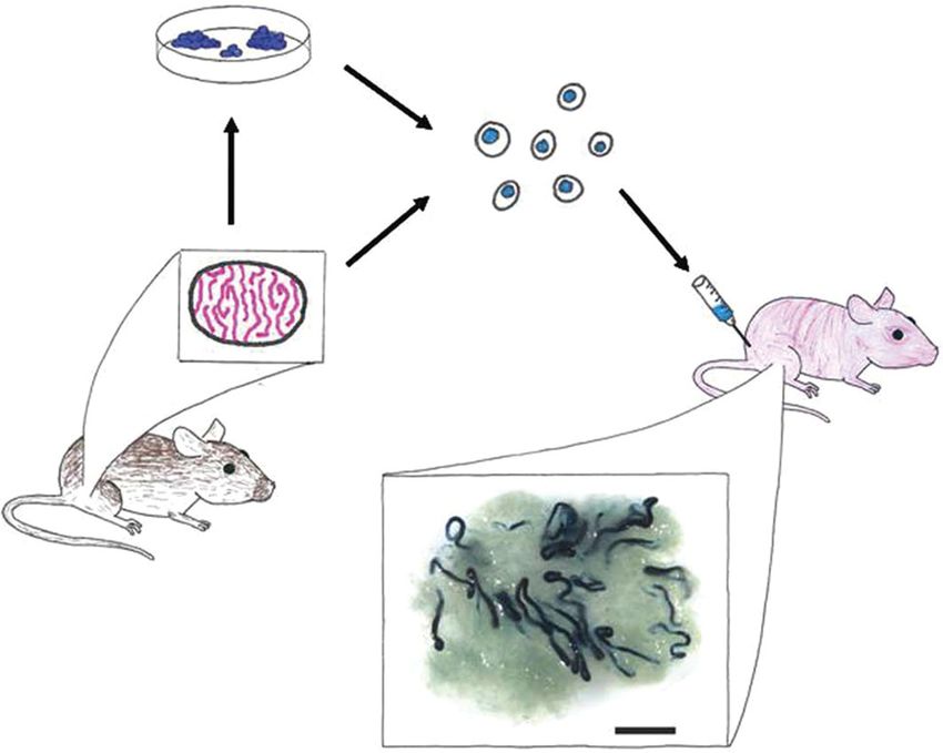
Downloaded from http://rstb.royalsocietypublishing.org/ on November 1, 2015

1664     B. T. Phillips et al.     Review. Spermatogonial stem cells

cytokinesis during mitotic divisions that lead to main-               division, see green clone in figure 1, stages IX, X
tenance of cytoplasmic bridges among germ cells.                      and XI) or (ii) remains connected by an intercellular
Proteins and messenger RNAs are exchanged via the                     cytoplasmic bridge and produces a chain of four Aal
cytoplasmic bridges and may help in coordinating                      spermatogonia at the next division (differentiating div-
the synchronized development of germ cell clones                      ision, see red clone in figure 1, stages IX and X).
(Braun et al. 1989). Each stage is characterized by a                 Further cell divisions lead to the formation of chains
combination of the types of spermatogonia, spermato-                  of 8, 16 and sometimes 32 Aal spermatogonia (see
cytes and spermatids that synchronously proceed                       red clone in stages XII and I and blue clone in stage
through the spermatogenic process (figure 1). For                     VII, figure 1). Chains of 4–16 Aal are generally con-
example, the basement membrane of a stage V semini-                   sidered committed to the differentiation process.
ferous tubule depicted in figure 1 is mostly filled with              Thus, the stem cell pool includes As and at least
preleptotene primary spermatocytes (blue cells of a                   some Apr spermatogonia. Some have argued that stem
large clone). By stage VI, these spermatocytes migrate                cell potential may extend to larger clones (e.g. Aal4 or
off the basement membrane and will be replaced by                     beyond; (Yoshida et al. 2007a; Morimoto et al.
spermatogonia. Thus, stage V can be distinguished                     2009)), but this is difficult to confirm experimentally.
from stage VI by the presence or absence of spermato-                 Note that while each clone can be observed in histo-
cytes on the basement membrane. The duration of                       logical sections as well as in whole-mount
each stage is precisely timed, and the complete sper-                 preparations of seminiferous tubules, clone size can
matogenic cycle was determined to be around 8.6                       only be observed in the whole-mount preparations
days in the mouse (Oakberg 1956b), and 12.8 days                      (figure 1).
in the rat (Hilscher et al. 1969). One complete cycle                    As, Apr and smaller chains of four Aal spermatogo-
(12 stages) of the mouse seminiferous epithelium is                   nia are evenly distributed along the seminiferous
depicted in figure 1.                                                 epithelium (Huckins 1971a,b; Tegelenbosch & de
                                                                      Rooij 1993). Larger chains of Aal (8, 16 and 32)
                                                                      become differentiating A1 spermatogonia between
4. SPERMATOGENIC LINEAGE DEVELOPMENT                                  stages IV and VIII of the seminiferous epithelium
In order to understand the regulation of spermato-                    (there is no cell division at this transition, see blue
gonial stem cells, it is important to understand them                 clone in figure 1, stages VII and VIII) and these give
in the context of the spermatogenic lineage that they                 rise to A2 spermatogonia at stage IX (see blue clone
produce. Spermatogonia are primitive diploid germ                     in figure 1, stage IX). Thus, in contrast to undifferen-
cells, located on the basement membrane of the semi-                  tiated spermatogonia, differentiating spermatogonia
niferous tubules. Three types of spermatogonia were                   (A1, A2, A3, A4, intermediate and B) divide in a syn-
initially described based on their nuclear morphology                 chronized manner and are found at specific stages of
(Roosen-Runge & Giesel 1950; Clermont & Leblond                       the seminiferous epithelium (for detailed description
1953; Monesi 1962). Type A spermatogonia were con-                    see Oakberg 1971). B spermatogonia give rise to
sidered the most primitive because heterochromatin is                 primary spermatocytes that progress into meiosis.
absent from the nucleus, a general characteristic of                  Two meiotic divisions lead to the formation of second-
undifferentiated cells. The nuclei of intermediate                    ary     spermatocytes     and     haploid    spermatids
type spermatogonia contain a small amount of                          respectively, which undergo 16 steps of morphological
heterochromatin and type B spermatogonia contain a                    changes to finally become spermatozoa ready to be
large amount of heterochromatin, indicating a more                    released from the seminiferous epithelium (Oakberg
differentiated state.                                                 1956a).
    Histological staining of whole-mount preparations                    An alternative to the As model of SSC self-renewal
of seminiferous tubules provided additional level of                  described above is the A0/A1 model (Clermont &
detail about spermatogonial morphometry compared                      Bustos-Obregon 1968; Dym & Clermont 1970;
with tissue sections alone and broadened the knowl-                   Clermont & Hermo 1975). This model is very similar
edge of the spermatogonial cell types in the testis.                  to the Adark and Apale model that has been used to
To facilitate the following discussion, figure 1 depicts              describe stem cell activity in non-human primates
one compete cycle of the mouse seminiferous epi-                      (Clermont & Leblond 1959; Clermont & Antar
thelium and represents whole-mount perspective as                     1973). Briefly, A0 spermatogonia were observed as
well as corresponding cross-section and longitudinal                  singles or pairs of cells that were present at all stage
section perspectives. Figure 1 traces the development                 of the seminiferous epithelium. Mitotic figures were
of three putative clones (green, red and blue) through                rarely observed in these cells, so they were considered
one cycle of the seminiferous epithelium. Based on                    ‘reserve stem cells’ not contributing to steady-state
whole-mount examination of seminiferous tubules,                      spermatogenesis. These reserve stem cells are only
Huckins & Oakberg (Huckins 1971c; Oakberg 1971)                       activated when spermatogenesis is destroyed by toxic
reported that undifferentiated type A spermatogonia                   insult (i.e. radiation). The ‘active stem cell’ pool is
can be subdivided into Asingle (As), Apaired (Apr) and                comprised of A1 – A4 spermatogonia. When A4 sper-
Aaligned (Aal) spermatogonia, which differ only in                    matogonia divide, they give rise either to new A1
their topographical arrangement on the seminiferous                   spermatogonia (self-renewal) or to intermediate sper-
tubule basement membrane. When an As (see green                       matogonia (differentiation). While there continues to
clone in figure 1, stage VII) spermatogonium divides,                 be vigorous debate about the merits of the As versus
it produces an Apr that either (i) completes cytokinesis              the A0/A1 models, the As model is currently favoured
to produce two new As spermatogonia (self-renewing                    by most investigators in the field and will be the basis
Phil. Trans. R. Soc. B (2010)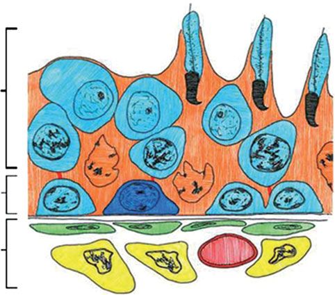
Downloaded from http://rstb.royalsocietypublishing.org/ on November 1, 2015
Review. Spermatogonial stem cells B. T. Phillips et al. 1665
cross-section whole-mount
VII VIII
IX
X
longitudinal section XI
XII
I II
III
VI IV
V
Figure 1. Mouse spermatogenic clone development by stage. The mouse spermatogenic cycle contains twelve stages (I– XII).
Each stage is temporally unique, and the stages in the diagram represent the relative time each stage lasts in the mouse. Each
stage in the diagram is shown in cross-sectional, longitudinal and whole-mount perspectives (labelled in stage VII). Three
putative spermatogonial clones are highlighted in blue, red and green. The dotted lines in the whole-mount perspective indi-
cate the planes of the cross section and longitudinal section views. For example, in stage VII, the red cell is in the vertical line
and therefore appears in the cross-sectional view. A green cell is in the horizontal line, so is observed in the longitudinal section
view. The development of three putative clones (blue, red and green) through one cycle of the seminiferous epithelium is
shown. Stage VII: Aal-16 (blue); Apair (red); Asingle (green); stage VIII: A1 (clone of 16) (blue); Apair (red); Asingle (green);
stage IX: A2 (clone of 32) (blue); Apair (red); Asingle (green); stage X: A2 (clone of 32) (blue); Aal-4 (red); Apair (green);
stage XI: A3 (clone of 64) (blue); Aal-4 (red); Asingle (x2) (green); stage XII: A3 (clone of 64) (blue); Aal-4 (red); Asingle
(x2) (green); stage I: A4 (clone of 128) (blue); Aal-8 (red); Asingle and Apair (green); stage II: intermediate spermatogonia
(clone of 256) (blue); Aal-8 (red); Asingle and Apair (green); stage III: intermediate spermatogonia (clone of 256) (blue);
Aal-8 (red); Asingle and Apair (green); stage IV: Type B Spermatagonia (clone of 512) (blue); Aal-8 (red); Asingle and Apair
(green); stage V: Type B Spermatagonia (clone of 512) (blue); Aal-8 (red); Asingle and Apair (green); stage VI: primary sperma-
tocytes (lifting off the basement membrane) (blue); Aal-8 (red); Asingle and Apair (green).
for further discussion of spermatogonial self-renewal 6. SPERMATOGONIAL STEM CELL
in this review. TRANSPLANTATION
A technique for transplanting SSCs was first described
by Brinster and colleagues in 1994 (Brinster & Avar-
5. EXPERIMENTAL TOOLS FOR STUDYING bock 1994; Brinster & Zimmermann 1994). Briefly,
SPERMATOGONIAL STEM CELLS germ cells are isolated from the testes of donor animals
As discussed above, experimental investigation of and transplanted into the testicular seminiferous
SSCs is complicated because these cells are rare and tubules of infertile recipients, where they produce
are difficult to distinguish from the differentiating pro- normal colonies of spermatogenesis and functional
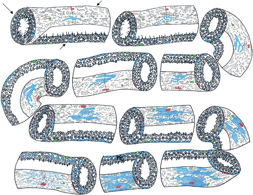
geny that they produce. Whole-mount analyses of sperm (figure 2). Infertility of recipients is because of
seminiferous tubules help in distinguishing As from genetic mutation (i.e. W mutant mice, (Ogawa et al.
Apr and Aal spermatogonia, but there is continuing 2000)) or induced experimentally (e.g. busulphan
debate about whether the stem cell pool is restricted treatment (Brinster & Zimmermann 1994)). In mice,
to As or might be expanded to include Apr and some these studies are facilitated by the availability of trans-
Aal (Nakagawa et al. 2007; Yoshida et al. 2007a). genic donors (e.g. lacZ and GFP) with germ cells that
Thus, the only way to definitively identify an SSC is can be readily identified in the testes of non-transgenic
by observing its biological capacity to produce and recipients. By definition, only a stem cell can produce
maintain spermatogenesis in a transplant paradigm. and maintain a colony of spermatogenesis and
Phil. Trans. R. Soc. B (2010)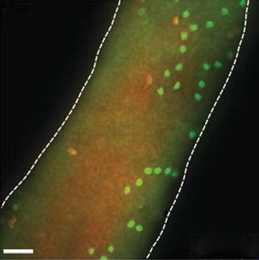
Downloaded from http://rstb.royalsocietypublishing.org/ on November 1, 2015

1666     B. T. Phillips et al.     Review. Spermatogonial stem cells

Figure 2. Spermatogonial stem cell (SSC) transplant assay. The functional analysis of SSCs is a retrospective assay of sperma-
togenic function. In this example, cells are isolated from a lacZ donor mouse testis and digested to produce a single cell
suspension. Cells can then be maintained in culture or injected into the testes of an infertile recipient mouse. Recipient
testes are typically analysed two to three months after transplantation for donor spermatogenesis (blue colonies in this
example). A typical recipient testis is shown with blue colonies of donor-derived spermatogenesis (scale bar, 2 mm).

each colony arises from the clonogenic proliferation                  that recognizes a cell surface antigen. Markerþ and
and differentiation of a single SSC (Dobrinski et al.                 marker2 cells are fractionated by FACS and each frac-
1999; Zhang et al. 2003; Kanatsu-Shinohara et al.                     tion is transplanted into the seminiferous tubules of
2006). Therefore, the SSC transplantation technique                   infertile recipient mice to determine the relative stem
provides a quantitative functional assay to characterize              cell activity. The first application of this approach for
stem cell activity in any donor cell population. SSC                  characterizing SSCs was reported by Shinohara and
transplantation remains the gold standard method                      co-workers, who demonstrated that SSCs specifically
for identifying SSCs, but this approach can be                        bind to laminin-coated plates. The laminin-binding
technically challenging. In addition, SSC transplan-                  cells were enriched for b1-integrin, making this sur-
tation is a retrospective assay with an inherent two                  face molecule a candidate for enriching SSCs
to three months timeframe between transplant and                      (Shinohara et al. 1999). Subsequent transplantation
analysis. To accelerate investigations of SSCs,                       of magnetic-activated cell-sorted (MACS) and
Nagano and co-workers recently suggested that the                     FACS-sorted testis fractions indicated that SSCs
SSC culture system (described below) may provide a                    express b1-integrin and a6-integrin, but are negative
shorter term, in vitro assay for SSCs (Yeh et al.                     for av-integrin and the c-KIT receptor tyrosine
2007). However, culture does not assess regenerative                  kinase (Shinohara et al. 1999, 2000). Based on several
activity.                                                             similar studies, mouse SSCs can now be described by
                                                                      the cell surface phenotype, a6-Integrin (CD49f)þ, b1-
                                                                      Integrin (CD29)þ, THY-1 (CD90)þ, CD9þ,
7. DISSECTING THE MOLECULAR PHENOTYPE                                 GFRa1þ, CDH1þ, av-Integrin (CD51)2, c-KIT
OF SPERMATOGONIAL STEM CELLS                                          (CD117)2, major histocompatibility complex class I
Fluorescence-activated cell sorting (FACS), combined                  (MHC-I)2, CD452 (Shinohara et al. 1999, 2000;
with SSC transplantation is a powerful tool that has                  Kubota et al. 2003; Kanatsu-Shinohara et al. 2004b;
enabled investigators to systematically characterize                  Buageaw et al. 2005; Fujita et al. 2005; Hofmann
cell surface molecules of SSCs. This experimental                     et al. 2005b; Lo et al. 2005; Tokuda et al. 2007,
approach is patterned after similar studies to charac-                table 1). Using combinations of positive and negative
terize and enrich haematopoietic stem cells                           markers, it is now possible to achieve significant
(Spangrude 1989; Smith et al. 1991; Osawa et al.                      enrichment (100- to 200-fold) of mouse SSCs
1996). Briefly, a heterogeneous testis cell suspension                (Shinohara et al. 2000; Kubota et al. 2003). However,
is stained with a fluorescent-conjugated antibody                     it should be noted that none of these markers are
Phil. Trans. R. Soc. B (2010)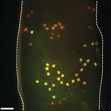
Downloaded from http://rstb.royalsocietypublishing.org/ on November 1, 2015
Review. Spermatogonial stem cells B. T. Phillips et al. 1667
Table 1. Germ cell markers in the rodent testis.
germ cell type
germ cell
markers in the experimental transplantable A1–
rodent testis evidence SSC?a As Apr Aal 4 In B Spc RS ES references
c-kit Mu, RT–PCR, no X X X X X X Manova et al.
ISH, IHC, Tr (1990), Yoshinaga
et al. (1991),
Schrans-Stassen
et al. (1999) and
Shinohara et al.
(2000)
GCNA1 WB, IHC not tested X X X X X X X X Enders & May
(1994)
VASA (MvH) ISH, WB, IHC, not tested X X X X X X X X Fujiwara et al.
KO (1994), Tanaka
et al. (2000) and
Toyooka et al.
(2000)
EE2 antigen WB, IHC, not tested X X X X X X X Koshimizu et al.
(1995)
DAZL RT–PCR, NB, not tested X X X X X X X Cooke et al. (1996),
ISH, KO, Niederberger et al.
IHC (1997) and Ruggiu
et al. (1997)
Stra8 RT–PCR, ISH, yes X X X X X X X X X Oulad-Abdelghani
IHC, WM, et al. (1996),
TG, MACS, Giuili et al. (2002)
Tr and Antonangeli
et al. (2009)
a6-integrin FC, MACS, Tr yes X X X X X X X Shinohara et al.
(CD49f) (1999, 2000)
b1-integrin FC, MACS, Tr yes X X X X X X Shinohara et al.
(CD29) (1999) and
Kanatsu-
Shinohara et al.
(2008)
Epcam IHC, MACS not tested X X X X X X Anderson et al.
(1999), van der
Wee et al. (2001)
and Tokuda et al.
(2007)
Pou5f1 (Oct4) IHC, WM, TG, yes X X X X Pesce et al. (1998),
FC, Tr, ISH Yoshimizu et al.
(1999), Ohbo
et al. (2003) and
Ohmura et al.
(2004)
GFR-a1 TG, ISH, IHC, yes X X X Meng et al. (2000),
MACS, TR, Dettin et al.
WM (2003), Buageaw
et al. (2005) and
Grisanti et al.
(2009)
CD24 FC not tested X X X Kubota et al. (2003)
Thy1 (CD90) FC, TR yes X X X Kubota et al. (2003)
Nanos2 ISH, KO, WB, yes X X Tsuda et al. (2003),
RT– PCR, Suzuki et al.
Tg, IHC, (2007, 2009) and
TR, WM Sada et al. (2009)
Nanos3 NB, ISH, KO, not tested X X X Tsuda et al. (2003),
WB, RT– Suzuki et al.
PCR, Tg, (2007, 2009) and
IHC, WM Lolicato et al.
(2008)
(Continued.)
Phil. Trans. R. Soc. B (2010)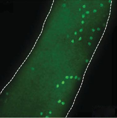
Downloaded from http://rstb.royalsocietypublishing.org/ on November 1, 2015

1668     B. T. Phillips et al.     Review. Spermatogonial stem cells

Table 1. (Continued.)

                                                            germ cell type
germ cell
markers in the      experimental        transplantable                     A1 –
rodent testis       evidence            SSC?a               As Apr     Aal 4         In B     Spc    RS ES    references

CD9                 FC, IHC,            yes                 X    X     X      X      X    X                   Kanatsu-Shinohara
                      MACS, Tr                                                                                  et al. (2004b)
EGR3                IVC, IHC            not tested          X    X                                            Hamra et al. (2004)
Ngn3                ISH, TG, WM,        yes (approx.        X    X     X                      X               Yoshida et al. (2004,
                      IHC, Tr             10% of                                                                2006) and Raverot
                                          SSCs)                                                                 et al. (2005)
PLZF                Mu, KO, Tr,         yes                 X    X     X                                      Buaas et al. (2004),
                     WM, FC,                                                                                    Costoya et al.
                     ISH, IHC                                                                                   (2004) and
                                                                                                                Grisanti et al.
                                                                                                                (2009)
RBM                 RT–PCR, IHC         not tested          X    X     X      X                               Jarvis et al. (2005)
Sox-3               KO, IHC             not tested          X    X     X                                      Raverot et al. (2005)
TAF4B               KO, IHC             not tested          X    X     X      X      X    X   X           X   Falender et al.
                                                                                                                (2005)
Bcl6b               siRNA               not tested          X    X     X                                      Oatley et al. (2006)
Numb                NB, WB, IHC         not tested          X    X     X      X      X    X   X               Corallini et al.
                                                                                                                (2006)
Lrp4                WB, IHC             not tested          X    X     X      X      X    X   X      X    X   Yamaguchi et al.
                                                                                                                (2006)
Ret                 IHC, MACS,          no                  X    X     X                                      Ebata et al. (2005)
                      Tr                                                                                        and Naughton
                                                                                                                et al. (2006)
Sohlh1              KO, RT–PCR,         not tested                     X      X      X    X   X               Ballow et al. (2006a)
                      IHC,
Sohlh2              RT–PCR, IHC         not tested          X    X     X                                      Ballow et al. (2006b)
CDH1                IHC, WM,            yes                 X    X     X                                      Tokuda et al. (2007)
  (CD324)             MACS, Tr
GPR125              TG, FC, IVC,        yes                 X    X     X                                      Seandel et al. (2007)
                      Tr
Nucleostemin        TG, IHC, FC,        yes                 X    X     X      X      X    X   X               Ohmura et al. (2008)
                      Tr, IVC,
                      siRNA
UTF1                RT–PCR, IHC         not tested          X    X     X                                      van Bragt et al.
                                                                                                                (2008)
Lin28 (Tex17)       IHC, WM,            not tested          X    X     X                                      Zheng et al. (2009)
                      WB, siRNA
As, A single spermatogonia; Apr, A paired spermatogonia; Aal, A aligned spermatogonia; A1–4, differentiating type A1 to A4
spermatogonia; In, intermediate type spermatogonia; B, type B spermatogonia; Spc, spermatocytes; RS, round spermatids; ES, elongated
spermatids; Mu, mutant mouse; TG, transgenic mouse; KO, Knockout mouse; Tr, germ cell transplantation; IHC,
immunohistochemistry; WM, whole mount immunostaining; FC, flow cytometry (including FACS); IVC, in vitro culture; WB, Western
blot; NB, Northern blot; ISH, in situ hybridization; RT–PCR, reverse transcriptase– PCR; siRNA, in vitro knockdown experiment using
siRNA; MACS, magnetic-activated cell sorting.
a
  As determined by the spermatogonial stem cell transplantation assay.

exclusive to SSCs and no marker or combination of                     faithful expression of lacZ and GFP transgenes
markers has produced a pure population of SSCs.                       (Yeom et al. 1996; Yoshimizu et al. 1999). The
Also, while FACS and MACS sorting followed by                         Oct-4 – GFP mouse is a valuable tool that enabled
transplantation are powerful tools for characterizing                 FACS-based isolation and transplantation of Oct4
the cell surface phenotype of SSCs, this approach                     expressing germ cells from a heterogeneous testis cell
has limited utility for characterizing cytoplasmic or                 suspension (Ohbo et al. 2003; Ohmura et al. 2004).
nuclear markers.                                                      Stem cell activity was significantly enriched in the
   Genetic mouse models in which GFP expression is                    Oct4 expressing (GFPþ) population compared with
driven by a promoter from a putative SSC gene pro-                    the Oct4 negative (GFP2) population of mouse
vide an alternative approach for characterizing SSCs.                 testis cells (Ohmura et al. 2004). Interestingly, gono-
For example, Schöler and co-workers reported that                    cytes and pre-spermatogonia from neonatal mice
the OCT-4 transcription factor is expressed by gono-                  with an Oct4 –EGFPþ/c-Kit2 phenotype had a greater
cytes and type A spermatogonia of newborn, pup                        repopulation capacity than Oct4 – EGFPþ/c-Kitþ cell
and adult mouse testes (Pesce et al. 1998). This                      fractions (Ohbo et al. 2003). These data suggest that
group subsequently characterized an 18 kb promoter/                   there is molecular heterogeneity among pre-spermato-
enhancer fragment of the Oct-4 gene that directed                     gonia. This observation is consistent with reports

Phil. Trans. R. Soc. B (2010)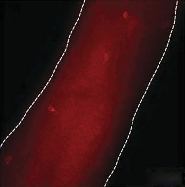
Downloaded from http://rstb.royalsocietypublishing.org/ on November 1, 2015

                                                              Review. Spermatogonial stem cells           B. T. Phillips et al.   1669

suggesting that some gonocytes/pre-spermatogonia                      male germ lineage. In this context, a candidate SSC
establish the initial pool of SSCs, while other gono-                 marker would be expressed by cells located on the
cytes/pre-spermatogonia differentiate to produce the                  basement membrane of seminiferous tubules and be
first round of spermatogenesis (Kluin & de Rooij                      co-expressed with confirmed markers of SSCs. This
1981; Yoshida et al. 2006).                                           histochemical approach is most convincing in whole-
    Transgenic and conditional knock-in approaches                    mount preparations of seminiferous tubules in which
were recently used to demonstrate that neurogenin 3                   it is possible to correlate marker expression with
(Ngn3) is expressed by the earliest spermatogonia                     clone size (i.e. As, Apr, Aal). Several established mar-
(Yoshida et al. 2004), including at least 11 per cent                 kers of stem, progenitor and differentiating
of transplantable SSCs (Nakagawa et al. 2007). The                    spermatogonia are listed in figure 3. Here we define
fact that Ngn3 was not expressed by all transplantable                progenitors as undifferentiated spermatogonia that
stem cells in that study provides additional evidence                 are committed to differentiate. An example of this
that there may be heterogeneity among SSCs. A con-                    approach is shown in figure 4 for the putative SSC
ditional knock-in approach was also used to                           marker, Spalt-like 4 (SALL4). SALL4 is a zinc finger
demonstrate that Nanos2 is expressed by SSCs (Sada                    transcription factor that is expressed in the inner cell
et al. 2009). Finally, transgenic models suggest that                 mass of the late blastocyst in a pattern similar to
Stra-8 (stimulated by retinoic acid-8) is expressed by                OCT4 and SOX2 (Elling et al. 2006). In vitro,
undifferentiated spermatogonia, including SSCs                        SALL4 stimulates embryonic stem (ES) cell prolifer-
(Giuili et al. 2002; Guan et al. 2006; Sadate-Ngatchou                ation (Sakaki-Yumoto et al. 2006) and maintains
et al. 2008), although the transplant data in the Stra-8              pluripotency by repressing trophectoderm differen-
studies were limited.                                                 tiation (Yuri et al. 2009), possibly by binding the
    Knock-out models have also been used to demon-                    Oct-4 proximal promoter (Zhang et al. 2006) and by
strate that specific genes/proteins are required for                  interacting with NANOG (Wu et al. 2006). Thus,
SSC function. Male mice carrying the luxoid (lu)                      SALL4 is an important stemness factor and together
mutation are subfertile and show abnormal sperm                       with OCT-4, SOX2 and NANOG constitutes a tightly
development. Progression of infertility is caused by                  regulated transcription circuit important for stem cell
gradual loss of SSCs (Buaas et al. 2004). The mutation                pluripotency (Lim et al. 2008; Yang et al. 2008). Post-
was shown to affect the Zfp145 locus, which encodes                   natally, Sall4 expression is restricted to the gonads and
the transcriptional repressor PLZF (promyelocytic                     is expressed by isolated spermatogonia (Wang et al.
leukaemia zinc-finger). PLZF is expressed during                      2001; Sall4 was identified as testis-expressed gene 20
embryogenesis and plays a crucial role during limb                    (Tex20) in that paper). Co-stained whole-mount semi-
and axial skeletal patterning. Targeted disruption of                 niferous tubules (figure 4) indicated that SALL4 is
Zfp145 resulted in a testicular phenotype similar to                  expressed by single, paired and aligned cells on the
that of luxoid mutant mice (Costoya et al. 2004). In                  seminiferous tubule basement membrane and overlaps
the testis, PLZF expression is restricted to As, Apr                  with consensus SSC markers, PLZF (figure 4a – c)
and Aal undifferentiated spermatogonia, including                     and GFRa1 (figure 4d – f ). However, these whole-
SSCs as demonstrated by transplantation experiments                   mount immunohistochemistry results highlight the
of testicular cells from luxoid or PLZF 2/2 mice that                 heterogeneity among undifferentiated spermatogonia,
failed to initiate donor-derived spermatogenesis in reci-             including As spermatogonia (see figure 4f with
pient mice (Buaas et al. 2004; Costoya et al. 2004). A                examples of SALL4þ/GFRa12 and SALL4þ/
possible role for PLZF in spermatogonia could be the                  GFRa1þ As spermatogonia). GFRa1 appears to have
maintenance of an undifferentiated state (Filipponi                   the most restricted expression (limited to singles,
et al. 2007), similar to the role suggested for Plzf in               pairs and chains of four), while PLZF and SALL4
haematopoietic precursor cells (Reid et al. 1995).                    are also expressed by larger chains of 8 and 16 Aal
    Similar knock-out and over-expression studies                     spermatogonia.
implicate glial cell line-derived neurotrophic factor                    Similar observations of molecular heterogeneity
(GDNF) and its receptor GFRa1 in stem cell self-                      among undifferentiated As, Apr and Aal spermatogonia
renewal (Meng et al. 2000). GDNF signalling has                       have been reported in several recent studies (Tokuda
since been shown to be required for in vitro expansion                et al. 2007; Grisanti et al. 2009; Sada et al. 2009;
of SSCs, and it has been demonstrated that a combi-                   Suzuki et al. 2009; Zheng et al. 2009). The functional
nation of GDNF and soluble GFRa1 is most                              significance of this heterogeneity remains to be deter-
favourable for the self-renewal of SSCs in vitro                      mined. Through the combination of FACS and
(Kubota et al. 2004a,b; see below). Finally, knock-out                MACS analyses, transplantation, genetic models and
studies implicate Sox3 in the differentiation of the ear-             histochemical approaches, the phenotype of rodent
liest germ cells (Raverot et al. 2005). The latter study              SSCs is beginning to emerge. A list of putative SSC
indicated that Ngn3 expression is dependent on SOX3                   and undifferentiated spermatogonia markers is pro-
and suggested that SOX3 may act directly or indirectly                vided in table 1 along with the experimental evidence
through Ngn3 to regulate spermatogonial differentiation               used to characterize each marker.
(Raverot et al. 2005).
    In addition to data derived from flow cytometry,
genetic models and transplantation, immunohisto-                      8. THE SPERMATOGONIAL STEM CELL NICHE
chemistry in tissue sections or intact seminiferous                   SSCs reside within a specialized microenvironment
tubules (whole mount) has been widely used to inves-                  called ‘niche’ that regulates testicular homeostasis by
tigate the expression patterns of various proteins in the             balancing SSC self-renewal and differentiation. A
Phil. Trans. R. Soc. B (2010)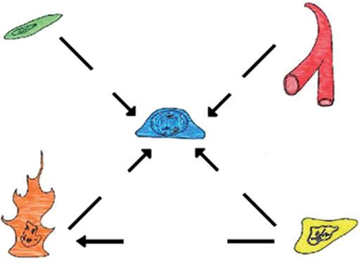
Downloaded from http://rstb.royalsocietypublishing.org/ on November 1, 2015

1670     B. T. Phillips et al.      Review. Spermatogonial stem cells

                                                                                                                 stem cell
                                    stem        As                                                        Oct4 +          Sohlh2 +
                                                                                                          Gfrα1 +         Cdh1 +
                                                                                                          Ngn3 +/–        Utf1 +
                                                                                                          Plzf +          Lin28 +
                                                Apair                                                      Bcl6b +        Sall4 +

      undifferentiated
                                                                                                                 progenitor
                                                                                                           Oct4 +            Sohlh2 +
                                                Aaligned(4)                                                Gfrα1 +           Cdh1 +
                                                                                                           Ngn3 +            Utf1 +
                                                                                                           Plzf +            Lin28 +
                                                                                                           Bcl6b +           Sall4 +
                                progenitor      Aaligned(16)                                               Sohlh1 +

       differentiating                                                                                         differentiating
                                                A1
                                                                                                               spermatogonia
                                                                                                           cKit +         Sohlh1 +
                                                                                                           Ngn3 +

Figure 3. Genes expressed by stem, progenitor and differentiating spermatogonia. The As, seen at the top of the diagram, is
responsible for self renewal and differentiation. Self-renewal is represented here by the Apair dividing to form two As. Differ-
entiation is indicated by colour change (from dark to light) and the lengthening chain of germ cells. Genes are listed with their
expression at the given stages of spermatogonial development. While stem cell activity is considered to reside in the pool of As
spermatogonia, the tapered triangle on the left indicates that stem cell activity may extend to Apr and some Aal spermatogonia.

stem cell niche is comprised of cells, extracellular                   (Chiarini-Garcia et al. 2003). Undifferentiated sper-
matrix components, and local soluble factors present                   matogonia are observed predominantly in tubule
in the vicinity of the stem cell that regulates cell fate.             areas adjacent to vasculature (Yoshida et al. 2007b;
The structural basis for the SSC niche in the                          figure 5a).
mammalian testis is the basal compartment of the                          The SSC niche mediates endocrine and paracrine
seminiferous tubules that is composed of Sertoli cells                 signals that regulate self-renewal and differentiation
and peritubular myoid cells (Dadoune 2007)                             (figure 5b). A key regulator of the SSC niche is
(figure 5). Together, Sertoli and peritubular myoid                    GDNF which is secreted by Sertoli cells and acts
cells secrete the basement membrane components to                      through Ret receptor tyrosine kinase and GFRa1
which the SSCs are connected via adhesion molecules                    co-receptor, which form a receptor complex on the sur-
(Tung et al. 1984). Sertoli cells are polarized columnar               face of As, APr and Aal (Meng et al. 2000). Downstream
epithelial cells that support SSCs and differentiating                 signalling pathways that are activated by GDNF in
germ cells by providing nutrients and mediating exter-                 undifferentiated spermatogonia are the PI3K/Akt path-
nal signals in order to support spermatogenesis                        way, members of the Src kinase family and the Ras/
(Griswold 1998). The importance of Sertoli cells for                   Erk1/2 pathway (Braydich-Stolle et al. 2007; Oatley
germ cell differentiation is demonstrated by the trans-                et al. 2007; He et al. 2008). GDNF is thought to act
plantation of normal Sertoli cells into the testis of                  through these pathways to regulate SSC self-renewal.
infertile mutant recipients with a Sertoli cell defect                    Targeted disruption of the Ets variant gene 5
and successful initiation of spermatogenesis by recipi-                (Etv5) results in defective maintenance of the SSC
ent-derived spermatogonia (Kanatsu-Shinohara et al.                    pool, whereas spermatogonial differentiation appears
2003b, 2005b). Tight junctions between adjacent                        to be unaffected by this mutation (Chen et al.
Sertoli cells constitute a protective blood –testis barrier            2005). The transcription factor Etv5 is expressed in
(BTB) that divides the seminiferous epithelium into                    Sertoli cells and loss of Etv5 appears to impair the
basal and adluminal compartments (figure 5a) and                       ability of Sertoli cells to support spermatogonia,
plays an important role in the regulation of germ cell                 possibly by disrupted BTB function as indicated by
differentiation (Cheng & Mruk 2002). The BTB                           decreased Claudin-5 (CLDN5) levels in mutant
maintains a selective substance flow between luminal                   mice (Morrow et al. 2009). In Sertoli cells, Etv5 is
fluid, blood plasma and interstitial fluid, thereby creat-             upregulated by FGF2 in vitro, which is important
ing an immune-privileged environment for haploid                       for SSC renewal in culture (Kanatsu-Shinohara et al.
germ cells in the adluminal compartment of the                         2003a; Kubota et al. 2004a; Yoon et al. 2009). There-
seminiferous tubules.                                                  fore, in addition to a direct effect of FGF2 on SSCs,
   Along the length of the tubule, SSCs are thought to                 an indirect paracrine effect of FGF2 on Sertoli cells
be localized in areas adjacent to the interstitial space               appears possible.

Phil. Trans. R. Soc. B (2010)
Downloaded from http://rstb.royalsocietypublishing.org/ on November 1, 2015
Review. Spermatogonial stem cells B. T. Phillips et al. 1671
(a) (b) (c)
Apr Apr
As As
As
Aal Aal Aal
Sall4 PLZF merge
(d ) (e) (f)
Aal Aal
As As
Aal
As As
Apr Apr
Apr
Apr
Sall4 Gfrα1 merge
Figure 4. Immunofluorescent co-staining of adult mouse whole-mount seminiferous tubules. (a) SALL4 labels undifferen-
tiated As, Apr and Aal spermatogonia. (b) PLZF labels undifferentiated As, Apr and Aal spermatogonia. (c) Merged picture
from (a,b). SALL4 and PLZF are mostly co-expressed in undifferentiated spermatogonia. (d–f ) Co-staining of SALL4 and
GFRa1 reveals heterogeneity within the population of undifferentiated spermatogonia. GFRa1 expression appears more
restricted than SALL4 or PLZF. Scale bar, 50 mm.
The importance of peritubular myoid cells for sper- testis cell populations (FACS or MACS sorting and/
matogonia maintenance has long been discussed. New or differential attachment and replating) resulted in
data now suggest a role for the peritubular cell product the enrichment of SSCs and the removal of somatic
colony-stimulating factor 1 (CSF1) on SSC mainten- cells that promote germ cell differentiation. Second,
ance (Oatley et al. 2009). Csf1 was found to be development of a serum-free, defined medium facili-
expressed in interstitial Leydig and peritubular myoid tated the discovery of essential growth factors.
cells, whereas the Csf1 receptor (Csf1r) was highly Specifically, GDNF is necessary to maintain and
enriched in THY1þ cell fractions from pre-pubertal expand rodent SSCs in culture (Kanatsu-Shinohara
and adult mouse testis. et al. 2003a; Kubota et al. 2004b). The trophic effects
of GDNF in both mice and rats is enhanced by the
addition of soluble GFRa1 (the receptor for GDNF)
9. SSC CULTURE and FGF2 (Kubota et al. 2004a; Ryu et al. 2005).
SSC culture provides a new approach for investigating Unlike mouse ES cells, the additions of leukaemia
the molecular mechanisms and cell-signalling path- inhibitory factor (LIF) and foetal bovine serum
ways that regulate SSC function. While methods for (FBS) to cultures are superfluous and detrimental,
maintaining and amplifying pluripotent ES and respectively, in SSC cultures (Kubota et al. 2004b).
embryonic germ cells in culture are routine, methods Third, STO or mouse embryonic fibroblast (MEF)
for culturing adult tissue stem cells (including SSCs) feeder cells are usually required. Whereas Shinohara’s
had been more difficult to establish. However, tremen- group has demonstrated that mouse SSCs can also
dous progress culturing mouse and rat SSCs has been be maintained in feeder-free conditions (Kanatsu-
reported during the past 5 – 6 years (Kanatsu- Shinohara et al. 2005a). SSC cultures are usually estab-
Shinohara et al. 2003a; Kubota et al. 2004a,b; lished from mouse pup testes (5–12 days postpartum)
Hamra et al. 2005; Ryu et al. 2005). Rodent SSCs because SSCs are enriched at this stage of develop-
can now be maintained for a very long time (perhaps ment. However, SSC cultures can be established from
indefinitely) with a significant amplification in neonate (Kanatsu-Shinohara et al. 2003a) and adult
numbers. Stem cell activity in these cultures was con- mouse testis cells (Kubota et al. 2004a). Immortalized
firmed by SSC transplantation, as diagrammatically SSC lines have been established by the introduction of
represented in figure 2. The doubling time for mouse a retroviral telomerase gene (Feng et al. 2002) or treat-
SSCs was determined to be 5.6 days (Kubota et al. ment with the SV40 large T-antigen (Hofmann et al.
2004b), while the doubling time for rat SSCs is 3– 4 2005a). Evidence that each of these immortalized cell
days (Hamra et al. 2005) or 11 days (Ryu et al. 2005). lines is spermatogonial-like is based primarily on gen-
Several factors were critical to the establishment of etic or immunocytochemical data, but transplantation
long-term SSC cultures. First, methods to fractionate data are lacking.
Phil. Trans. R. Soc. B (2010)Downloaded from http://rstb.royalsocietypublishing.org/ on November 1, 2015
1672 B. T. Phillips et al. Review. Spermatogonial stem cells
(a) containing on Oct-4-targeted shRNA. The treatment
caused a significant reduction in OCT-4 expression
and reduced colonizing activity in the transplant
assay by sixfold. Thus, through genetic manipulation
adluminal and transplantation of SSC cultures, studies will con-
tinue to unravel regulatory pathways required for
SSC self-renewal and differentiation.
basal
10. FUTURE DIRECTIONS
We have attempted to review the current state of
interstitial knowledge and research in the biology of SSCs,
focused primarily on the rodent model. Many areas
of research are only beginning to be thoroughly inves-
tigated in SSCs, such as the molecular regulation of
(b) myoid cell blood vessel stem cell fate decisions and SSC heterogeneity.
Recent progress characterizing, manipulating and cul-
turing SSCs has opened the door to new experimental
approaches for fundamental investigation and possible
CSF-1 SSC ? practical applications discussed below.
In vitro derivation of haploid gametes (elongated
spermatids or sperm) may help to overcome spermato-
genic barriers in infertile men. Feng et al. (2002)
GDNF testosterone (?) reported the production of spermatocytes and sperma-
tids from a stable mouse SSC line, though the
fertilization potential of these cells was not tested.
testosterone
Haploid male germ cells have also been generated by
Sertoli cell Leydig cell differentiation of ES cells in mice (Toyooka et al.
2003; Geijsen et al. 2004; Nayernia et al. 2006) and
Figure 5. SSC niche. The SSC (dark blue) is diagrammed in humans (Kee et al. 2009). Two studies demonstrated
its physical niche (a) surrounded by Sertoli cells (orange) that in vitro, ESC-derived spermatids were competent
and differentiating germ cells (light blue) within the semini- to fertilize mouse eggs, generating blastocysts (Geijsen
ferous tubule. Niche components outside the tubule itself
2004) and live progeny (Nayernia et al. 2006), respect-
include myoid cells (green), blood vessels (red) and Leydig
cells (yellow). The components of the niche and the some ively. However, because of the epigenetic
factors known to be provided by each are shown in (b). reprogramming that occurs during in vivo germ cell
While some factors are known to act directly on the SSC, development, the epigenetic regulation of in vitro
such as GDNF, others, like testosterone are important for gametogenesis must be carefully assessed before clini-
spermatogenesis but may not act on the SSC. cal applications ensue (Georgiou et al. 2007).
Generation of haploid germ cells from primary SSC
cultures has not yet been reported, but this approach
Stable SSC culture provides a valuable tool for dis- may have epigenetic advantages over ESC-derived
secting mechanisms that regulate SSC renewal and gametes. Furthermore, progress establishing human
differentiation. GDNF is required for SSC renewal SSC cultures will be an important experimental tool
in vitro (Kubota et al. 2004a) and in vivo (Meng in a species where transplantation is not an option
et al. 2000). Through withdrawal and/or addition of for characterizing SSCs.
GDNF to SSC cultures, two groups have now demon- In addition to trying to drive SSCs towards their
strated that GDNF action is mediated by Src family typical biological end, there is evidence that SSCs
kinases acting through PI3 kinase/Akt-dependent are a source of pluripotent stem cells (Kanatsu-
pathways (Braydich-Stolle et al. 2007; Oatley et al. Shinohara et al. 2004a; Guan et al. 2006; Seandel
2007). In addition, microarray analysis identified et al. 2007). The ability to derive pluripotent stem
genes that are regulated by GDNF withdrawal in cells from adult tissues, with the consent of the
SSC cultures. The importance of three of these donor, may have some advantages over other
genes (Bcl6b, Erm and Lhx1) was confirmed by trans- approaches to pluripotency. However, more details
fecting SSC cultures with siRNAs specific for each are needed to understanding the genetic, epigenetic
gene. siRNA treatment caused decreased clump for- constitution of these cells, as well as their developmen-
mation in vitro and decreased colonization of tal potential.
recipient testes after transplantation (Oatley et al. Testicular tissues or testicular cell suspensions (con-
2006, 2007). taining SSCs) can be cryopreserved and may provide
Transfection of SSC cultures with siRNA, as an avenue for preservation of valuable strains or
described above, enables temporary knockdown of species. Honaramooz et al. (2002) recently demon-
the target gene. To achieve stable knockdown of a strated that testicular tissues from newborn mice,
target gene, short hairpin RNAs (shRNAs) can be pigs or goats can be grafted under the skin of
coupled with lentiviral vectors. Dann et al. (2008) immune-deficient mice and generate complete sper-
recently treated cultured SSCs with a lentiviral vector matogenesis. This approach has now been reported
Phil. Trans. R. Soc. B (2010)Downloaded from http://rstb.royalsocietypublishing.org/ on November 1, 2015
Review. Spermatogonial stem cells B. T. Phillips et al. 1673
for several species (Oatley et al. 2004, 2005; Snedaker REFERENCES
et al. 2004; Zeng et al. 2006; Kim et al. 2007; Arregui Anderson, R., Schaible, K., Heasman, J. & Wylie, C. 1999
et al. 2008; Rodriguez-Sosa et al. 2010) and may allow Expression of the homophilic adhesion molecule, Ep-
germline preservation for endangered species or valu- CAM, in the mammalian germ line. J. Reprod. Fertil.
116, 379–384.
able domestic strains. Alternatively, valuable
Antonangeli, F., Giampietri, C., Petrungaro, S., Filippini, A.
germlines can be preserved by freezing testis cell sus- & Ziparo, E. 2009 Expression profile of a 400-bp Stra8
pensions (containing SSCs) for future SSC promoter region during spermatogenesis. Microsc. Res.
transplantation. The proof in principle for this Tech. 72, 816 –822.
approach is already established for mice, rats, goats Arregui, L., Rathi, R., Megee, S. O., Honaramooz, A.,
and dogs (Brinster & Avarbock 1994; Brinster et al. Gomendio, M., Roldan, E. R. & Dobrinski, I. 2008
2003; Honaramooz et al. 2003; Ryu et al. 2003; Kim Xenografting of sheep testis tissue and isolated cells as a
et al. 2008). model for preservation of genetic material from endan-
SSC transplantation may have application for gered ungulates. Reproduction 136, 85–93. (doi:10.1530/
treating some cases of male infertility. For example, REP-07-0433)
high-dose chemotherapy and total body radiation Ballow, D., Meistrich, M. L., Matzuk, M. & Rajkovic, A.
2006a Sohlh1 is essential for spermatogonial differen-
treatment of cancer can cause permanent infertility.
tiation. Dev. Biol. 294, 161– 167. (doi:10.1016/j.ydbio.
While adult men can cryopreserve a semen sample 2006.02.027)
prior to their oncologic treatment, this is not an Ballow, D. J., Xin, Y., Choi, Y., Pangas, S. A. & Rajkovic, A.
option for pre-adolescent boys who are not yet 2006b Sohlh2 is a germ cell-specific bHLH transcription
making sperm. Using methods similar to those already factor. Gene Expr. Patterns 6, 1014 –1018. (doi:10.1016/j.
established for other species, it may be possible for modgep.2006.04.007)
these young cancer patients to cryopreserve testis Bendel-Stenzel, M., Anderson, R., Heasman, J. & Wylie, C.
cells or tissue prior to cancer treatment and use 1998 The origin and migration of primordial germ cells in
those tissues to achieve fertility after they are cured the mouse. Semin. Cell Dev. Biol. 9, 393 –400.
(Orwig & Schlatt 2005; Goossens et al. 2008; Braun, R. E., Behringer, R. R., Peschon, J. J., Brinster, R. L. &
Hermann et al. 2009). We have recently established a Palmiter, R. D. 1989 Genetically haploid spermatids are
phenotypically diploid. Nature 337, 373–376. (doi:10.
non-human primate model of cancer survivorship to
1038/337373a0)
test the safety and feasibility of SSC transplantation Braydich-Stolle, L., Kostereva, N., Dym, M. & Hofmann,
in a species that is relevant to human physiology M. C. 2007 Role of Src family kinases and N-Myc in
(Hermann et al. 2007). Although SSC transplantation spermatogonial stem cell proliferation. Dev. Biol. 304,
is not yet ready for the human fertility clinic, it may be 34–45.
reasonable for young cancer patients, with no other Brinster, R. L. & Avarbock, M. R. 1994 Germline trans-
options to preserve their fertility, to cryopreserve mission of donor haplotype following spermatogonial
testicular cells (Schlatt et al. 2009). Ginsberg and co- transplantation. Proc. Natl Acad. Sci. USA 91, 11 303–
workers have been cryopreserving testicular tissue for 11 307. (doi:10.1073/pnas.91.24.11303)
young cancer patients since 2008 and report that Brinster, R. L. & Zimmermann, J. W. 1994 Spermatogenesis
following male germ-cell transplantation. Proc. Natl Acad.
this intervention is acceptable to parents and that
Sci. USA 91, 11 298– 11 302. (doi:10.1073/pnas.91.24.
testicular biopsies caused no acute adverse effects 11298)
(Ginsberg et al. 2010). A human SSC culture system Brinster, C. J., Ryu, B. Y., Avarbock, M. R., Karagenc, L.,
would be particularly useful in this setting because a Brinster, R. L. & Orwig, K. E. 2003 Restoration of ferti-
few SSCs could be obtained in a small biopsy and lity by germ cell transplantation requires effective
expanded to a number sufficient for transplant recipient preparation. Biol. Reprod. 69, 412– 420.
therapy. (doi:10.1095/biolreprod.103.016519)
Progress studying SSC origins, regulation and Buaas, F. W., Kirsh, A. L., Sharma, M., McLean, D. J.,
activity over the past half century, has laid the foun- Morris, J. L., Griswold, M. D., de Rooij, D. G. &
dation to pursue the clinical and veterinary options Braun, R. E. 2004 Plzf is required in adult male germ
described in the preceding paragraphs. The field of cells for stem cell self-renewal. Nat. Genet. 36, 647–
652. (doi:10.1038/ng1366)
SSC biology has grown substantially in the past two Buageaw, A., Sukhwani, M., Ben-Yehudah, A., Ehmcke, J.,
decades, fuelled in part by development of the SSC Rawe, V. Y., Pholpramool, C., Orwig, K. E. & Schlatt, S.
transplantation technique (Brinster & Avarbock 2005 GDNF family receptor alpha1 phenotype of sperma-
1994; Brinster & Zimmermann 1994), which togonial stem cells in immature mouse testes. Biol. Reprod.
impacted fundamental investigations as well as clinical 73, 1011–1016. (doi:10.1095/biolreprod.105.043810)
application. Growth was also fuelled by the explosive Chen, C. et al. 2005 ERM is required for transcriptional
development of the pluripotent stem cell and regenera- control of the spermatogonial stem cell niche. Nature
tive medicine fields. The next half century should 436, 1030–1034. (doi:10.1038/nature03894)
bring many new discoveries about the biology and Cheng, C. Y. & Mruk, D. D. 2002 Cell junction dynamics
regenerative potential of SSCs that parallels the devel- in the testis: sertoli–germ cell interactions and male
contraceptive development. Physiol. Rev. 82, 825 –874.
opment of the haematopoietic stem cell field in the
Chiarini-Garcia, H., Raymer, A. M. & Russell, L. D. 2003
1980s and 1990s. Non-random distribution of spermatogonia in rats: evi-
The authors would like to thank Dr Brian Hermann for dence of niches in the seminiferous tubules.
critically reviewing the chapter. B.T.P. produced the Reproduction 126, 669 –680. (doi:10.1530/rep.0.1260669)
artwork in figures 1, 2, 3 and 5. K.E.O. is supported by Clermont, Y. 1972 Kinetics of spermatogenesis in mammals:
NIH grants HD055475 and HD008610 and the Magee- seminiferous epithelium cycle and spermatogonial
Womens Research Institute and Foundation. renewal. Physiol. Rev. 52, 198–236.
Phil. Trans. R. Soc. B (2010)Downloaded from http://rstb.royalsocietypublishing.org/ on November 1, 2015 1674 B. T. Phillips et al. Review. Spermatogonial stem cells Clermont, Y. & Antar, M. 1973 Duration of the cycle of the female mice. Dev. Biol. 163, 331–340. (doi:10.1006/ seminiferous epithelium and the spermatogonial renewal dbio.1994.1152) in the monkey, Macaca arctoides. Am. J. Anat. 136, Falender, A. E., Freiman, R. N., Geles, K. G., Lo, K. C., 153 –165. (doi:10.1002/aja.1001360204) Hwang, K., Lamb, D. J., Morris, P. L., Tjian, R. & Clermont, Y. & Bustos-Obregon, E. 1968 Re-examination Richards, J. S. 2005 Maintenance of spermatogenesis of spermatogonial renewal in the rat by means of semi- requires TAF4b, a gonad-specific subunit of TFIID. niferous tubules mounted ‘in toto’. Am. J. Anat. 122, Genes Dev. 19, 794 –803. (doi:10.1101/gad.1290105) 237 –247. (doi:10.1002/aja.1001220205) Feng, L. X., Chen, Y., Dettin, L., Pera, R. A., Herr, J. C., Clermont, Y. & Hermo, L. 1975 Spermatogonial stem cells Goldberg, E. & Dym, M. 2002 Generation and in vitro in the albino rat. Am. J. Anat. 142, 159– 175. (doi:10. differentiation of a spermatogonial cell line. Science 297, 1002/aja.1001420203) 392 –395. (doi:10.1126/science.1073162) Clermont, Y. & Leblond, C. P. 1953 Renewal of spermatogo- Filipponi, D., Hobbs, R. M., Ottolenghi, S., Rossi, P., Jan- nia in the rat. Am. J. Anat. 93, 475–501. (doi:10.1002/ nini, E. A., Pandolfi, P. P. & Dolci, S. 2007 Repression aja.1000930308) of kit expression by Plzf in germ cells. Mol. Cell. Biol. Clermont, Y. & Leblond, C. P. 1959 Differentiation and 27, 6770–6781. (doi:10.1128/MCB.00479-07) renewal of spermatogonia in the monkey, Macacus Fujita, K., Ohta, H., Tsujimura, A., Takao, T., Miyagawa, rhesus. Am. J. Anat. 104, 237 –273. (doi:10.1002/aja. Y., Takada, S., Matsumiya, K., Wakayama, T. & 1001040204) Okuyama, A. 2005 Transplantation of spermatogonial Clermont, Y. & Perey, B. 1957 Quantitative study of the cell stem cells isolated from leukemic mice restores fertility population of the seminiferous tubules in immature rats. without inducing leukemia. J. Clin. Invest. 115, 1855– Am. J. Anat. 100, 241 –267. (doi:10.1002/aja. 1861. (doi:10.1172/JCI24189) 1001000205) Fujiwara, Y., Komiya, T., Kawabata, H., Sato, M., Fujimoto, Cooke, H. J., Lee, M., Kerr, S. & Ruggiu, M. 1996 A murine H., Furusawa, M. & Noce, T. 1994 Isolation of a DEAD- homologue of the human DAZ gene is autosomal and family protein gene that encodes a murine homolog of expressed only in male and female gonads. Hum. Mol. Drosophila vasa and its specific expression in germ cell Genet. 5, 513–516. (doi:10.1093/hmg/5.4.513) lineage. Proc. Natl Acad. Sci. USA 91, 12 258–12 262. Corallini, S., Fera, S., Grisanti, L., Falciatori, I., Muciaccia, (doi:10.1073/pnas.91.25.12258) B., Stefanini, M. & Vicini, E. 2006 Expression of the Geijsen, N., Horoschak, M., Kim, K., Gribnau, J., Eggan, adaptor protein m-Numb in mouse male germ cells. K. & Daley, G. Q. 2004 Derivation of embryonic germ Reproduction 132, 887– 897. (doi:10.1530/REP-06-0062) cells and male gametes from embryonic stem cells. Costoya, J. A., Hobbs, R. M., Barna, M., Cattoretti, G., Nature 427, 148–154. (doi:10.1038/nature02247) Manova, K., Sukhwani, M., Orwig, K. E., Wolgemuth, Georgiou, I. et al. 2007 In vitro spermatogenesis as a method D. J. & Pandolfi, P. P. 2004 Essential role of Plzf in main- to bypass pre-meiotic or post-meiotic barriers blocking tenance of spermatogonial stem cells. Nat. Genet. 36, the spermatogenetic process: genetic and epigenetic 653 –659. (doi:10.1038/ng1367) implications in assisted reproductive technology. Dadoune, J. P. 2007 New insights into male gametogenesis: Andrologia 39, 159 –176. (doi:10.1111/j.1439-0272. what about the spermatogonial stem cell niche? Folia 2007.00778.x) Histochem. Cytobiol. 45, 141 –147. Ginsberg, J. P., Carlson, C. A., Lin, K., Hobbie, W. L., Dann, C. T., Alvarado, A. L., Molyneux, L. A., Denard, Wigo, E., Wu, X., Brinster, R. L. & Kolon, T. F. 2010 B. S., Garbers, D. L. & Porteus, M. H. 2008 Spermato- An experimental protocol for fertility preservation in pre- gonial stem cell self-renewal requires OCT4, a factor pubertal boys recently diagnosed with cancer: a report of downregulated during retinoic acid-induced differen- acceptability and safety. Hum. Reprod. 25, 37–41. tiation. Stem Cells 26, 2928– 2937. (doi:10.1634/ (doi:10.1093/humrep/dep371) stemcells.2008-0134) Ginsburg, M., Snow, M. H. & McLaren, A. 1990 Primordial Dettin, L., Ravindranath, N., Hofmann, M. C. & Dym, M. germ cells in the mouse embryo during gastrulation. 2003 Morphological characterization of the spermato- Development 110, 521 –528. gonial subtypes in the neonatal mouse testis. Biol. Giuili, G., Tomljenovic, A., Labrecque, N., Oulad- Reprod. 69, 1565–1571. (doi:10.1095/biolreprod.103. Abdelghani, M., Rassoulzadegan, M. & Cuzin, F. 2002 016394) Murine spermatogonial stem cells: targeted transgene Dobrinski, I., Ogawa, T., Avarbock, M. R. & Brinster, R. L. expression and purification in an active state. EMBO 1999 Computer assisted image analysis to assess coloniza- Rep. 3, 753–759. (doi:10.1093/embo-reports/kvf149) tion of recipient seminiferous tubules by spermatogonial Goossens, E., Geens, M., De Block, G. & Tournaye, H. stem cells from transgenic donor mice. Mol. Reprod. 2008 Spermatogonial survival in long-term human pre- Dev. 53, 142–148. (doi:10.1002/(SICI)1098- pubertal xenografts. Fertil. Steril. 90, 2019 –2022. 2795(199906)53:2,142::AID-MRD3.3.0.CO;2-O) (doi:10.1016/j.fertnstert.2007.09.044) Dym, M. & Clermont, Y. 1970 Role of spermatogonia in the Grisanti, L. et al. 2009 Identification of spermatogonial stem repair of the seminiferous epithelium following x- cell subsets by morphological analysis and prospective irradiation of the rat testis. Am. J. Anat. 128, 265 –282. isolation. Stem Cells 27, 3043 –3052. (doi:10.1002/aja.1001280302) Griswold, M. D. 1998 The central role of Sertoli cells in Ebata, K. T., Zhang, X. & Nagano, M. C. 2005 Expression spermatogenesis. Semin. Cell Dev. Biol. 9, 411 –416. patterns of cell-surface molecules on male germ line stem (doi:10.1006/scdb.1998.0203) cells during postnatal mouse development. Mol. Reprod. Guan, K. et al. 2006 Pluripotency of spermatogonial stem Dev. 72, 171– 181. (doi:10.1002/mrd.20324) cells from adult mouse testis. Nature 440, 1199– 1203. Elling, U., Klasen, C., Eisenberger, T., Anlag, K. & Treier, (doi:10.1038/nature04697) M. 2006 Murine inner cell mass-derived lineages Hamra, F. K., Schultz, N., Chapman, K. M., Grellhesl, depend on Sall4 function. Proc. Natl Acad. Sci. USA D. M., Cronkhite, J. T., Hammer, R. E. & Garbers, 103, 16 319– 16 324. (doi:10.1073/pnas.0607884103) D. L. 2004 Defining the spermatogonial stem cell. Enders, G. C. & May 2nd, J. J. 1994 Developmentally regu- Dev. Biol. 269, 393 –410. lated expression of a mouse germ cell nuclear antigen Hamra, F. K., Chapman, K. M., Nguyen, D. M., Williams- examined from embryonic day 11 to adult in male and Stephens, A. A., Hammer, R. E. & Garbers, D. L. 2005 Phil. Trans. R. Soc. B (2010)
You can also read