PAI-1 in Diabetes: Pathophysiology and Role as a Therapeutic Target - MDPI
←
→
Page content transcription
If your browser does not render page correctly, please read the page content below
International Journal of
               Molecular Sciences

Review
PAI-1 in Diabetes: Pathophysiology and Role as a
Therapeutic Target
Rawan Altalhi 1,2 , Nikoletta Pechlivani 2                  and Ramzi A. Ajjan 2, *

                                          1   Biochemistry Department, Faculty of Science, Jeddah University, Jeddah 23235, Saudi Arabia;
                                              ml18rmqa@leeds.ac.uk
                                          2   Division of Cardiovascular & Diabetes Research, Leeds Institute of Cardiovascular and Metabolic
                                              Medicine (LICAMM), University of Leeds, Leeds LS2 9JT, UK; N.pechlivani@leeds.ac.uk
                                          *   Correspondence: R.Ajjan@leeds.ac.uk

                                          Abstract: Hypofibrinolysis is a key abnormality in diabetes and contributes to the adverse vascular
                                          outcome in this population. Plasminogen activator inhibitor (PAI)-1 is an important regulator of
                                          the fibrinolytic process and levels of this antifibrinolytic protein are elevated in diabetes and insulin
                                          resistant states. This review describes both the physiological and pathological role of PAI-1 in health
                                          and disease, focusing on the mechanism of action as well as protein abnormalities in vascular disease
                                          with special focus on diabetes. Attempts at inhibiting protein function, using different techniques,
                                          are also discussed including direct and indirect interference with production as well as inhibition
                                          of protein function. Developing PAI-1 inhibitors represents an alternative approach to managing
                                          hypofibrinolysis by targeting the pathological abnormality rather than current practice that relies on
                                          profound inhibition of the cellular and/or acellular arms of coagulation, and which can be associated
                                          with increased bleeding events. The review offers up-to-date knowledge on the mechanisms of action
                                of PAI-1 together with the role of altering protein function to improve hypofirbinolysis. Developing

                                          PAI-1 inhibitors may form for the basis of future new class of antithrombotic agents that reduce
Citation: Altalhi, R.; Pechlivani, N.;    vascular complications in diabetes.
Ajjan, R.A. PAI-1 in Diabetes:
Pathophysiology and Role as a             Keywords: plasminogen activator inhibitor 1 (PAI-1); PAI-1 inhibitors; diabetes; hypofibrinolysis;
Therapeutic Target. Int. J. Mol. Sci.     cardiovascular disease; therapeutics
2021, 22, 3170. https://doi.org/
10.3390/ijms22063170

Academic Editor: Manfredi Rizzo
                                          1. Introduction
Received: 25 February 2021                      Cardiovascular disease (CVD) remains the primary cause of death in individuals with
Accepted: 17 March 2021                   diabetes and it also results in significant morbidity, thus compromising quality of life [1].
Published: 20 March 2021                  The Framingham Heart Study has shown a 2–3-fold excess in risk of coronary artery disease
                                          (CAD), stroke, heart failure, and death from CVD among subjects with diabetes compared
Publisher’s Note: MDPI stays neutral      to individuals with normal glucose metabolism [2].
with regard to jurisdictional claims in         Acute vascular occlusion is usually due to the formation of an obstructive thrombus
published maps and institutional affil-   in a diseased blood vessel. Diabetes is characterised by early and more severe atheroscle-
iations.                                  rosis being responsible for the high rate of vascular occlusive events in this population.
                                          Moreover, diabetes is associated with a thrombotic environment, as a result of enhanced
                                          activation of platelets and prothrombotic coagulation factors, coupled with impairment in
                                          the fibrinolytic system [3,4]. In particular, hypofibrinolysis is a key abnormality in diabetes
Copyright: © 2021 by the authors.         and appears to directly contribute to the enhanced vascular risk and the adverse outcome
Licensee MDPI, Basel, Switzerland.        in this population [5]. Notably, hypofibrinolysis can occur at an early age in diabetes [6]
This article is an open access article    and, therefore, this abnormality warrants closer scrutiny to understand the mechanistic
distributed under the terms and           pathways responsible and devise more effective treatment strategies. While a number of
conditions of the Creative Commons        pathways that control fibrinolysis are affected in diabetes, a central mechanism is related to
Attribution (CC BY) license (https://     alteration in plasminogen activator inhibitor (PAI)-1 levels and/or function. The current
creativecommons.org/licenses/by/          review summarises the role of PAI-1 in impaired fibrinolysis in diabetes and highlights
4.0/).

Int. J. Mol. Sci. 2021, 22, 3170. https://doi.org/10.3390/ijms22063170                                          https://www.mdpi.com/journal/ijms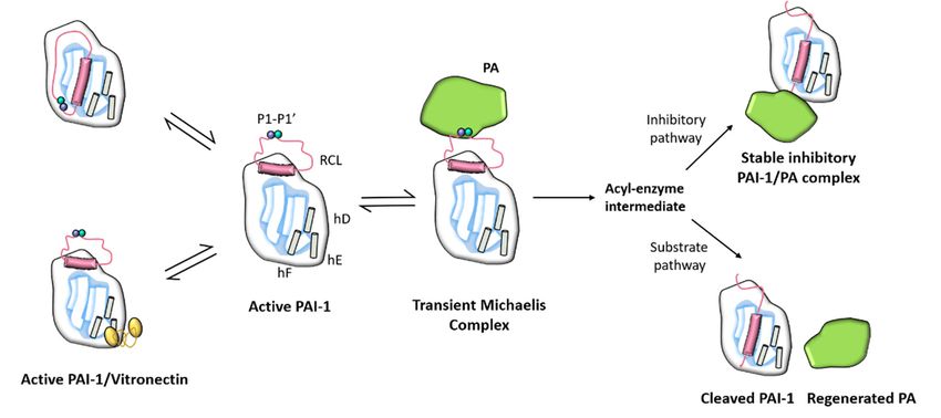
Int. J. Mol. Sci. 2021, 22, x FOR PEER REVIEW 2 of 16
Int. J. Mol. Sci. 2021, 22, 3170 2 of 15
and highlights strategies to modulate PAI-1 levels or activity as a mean to improve the
fibrinolytic process and reduce thrombosis risk.
strategies to modulate PAI-1 levels or activity as a mean to improve the fibrinolytic process
andFibrinolysis
1.1. reduce thrombosis risk.
in Diabetes
The fibrinolytic process starts with the conversion of plasminogen into plasmin after
1.1. Fibrinolysis in Diabetes
activation by tissue-type plasminogen activator (t-PA) or urokinase-type plasminogen ac-
tivatorThe fibrinolytic
(u-PA). process
Plasmin is thestarts
mainwith the conversion
protein that cleavesofthe
plasminogen into
fibrin fibres plasmininafter
resulting the
activation by tissue-type plasminogen activator (t-PA) or urokinase-type plasminogen
formation of fibrin degradation products [7]. Plasmin generation is tightly controlled not
activator
only (u-PA). Plasmin
by activators but alsois the maintoprotein
inhibitors that cleaves
avoid excessive clotthe fibrin
lysis. fibres
PAI-1 resulting
is one of thein the
most
formation of fibrin degradation products [7]. Plasmin generation is tightly controlled not
powerful antifibrinolytic proteins that binds to t-PA or u-PA, inhibiting their function and
only by activators but also inhibitors to avoid excessive clot lysis. PAI-1 is one of the most
reducing plasmin generation [7].
powerful antifibrinolytic proteins that binds to t-PA or u-PA, inhibiting their function and
Importantly, in patients with metabolic syndrome and/or type 2 diabetes, plasma
reducing plasmin generation [7].
concentrations of PAI-1 are elevated, thus contributing to the hypofibrinolytic environ-
Importantly, in patients with metabolic syndrome and/or type 2 diabetes, plasma
ment [8,9]. In addition to the effect on clot lysis, recent evidence suggests that increased
concentrations of PAI-1 are elevated, thus contributing to the hypofibrinolytic environ-
vascular PAI-1 can directly accelerate the atherothrombotic process by promoting neoin-
ment [8,9]. In addition to the effect on clot lysis, recent evidence suggests that increased
timal plaque formation [10]. This indicates that abnormalities in the coagulation system
vascular PAI-1 can directly accelerate the atherothrombotic process by promoting neointimal
do not only affect thrombosis potential but can also contribute to the progression of ath-
plaque formation [10]. This indicates that abnormalities in the coagulation system do not
erosclerosis.
only affect thrombosis potential but can also contribute to the progression of atherosclerosis.
1.2.
1.2. PAI-1
PAI-1 Structure
Structure and
and Function
Function
1.2.1.
1.2.1. PAI-1
PAI-1 Structure
Structure
PAI-1,
PAI-1, a member of the the superfamily
superfamilyof ofserine
serineprotease
proteaseinhibitors
inhibitors(SERPIN)
(SERPIN)[11,12],
[11,12],isisa
asingle-chain
single-chainglycoprotein
glycoproteinof ofapproximately
approximately52 52kDa
kDa consisting
consisting of of 379 or 381 amino acids
depending
depending on on heterogeneity
heterogeneity of of the
the N-terminal
N-terminal caused
caused by by two
two potential
potential cleavage
cleavage sites
sites for
for
signal peptidase
signal peptidase [13].
[13]. PAI-1
PAI-1 contains
contains two
two distinct
distinct interactive
interactive domains;
domains; aa reactive
reactive centre
loop (RCL)
loop (RCL) and
and aa flexible
flexible joint
joint region
region with
with helix
helix DD (hD),
(hD), helix
helix EE (hE),
(hE), and
and helix
helix FF (hF)
(hF)
binding sites as detailed
binding detailedin inFigure
Figure11[14].
[14].The
TheRCL
RCLdomain
domain is is
thethe
primary
primarysitesite
forfor
u-PA/t-PA
u-PA/t-
binding
PA andand
binding contains a P1-P1’
contains peptide
a P1-P1’ bondbond
peptide that interacts with these
that interacts proteases
with these [15]. PAI-1
proteases [15].
lacks cysteine
PAI-1 residues
lacks cysteine and hence
residues and there
henceisthere
an absence of disulfide
is an absence bonds that
of disulfide can that
bonds account
can
for its instability
account in solution.
for its instability in It includesItseveral
solution. residues
includes severalof residues
methionine, which may explain
of methionine, which
its susceptibility
may to irreversible
explain its susceptibility toinactivation
irreversible by oxidising by
inactivation agents.
oxidising agents.
Figure
Figure 1.1. Schematic
Schematic depiction of the
depiction of the PAI-1
PAI-1conformation
conformationasaswell
wellasasitsitsinteraction
interactionwith
with vitronectin
vitronectin cofactor
cofactor and
and plasmino-
plasminogen
gen activators (PA). PAI-1 contains two distinct interactive domains: a reactive centre loop (RCL) and a flexible
activators (PA). PAI-1 contains two distinct interactive domains: a reactive centre loop (RCL) and a flexible joint region joint region
with
with helix D (hD), helix E (hE), and helix F (hF) binding sites. The P1-P1’ bond is broken to create an acyl–enzyme
helix D (hD), helix E (hE), and helix F (hF) binding sites. The P1-P1’ bond is broken to create an acyl–enzyme intermediate
inter-
mediate following the creation of a non-covalent PAI-1/PA Michaelis complex. The reaction takes place through a
following the creation of a non-covalent PAI-1/PA Michaelis complex. The reaction takes place through a branched pathway,
branched pathway, leading either to the formation of an irreversible inhibitory complex or to the generation of cleaved
leading
PAI-1 dueeither tointermediate
to the the formation of an irreversible
acyl–enzyme inhibitory complex or to the generation of cleaved PAI-1 due to the
hydrolysis.
intermediate acyl–enzyme hydrolysis.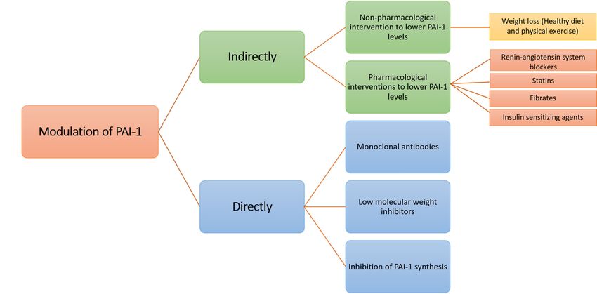
Int. J. Mol. Sci. 2021, 22, 3170                                                                                             3 of 15

                                        PAI-1 exists in three structurally and functionally distinct conformations, active, latent,
                                   and cleaved (substrate) [16]. Unlike other serpins, PAI-1 is readily converted from the active
                                   to the latent state. In vitro, the conformation of active PAI-1 is spontaneously converted to
                                   an energetically more favourable inactive latent state by moving the RCL into the central
                                   β-sheet [15]. Inhibitory activity relies on the active state exposure of the RCL, so the latent
                                   form is unable to inhibit proteases. The P1-P1’ bond in the latent conformation is also
                                   inaccessible to proteolytic attack [15].
                                        The half-life of native PAI-1 is approximately 2 h in vitro at 37 ◦ C and is slightly
                                   longer in vivo since most of the circulating PAI-1 is in complex with vitronectin (VN) (a
                                   relatively thermostable glycoprotein capable of stabilising and transforming PAI-1 into
                                   an active form) [15,17]. It has been found that PAI-1 binds to the N-terminal ~50- amino
                                   acid somatomedin B domain of VN [18,19] and X-ray structure evidence indicates that
                                   binding to this domain induces conformational changes in PAI-1 that improves stability of
                                   the protein [15,17]. The cleaved form of PAI-1 is discussed in the next section.

                                   1.2.2. PAI-1 Function
                                        PAI-1 is physiologically one of the most effective endogenous regulators of fibrinol-
                                   ysis [20]. As alluded to earlier, PAI-1 is the main inhibitor of plasminogen activation
                                   by interacting with t-PA and u-PA. This interaction involves the formation of an initial
                                   non-covalent Michaelis-like complex, followed by an acyl-enzyme intermediate and finally
                                   by the formation of an ester bond between the carboxyl group of the P1 residue of PAI-1
                                   with the serine residue of the protease (Figure 1). With the formation of this covalent
                                   complex, the Arg346 and Met347 (P1-P1’) bond of PAI-1 is cleaved and the target protease is
                                   translocated to the opposite side of the PAI-1 molecule, thereby irreversibly inhibiting the
                                   activity of the protease [21].

                                   1.3. PAI-1 Production and Regulation
                                   1.3.1. Source of PAI-1
                                         A large number of cells have the ability to produce PAI-1 including platelets, megakary-
                                   ocytes, monocytes/macrophages, adipocytes, cardiac myocytes and vascular smooth mus-
                                   cle cells, as well as cells of the endometrium, peritoneum, liver, mesothelium and endothe-
                                   lium [22]. Once synthesised, more than 90% of PAI-1 is stored in platelet α-granule, with
                                   the rest circulating in blood or deposited on the subendothelial matrix [23]. Therefore,
                                   PAI-1 levels in the blood do not reflect protein concentrations at the thrombus site given
                                   protein release from platelets when the coagulation system is activated.
                                         PAI-1 can be differentially regulated in various tissues and is also affected by patho-
                                   logical conditions such as vascular disease, sepsis, inflammation, and metabolic disorders
                                   including obesity and diabetes. Since PAI-1 may show cell type-specific glycosylation and
                                   activity, the glycosylation patterns of PAI-1 may serve as potential biomarkers to predict
                                   cellular dysfunction and thrombotic risk [24].
                                         Under physiological conditions, PAI-1 circulates in the plasma at concentrations of
                                   10–50 ng/mL [25], rising to over 100 ng/mL in the presence of obesity, insulin resistance
                                   and diabetes [24]. There is a circadian pattern to PAI-1 plasma levels, which peak in
                                   the early morning corresponding to a nadir in fibrinolytic activity, while they fall in the
                                   afternoon [25,26]. This seems to be related to stimulation of PAI-1 production by cortisol,
                                   which peaks in the early morning and clinically may contribute to the increased risk
                                   of myocardial infarction at this time of the day. Moreover, PAI-1 plasma levels vary
                                   according to race/ethnicity [27,28] and gender [29], although variations in body structure
                                   and distribution of adipose tissue can account for much of this variability.

                                   1.3.2. Regulation of PAI-1
                                        A variety of factors regulate the expression of PAI-1 (Figure 2), including glucocor-
                                   ticoids [30], insulin, glucose and inflammatory cytokines [31]. It is worth noting that the
                                   expression of transforming growth factor-β (TGF-β), tumour necrosis factor-α (TNF-α),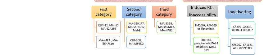
Mol. Sci. 2021, 22, x FOR PEER REVIEW 4 of 16
Int. J. Mol. Sci. 2021, 22, 3170 4 of 15
expression of transforming growth factor-β (TGF-β), tumour necrosis factor-α (TNF-α),
and interleukin-1β
and interleukin-1β (IL-1β) isinupregulated
(IL-1β) is upregulated adipocytes in of adipocytes of obeseand
obese individuals individuals
those and those
with diabetes [32], providing a mechanism for increased PAI-1
with diabetes [32], providing a mechanism for increased PAI-1 levels in obesity and insu- levels in obesity and in-
sulin resistant states. The synthesis of PAI-1 is also increased by
lin resistant states. The synthesis of PAI-1 is also increased by angiotensin II (Ang II), angiotensin II (Ang II),
which acts via type 1 receptor of angiotensin II expressed in adipocytes
which acts via type 1 receptor of angiotensin II expressed in adipocytes [33]. In contrast, [33]. In contrast,
catecholamines catecholamines
down-regulatedown-regulate PAI-1 gene
PAI-1 gene expression [9].expression [9].
Figure 2.expression
Figure 2. Adipocyte Adipocyteand expression
secretionand secretion of plasminogen
of plasminogen activator(PAI-1)
activator inhibitor-1 inhibitor-1 (PAI-1) is
is stimulated bystim-
glucocorticoids,
ulated
angiotensin II (Angby
II),glucocorticoids, angiotensin
insulin, glucose and a varietyIIof(Ang II), insulin,
cytokines, glucose
including and necrosis
tumour a varietyfactor-α
of cytokines, in- transforming
(TNF-α),
cluding tumour necrosis factor-α (TNF-α), transforming growth factor-β (TGF-β), and interleukin-
growth factor-β (TGF-β), and interleukin-1β (IL-1β). Catecholamines, such as norepinephrine inhibit production of PAI-1.
1β (IL-1β). Catecholamines, such as norepinephrine inhibit production of PAI-1.
1.4. Role of PAI-1
1.4.inRole
Diabetes
of PAI-1 in Diabetes
Plasma levels of PAI-1levels
Plasma are elevated
of PAI-1inareindividuals
elevated in with type 2 diabetes,
individuals with typealthough
2 diabetes,this although this
does not necessarily hold true for type 1 diabetes (Table 1) [34]. These findings
does not necessarily hold true for type 1 diabetes (Table 1) [34]. These findings suggest suggest
the main mechanism
the main formechanism
elevated PAI-1 levels inPAI-1
for elevated diabetes is related
levels to obesity
in diabetes andto
is related insulin
obesity and insulin
resistance, rather than elevated
resistance, glucose
rather than levelsglucose
elevated [35]. However, hyperglycaemia
levels [35]. may still
However, hyperglycaemia may still
have an effect have
in thean effect inofthe
presence presence
insulin of insulin
resistance. resistance.
A role for PAI-1 A inrole for PAI-1
vascular in vascular pathology
pathology
is supported by is protein
supported by proteinataccumulation
accumulation vascular atheroma at vascular atheroma
sites, which appearsites,
to bewhich
par- appear to be
particularly
ticularly pronounced pronounced
in those in those
with diabetes [25].with diabetes [25].
Insulin was foundInsulin was found
to increase to increase
PAI-1 expressionPAI-1in aexpression in a HepG2
HepG2 hepatocyte cellhepatocyte
line [36]. cell line [36].
The combination Theof combination
insulin andofinsulin-like
insulin andgrowth
insulin-like
factorgrowth
1 furtherfactor 1 further demonstrated
demonstrated a syn- a syner-
gistic
ergistic effect on effect
PAI-1 on PAI-1[37].
expression expression
Moreover, [37]. Moreover,
insulin insulinboth
precursors, precursors,
proinsulin both
andproinsulin and
split products split products of
of proinsulin, proinsulin,
also enhancedalso PAI-1 enhanced
expressionPAI-1andexpression and levels
levels of these proteinsof these proteins
are known to be elevated in type 2 diabetes (T2D) [38]. Therefore, insulin resistance, a resistance, a
are known to be elevated in type 2 diabetes (T2D) [38]. Therefore, insulin
hallmark of T2D,hallmark of T2D,
increases PAI-1 increases
expressionPAI-1 expression
secondary secondarylevels
to increased to increased
of insulin levels
andof insulin and
its precursors. In addition to increased PAI-1 mRNA expression,
its precursors. In addition to increased PAI-1 mRNA expression, insulin may also decrease insulin may also decrease
the rate of mRNA degradation thus resulting in
the rate of mRNA degradation thus resulting in sustained protein production [31].sustained protein production [31].
Insulin
Insulin resistance resistance
causes causesin
a disruption a disruption in the phosphoinositide-3
the phosphoinositide-3 kinase (PI3-K/Akt) kinase (PI3-K/Akt)
signalling pathway, which results in insufficient tissue insulin sensitivity. During com- During com-
signalling pathway, which results in insufficient tissue insulin sensitivity.
pensatory hyperinsulinemia,
pensatory hyperinsulinemia, the paradox ofthe paradox ofinpathologies
pathologies in molecular
molecular insulin insulin signalling
signalling
leads to decreased activity of the PI3-K/Akt pathway
leads to decreased activity of the PI3-K/Akt pathway together with upregulation of the together with upregulation of the
mitogen-activated protein kinase/extracellular signal-regulated
mitogen-activated protein kinase/extracellular signal-regulated kinase (MAPK/ERK) kinase (MAPK/ERK) path-
way [39]. Insulin resistance is linked to glycotoxicity, lipotoxicity,
pathway [39]. Insulin resistance is linked to glycotoxicity, lipotoxicity, and inflammation, and inflammation, which
which contributecontribute to the initiation
to the initiation and progression
and progression of atherogenesis
of atherogenesis and vascular
and vascular diseasedisease [40,41].
Insulin resistance and endothelial dysfunction are linked by changes in the balance between
[40,41]. Insulin resistance and endothelial dysfunction are linked by changes in the bal-
the PI3-K/Akt and MAPK/ERK pathways, explaining the high risk of atherothrombosis
in insulin-resistant states [42]. Furthermore, when the balance of insulin resistance is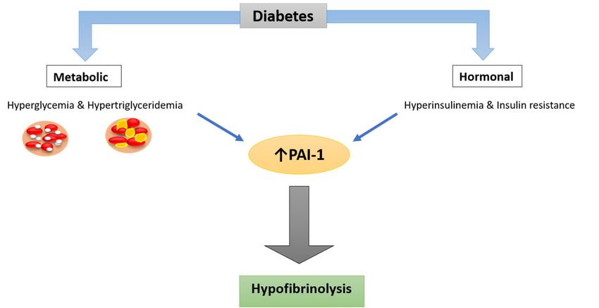
Int. J. Mol. Sci. 2021, 22, 3170                                                                                                  5 of 15

                                     transferred towards the MAPK/ERK pathway, insulin releases inflammatory markers such
                                     as PAI-1, intercellular adhesion molecule 1 (ICAM-1), vascular cell adhesion molecule 1
                                     (VCAM-1), and E-selectin, which leads to endothelial dysfunction, which in turn predis-
                                     poses to vascular pathology [43].
                                           Hyperglycemia and hypertriglyceridemia are metabolic disorders caused by insulin
                                     deficiency, either relative or absolute. In vitro, increased glucose concentrations enhance
                                     PAI-1 expression in both endothelial and vascular smooth muscle (VSM) cells [44]. In
                                     HepG2 cells, triglycerides and their constituents (fatty acids) increase PAI-1 expression [45].
                                     Furthermore, the combination of elevated insulin and triglyceride levels induces a syner-
                                     gistic increase in PAI-1 production [46].
                                           Insulin resistance is also associated with lower very low-density lipoprotein (VLDL)
                                     clearance [47]. By inducing transcription of the PAI-1 gene promoter, VLDL particles
                                     have been shown to increase PAI-1 biosynthesis in endothelial cells while also stabilising
                                     the mRNA transcripts of the protein. The main signalling pathway involved in VLDL-
                                     induced PAI-1 biosynthesis is related to MAPK activation [48]. Therefore, a combination of
                                     factors, related to hyperglycemia and insulin resistance, are responsible for increased PAI-1
                                     production in T2D, through well-defined pathways.

      Table 1. Summary of main studies investigating PAI-1 levels in individuals with type 2 diabetes (T2D). Values are presented
      as mean ± SD or median interquartile range (IQR). Variation in PAI-1 levels is likely due to the different methodologies
      used to measure protein levels and potential differences between study populations.

                                                                     Mean ± SD Median
                                    Country            No. of                                Mean ± SD Median
             Study                                                         (IQR)                                         p-Value
                                                    T2D/Controls                              (IQR) of Control
                                                                          of T2D
                                                                        82.7 ± 54.5               52.9 ± 51.7
    Kitagawa (2006) [49]             Japan             47/31
Int. J. Mol. Sci. 2021, 22, 3170 6 of 15
Int. J. Mol. Sci. 2021, 22, x FOR PEER REVIEW 6 of 16
Figure 3.
Figure 3. Mechanism
Mechanismofofincreased
increasedPAI-1 levels
PAI-1 in in
levels diabetes. Hormonal
diabetes. (hyperinsulinemia)
Hormonal and met-
(hyperinsulinemia) and
abolic (hyperglycemia and hypertriglyceridemia) derangements in T2D patients, seem
metabolic (hyperglycemia and hypertriglyceridemia) derangements in T2D patients, seem to have to have a a
role in elevating PAI-1 levels in this population, resulting in hypofibrinolysis that causes patholog-
role in elevating PAI-1 levels in this population, resulting in hypofibrinolysis that causes pathological
ical deposition of fibrin and damage to the tissues.
deposition of fibrin and damage to the tissues.
1.5. Role
1.5. Role of
of PAI-1
PAI-1ininCVD
CVD
Prospective studies
Prospective studies have
have shown
shown that
that impaired
impaired fibrinolysis
fibrinolysisand
andspecifically
specificallyelevated
elevated
PAI-1concentrations
PAI-1 concentrationsare areindependent
independentrisk riskfactors
factorsfor
forboth
botharterial
arterialand
andvenous
venous thrombotic
thrombotic
occlusive vascular
occlusive vascular disease
disease [30,58].
[30,58]. Moreover,
Moreover, itit is
is suggested
suggested that
that patients
patients with
withelevated
elevated
plasma PAI-1
plasma PAI-1have
havethe
thehighest
highestrisk
riskof
ofearly
earlyre-infarction
re-infarction[59][59]and
andtherefore
thereforeprotein
proteinlevels
levels
have prognostic significance following cardiac ischaemia. Importantly, a positive
have prognostic significance following cardiac ischaemia. Importantly, a positive associa- associ-
ationbetween
tion betweenPAI-1
PAI-1activity
activityofoffathers
fatherswith
withpremature
prematureMI MIandandtheir
theirchildren
childrenhashasrecently
recently
been reported
been reportedand andtherefore
thereforePAI-1
PAI-1levels
levelscan
can also
also have
have a prognostic
a prognostic role
role in familial
in familial pre-
predis-
position to CVD
disposition [23,60].
to CVD [23,60].
1.6.
1.6. Role
Role ofof PAI-1
PAI-1ininDiabetic
DiabeticRetinopathy
RetinopathyandandChronic
ChronicKidney
KidneyDisease
Disease
A
A number of studies have documented an association between
number of studies have documented an association between PAI-1
PAI-1 and
and microvas-
microvas-
cular
cular complications
complicationssuch suchasas retinopathy
retinopathyand andchronic
chronickidney
kidneydisease.
disease.One
Onestudy
studysuggested
suggested
that
that elevated
elevatedPAI-1
PAI-1plasma
plasmalevels areare
levels independently
independently associated withwith
associated lower risk of
lower retinopa-
risk of reti-
thy [61]. However, a much larger longitudinal study involving 858 individuals
nopathy [61]. However, a much larger longitudinal study involving 858 individuals from from the
Veterans Affairs Diabetes Trial (VADT) cohort has shown that PAI-1 plasma
the Veterans Affairs Diabetes Trial (VADT) cohort has shown that PAI-1 plasma levels levels predict
the development
predict of retinopathy
the development in T2D withina 12%
of retinopathy T2D increased
with a 12%riskincreased
for every 10 ng/dl
risk increase
for every 10
in PAI-1 levels [62]. Moreover, PAI-1 levels were elevated in 153 Chinese diabetes
ng/dl increase in PAI-1 levels [62]. Moreover, PAI-1 levels were elevated in 153 Chinese patients
with advanced
diabetes patientsdiabetic retinopathy,
with advanced suggesting
diabetic an association
retinopathy, suggestingwith
andisease severity
association with[63].
dis-
Interestingly, animal work has shown increased PAI-1 expression in the retina with the de-
ease severity [63]. Interestingly, animal work has shown increased PAI-1 expression in the
velopment of proliferative changes, directly implicating PAI-1 in disease pathogenesis [64].
retina with the development of proliferative changes, directly implicating PAI-1 in disease
Taken together, studies generally agree that elevated PAI-1 levels carry a higher risk of
pathogenesis [64]. Taken together, studies generally agree that elevated PAI-1 levels carry
retinopathy, although the exact mechanisms remain an area for future research.
a higher risk of retinopathy, although the exact mechanisms remain an area for future
A number of studies have also reported a relationship between elevated plasma PAI-1
research.
levels and chronic kidney disease (CKD). Many factors can induce PAI-1 expression in CKD,
A number of studies have also reported a relationship between elevated plasma PAI-
such as TGF-β and angiotensin-II. The most compelling evidence for a role of PAI-1 in renal
1 levels and chronic kidney disease (CKD). Many factors can induce PAI-1 expression in
disease is derived from prevention of CKD, and even disease regression, with reduction
CKD, such as TGF-β and angiotensin-II. The most compelling evidence for a role of PAI-
in PAI-1 production in animal models [65,66]. Moreover, data investigating the effects
1 in renal disease is derived from prevention of CKD, and even disease regression, with
of two PAI-1 inhibitors on kidney injury in diabetic mice suggested that this approach
reduction in PAI-1 production in animal models [65,66]. Moreover, data investigating the
improved kidney function [67]. Another study on PAI-1 deficient mice demonstrated
effects
that of two
PAI-1 PAI-1regulates
directly inhibitors on kidney
TGF-β injuryby
expression in binding
diabetic to
mice suggested
u-PAR that this the
and activating ap-
proach improved kidney function [67]. Another study on PAI-1 deficient
extracellular-regulated signal kinase (ERK)/MAPK pathway thus contributing to renal mice demon-
strated that
disease [68].PAI-1 directly regulates TGF-β expression by binding to u-PAR and activatingInt. J. Mol. Sci. 2021, 22, x FOR PEER REVIEW 7 of 16
Int. J. Mol. Sci. 2021, 22, 3170 7 of 15
the extracellular-regulated signal kinase (ERK)/MAPK pathway thus contributing to renal
disease [68].
Targeting PAI-1 for Potential
2. Targeting Potential Therapies
Therapies
Since elevated
Since elevated PAI-1
PAI-1 plasma levels and activity contribute
contribute to
to cardiovascular
cardiovascular pathol-
pathol-
ogy, several groups attempted to use PAI-1 as a therapeutic target in order to improve
plasmin generation and alleviate the hypofibrinolytic environment [23]. We We provide here
an overview
an overview ofof the different approaches used to modulate PAI-1 production or function
(summarised in Figure
(summarised in Figure 4).4).
Figure
Figure 4.
4. Summary
Summary of
of the
the strategies
strategies for
for modulating
modulating PAI-1
PAI-1 indirectly and directly.
indirectly and directly.
2.1.
2.1. Indirect
Indirect Modulation
Modulation of of PAI-1 Production or
PAI-1 Production or Function
Function
2.1.1. A.
A. Non-Pharmacological
Non-Pharmacological Intervention to Lower PAI-1 Levels
Weight Loss (Healthy Diet and Physical Exercise)
Weight loss
lossisisone
oneofofthethe best
best methods
methods to improve
to improve obesity-related
obesity-related fibrinolytic
fibrinolytic im-
impair-
pairment [9], partly related to a marked decrease in PAI-1 production,
ment [9], partly related to a marked decrease in PAI-1 production, which has been demon- which has been
demonstrated
strated in a number
in a number of studies
of studies [69]. It[69].
has Italso
hasbeen
also been
shown shown thatonly
that not not only weight
weight loss loss
but
but also
also increased
increased physical
physical activity
activity are linked
are linked to decreased
to decreased PAI-1PAI-1 levels
levels [22].[22]. Importantly,
Importantly, the
the decrease
decrease in PAI-1
in PAI-1 levels
levels is associated
is associated withwith improved
improved clinical
clinical parameters;
parameters; Killewich
Killewich et al.
et al.found
[70] [70] found that patients
that patients with with intermittent
intermittent claudication,
claudication, due todue to peripheral
peripheral artery
artery dis-
disease,
ease, had improvement in their symptoms after a programme of
had improvement in their symptoms after a programme of physical exercise, which was physical exercise, which
was associated
associated with with decreased
decreased PAI-1PAI-1 activity
activity and enhanced
and enhanced fibrinolytic
fibrinolytic activity.
activity. Interest-
Interestingly,
ingly, those with higher baseline PAI-1 levels gained the most clinical
those with higher baseline PAI-1 levels gained the most clinical benefit from the interven- benefit from the
intervention,
tion, directly implicating
directly implicating protein
protein levels in levels
disease in presentation
disease presentation
[71]. [71].
2.1.2. Pharmacological Interventions to Lower PAI-1 Levels
2.1.2. Pharmacological Interventions to Lower PAI-1 Levels
Renin-Angiotensin System Blockers
Renin-Angiotensin System Blockers
The activation of the renin–angiotensin system is closely related to PAI-1 [72]. An-
giotensinactivation
The of the renin–angiotensin
II (also produced system
by adipose tissue) is a is closely related
biologically to PAI-1
active [72]. Angi-
angiotensinogen
otensin
processingII (also produced
agent by adipose
and has been showntissue) is a biologically
to stimulate active angiotensinogen
PAI-1 expression pro-
at the transcriptional
cessing agent and has been shown to stimulate PAI-1 expression at the
level in human adipocytes [33]. The effects of angiotensin-converting enzyme (ACE) transcriptional
level in human
inhibitors adipocytes
on cardio [33].appear
protection The effects
to beofmediated
angiotensin-converting
by pathways that enzyme (ACE) in-
are partially in-
dependent of the reduction in blood pressure. Vaughan et al. reported that one proposed
mechanism is related to modulation of PAI-1 concentrations [73]. However, others failed to
show an effect of ACE inhibitors on PAI-1 levels [74]. Therefore, this remains an unresolvedInt. J. Mol. Sci. 2021, 22, 3170 8 of 15
area and future work is required to understand the role of ACE inhibitors on fibrinolysis in
general and PAI-1 activity/levels in particular [75].
Statins
Anti-hyperlipidaemic agents, in particular statins, have been shown to modulate
the prothrombotic environment and affect the fibrinolytic system [76]. Statins inhibit the
expression of PAI-1 and their use is associated with reduction in PAI-1 levels, which may
explain improved t-PA activity in those treated with these agents [16,77,78].
Fibrates
Fibrates can decrease the in vitro production of PAI-1 independently of the triglyceride
reducing effect [79]. In vivo use of fibrates also decreases PAI-1 levels and reduces protein
expression in human arterial smooth muscle cells (SMC) [79,80]. Different fibrates showed
different potencies to suppress PAI-1 synthesis, but these effects are generally mild and the
clinical role of these observations remain unclear [81].
Insulin-Sensitising Agents
Insulin-sensitising agents, such as metformin and thiazolidinediones have been shown
to have beneficial effects on the fibrinolytic system, mediated, at least in part, by a reduction
in PAI-1 levels [82]. Glucagon-like peptide-1 receptor agonists inhibit TNF-α stimulated
PAI-1 production in vitro [83], while metformin reduces PAI-1 levels in vivo, an effect
believed to be related to enhanced insulin sensitivity and/or reduction in proinsulin
production [8,84,85].
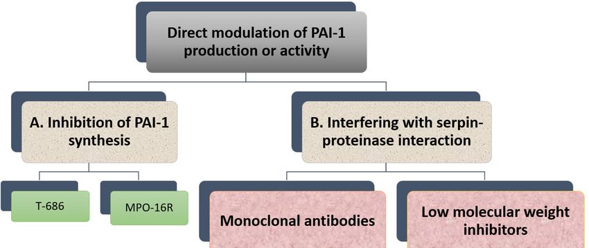
2.2. Direct Modulation of PAI-1 Production or Activity
Many research groups have invested in the development of PAI-1 inhibitors over
the past three decades [86] either through inhibiting PAI-1 synthesis or interfering with
serpin-proteinase
Int. J. Mol. Sci. 2021, 22, x FOR PEER REVIEW interaction (Figure 5). Despite large volume of research in9 this
of 16 area, no
PAI-1 inhibitor is yet available for routine clinical use.
Figure 5. Overview of direct modulation of PAI-1 production or activity by (A) inhibition of PAI-1 synthesis or (B) inter-
Overview
Figure 5. fering of direct modulation of PAI-1 production or activity by (A) inhibition of PAI-1 synthesis or (B) interfering
with serpin–proteinase interaction.
with serpin–proteinase interaction.
2.2.1. Inhibition of PAI-1 Synthesis
Reduction in PAI-1 synthesis represents an attractive approach to enhancing fibrinol-
ysis and reducing thrombosis risk. However, this needs to be undertaken in a controlled
manner as full inhibition of PAI-1 synthesis predisposes to mild-severe bleeding events,
supported by evidence from individuals with complete PAI-1 deficiency [87]. Therefore,Int. J. Mol. Sci. 2021, 22, 3170 9 of 15
2.2.1. Inhibition of PAI-1 Synthesis
Reduction in PAI-1 synthesis represents an attractive approach to enhancing fibrinoly-
sis and reducing thrombosis risk. However, this needs to be undertaken in a controlled
manner as full inhibition of PAI-1 synthesis predisposes to mild-severe bleeding events,
supported by evidence from individuals with complete PAI-1 deficiency [87]. Therefore,
ideally agents suppressing PAI-1 production should reduce excess PAI-1 levels to attain
normal levels and restore normal haemostasis [88].
In vitro studies have shown that TGF-β-stimulated PAI-1 production in cultured
endothelial cells is attenuated by a butadiene derivative, T-686. Furthermore, T-686 also
diminished the rise in PAI-1 activity in aortic atherosclerotic lesions of rabbits. This was
accompanied by decreased aortic PAI-1 mRNA expression, associated with a reduction
in the development of atherosclerotic lesions [89]. Pawlowska et al. [90] showed that an
oligonucleotide antisense to PAI-1 mRNA (MPO-16R) decreased PAI-1 concentration in
rat plasma and platelets, while MPO-16R induced a large delay in occlusion time in an
experimental model of rat arterial thrombosis. Therefore, reducing PAI-1 production is
possible and preliminary evidence suggests this strategy can alter the atherothrombotic
process. However, findings from these studies have not been translated into work in man
and the clinical future of this antithrombotic strategy is uncertain.
2.2.2. Interfering with Serpin-Proteinase Interaction
Although the available evidence is very preliminary, studies have shown that inhibi-
tion of PAI-1 enhances endogenous fibrinolytic activity without directly affecting blood
coagulation or platelet function [91]. Several PAI-1 inhibitors have been studied in vitro
and in vivo and have shown variable antithrombotic efficacy.
Monoclonal Antibodies
A variety of monoclonal antibodies against human PAI-1 has been developed to mod-
ulate protein activity [11]. These can be subdivided into at least three different categories.
First, monoclonal antibodies may prevent the formation of the initial Michaelis complex
formed between PAI-1 and its target protease. It was found that monoclonal antibodies
against PAI-1, including ESPI-12, MAI-12, MA-42A2F6, MA-44E4 and MA-56A7C10, with
their epitopes located near the RCL of PAI-1, interfere directly with the PAI-1/protease
interaction [92]. MAI-12, one of the PAI-1 binding antibodies that inhibit complex for-
mation with the target, has been studied extensively in vitro and in an in vivo venous
thrombosis model [93]. MAI-12 inhibited human and rabbit PAI-1 in vitro and significantly
enhanced endogenous thrombolysis in a rabbit model of jugular vein thrombosis and
partially prevented thrombus extension [93].
A second category of PAI-1 inhibiting antibodies are the so-called “switching antibod-
ies”, which inhibit PAI-1 activity by cleaving and inactivating the protein. These antibodies
include MA-33H1F7, MA-55F4C12 [94], Mab2 [95], CLB-2C8 [96] and MA-MP2D2 [97].
The murine monoclonal antihuman PAI-1 antibody CLB-2C8 showed great promise as it
inhibited protein activity in different species [98], while in vivo administration significantly
enhanced endogenous thrombolysis, and reduced thrombus growth in a rabbit jugular
vein thrombosis model [98]. Despite the early promise, CLB-2C8 was not followed up with
human studies for reasons that are unclear.
As for the third category of PAI-1 inhibiting antibodies, these are known for ac-
celerating conversion of active PAI-1 to its latent conformation and include MA-H4B3,
MA-159M12 and MA-33B8 antibodies [99]. The monoclonal antibody, MA-33B8, was
reported to convert PAI-1 to latent conformation and random mutagenesis was used to
identify the MA-33B8 epitope in the PAI-1 molecule to comprehend the mechanism of
action [100]. Surface plasmon resonance showed 100-fold higher affinity of MA-33B8 to
latent PAI-1 than active protein [100]. Additionally, structural modelling results specified
the presence of a particular intermediate structure of PAI-1 that is stabilised by MA-33B8
binding [100]. It was suggested that this intermediate form of PAI-1 has a partial insertionInt. J. Mol. Sci. 2021, 22, 3170 10 of 15
into β-sheet A of the reactive centre loop, supporting the hypothesis that active serpins
have flexible RCLs, and that this flexibility is crucial for the role of the inhibitor [101]. As a
result, the activity of the serpin may also be blocked by synthetic peptides homologous to
the RCL serpin converting PAI-1 to a shape similar to the latent form [102]. Moreover, Ngo
et al. [103] reported that antibody MA-159M12 promoted the conversion of active to latent
conformation, further indicating this is a viable approach to alter PAI-1 activity. Antibody
MA-H4B3 has been shown to inactivate recombinant PAI-1 in a time-dependent way [104],
which indicates that, in the presence of MA-H4B3, incorporation of the RCL is accelerated,
suggesting that the loss of activity is the result of the transition of latency [104].
Low Molecular Weight Inhibitors
Several PAI-1 low molecular weight antagonists have been discovered and charac-
terised. The first were diketopiperazines (XR330 and XR334) and, from that template, more
potent antagonists were designed (XR1853, XR5082, XR5967, XR1121 and XR5118) which
inhibit PAI-1 by inducing the transition from active PAI-1 to non-reactive PAI-1 [105,106].
XR334, XR1853 and XR5082 effectively increased fibrinolysis in vivo by inhibiting the
interaction of t-PA/u-PA and PAI-1 in a rat carotid artery thrombosis model [107].
XR5118 administration improved ex vivo fibrinolysis and protected against throm-
bus formation in a rat electrically stimulated carotid artery (ESCA) model [23,108]. The
protection in this arterial thrombosis was associated with a significant decrease in PAI-1
activity and a significant increase in t-PA activity in plasma [23,108]. Importantly, inhibition
of PAI-1 by XR5118 did not contribute to any increase in rat bleeding time, which held
promise for the safety of this compound [108].
Furthermore, TM5007, a derivative of indole oxoacetic acid was shown to effectively
inhibit PAI-1 activity [91]. This compound is metabolically stable, non-toxic, and has
demonstrated strong oral bioavailability and in vivo efficacy in a rat thrombosis model [91].
Björquist et al. [109] developed the flufenamic acid-based AR-H029953XX after it was
reported that flufenamic acid and its derivatives enhanced in vitro plasma clot lysis in an
assay of plasmin generation [110].
As for PAI-039 or Tiplaxtinin, an indole derivative, seemed a very promising PAI-1
inhibitor [111]. It has been examined in several animal models confirming a putative value
of PAI-1 inhibition for therapeutic purposes [112]. In another study, however, Tiplaxtinin
did not inhibit the activity of PAI-1 in rat plasma 15 min after intravenous injection, which
was suggested to be due to the fact that Tiplaxtinin is unable to inhibit vitronectin bound
PAI-1 because of the overlapping epitopes of vitronectin and Tiplaxtinin on PAI-1 [113].
A novel class of polyphenolic PAI-1 inhibitors was identified by Cale et al. [114],
representing an enhanced 10–1000-fold potency over previously described PAI-1 inhibitors.
This class exerted its PAI-1-inhibitor effect by blocking the initial bond between the protein
and protease. These compounds inactivate PAI-1 in the presence of vitronectin, giving
them an advantage over other PAI-1 inhibitors. Two of these compounds demonstrated
efficacy in plasma and one other blocked PAI-1 activity in in vivo mice studies. The authors
proposed that the known cardiovascular advantages of dietary polyphenols may be due
in part to PAI-1 inactivation, although this is only a hypothesis that is yet to be tested. A
human antibody, MEDI-579, has recently been shown to bind to the active form of human
PAI-1 with high affinity and specificity [115]. Importantly, MEDI-579 effects were still
evident in vitronectin bound PAI-1. Crystallography demonstrated that this specificity
is achieved due to the attachment of MEDI-579 Fab to the RCL of PAI-1 and to the same
exosite used by both t-PA and u-PA [115]. This indicates that MEDI-579 is able to modulate
the interaction of PAI-1 with t-PA and u-PA in a manner not previously defined for a human
PAI-1 inhibitor by directly competing with proteases for RCL binding.
3. Conclusions
People with diabetes suffer from premature atherothrombosis, resulting in a high rate
of morbidity and mortality. Hypofibrinolysis contributes to the risk of vascular eventsInt. J. Mol. Sci. 2021, 22, 3170 11 of 15
and is partly mediated by elevated levels/activity of PAI-1. Therefore, the development of
agents that modulate the hypofibrinolytic milieu in diabetes is one alternative strategy to
reduce the risk of vascular thrombosis in this population.
Research over the past few decades has shown that PAI-1 is a complex protein with
different conformations and associations with other proteins, which can affect its function.
This contributed to the difficulties in identifying a reliable agent that inhibits protein
function to ameliorate the hypofibrinolytic environment. Different strategies have been
explored over the years to reduce PAI-1 production and/or modulate protein activity. A
large number of compounds have been identified, ranging from chemicals, monoclonal
antibodies to small molecules. Despite favourable in vitro and animal in vivo effects for
some of these agents, none made it into human studies for reasons that are not entirely clear.
The suspicion is that human ex vivo work was disappointing, or the various compounds
were found to have “off target” or toxic effects preventing human use. This highlights the
importance of publishing “negative studies” in order to make the scientific community
better informed and avoid duplicating work that has already been undertaken.
The newer and more refined PAI-1 inhibitors offer the promise of having effective
agents that modulate hypofibrinolysis and reduce clinical thrombotic events while potentially
limiting bleeding risk; the quest to find an effective and safe PAI-1 inhibitor in man continues.
Author Contributions: R.A. wrote the draft of the manuscript and prepared the original figures.
R.A.A. and N.P. contributed substantially in reviewing and editing the manuscript. All authors have
read and agreed to the published version of the manuscript.
Funding: This research received no external funding.
Conflicts of Interest: The authors declare no conflict of interest.
References
1. Narayan, K.M.V.; Boyle, J.P.; Thompson, T.J.; Sorensen, S.W.; Williamson, D.F. Lifetime Risk for Diabetes Mellitus in the United
States. JAMA 2003, 290, 1884–1890. [CrossRef]
2. Kannel, W.B.; McGee, D.L. Diabetes and Glucose Tolerance as Risk Factors for Cardiovascular Disease: The Framingham Study.
Diabetes Care 1979, 2, 120–126. [CrossRef]
3. Alzahrani, S.H.; Ajjan, R. Review article: Coagulation and fibrinolysis in diabetes. Diabetes Vasc. Dis. Res. 2010, 7, 260–273.
[CrossRef]
4. Kearney, K.; Tomlinson, D.; Smith, K.; Ajjan, R. Hypofibrinolysis in diabetes: A therapeutic target for the reduction of cardiovas-
cular risk. Cardiovasc. Diabetol. 2017, 16, 1–17. [CrossRef]
5. Sumaya, W.; Wallentin, L.; James, S.K.; Siegbahn, A.; Gabrysch, K.; Himmelmann, A.; Ajjan, R.A.; Storey, R.F. Impaired Fibrinolysis
Predicts Adverse Outcome in Acute Coronary Syndrome Patients with Diabetes: A PLATO Sub-Study. Thromb. Haemost. 2020,
120, 412–422. [CrossRef]
6. Hess, K.; Alzahrani, S.H.; Mathai, M.; Schroeder, V.; Carter, A.M.; Howell, G.J.; Koko, T.; Strachan, M.W.J.; Price, J.; Smith, K.A.; et al.
A novel mechanism for hypofibrinolysis in diabetes: The role of complement C3. Diabetologia 2012, 55, 1103–1113. [CrossRef]
7. Sprengers, E.; Kluft, C. Plasminogen activator inhibitors. Blood 1987, 69, 381–387. [CrossRef]
8. Schneider, D.J. Abnormalities of coagulation, platelet function, and fibrinolysis associated with syndromes of insulin resistance.
Coron. Artery Dis. 2005, 16, 473–476. [CrossRef]
9. Skurk, T.; Hauner, H. Obesity and impaired fibrinolysis: Role of adipose production of plasminogen activator inhibitor-1.
Int. J. Obes. 2004, 28, 1357–1364. [CrossRef]
10. Aso, Y. Plasminogen activator inhibitor (PAI)-1 in vascular inflammation and thrombosis. Front. Biosci. 2007, 12, 2957–2966.
[CrossRef]
11. Gils, A.; Declerck, P.J. The structural basis for the pathophysiological relevance of PAI-1 in cardiovascular diseases and the
development of potential PAI-1 inhibitors. Thromb. Haemost. 2004, 91, 425–437. [CrossRef] [PubMed]
12. Durand, M.K.; Bødker, J.S.; Christensen, A.; Dupont, D.M.; Hansen, M.; Jensen, J.K.; Kjelgaard, S.; Mathiasen, L.; Pedersen, K.E.;
Skeldal, S.; et al. Plasminogen activator inhibitor-1 and tumour growth, invasion, and metastasis. Thromb. Haemost. 2004, 91,
438–449. [CrossRef] [PubMed]
13. Binder, B.R.; Christ, G.; Gruber, F.; Grubic, N.; Hufnagl, P.; Krebs, M.; Mihaly, J.; Prager, G.W. Plasminogen activator inhibitor 1:
Physiological and pathophysiological roles. Physiology 2002, 17, 56–61. [CrossRef]
14. Placencio, V.R.; Declerck, Y.A. Plasminogen Activator Inhibitor-1 in Cancer: Rationale and Insight for Future Therapeutic Testing.
Cancer Res. 2015, 75, 2969–2974. [CrossRef]Int. J. Mol. Sci. 2021, 22, 3170 12 of 15
15. Fjellström, O.; Deinum, J.; Sjögren, T.; Johansson, C.; Geschwindner, S.; Nerme, V.; Legnehed, A.; McPheat, J.; Olsson, K.;
Bodin, C.; et al. Characterization of a Small Molecule Inhibitor of Plasminogen Activator Inhibitor Type 1 That Accelerates the
Transition into the Latent Conformation. J. Biol. Chem. 2013, 288, 873–885. [CrossRef]
16. Simone, T.M.; Higgins, P.J. Low Molecular Weight Antagonists of Plasminogen Activator Inhibitor-1: Therapeutic Potential in
Cardiovascular Disease. Mol. Med. Ther. 2012, 1, 101. [CrossRef] [PubMed]
17. Zhou, A.; A Huntington, J.; Pannu, N.S.; Carrell, R.W.; Read, R.J. How vitronectin binds PAI-1 to modulate fibrinolysis and cell
migration. Nat. Struct. Mol. Biol. 2003, 10, 541–544. [CrossRef] [PubMed]
18. Blouse, G.E.; Dupont, D.M.; Schar, C.R.; Jensen, J.K.; Minor, K.H.; Anagli, J.Y.; Gårdsvoll, H.; Ploug, M.; Peterson, C.B.;
Andreasen, P.A. Interactions of Plasminogen Activator Inhibitor-1 with Vitronectin Involve an Extensive Binding Surface and
Induce Mutual Conformational Rearrangements. Biochemistry 2009, 48, 1723–1735. [CrossRef]
19. Schar, C.R.; Blouse, G.E.; Minor, K.H.; Peterson, C.B. A Deletion Mutant of Vitronectin Lacking the Somatomedin B Domain
Exhibits Residual Plasminogen Activator Inhibitor-1-binding Activity. J. Biol. Chem. 2008, 283, 10297–10309. [CrossRef]
20. Mertens, I.; van Gaal, L.F. Obesity, haemostasis and the fibrinolytic system. Obes. Rev. 2002, 3, 85–101. [CrossRef]
21. van de Craen, B.; Declerck, P.J.; Gils, A. The Biochemistry, Physiology and Pathological roles of PAI-1 and the requirements for
PAI-1 inhibition in vivo. Thromb. Res. 2012, 130, 576–585. [CrossRef]
22. Cesari, M.; Pahor, M.; Incalzi, R.A. REVIEW: Plasminogen Activator Inhibitor-1 (PAI-1): A Key Factor Linking Fibrinolysis and
Age-Related Subclinical and Clinical Conditions. Cardiovasc. Ther. 2010, 28, e72–e91. [CrossRef]
23. Charlton, P. The status of plasminogen activator inhibitor-1 as a therapeutic target. Expert Opin. Investig. Drugs 1997, 6, 539–554.
[CrossRef]
24. Tjärnlund-Wolf, A.; Brogren, H.; Lo, E.H.; Wang, X. Plasminogen Activator Inhibitor-1 and Thrombotic Cerebrovascular Diseases.
Stroke 2012, 43, 2833–2839. [CrossRef] [PubMed]
25. Aso, Y. Fibrinolysis and diabetic vascular disease: Roles of plasminogen activator inhibitor-1 and thrombin-activatable fibrinolysis
inhibitor. Future Lipidol. 2006, 1, 429–440. [CrossRef]
26. Mansfield, M.W.; Stickland, M.H.; Grant, P.J. Plasminogen Activator Inhibitor-1 (PAI-1) Promoter Polymorphism and Coronary
Artery Disease in Non-Insulin-Dependent Diabetes. Thromb. Haemost. 1995, 74, 1032–1034. [CrossRef]
27. Raji, M.A.; Snih, S.A.; Ray, L.A.; Patel, K.V.; Markides, K.S. Cognitive status and incident disability in older Mexican Americans:
Findings from the Hispanic established population for the epidemiological study of the elderly. Ethn. Dis. 2004, 14, 26–31.
[PubMed]
28. Lutsey, P.L.; Cushman, M.; Steffen, L.M.; Green, D.; Barr, R.G.; Herrington, D.; Ouyang, P.; Folsom, A.R. Plasma hemostatic
factors and endothelial markers in four racial/ethnic groups: The MESA study. J. Thromb. Haemost. 2006, 4, 2629–2635. [CrossRef]
[PubMed]
29. Krishnamurti, C.; Tang, D.B.; Barr, C.F.; Alving, B.M. Plasminogen Activator and Plasminogen Activator Inhibitor Activities in a
Reference Population. Am. J. Clin. Pathol. 1988, 89, 747–752. [CrossRef]
30. Trost, S.; Pratley, R.E.; Sobel, B.E. Impaired fibrinolysis and risk for cardiovascular disease in the metabolic syndrome and type 2
diabetes. Curr. Diabetes Rep. 2006, 6, 47–54. [CrossRef]
31. Fattal, P.; Schneider, D.; Sobel, B.; Billadello, J. Post-transcriptional regulation of expression of plasminogen activator inhibitor
type 1 mRNA by insulin and insulin-like growth factor 1. J. Biol. Chem. 1992, 267, 12412–12415. [CrossRef]
32. Lee, Y.H.; Nair, S.; Rousseau, E.; Allison, D.B.; Page, G.P.; Tataranni, P.A.; Bogardus, C.; Permana, P.A. Microarray profiling of
isolated abdominal subcutaneous adipocytes from obese vs. non-obese Pima Indians: Increased expression of inflammation-
related genes. Diabetologia 2005, 48, 1776–1783. [CrossRef]
33. Skurk, T.; Lee, Y.-M.; Hauner, H. Angiotensin II and Its Metabolites Stimulate PAI-1 Protein Release From Human Adipocytes in
Primary Culture. Hypertension 2001, 37, 1336–1340. [CrossRef]
34. Aso, Y.; Matsumoto, S.; Fujiwara, Y.; Tayama, K.; Inukai, T.; Takemura, Y. Impaired fibrinolytic compensation for hypercoagulabil-
ity in obese patients with type 2 diabetes: Association with increased plasminogen activator inhibitor-1. Metab. Clin. Exp. 2002,
51, 471–476. [CrossRef]
35. Ågren, A.; Jörneskog, G.; Elgue, G.; Henriksson, P.; Wallén, H.; Wiman, B. Increased Incorporation of Antiplasmin into the Fibrin
Network in Patients with Type 1 Diabetes. Diabetes Care 2014, 37, 2007–2014. [CrossRef]
36. Alessi, M.C.; Juhan-Vague, I.; Kooistra, T.; Declerck, P.J.; Collen, D. Insulin Stimulates the Synthesis of Plasminogen Activator
Inhibitor 1 by the Human Hepatocellular Cell Line Hep G2. Thromb. Haemost. 1988, 60, 491–494. [CrossRef]
37. Schneider, D.J.; Sobel, B.E. Augmentation of synthesis of plasminogen activator inhibitor type 1 by insulin and insulin-like
growth factor type I: Implications for vascular disease in hyperinsulinemic states. Proc. Natl. Acad. Sci. USA 1991, 88, 9959–9963.
[CrossRef]
38. Nordt, T.K.; Schneider, D.J.; E Sobel, B. Augmentation of the synthesis of plasminogen activator inhibitor type-1 by precursors of
insulin. A potential risk factor for vascular disease. Circulation 1994, 89, 321–330. [CrossRef]
39. Cusi, K.; Maezono, K.; Osman, A.; Pendergrass, M.; Patti, M.E.; Pratipanawatr, T.; DeFronzo, R.A.; Kahn, C.R.; Mandarino, L.J.
Insulin resistance differentially affects the PI 3-kinase—And MAP kinase—Mediated signaling in human muscle. J. Clin. Investig.
2000, 105, 311–320. [CrossRef]
40. Brown, M.S.; Goldstein, J.L. Selective versus Total Insulin Resistance: A Pathogenic Paradox. Cell Metab. 2008, 7, 95–96. [CrossRef]Int. J. Mol. Sci. 2021, 22, 3170 13 of 15
41. Muniyappa, R.; Montagnani, M.; Koh, K.K.; Quon, M.J. Cardiovascular Actions of Insulin. Endocr. Rev. 2007, 28, 463–491.
[CrossRef] [PubMed]
42. Hsueh, W.A.; Quiñones, M.J. Role of endothelial dysfunction in insulin resistance. Am. J. Cardiol. 2003, 92, 10–17. [CrossRef]
43. Montagnani, M.; Golovchenko, I.; Kim, I.; Koh, G.Y.; Goalstone, M.L.; Mundhekar, A.N.; Johansen, M.; Kucik, D.F.; Quon, M.J.;
Draznin, B. Inhibition of Phosphatidylinositol 3-Kinase Enhances Mitogenic Actions of Insulin in Endothelial Cells. J. Biol. Chem.
2002, 277, 1794–1799. [CrossRef]
44. Chen, Y.-Q.; Su, M.; Walia, R.R.; Hao, Q.; Covington, J.W.; Vaughan, D.E. Sp1 Sites Mediate Activation of the Plasminogen
Activator Inhibitor-1 Promoter by Glucose in Vascular Smooth Muscle Cells. J. Biol. Chem. 1998, 273, 8225–8231. [CrossRef]
45. Chen, Y.; Billadello, J.J.; Schneider, D.J. Identification and localization of a fatty acid response region in the human plasminogen
activator inhibitor-1 gene. Arterioscler. Thromb. Vasc. Biol. 2000, 20, 2696–2701. [CrossRef]
46. Schneider, D.J.; Sobel, B.E. Synergistic augmentation of expression of plasminogen activator inhibitor type-1 induced by insulin,
very-low-density lipoproteins, and fatty acids. Coron. Artery Dis. 1996, 7, 813–818. [CrossRef]
47. Miyashita, Y.; Shirai, K.; Itoh, Y.; Sasaki, H.; Totsuka, M.; Murano, T.; Watanabe, H. Low lipoprotein lipase mass in preheparin
serum of type 2 diabetes mellitus patients and its recovery with insulin therapy. Diabetes Res. Clin. Pract. 2002, 56, 181–187.
[CrossRef]
48. Banfi, C.; Mussoni, L.; Risé, P.; Cattaneo, M.G.; Vicentini, L.; Battaini, F.; Galli, C.; Tremoli, E. Very Low Density Lipoprotein—
Mediated Signal Transduction and Plasminogen Activator Inhibitor Type 1 in Cultured HepG2 Cells. Circ. Res. 1999, 85, 208–217.
[CrossRef]
49. Kitagawa, N.; Yano, Y.; Gabazza, E.C.; Bruno, N.E.; Araki, R.; Matsumoto, K.; Katsuki, A.; Hori, Y.; Nakatani, K.; Taguchi, O.; et al.
Different metabolic correlations of thrombin-activatable fibrinolysis inhibitor and plasminogen activator inhibitor-1 in non-obese
type 2 diabetic patients. Diabetes Res. Clin. Pract. 2006, 73, 150–157. [CrossRef]
50. Soares, A.L.; Rosário, P.W.; Borges, M.A.R.; Sousa, M.O.; Fernandes, A.P.S.M.; Carvalho, M.D.G.M. PAI-1 and D-Dimer in Type 2
Diabetic Women With Asymptomatic Macrovascular Disease Assessed by Carotid Doppler. Clin. Appl. Thromb. Hemost. 2009, 16,
204–208. [CrossRef] [PubMed]
51. Le, D.S.N.; Miles, R.; Savage, P.J.; Cornell, E.; Tracy, R.P.; Knowler, W.C.; Krakoff, J. The association of plasma fibrinogen concen-
tration with diabetic microvascular complications in young adults with early-onset of type 2 diabetes. Diabetes Res. Clin. Pract.
2008, 82, 317–323. [CrossRef] [PubMed]
52. Romuk, E.; Jagosz, J.; Skrzep-Poloczek, B.; Wojciechowska, C.; Strojek, K.; S˛edek, Ł.; Birkner, E. Evaluation of VCAM-1 and PAI-1
concentration in diabetes mellitus patients. Exp. Clin. Diabetol. 2008, 8, 85–88.
53. Sahli, D.; Eriksson, J.W.; Boman, K.; Svensson, M.K. Tissue plasminogen activator (tPA) activity is a novel and early marker of
asymptomatic LEAD in type 2 diabetes. Thromb. Res. 2009, 123, 701–706. [CrossRef]
54. Erem, C.; Hacıhasanoğlu, A.; Çelik, Ş.; Ovalı, E.; Ersöz, H.Ö.; Ukinç, K.; Deger, O.; Telatar, M. Coagulation and Fibrinolysis
Parameters in Type 2 Diabetic Patients with and without Diabetic Vascular Complications. Med. Princ. Pract. 2004, 14, 22–30.
[CrossRef]
55. Verkleij, C.J.N.; de Bruijn, R.E.; Meesters, E.W.; Gerdes, V.E.; Meijers, J.C.M.; Marx, P.F. The Hemostatic System in Patients with
Type 2 Diabetes with and without Cardiovascular Disease. Clin. Appl. Thromb. 2010, 17, E57–E63. [CrossRef] [PubMed]
56. Krekora, K.; Vitacolonna, E.; Di Castelnuovo, A.; D’Orazio, A.; de Lucia, D.; Dooijewaard, G.; Capani, F.; Donati, M.; Iacoviello, L.
Decrease in urokinase-type plasminogen activator (u-PA) levels in patients with non-insulin dependent diabetes mellitus.
Fibrinolysis Proteolysis 1997, 11, 215–219. [CrossRef]
57. Hernández, C.; Chacón, P.; García-Pascual, L.; Mesa, J.; Simó, R. Relationship Between Lipoprotein(a) Phenotypes and Plaminogen
Activator Inhibitor Type 1 in Diabetic Patients. Thromb. Res. 2000, 99, 119–127. [CrossRef]
58. Johansson, L.; Jansson, J.-H.; Boman, K.; Nilsson, T.K.; Stegmayr, B.; Hallmans, G. Tissue Plasminogen Activator, Plasminogen
Activator Inhibitor-1, and Tissue Plasminogen Activator/Plasminogen Activator Inhibitor-1 Complex as Risk Factors for the
Development of a First Stroke. Stroke 2000, 31, 26–32. [CrossRef] [PubMed]
59. Spiess, B.D. Ischemia-A Coagulation Problem? J. Cardiovasc. Pharmacol. 1996, 27, 38–41. [CrossRef] [PubMed]
60. Hamsten, A.; Eriksson, P. Fibrinolysis and atherosclerosis: An update. Fibrinolysis 1994, 8, 253–262. [CrossRef]
61. Brazionis, L.; Rowley, K.; Jenkins, A.; Itsiopoulos, C.; O’Dea, K. Plasminogen activator inhibitor-1 activity in type 2 diabetes: A
different relationship with coronary heart disease and diabetic retinopathy. Arterioscler. Thromb. Vasc. Biol. 2008, 28, 786–791.
[CrossRef]
62. Azad, N.; Agrawal, L.; Emanuele, N.V.; Klein, R.; Bahn, G.D.; McCarren, M.; Reaven, P.; Hayward, R.; Duckworth, W. Association
of PAI-1 and Fibrinogen with Diabetic Retinopathy in the Veterans Affairs Diabetes Trial (VADT). Diabetes Care 2013, 37, 501–506.
[CrossRef]
63. Zhong, Z.-L.; Chen, S. Plasma Plasminogen Activator Inhibitor-1 Is Associated with End-Stage Proliferative Diabetic Retinopathy
in the Northern Chinese Han Population. Exp. Diabetes Res. 2012, 2012, 350852. [CrossRef]
64. Das, A.; Menicucci, G.; Giebel, S.; Colombo, E.; McGuire, P. Plasminogen Activator Inhibitor-1 (PAI-1) in Early Diabetic
Retinopathy and Retinal Neovascularization. Investig. Ophthalmol. Vis. Sci. 2005, 46, 2367.
65. Eddy, A.A.; Fogo, A.B. Plasminogen Activator Inhibitor-1 in Chronic Kidney Disease: Evidence and Mechanisms of Action. J. Am.
Soc. Nephrol. 2006, 17, 2999–3012. [CrossRef]You can also read