Fruit ripening-associated leucylaminopeptidase with cysteinylglycine dipeptidase activity from durian suggests its involvement in glutathione ...
←
→
Page content transcription
If your browser does not render page correctly, please read the page content below
Fruit ripening-associated leucylaminopeptidase with
cysteinylglycine dipeptidase activity from durian
suggests its involvement in glutathione recycling
Pawinee Panpetch
Chulalongkorn University
Supaart Sirikantaramas ( supaart.s@chula.ac.th )
Chulalongkorn University Faculty of Science https://orcid.org/0000-0003-0330-0845
Research article
Keywords: Cys-Gly, durian, fruit ripening, LAP, leucylaminopeptidase, sulphur compound
Posted Date: December 3rd, 2020
DOI: https://doi.org/10.21203/rs.3.rs-25816/v2
License: This work is licensed under a Creative Commons Attribution 4.0 International License.
Read Full License
Version of Record: A version of this preprint was published on February 1st, 2021. See the published
version at https://doi.org/10.1186/s12870-021-02845-6.
Page 1/46
Abstract
Background: Durian ( Durio zibethinus L.) is a highly popular fruit in Thailand and several other
Southeast Asian countries. It is abundant in essential nutrients and sulphur-containing compounds such
as glutathione (GSH) and γ-glutamylcysteine (γ-EC). Cysteinylglycine (Cys-Gly) is produced by GSH
catabolism and occurs in durian fruit pulp. Cysteine (Cys) is a precursor of sulphur-containing volatiles
generated during fruit ripening. The aforementioned substances contribute to the strong odour and
flavour of the ripe fruit. However, the genes encoding plant Cys-Gly dipeptidases are unknown. The aim of
this study was to measure leucylaminopeptidase (LAP) activity in durian fruit pulp.

Results: We identified DzLAP1 and DzLAP2, which the former was highly expressed in the fruit pulp.
DzLAP1 was expressed at various ripening stages and in response to ethephon/1-MCP treatment. Hence,
DzLAP1 is active at the early stages of fruit ripening. DzLAP1 is a metalloenzyme ~ 63 kDa in size. It is
activated by Mg 2+ or Mn 2+ and, like other LAPs, its optimal alkaline pH is 9.5. Kinetic studies revealed
that DzLAP1 has K m = 1.62 mM for its preferred substrate Cys-Gly. DzLAP1-GFP was localised to the
cytosol and targeted the plastids. In planta Cys-Gly hydrolysis was confirmed for Nicotiana benthamiana
leaves co-infiltrated with Cys-Gly and expressing DzLAP1 .

Conclusions: DzLAP1 has Cys-Gly dipeptidase activity in the γ-glutamyl cycle. The present study revealed
that the LAPs account for the high sulphur-containing compound levels identified in fully ripened durian
fruit pulp.

Background
Durian (Durio zibethinus L.) is a highly flavourful fruit grown in Thailand and other Southeast Asian
countries. Fresh durian production and export are highly profitable. Several studies showed that durian is
rich in protein, carbohydrate, fat [1], and bioactive phenolic and anti-proliferative compounds [2-4]. Durian
fruit has unique texture, flavour, and odour. During ripening, the sulphur-containing volatile-associated
gene methionine γ-lyase (MGL) and the ethylene-related gene aminocyclopropane-1-carboxylic acid
synthase (ACS) are upregulated. Correspondingly, their respective metabolites (sulphur volatiles and
ethylene) accumulate. Hence, there is a correlation between ethylene biosynthesis and sulphur volatile
production during ripening [5]. After the publication of the draft durian genome in 2017 [5], our group
identified several transcription factors associated with ripening process. For example, DzDof2.2 is
involved in the regulation of auxin biosynthesis [6] and DzARF2A could bind to the promoters of several
ethylene biosynthetic genes and, thus, controls durian fruit ripening [7].

Several sulphur-containing volatiles are responsible for the pungent odour of ripe durian fruit. Glutathione
(GSH), gamma-glutamylcysteine (γ-EC), S-adenosylmethionine (SAM), cysteine (Cys), and
cysteinylglycine (Cys-Gly) were detected in the ripe fruit pulp of the Thai cultivars Chanee and Monthong
[8]. The former has a stronger odour and faster postharvest ripening than the latter. Moreover, ripe Chanee
fruit has higher Cys and Cys-Gly levels than ripe Monthong fruit [8].

                                                  Page 2/46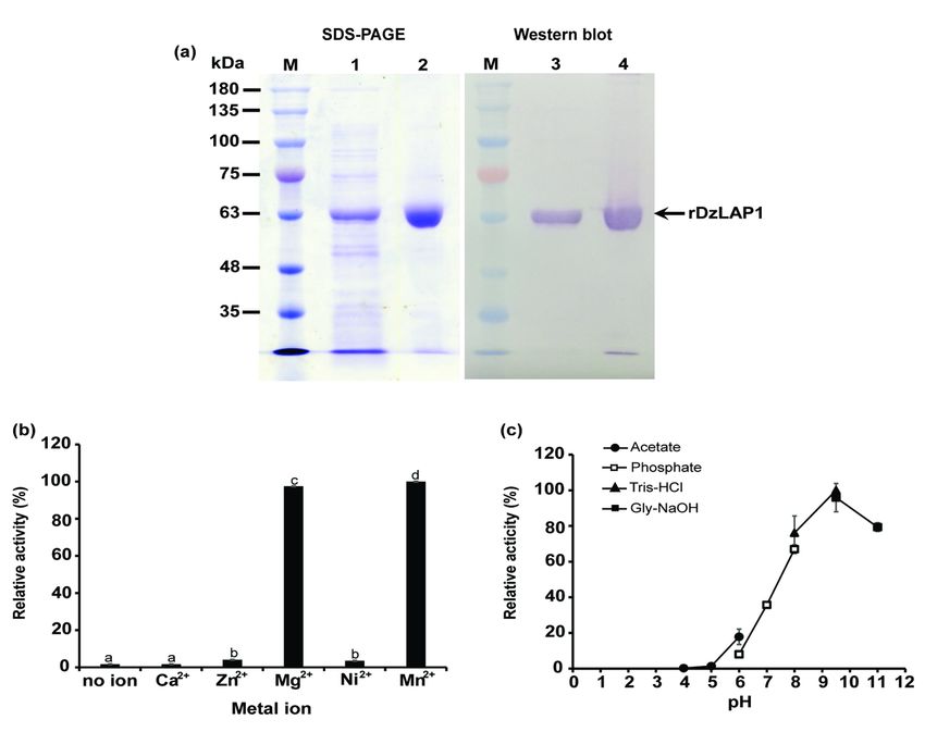
Cys-Gly is an important intermediate in the γ-glutamyl cycle of sulphur metabolism. It participates in
redox homeostasis and recycles amino acids in living cells. In mammals, Cys-Gly is the product of GSH
degradation via sequential γ-glutamyl transpeptidase (GGT), γ-glutamyl cyclotransferase (GGCT), and 5-
oxoprolinase (OPase) reactions [9]. Nevertheless, only the concerted actions of GGCT and OPase or a
GGT-independent pathway governed Cys-Gly production in Arabidopsis [10]. Cys-Gly is hydrolysed to Cys
and Gly by a dipeptidase. Cys-Gly is a pro-oxidant and may contribute to the intracellular redox
environment. Excess Cys-Gly was toxic to yeast cells [11, 12]. Therefore, enzymatic Cys-Gly regulation
may be vital. Moreover, the release of Cys is important as this amino acid can be converted to methionine
used in sulphur volatile production during fruit ripening [5].

Leucylaminopeptidases (LAPs, EC. 3.4.11.1) are members of the M17 enzyme family. They might process
intracellular proteins but their precise metabolic functions are still unknown. They preferentially hydrolyse
Cys-Gly in Bos taurus (cow) [13], Treponema denticola [14], and Arabidopsis [15]. They were originally
named LAPs as early reports suggested that they react with N-terminal leucines [16]. The cytosolic M20
metallopeptidase Dug1p has Cys-Gly peptidase activity. Its homologues occur in mammals and yeast but
not plants [17]. LAPs have six identical monomers comprising two conserved Zn-binding sites per subunit
[18]. They all participate in amino acid turnover but their other biological functions are complex and
species-specific. Escherichia coli LAP, also known as XerB, PepA, or CarP, is an aminopeptidase-
independent DNA-binding protein [19, 20]. It mediates site-specific plasmid and phage recombination [21,
22] and activates transcriptional carAB operons [20]. Multiple functions have been reported for
mammalian LAPs. Interferon-γ (IFN-γ) induction promoted high LAP accumulation. LAP may participate
in antigen presentation in humans [23, 24]. Animal LAPs have been implicated in oxidative lens aging
[25]. Plant LAP-A is a defence protein that plays important roles in floral development in Solanaceae [26-
29].

Plant LAPs are either acidic LAP-A or neutral LAP-N depending on their pI. LAP-A and LAP-N have distinct
biochemical properties and respond differently to developmental and environmental cues. LAP-A occurs
only in the Solanaceae, is induced by biotic and abiotic stress [30-32], and accumulates in reproductive
organs [33, 34]. In contrast, LAP-N is constitutively produced in all plants [32, 34]. LAP-A-silenced
tomatoes were relatively more susceptible to Manduca sexta (tomato hornworm) invasion than their wild
type counterparts [35]. LAP-A and LAP-N are molecular chaperones protecting proteins from heat damage
[36]. Here, we identified two LAP isoforms in durian fruit pulp. Of these, the LAP-N DzLAP was highly
expressed in the pulp. We biochemically characterised DzLAP1 expressed as a His-tagged protein in E.
coli. Our objective was to clarify its cooperative function in sulphur-volatile production via Cys liberation
from Cys-Gly cleavage. We discovered that DzLAP1 was localised to both the cytoplasm and chloroplast.
We examined its physiological roles in cytosolic GSH and plastidial peptide catabolism. To the best of
our knowledge, the present work is the first to report the involvement of LAPs and associate DzLAP1 with
durian fruit ripening.

Results
                                                   Page 3/46
LAP identification, protein sequence alignment, and phylogenetic analysis

According to the open-source Musang King durian cultivar genome database, DzLAP1_MK and
DzLAP2_MK were identified. Only one Chanee DzLAP isoform was present in the in-house RNA-seq data
obtained from ripe fruit pulp tissues (data not shown). Full-length Chanee DzLAP was aligned with the
LAPs of Musang King and tomato (Solanum lycopersicum; SlLAP1 and SlLAP2), potato (Solanum
tuberosum; StLAP1 and StLAP2), and Arabidopsis (Arabidopsis thaliana; AtLAP1 to AtLAP3). The
putative Chanee DzLAP was annotated as DzLAP1 (accession no. MN879753) and encoded DzLAP1. It
was represented as DzLAP1_CN in multiple alignment and phylogenetic neighbour-joining (NJ).
DzLAP1_CN clustered with Musang King DzLAP1_MK. DzLAP1 shared 99.3% and 89.7% identity with
DzLAP1_MK and DzLAP2_MK, respectively. The essentially high % identity between DzLAP1 and
DzLAP1_MK is possibly resulted from the SNP present among different cultivars. There were eight highly
conserved residues (K350, D355, K362, D375, D435, E437, R439, and L463 of DzLAP1) involved in
substrate binding or catalytic function (Fig. 1, arrowheads). Five conserved residues (K350, D355, D375,
D435, and E437) interacted with metal ions [37, 38] which are vital enzyme cofactors. These conserved
residues comprise a subset of the catalytic residues (Fig. 1, boxes). All protein sequences including
DzLAP1 but neither SlLAP1 nor StLAP1 harboured substitutions of all 28 LAP-A signature residues (Fig. 1,
highlights). Fully conserved residues were detected in the C-terminal region containing all essential active
residues participating in catalysis (Fig. 1, asterisks). In contrast, the N-terminal region harboured highly
variable amino acid sequences [34]. AtLAP1 lacked the signal peptide sequence but the N-terminal
regions of SlLAPs and StLAPs were slightly shorter than the other sequences. Only ~ 64 – 77% identity
was established when comparing two isoforms of both SlLAP and StLAP with two and three isoforms
of DzLAP_MK and AtLAP, respectively.

A phylogenetic analysis revealed that all plant LAPs clustered together and were separate from those of
bacteria (Fig. 2). However, Cys-Gly peptidase activity was detected in certain plant and bacterial LAPs.
Tomato SlLAPs and potato StLAPs were separate from those of Arabidopsis and durian and participated
in defence responses and protein catabolism (Fig. 2). In contrast, they had little activity towards Cys-Gly
[30]. Various LAPs resided in different cellular compartments and may have had divergent putative
functions (Fig. 2).

Gene expression analysis by qRT-PCR

In silico analysis disclosed that DzLAP_MK expression substantially varied among tissue types.
DzLAP1_MK expression was highest in durian pulp whereas DzLAP2_MK was expressed in other tissues
(Supplementary Fig. S1). Hence, we focused on DzLAP1_MK as it was nearly identical to Chanee DzLAP1.
The qRT-PCR revealed differential DzLAP1 expression at the unripe, midripe, and ripe stages of Chanee
and Monthong fruit pulp. DzLAP1 was significantly upregulated from the unripe to midripe stages but
downregulated by the ripe stage (Fig. 3a). DzLAP1 expression in the Chanee cultivar (white bars)
somewhat resembled that in the Monthong cultivar (black bars) (Fig. 3a). The phytohormones ethephon
and 1-MCP were applied to validate the association between DzLAP1 and fruit ripening. DzLAP1 was

                                                  Page 4/46
significantly downregulated in the fruit pulp treated with 1-MCP compared to the fruit pulp undergoing
natural ripening (control) or treated with the ripening agent ethephon (Fig. 3b). However, ethephon
treatment had relatively little influence on DzLAP1 expression.
In planta Cys-Gly dipeptidase activity analysis
Cys-Gly dipeptidase activity was assessed for crude fruit pulp extracts at the unripe and midripe stages
(Supplementary Fig. S3). Cys-Gly dipeptidase activity was significantly higher in the midripe (white bars)
than the unripe (black bars) sample (p < 0.05). This finding was consistent with that obtained from the
gene expression analysis (Fig. 3a). Cys-Gly dipeptidase activity was also confirmed for Nicotiana
benthamiana leaves co-infiltrated with 15 mM Cys-Gly and harbouring either pEAQ-DzLAP1 or pEAQ
(control). The leaves were collected and their Cys-Gly levels were quantified by HPLC. The Cys-Gly was
significantly higher in the control than the DzLAP1-overexpressing leaves (p < 0.05) (Fig. 4).
In vitro rDzLAP1 production and biochemical characterisation
Purified soluble rDzLAP1 produced by E. coli appeared as a single-band protein on SDS gel. Its molecular
weight was ~ 63 kDa and was confirmed by western blot (Fig. 5a). Relative to the BSA standard, the
purified DzLAP1 concentration was ~ 1.16 mg mL-1.
The metalloenzyme activity of DzLAP1 was tested by incubating the enzyme with Cys-Gly substrate in
the presence of Ca2+, Zn2+, Mg2+, Ni2+, and Mn2+. DzLAP1 activity was measured by a modified acidic
ninhydrin method detecting released cysteines are detected. The highest DzLAP1 activity was obtained
when the reaction systems contained Mg2+ and Mn2+. Conversely, Ca2+, Zn2+, and Ni2+ had minimal
effect as DzLAP1 activity in their presence did not significantly differ from that measured for the metal
ion-free control (Fig. 5b). Mg2+ was selected as the DzLAP1 cofactor in the enzyme kinetics assay.
Optimal DzLAP1 pH was established by incubating the enzyme with Cys-Gly at pH 4.0 – 11.0. DzLAP1
had maximum activity against Cys-Gly at pH 9.5 (Fig. 5c) and ~ 80% of the enzyme activity occurred in a
pH range of 8.0 – 11.0. At pH < 7.0, DzLAP1 activity was < 50% and at pH 4.0 – 5.0, the enzyme was
inactive.
To identify enzyme substrate specificity, 3.5 µg purified rDzLAP1 was incubated with various substrate
concentrations in the presence of 1 mM MgCl2 + 25 mM K3PO4 buffer (pH 8.0) at 37 °C for a specific
length of time. There was no DzLAP1 activity in the presence of GSH or γ-Glu-Cys (Table 1). DzLAP1 had
positive activity against various α-linked dipeptides. Therefore, it was proposed that this enzyme is a Cys-
Gly dipeptidase as it had maximum catalytic efficiency in the presence of Cys-Gly. The kcat/Km for Cys-
Gly was ~ 118× and ~ 6× higher than those for Met-Gly and Leu-Gly, respectively (Table 1). However, a
previous report indicated that LAP preferred N-terminal Leu peptides and had high affinity for Leu-Gly (Km
= 0.35 mM) [30].
Table 1. DzLAP1 kinetics on different substrates.
Page 5/46Substrate kcat (min-1) Km (mM) kcat/Km (min-1 mM-1)
GSH N.D. N.D. N.D.
γ-Glu-Cys N.D. N.D. N.D.
Cys-Gly 74.8 ± 8.1 1.6 ± 0.3 46.2
Met-Gly 2.1 ± 0.4 5.2 ± 1.3 0.4
Leu-Gly 2.9 ± 0.4 0.4 ± 0.1 8.2
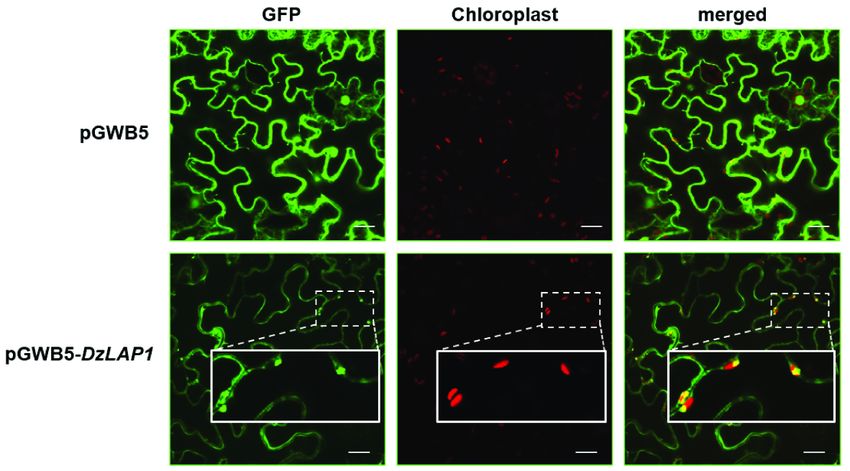
Subcellular DzLAP1 localisation in N. benthamiana
An in silico analysis predicted that DzLAP1 is a chloroplast-localised protein. The pGWB5-DzLAP1 and
the silencing suppressor p19 were co-expressed in Agrobacterium tumefaciens GV3101 infiltrated in 4-wk
N. benthamiana leaves. The in planta assay showed that GFP-tagged DzLAP1 was a soluble protein
probably localised to the cytosol (Fig. 6). Fluorescence signals were detected in the chloroplasts (Fig. 6,
insets). Hence, durian DzLAP1 may be localised either to cytosol or the chloroplasts.
Discussion
Durian fruit pulp accumulates large quantities of GSH [8] which is vital for plant cell homoeostasis [39]
and stores Cys via incorporation with Glu and Gly in the γ-glutamyl cycle [40]. Cys is recycled from the
hydrolysis of Cys-Gly which is a by-product of GSH breakdown. This mechanism may generate abundant
ethylene precursors and sulphur-volatile compounds via Met synthesis. Both pathways promote durian
fruit ripening and the malodour associated with it. Cys-Gly was detected in durian fruit pulp [8]. Hence, the
γ-glutamyl cycle must be activated in durian fruit ripening. In the present study, we aimed to identify and
characterise the DzLAPs involved in the aforementioned biochemical processes. There has been little
published information on the involvement of Cys-Gly dipeptidase in the γ-glutamyl cycle [15, 17].
LAPs are highly conserved metallopeptidases in animals, plants, and microorganisms [41]. Several LAPs
were identified in Arabidopsis [15] and durian (this work). Arabidopsis has three LAPs encoding AtLAP1–
AtLAP3. DzLAP1_MK and DzLAP2_MK were detected in the durian cultivar Musang King genome [5]. We
focused on DzLAP1_MK as it was highly expressed in durian fruit pulp (Supplementary Fig. S1) and GSH
and γ-EC accumulated there [8]. DzLAP1_MK showed 99.3% identity with DzLAP1 in the durian cultivar
Chanee. DzLAP1 was the only isoform detected in our in-house RNA-seq data. A primary protein sequence
analysis (Fig. 1) confirmed that DzLAP1 is LAP-N as it contains the conserved substrate binding,
catalytic, and metal ion-binding residues associated with the enzyme mechanism.
Postharvest DzLAP1 expression analyses showed upregulation at the midripe stage. Thus, DzLAP1 may
hydrolyse Cys-Gly to Cys and Gly and cause the strong malodour associated with durian fruit pulp
ripening. Relative DzLAP1 expression was similar for Chanee and Monthong at all ripening stages (Fig.
Page 6/463a). However, the Cys-Gly content was significantly higher in Chanee than Monthong (p < 0.01) [8].
Therefore, DzLAP1 is not cultivar-dependent and does not account for the relative differences between
Chanee and Monthong in terms of fruit odour intensity. The competitive ethylene inhibitor 1-MCP
significantly repressed DzLAP1 during postharvest ripening (p < 0.05) (Fig. 3b). Thus, DzLAP1 might play
an important role in this process and LAP1 may be associated with the early stages of ripening. In
contrast, LAP-Ns in other plant species are constitutively expressed in all organs [34, 42]. We compared
LAP expression during tomato fruit ripening and found that the LAP-A levels were slightly lower at the
breaker or ethylene-producing stages than they were in mature green fruit (Supplementary Fig. S2a).
Tomato LAP-N is constitutively expressed at all fruit developmental stages (Supplementary Fig. S2b).
Therefore, LAPs are not implicated in tomato fruit development or ripening.
Cys-Gly dipeptidase activity was observed in crude durian fruit extract (Supplementary Fig. S3) and in co-
infiltrated N. benthamiana leaves overexpressing DzLAP1 (Fig. 4). Cys-Gly hydrolytic activity
was higher in midripe than unripe durian pulp extracts (Supplementary Fig. S3). This finding
was consistent with DzLAP1 upregulation at the midripe stage (Fig. 3a). Thus, Cys-Gly dipeptidase was
functional in planta. The observed Cys-Gly dipeptidase activity of DzLAP1 in
planta upholds our aforementioned hypothesis and suggests that the Cys generated by Cys-
Gly hydrolysis is converted into sulphur volatiles contributing to the malodour of ripening durian
fruit. Hence, the γ-glutamyl cycle is essential for recycling GSH and its amino acid constituents and
for durian fruit ripening. Nevertheless, an alternative pathway to GSH recycling has been suggested in
other plants. In ripening tomato fruit, the oxidised glutathione (GSSG)-catabolising enzyme γ-glutamyl
transpeptidase (GGT) plays a major role in glutathione degradation that releases Cys-Gly and Glu [43]. In
Arabidopsis, an increased GSH content and a drop in Cys-Gly content were observed in the ggt1 knockout
line when compared with those of wild type plants [44]. Unlike tomato and Arabidopsis, however, durian
fruit is rich in sulphur. For this reason, γ-glutamyl cycle in durian might have evolved to be more active
during ripening.
An in vitro biochemical assay of rDzLAP1 (Fig. 5) disclosed that LAPs have pH optima in the range of
8.0–11.0 and metal ion dependency [29, 45, 46]. DzLAP1 has high catalytic efficiency (kcat/Km ) for Cys-
Gly (Km = 1.6 mM). Therefore, DzLAP1 is a metalloenzyme and a Cys-Gly dipeptidase. The
aforementioned Km resembles those for other Cys-Gly peptidases such as AtLAP1 [15], yeast Dug1p [17],
and bacterial TdLAP [14] all of which were ≤ 1.3 mM. Thus, the affinity of LAP for Cys-Gly corresponds to
the millimolar physiological GSH concentration range in living cells [47, 48]. Durian is more abundant in
GSH than many other fruits and vegetables [8, 49]. For this reason, the cellular GSH level in durian should
be higher than the general physiological concentration of this substance and Cys-Gly dipeptidases have
highly conserved activity across species. As for other degradable α-linked dipeptides, DzLAP1 could not
hydrolyse γ-Glu-Cys-Gly (Table 1). Consequently, this enzyme might have specificity for α-peptide bonds.
However, earlier studies did not investigate the affinities of LAPs for γ-linked dipeptides.
Subcellular protein localisation can elucidate the correlations between the native functions and the
physiological substrates of an enzyme. DzLAP1 is a dual-target protein in the chloroplasts and the
Page 7/46cytosol (Fig. 6). However, it harbours a plastidial transit peptide sequence. DzLAP1 transcripts may have
an alternative codon to initiate ribosomal translation that bypasses the first start codon [34] and/or forms
secondary RNA structures in the sequences adjacent to it [50]. Delta-2-isopentenyl pyrophosphate:tRNA
isopentenyl transferase (MOD5) is encoded by a single gene, has two translational initiation sites, and is
localised to the mitochondria, cytosol, and nucleus [51]. Plastidial DzLAP1 may recycle protein and
perform other tasks. As chloroplasts participate in cellular metabolism, they may respond to various
stressors [52]. The present study did not clarify the functions of plastidial DzLAP1. However, we propose
that, like AtLAPs, it is a molecular chaperone preventing misfolded protein accumulation and other
adverse effects [36]. Partial DzLAP1 localisation to the chloroplasts enhances its chaperone and/or
protease activity in specific suborganellar compartments. Similar observations were reported for CRP-like
protein (Clp) and filamentous temperature-sensitive protein (FtsH). These are major conserved ATP-
dependent chaperone and degradative proteases in the stroma and on the thylakoid membranes,
respectively [53]. Further experimentation is needed to confirm the role of DzLAP1 in the plastids during
fruit ripening.
As Cys-Gly has pro-oxidant activity, its concentration must be regulated. Cys-Gly promotes the formation
of reactive oxygen species (ROS) such as hydrogen peroxide and superoxide anion in the presence of
certain metal ions [54]. The next step is the oxidation of highly reduced thiols such as GSH [55]. Cytosolic
localisation and the enzyme kinetics of DzLAP1 suggest that it hydrolyses Cys-Gly in the cytoplasmic γ-
glutamyl cycle and controls cytosolic Cys-Gly levels. Cys-Gly may also be a substrate for cytosolic
DzLAP1 in vivo. In this manner, DzLAP1 regulates Cys-Gly concentration. As DzLAP1 is localised to the
cytosol and expressed where ethylene and sulphur volatiles accumulate, it might participate in durian
fruit ripening. In fact, the slightly alkaline pH of the cytoplasm [56] and the chloroplast stroma [57, 58] is
conducive to DzLAP1 activity.
DzLAP1 and the LAP-N of Arabidopsis and Solanaceae all have similar catalytic, substrate, and metal
ion-binding residues. However, DzLAP1 expression differs from those of the latter genes. Durian
accumulates high levels of sulphur volatiles that impart a strong flavour and unique odour to the fruit.
However, the levels of these thiols must be tightly controlled. Expression of the genes encoding sulphur-
metabolising enzymes must be regulated during fruit ripening. The DzLAP1 promoter region may have
been modified to produce the optimal DzLAP1 content, limit the amount of Cys-Gly, catalyse Cys
recycling, and generate sulphur volatiles and ethylene during fruit ripening. A similar finding was reported
for durian-specific upregulation of the methionine γ-lyase gene [5]. The latter is responsible for sulphur
volatile production in plants and bacteria [59, 60]. An isoform of this enzyme associated with fruit
ripening has been identified [5]. Figure 7 is a schematic diagram summarising the putative functions of
DzLAP1 in durian fruit pulp cytoplasm and chloroplasts.
Conclusions
Based on durian cultivar Musang King genomic data, we identified and characterised the LAP-N gene
encoding leucylaminopeptidase (LAP). This enzyme is highly expressed in durian fruit pulp and was
Page 8/46designated DzLAP1. We established that DzLAP1 is active in the early stages of durian fruit ripening.
However, it is not cultivar-dependent and may not be responsible for the fact that ripe Chanee durian fruit
has a stronger odour than ripe Monthong durian fruit. An in planta assay in Nicotiana benthamiana
leaves demonstrated Cys-Gly dipeptidase activity. The enzyme kinetics and subcellular localisation of
DzLAP1 indicate that it has in vivo Cys-Gly dipeptidase activity. Its presence in the cytosol suggests that it
participates in the γ-glutamyl cycle and is adjacent to intracellular ethylene and sulphur volatile
production sites. The plastidial DzLAP1 isoform may participate in protein turnover and/or protection.
The present study partially elucidated the mechanisms of sulphur metabolism in plant tissues
accumulating high levels of sulphur volatiles.
Methods
Plant materials and growth conditions
Mature durian (Durio zibethinus L. ‘Chanee’) fruits were harvested 95 d after anthesis from a commercial
orchard in Trat Province, Thailand in early April 2017. The samples were maintained at room temperature
(30 °C) before being peeled on days 1, 2, and 4 (unripe, midripe, and ripe postharvest stages, respectively).
Monthong served as a representative sample for gene expression analysis. Its three different ripening
stages were also evaluated but their timings (days) slightly differed from those of Chanee. Monthong
fruits were harvested at day 105 after anthesis. Unripe, midripe, and ripe Monthong fruit samples were
peeled and analysed at 1 d, 3 d, and 5 d after storage at room temperature, respectively [6].
To investigate the association between DzLAP1 and durian fruit ripening, unripe Monthong samples were
treated either with ethephon or 1-methylcyclopropene (1-MCP). These synthetic phytohormones have
opposite modes of action. Ethephon is converted to ethylene which enhances ripening. In contrast, 1-MCP
is an ethylene antagonist and delays ripening. The treated samples were compared with controls
naturally ripened according to the method of Khaksar et al. (2019). Three biological replicates were used
and each comprised a single durian fruit harvested from a separate tree.
Crude extract from durian pulp was used to determine Cys-Gly dipeptidase activity. Durian cultivar
Chanee was obtained from a local market in Nonthaburi Province, Thailand. Postharvest samples were
collected at the unripe and midripe stages. Three biological replicates were used and each consisted of a
single lobe harvested from a separate durian fruit.
Nicotiana benthamiana plants were raised for agroinfiltration. Seeds were sown on peat moss and the
seedlings were grown at 25 °C under a 16 h/8 h light/dark photoperiod and 4,500 lx (artificial light). Two-
week-old plants were individually transplanted to pots and raised under the same conditions for another 2
wks.
Phylogenetic analysis and putative LAP identification in durian fruit
Page 9/46The protein sequences of LAPs harbouring Cys-Gly dipeptidase activity in Treponema denticola
(accession no. WP_010698434.1) [14] and Arabidopsis thaliana (AtLAP1 and AtLAP3; accession nos.
P30184.1 and Q8RX72.1, respectively) [15] served as queries for a BLAST search against the D.
zibethinus cultivar Musang King NCBI database. The MaGenDB database [61] confirmed DzLAP
isoforms.
To establish the phylogenetic relationships among LAPs, the amino acid sequences of the putative
DzLAPs and other LAPs deposited in NCBI were subjected to ClustalW multiple alignment. A neighbour
joining (NJ) tree was created with MEGA v. 7 [62] using 1,000 bootstrap replicates.
Determination of tissue-specific DzLAP1 expression
A search of the DzLAPs against the genomic data for durian cultivar Musang King disclosed two
candidate genes (accession nos. XM_022894525.1 (LOC111299369) and XM_022874012.1
(LOC111284913)) annotated as DzLAP1-like and named DzLAP1_MK and DzLAP2_MK, respectively.
Attention was directed to the durian fruit pulp as it accumulated several sulphur volatiles. DzLAP1
expression was analysed in silico in various fruit tissues. To compare relative DzLAP expression in
different tissues, normalised total read counts (RCs) derived from Illumina reads were obtained from the
Sequence Read Archive (SRA) resource and processed according to the method of Khaksar et al. (2019).
RNA-seq data were obtained for SRX3188225 (root), SRX3188222 (stem), SRX3188226 (leaf), and
SRX3188223 (aril/pulp) [5]. A heatmap based on the RCs was constructed using MetaboAnalyst v. 4.0
[63].
Gene expression analysis by qRT-PCR
Total RNA was isolated from Chanee and Monthong durian cultivar pulps with PureLink® plant RNA
reagent (Thermo Fisher Scientific, Waltham, MA, USA) following the manufacturer’s instructions. DNase-
treated RNA sample quantity and integrity were assessed. Approximately 1 µg total RNA was reverse-
transcribed to cDNA with a RevertAid first-strand cDNA synthesis kit (Thermo Fisher Scientific, Waltham,
MA, USA). Gene-specific primers listed in Supplementary Table S1. The qRT-PCR elucidated DzLAP1
expression in unripe, midripe, and ripe durian fruit. The reactions were performed in 10 μL total volume in
a 96-well PCR plate. The cDNA and primers were combined with Luna® universal qPCR master mix (New
England Biolabs, Ipswich, MA, USA). PCR was run in a CFX Connect™ real-time PCR detection system
coupled to CFX Manager™ (Bio-Rad Laboratories, Hercules, CA, USA). Single amplicon production was
verified by melting curve analysis. Relative gene expression levels were calculated by the 2−ΔΔCt method
[64] based on the cycle threshold (Ct) of the gene relative to the reference gene elongation factor 1 alpha
(EF-1α). There were three independent biological replicates in the qRT-PCR. Gene expression analyses
were also conducted on the ethephon- and 1-MCP-treated samples and the naturally un/ripened samples
(controls).
In planta Cys-Gly dipeptidase activity assay
Page 10/46To determine Cys-Gly dipeptidase activity in durian fruit pulp, crude enzyme was extracted from it at the
unripe and midripe stages. Pulp samples were separately collected, frozen in liquid nitrogen, and
pulverised in the MM400 mixer mill (Retsch GmbH, Haan, Germany) at 30 Hz for 1 min. Then 250 mg of
each sample was dissolved in 2.5 mL lysis buffer (50 mM K3PO4, pH 8.0) and gently mixed at 4 °C for 1
h. The samples were centrifuged at 14,000 × g and 4 °C for 5 min and the supernatant was collected. An
enzyme activity assay was performed by incubating durian fruit pulp extract with 20 mM Cys-Gly in
reaction buffer (50 mM K3PO4 buffer (pH 8.0) + 1 mM MgCl2) at 30 min, 1 h, and 3 h. The enzyme activity
was measured by a modified acidic ninhydrin method [65]. Briefly, 50 µL of each enzyme reaction system
was terminated with 50 µL glacial acetic acid followed by 50 µL acidic ninhydrin solution (250 mg
ninhydrin in 6 mL glacial acetic acid + 4 mL HCl), boiled for 9 min, and cooled with tap water. The pink
endpoint indicated the reaction between the released Cys and ninhydrin under acidic conditions. Colour
intensity was measured spectrophotometrically at A560 (BioTex, Winooski, VT, USA). Three independent
biological replicates were used.
In planta Cys-Gly dipeptidase activity was also investigated. Full-length DzLAP1 was amplified with
Phusion Hot Start II high-fidelity DNA polymerase (Thermo Fisher Scientific, Waltham, MA, USA) using
Chanee cDNA as a template. The gene-specific primers (excluding the stop codon) listed in
Supplementary Table S1 were used in the PCR. The PCR product was cloned into a pCR™8/GW/TOPO®
TA cloning vector (Invitrogen, Carlsbad, CA, USA) and generated pTOPO-DzLAP1. The latter was then
sequenced. DzLAP1 was transferred to the pEAQ3 destination vector and fused at the C-terminal with 6×
His [66] via Gateway® LR Clonase® II (Invitrogen, Carlsbad, CA, USA). The pEAQ-DzLAP1 product was
transformed into Agrobacterium tumefaciens GV3101 by electroporation.
A. tumefaciens bearing the DzLAP1-6xHis or the empty pEAQ vector (control) was infiltrated into 4 wk
plants. Briefly, cells from each culture were washed and suspended in MM buffer (10 mM MES buffer +
10 mM MgCl2; pH 5.6). The cell suspension was adjusted at A600 to OD = 0.6. Acetosyringone was added
to make up 200 µM final concentration and the suspension was kept in the dark at 30 °C for 2 h before
infiltration. At day 3 after agroinfiltration, the leaves were infiltrated with 15 mM Cys-Gly, incubated for 1 h
[67], freeze-dried, and kept in a dry place at 30 °C until subsequent metabolite analysis by HPLC. To
measure the foliar Cys-Gly levels, the co-infiltrated leaves were pulverised in the MM400 mixer mill
(Retsch GmbH, Haan, Germany) at 30 Hz for 1 min. Each sample was suspended in 0.1 M HCl extraction
buffer at 1 mg/50 µL. Then, 10 mM N-acetylcysteine (internal standard) was added and mixed by
shaking at 250 rpm and 37 °C for 2.5 h. The samples were centrifuged at 14,000 × g and 30 °C for 5 min.
The soluble fraction was transferred to a new microcentrifuge tube, combined with acetonitrile (1:1), and
centrifuged at 14,000 × g and 30 °C for 5 min. The pellet was removed and the supernatant was dried
with a CentriVap benchtop vacuum concentrator (Labconco Corp., Kansas City, MO, USA). The dried
samples were re-dissolved in 200 µL deionised H2O, passed through a 0.22-µm syringe filter, and
subjected to HPLC analysis. Ten microlitres of each sample was injected into a C18 column (250 mm ×
4.6 mm; Phenomenex, Torrance, CA, USA) using acetonitrile (5%) in 2% perchloric acid as a mobile phase.
Page 11/46The flow rate was 1 mL/min and the temperature was 40 °C. The Cys-Gly peak was detected at 210 nm
[15]. Three independent biological replicates were used and each consisted of a separate leaf.
DzLAP1 cloning and expression in Escherichia coli
The putative DzLAP1 was amplified with Phusion Hot Start II DNA polymerase (Thermo Fisher Scientific,
Waltham, MA, USA) and midripe Chanee durian cultivar cDNA served as a template. The PCR temperature
profile was as follows: initial denaturation at 98 °C for 30 s; 30 cycles of 98 °C for 10 s; 57 °C for 10 s; 72
°C for 1 min; and a final extension at 72 °C for 5 min. Gene-specific forward and reverse primers were
designed according to the DzLAP1_MK sequence (Supplementary Table S1). The signal sequences
predicted by the ChloroP1.1 and TargetP servers were excluded. The putative DzLAP1 PCR product was
excised with restriction enzymes (FastDigest™; Thermo Fisher Scientific, Waltham, MA, USA) and cloned
into a pET21b vector (Merck KGaA, Darmstadt, Germany). The product was pET21b-DzLAP1 in-frame
with 18 nucleotides encoding 6× His residues at the C-terminus. It was transformed into E. coli TOP10
(K2500-20; Invitrogen, Carlsbad, CA, USA). Bacterial colonies were raised on LB agar supplemented with 1
mg mL-1 ampicillin and analysed by colony PCR. The nucleotide sequences of the positive clones were
verified by 1st BASE DNA sequencing.
Recombinant DzLAP1 (rDzLAP1) production and purification
The pET21b-DzLAP1 was transformed into the T7 host E. coli SoluBL21 (DE3). Cells harbouring the
recombinant plasmid were incubated overnight in LB broth supplemented with 1 mg mL-1 ampicillin. The
starter culture was inoculated into fresh LB broth supplemented with 1 mg mL-1 ampicillin and incubated
at 37 °C with shaking at 250 rpm until OD 600 = 0.4 – 0.5. The rDzLAP1 was generated by induction with 1
mM isopropyl β-D-1-thiogalactopyranoside (IPTG) at 30 °C and shaking at 250 rpm for 17 h. The cells
were harvested by centrifugation at 5,000 × g and 37 °C for 10 min, suspended in buffer A (25 mM K3PO4
buffer + 0.3 M NaCl; pH 7.2), and lysed by ultrasonication. Soluble intracellular proteins were collected by
centrifugation at 7,500 × g and 4 °C for 30 min, analysed by western blot (6× His epitope tag antibody;
Thermo Fisher Scientific, Waltham, MA, USA), and stored at 4 °C until purification.
The crude extract was loaded onto a HisTrapTM column (Merck, Darmstadt, GER) pre-equilibrated with
buffer A. The column was washed with excess buffer A and the rDzLAP1 was eluted with buffer B (25
mM K3PO4 buffer + 0.3 M NaCl + 150 mM imidazole; pH 7.2). The samples were analysed by SDS-PAGE
and western blot. The pooled purified rDzLAP1 fraction was dialysed against 25 mM K3PO4 buffer (pH
7.2). The protein concentration was determined by a modified Bradford assay [68]. The reference protein
standard was BSA.
Enzymatic rDzLAP1 assay
The metal ion dependency of rDzLAP1 was evaluated. The enzyme was incubated at 37 °C for 15 min
with 7.5 mM Cys-Gly in 25 mM K3PO4 buffer (pH 7.2) in the presence of 0 mM or 1 mM Ca2+, Zn2+, Mg2+,
Page 12/46Ni2+, or Mn2+. The total reaction volume was 50 µL. Enzyme activity was measured by a modified acidic
ninhydrin method [65]. To determine the optimum enzyme pH, 50-µL reaction systems were prepared by
incubating rDzLAP1 with 7.5 mM Cys-Gly and 1 mM Mg2+ at various pH (acetate buffer, pH 4 – 6;
phosphate buffer, pH 6 – 8; Tris-HCl buffer, pH 8 – 9.5; glycine-NaOH buffer, pH 9.5 – 11) at 37 °C for 15
min. Maximum enzyme activity was defined as 100% relative activity.
To assess enzyme kinetics, 3.5 µg purified rDzLAP1 was incubated with 0–10 mM Cys-Gly, γ-Glu-Cys,
GSH, Met-Gly, or Leu-Gly in the presence of 1 mM MgCl2 in 25 mM K3PO4 buffer (pH 8.0) at 37 °C for a
specific length of time. The reactions were terminated with 0.13 N HCl. For γ-Glu-Cys and GSH, the
reactions were evaluated by a modified acidic ninhydrin assay. For Cys-Gly, Met-Gly, and Leu-Gly, the
reactions were analysed by monitoring the decrease in absorbance of the free peptides at A230, with
minor modifications [69]. Enzyme kinetics were measured with OriginPro® 2017 (OriginLab Corp.,
Northampton, MA, USA).
Subcellular localisation
Full-length DzLAP1 was amplified and cloned into a pCR™8/GW/TOPO® TA vector (Invitrogen, Carlsbad,
CA, USA) according to the in planta Cys-Gly dipeptidase activity assay. The pTOPO-DzLAP1 product was
then sequenced. The DzLAP1 was transferred to the pGWB5 destination vector and fused at the C-
terminal with green fluorescent protein (GFP) [70] via Gateway® LR Clonase® II (Invitrogen, Carlsbad, CA,
USA). The pGWB5-DzLAP1 product was transformed into A. tumefaciens GV3101 by electroporation.
Agrobacterium tumefaciens bearing the DzLAP1-GFP construct and A. tumefaciens with the silencing
suppressor p19 gene [71] were co-infiltrated into 4-wk plants as previously described, with some
modifications. Briefly, cells from each culture were washed and suspended in MM buffer (10 mM MES
buffer + 10 mM MgCl2; pH 5.6). Cell suspensions harbouring DzLAP1 and p19 were adjusted at A600 to
OD = 0.8 and 0.6, respectively, and combined in a 1:1 ratio. Acetosyringone was added to the mixture to a
final concentration of 200 µM and the suspension was maintained in the dark at room temperature for 2
h before infiltration. At day 3 after infiltration, autofluorescence was visualised under a FluoView® FV10i-
DOC confocal laser scanning microscope (Olympus Corp., Tokyo, Japan). GFP, chloroplast
autofluorescence, and phase-contrast detection excitation/emission were recorded at 473/510 nm,
559/600 nm, and 559/600 nm, respectively.
Statistical analysis
All data were analysed with SPSS Statistics® v. 22.0 (IBM Corp., Armonk, NY, USA). One-way ANOVA
identified significant differences among mean enzyme activity levels in the absence and presence of
metal ions. Cys-Gly dipeptidase gene expression levels and enzyme activity at various ripening stages
and in response to different ripening regulators were analysed by Tukey’s HSD multiple comparisons test
(p < 0.05). Student’s t-test (p < 0.05) identified significant differences between N. benthamiana leaves
overexpressing DzLAP1 and the control in terms of in planta Cys-Gly dipeptidase activity.
Page 13/46Abbreviations
GSH: glutathione
γ-EC: γ-glutamylcysteine
Cys-Gly: cysteinylglycine
Cys: cysteine
LAP: leucylaminopeptidase
DzLAP: Durio zibethinus L. leucylaminopeptidase gene
Declarations
Ethics approval and consent to participate
Not applicable
Consent for publication
Not applicable
Availability of data and material
The datasets used and/or analysed during the current study are available from the corresponding author
on reasonable request.
Competing interests
The authors declare that they have no competing interests.
Funding
This study was supported by the Thailand Research Fund (No. RSA6080021), Chulalongkorn University
(No. GRU 6203023003-1 to S.S.). P.P received the Second Century Fund (C2F) Postdoctoral Fellowship of
Chulalongkorn University. The funding bodies were not involved in the design of the study and plant
sample collection, data analyses, data interpretation, and writing of the manuscript.
Authors’ contributions
S.S. conceived the study. P.P. and S.S. designed the experiments. P.P. conducted the experiments. P.P. and
S.S. analysed the data and prepared the manuscript. All authors have read and approved the manuscript.
Acknowledgements
Page 14/46The authors thank Kittiya Tantisuwanichkul and Pinnapat Pinsorn for providing the durian cDNA
samples, Drs. Gholamreza Khaksar and Lalida Sangpong for their assistance with the RNA-seq data
analysis, Tsuyoshi Nakagawa of Shimane University, Shimane, Japan, for providing the Gateway®
expression vector pGWB5, Sophien Kamoun of Sainsbury Laboratory, Norwich, Norfolk, UK for providing
A. tumefaciens GV3101 harbouring pJL3:p19, and Plant Bioscience Ltd. (Norwich, UK) for supplying the
pEAQ vector.
References
1. Charoenkiatkul S, Thiyajai P, Judprasong K: Nutrients and bioactive compounds in popular and
indigenous durian (Durio zibethinus murr.). Food Chem. 2016;193:181-186.
2. Arancibia-Avila P, Toledo F, Park Y-S, Jung S-T, Kang S-G, Heo BG, Lee S-H, Sajewicz M, Kowalska T,
Gorinstein S: Antioxidant properties of durian fruit as influenced by ripening. LWT-food Sci Technol.
2008;41:2118-2125.
3. Maninang JS, Lizada MCC, Gemma H: Inhibition of aldehyde dehydrogenase enzyme by Durian
(Durio zibethinus Murray) fruit extract. Food Chem. 2009;117:352-355.
4. Haruenkit R, Poovarodom S, Vearasilp S, Namiesnik J, Sliwka-Kaszynska M, Park Y-S, Heo B-G, Cho
J-Y, Jang HG, Gorinstein S: Comparison of bioactive compounds, antioxidant and antiproliferative
activities of Mon Thong durian during ripening. Food Chem. 2010;118:540-547.
5. Teh BT, Lim K, Yong CH, Ng CCY, Rao SR, Rajasegaran V, Lim WK, Ong CK, Chan K, Cheng VKY: The
draft genome of tropical fruit durian (Durio zibethinus). Nat Genet. 2017;49:1633.
6. Khaksar G, Sangchay W, Pinsorn P, Sangpong L, Sirikantaramas S: Genome-wide analysis of the Dof
gene family in durian reveals fruit ripening-associated and cultivar-dependent Dof transcription
factors. Sci Rep. 2019;9:1-13.
7. Khaksar G, Sirikantaramas S: Auxin response factor 2A is part of the regulatory network mediating
fruit ripening through auxin-ethylene crosstalk in durian. Front Plant Sci. 2020;11.
8. Pinsorn P, Oikawa A, Watanabe M, Sasaki R, Ngamchuachit P, Hoefgen R, Saito K, Sirikantaramas S:
Metabolic variation in the pulps of two durian cultivars: Unraveling the metabolites that contribute to
the flavor. Food Chem. 2018;268:118-125.
9. Martin MN, Saladores PH, Lambert E, Hudson AO, Leustek T: Localization of members of the γ-
glutamyl transpeptidase family identifies sites of glutathione and glutathione s-conjugate hydrolysis.
Plant Physiol. 2007;144:1715-1732.
10. Ohkama-Ohtsu N, Oikawa A, Zhao P, Xiang C, Saito K, Oliver DJ: A γ-glutamyl transpeptidase-
independent pathway of glutathione catabolism to glutamate via 5-oxoproline in Arabidopsis. Plant
Physiol. 2008;148:1603-1613.
11. Kumar C, Igbaria A, D'autreaux B, Planson AG, Junot C, Godat E, Bachhawat AK, Delaunay‐Moisan A,
Toledano MB: Glutathione revisited: a vital function in iron metabolism and ancillary role in thiol‐
redox control. EMBO J. 2011;30:2044-2056.
Page 15/4612. Srikanth CV, Vats P, Bourbouloux A, Delrot S, Bachhawat AK: Multiple cis-regulatory elements and the
yeast sulphur regulatory network are required for the regulation of the yeast glutathione transporter,
Hgt1p. Curr Genet. 2005;47:345-358.
13. Cappiello M, Lazzarotti A, Buono F, Scaloni A, D'ambrosio C, Amodeo P, Méndez BL, Pelosi P, Del
Corso A, Mura U: New role for leucyl aminopeptidase in glutathione turnover. Biochem J.
2004;378:35-44.
14. Chu L, Lai Y, Xu X, Eddy S, Yang S, Song L, Kolodrubetz D: A 52-kDa leucyl aminopeptidase from
Treponema denticola is a cysteinylglycinase that mediates the second step of glutathione
metabolism. J Biol Chem. 2008;283:19351-19358.
15. Kumar S, Kaur A, Chattopadhyay B, Bachhawat AK: Defining the cytosolic pathway of glutathione
degradation in Arabidopsis thaliana: role of the ChaC/GCG family of γ-glutamyl cyclotransferases as
glutathione-degrading enzymes and AtLAP1 as the Cys-Gly peptidase. Biochem J. 2015;468:73-85.
16. Spackman DH, Smith EL, Brown DM: Leucine aminopeptidase IV. Isolation and properties of the
enzyme from swine kidney. J Biol Chem. 1955;212:255-269.
17. Kaur H, Kumar C, Junot C, Toledano MB, Bachhawat AK: Dug1p is a cys-gly peptidase of the γ-
glutamyl cycle of Saccharomyces cerevisiae and represents a novel family of cys-gly peptidases. J
Biol Chem. 2009;284:14493-14502.
18. Carpenter FH, Vahl JM: Leucine aminopeptidase (bovine lens) mechanism of activation by Mg2+ and
Mn2+ of the zinc metalloenzyme, amino acid composition, and sulfhydryl content. J Biol Chem.
1973;248:294-304.
19. McCulloch R, Burke ME, Sherratt DJ: Peptidase activity of Escherichia coli aminopeptidase A is not
required for its role in Xer site‐specific recombination. Mol Microbiol. 1994;12:241-251.
20. Charlier D, Kholti A, Huysveld N, Gigot D, Maes D, Thia-Toong T-L, Glansdorff N: Mutational analysis
of Escherichia coli PepA, a multifunctional DNA-binding aminopeptidase. J Mol Biol. 2000;302:409-
424.
21. Stirling C, Colloms S, Collins J, Szatmari G, Sherratt D: xerB, an Escherichia coli gene required for
plasmid ColE1 site‐specific recombination, is identical to pepA, encoding aminopeptidase A, a protein
with substantial similarity to bovine lens leucine aminopeptidase. EMBO J. 1989;8:1623-1627.
22. Colloms SD: Leucyl aminopeptidase PepA. In: Handbook of proteolytic enzymes. Elsevier; 2004: 905-
910.
23. Harris C, Hunte B, Krauss M, Taylor A, Epstein L: Induction of leucine aminopeptidase by interferon-
gamma. Identification by protein microsequencing after purification by preparative two-dimensional
gel electrophoresis. J Biol Chem. 1992;267:6865-6869.
24. Towne CF, York IA, Neijssen J, Karow ML, Murphy AJ, Valenzuela DM, Yancopoulos GD, Neefjes JJ,
Rock KL: Leucine aminopeptidase is not essential for trimming peptides in the cytosol or generating
epitopes for MHC class I antigen presentation. J Immunol. 2005;175:6605-6614.
25. Sharma KK, Kester K: Peptide hydrolysis in lens: role of leucine aminopeptidase, aminopeptidase III,
prolyloligopeptidase and acylpeptidehydrolase. Curr Eye Res. 1996;15:363-369.
Page 16/4626. Hildmann T, Ebneth M, Peña-Cortés H, Sánchez-Serrano JJ, Willmitzer L, Prat S: General roles of
abscisic and jasmonic acids in gene activation as a result of mechanical wounding. Plant Cell. 1992;
4:1157-1170.
27. Herbers K, Prat S, Willmitzer L: Functional analysis of a leucine aminopeptidase from Solatium
tuberosum L. Planta. 1994;194:230-240.
28. Milligan SB, Gasser CS: Nature and regulation of pistil-expressed genes in tomato. Plant Mol Biol.
1995;28:691-711.
29. Chao WS, Gu Y-Q, Pautot V, Bray EA, Walling LL: Leucine aminopeptidase RNAs, proteins, and
activities increase in response to water deficit, salinity, and the wound signals systemin, methyl
jasmonate, and abscisic acid. Plant Physiol. 1999;120:979-992.
30. Gu YQ, Walling LL: Specificity of the wound‐induced leucine aminopeptidase (LAP‐A) of tomato:
Activity on dipeptide and tripeptide substrates. Eur J Biochem. 2000;267:1178-1187.
31. Gu Y-Q, Chao WS, Walling LL: Localization and post-translational processing of the wound-induced
leucine aminopeptidase proteins of tomato. J Biol Chem. 1996;271:25880-25887.
32. Chao WS, Pautot V, Holzer FM, Walling LL: Leucine aminopeptidases: the ubiquity of LAP-N and the
specificity of LAP-A. Planta. 2000;210:563-573.
33. Pautot V, Holzer FM, Chaufaux J, Walling LL: The induction of tomato leucine aminopeptidase genes
(LapA) after Pseudomonas syringae pv. tomato infection is primarily a wound response triggered by
coronatine. Mol Plant Microbe Interac. 2001;14:214-224.
34. Tu C-J, Park S-Y, Walling LL: Isolation and characterization of the neutral leucine aminopeptidase
(LapN) of tomato. Plant Physiol. 2003;132:243-255.
35. Hartl M, Merker H, Schmidt DD, Baldwin IT: Optimized virus‐induced gene silencing in Solanum
nigrum reveals the defensive function of leucine aminopeptidase against herbivores and the
shortcomings of empty vector controls. New Phytol. 2008;179:356-365.
36. Scranton MA, Yee A, Park S-Y, Walling LL: Plant leucine aminopeptidases moonlight as molecular
chaperones to alleviate stress-induced damage. J Biol Chem. 2012;287:18408-18417.
37. Kim H, Lipscomb WN: Differentiation and identification of the two catalytic metal binding sites in
bovine lens leucine aminopeptidase by X-ray crystallography. Proc Nat Acad Sci. 1993;90:5006-5010.
38. Dorus S, Wilkin EC, Karr TL: Expansion and functional diversification of a leucyl aminopeptidase
family that encodes the major protein constituents of drosophila sperm. BMC Genom. 2011;12:177.
39. Foyer CH, Noctor G: Redox homeostasis and antioxidant signaling: a metabolic interface between
stress perception and physiological responses. Plant Cell. 2005;17:1866-1875.
40. May MJ, Vernoux T, Leaver C, Montagu MV, Inze D: Glutathione homeostasis in plants: implications
for environmental sensing and plant development. J Exp Bot. 1998;49:649-667.
41. Rawlings ND, Barrett AJ: Introduction: metallopeptidases and their clans. In: Handbook of proteolytic
enzymes. Elsevier; 2004:231-267.
Page 17/4642. Bartling D, Nosek J: Molecular and immunological characterization of leucine aminopeptidase in
Arabidopsis thaliana: a new antibody suggests a semi-constitutive regulation of a phylogenetically
old enzyme. Plant Sci. 1994;99:199-209.
43. Sorrequieta A, Ferraro G, Boggio SB, Valle EM: Free amino acid production during tomato fruit
ripening: a focus on L-glutamate. Amino Acids. 2010;38:1523-1532.
44. Tolin S, Arrigoni G, Trentin AR, Veljovic‐Jovanovic S, Pivato M, Zechman B, Masi A: Biochemical and
quantitative proteomics investigations in Arabidopsis ggt1 mutant leaves reveal a role for the
gamma‐glutamyl cycle in plant's adaptation to environment. Proteomics. 2013;13:2031-2045.
45. Mathew Z, Knox TM, Miller CG: Salmonella enterica serovar Typhimurium peptidase B is a leucyl
aminopeptidase with specificity for acidic amino acids. J Bacteriol. 2000;182:3383-3393.
46. Walling LL: Leucyl aminopeptidase (plant). In: Handbook of Proteolytic enzymes. Elsevier;2004:901-
905.
47. Fahey R, Brown W, Adams W, Worsham M: Occurrence of glutathione in bacteria. J Bacteriol.
1978;133:1126-1129.
48. Koffler BE, Bloem E, Zellnig G, Zechmann B: High resolution imaging of subcellular glutathione
concentrations by quantitative immunoelectron microscopy in different leaf areas of Arabidopsis.
Micron. 2013;45:119-128.
49. Wierzbicka GT, Hagen TM, Tones DP: Glutathione in food. J Food Compos Anal. 1989;2:327-337.
50. Yogev O, Pines O: Dual targeting of mitochondrial proteins: mechanism, regulation and function.
BBA-Biomembranes. 2011;1808:1012-1020.
51. Slusher LB, Gillman EC, Martin NC, Hopper AK: mRNA leader length and initiation codon context
determine alternative AUG selection for the yeast gene MOD5. Proc Nat Acad Sci. 1991;88:9789-
9793.
52. Van Aken O, De Clercq I, Ivanova A, Law SR, Van Breusegem F, Millar AH, Whelan J: Mitochondrial
and chloroplast stress responses are modulated in distinct touch and chemical inhibition phases.
Plant Physiol. 2016;171:2150-2165.
53. Nishimura K, Kato Y, Sakamoto W: Chloroplast proteases: updates on proteolysis within and across
suborganellar compartments. Plant Physiol. 2016;171:2280-2293.
54. Del Corso A, Vilardo PG, Cappiello M, Cecconi I, Dal Monte M, Barsacchi D, Mura U: Physiological
thiols as promoters of glutathione oxidation and modifying agents in protein s-thiolation. Arch
Biochem Biophys. 2002;397:392-398.
55. Noctor G, Queval G, Mhamdi A, Chaouch S, Foyer CH: Glutathione. The Arabidopsis Book/American
Society of Plant Biologists. 2011;9.
56. Martinière A, Bassil E, Jublanc E, Alcon C, Reguera M, Sentenac H, Blumwald E, Paris N: In vivo
intracellular pH measurements in tobacco and Arabidopsis reveal an unexpected pH gradient in the
endomembrane system. Plant Cell. 2013;25:4028-4043.
Page 18/4657. Höhner R, Aboukila A, Kunz H-H, Venema K: Proton gradients and proton-dependent transport
processes in the chloroplast. Front Plant Sci. 2016;7:218.
58. Su P-H, Lai Y-H: A Reliable and Non-destructive method for monitoring the stromal pH in isolated
chloroplasts using a fluorescent pH probe. Front Plant Sci. 2017;8:2079.
59. Rébeillé F, Jabrin S, Bligny R, Loizeau K, Gambonnet B, Van Wilder V, Douce R, Ravanel S: Methionine
catabolism in Arabidopsis cells is initiated by a γ-cleavage process and leads to s-methylcysteine
and isoleucine syntheses. Proc Nat Acad Sci. 2006;103:15687-15692.
60. Gonda I, Lev S, Bar E, Sikron N, Portnoy V, Davidovich‐Rikanati R, Burger J, Schaffer AA, Tadmor Ya,
Giovannonni JJ: Catabolism of l–methionine in the formation of sulfur and other volatiles in melon
(Cucumis melo L.) fruit. Plant J. 2013;74:458-472.
61. Wang D, Fan W, Guo X, Wu K, Zhou S, Chen Z, Li D, Wang K, Zhu Y, Zhou Y: MaGenDB: a functional
genomics hub for Malvaceae plants. Nucleic Acids Res. 2020;48:1076-1084.
62. Kumar S, Stecher G, Tamura K: MEGA7: molecular evolutionary genetics analysis version 7.0 for
bigger datasets. Mol Biol Evol. 2016;33:1870-1874.
63. Chong J, Soufan O, Li C, Caraus I, Li S, Bourque G, Wishart DS, Xia J: MetaboAnalyst 4.0: towards
more transparent and integrative metabolomics analysis. Nucleic Acids Res. 2018;46:486-494.
64. Schmittgen TD, Livak KJ: Analyzing real-time PCR data by the comparative C T method. Nat Protoc.
2008;3:1101.
65. Gaitonde M: A spectrophotometric method for the direct determination of cysteine in the presence of
other naturally occurring amino acids. Biochem J. 1967;104:627.
66. Peyret H, Lomonossoff GP: The pEAQ vector series: the easy and quick way to produce recombinant
proteins in plants. Plant Mol Biol. 2013;83:51-58.
67. Hellens RP, Allan AC, Friel EN, Bolitho K, Grafton K, Templeton MD, Karunairetnam S, Gleave AP, Laing
WA: Transient expression vectors for functional genomics, quantification of promoter activity and
RNA silencing in plants. Plant Methods. 2005;1:1-14.
68. Bradford MM: A rapid and sensitive method for the quantitation of microgram quantities of protein
utilizing the principle of protein-dye binding. Anal Biochem. 1976;72:248-254.
69. Tombs M, Souter F, Maclagan N: The spectrophotometric determination of protein at 210 mμ.
Biochem J. 1959;73:167.
70. Nakagawa T, Kurose T, Hino T, Tanaka K, Kawamukai M, Niwa Y, Toyooka K, Matsuoka K, Jinbo T,
Kimura T: Development of series of gateway binary vectors, pGWBs, for realizing efficient
construction of fusion genes for plant transformation. J Biosci Bioeng. 2007;104:34-41.
71. Lindbo JA: High-efficiency protein expression in plants from agroinfection-compatible Tobacco
mosaic virusexpression vectors. BMC Biotechnol. 2007;7:52.
72. Consortium TG: The tomato genome sequence provides insights into fleshy fruit evolution. Nature.
2012;485:635.
Page 19/46Supplementary Figure Legends
Supplementary Fig. S1. Tissue-specific DzLAP expression profiles in Musang King durian fruit pulp.
DzLAP1_MK and DzLAP2_MK expression levels in fruit pulp, stem, leaf, and root were analysed by RNA-
seq. Red: higher gene expression level; blue: lower gene expression level. Data were sum-normalised, log-
transformed, and autoscaled.
Supplementary Fig. S2. Relative LAP-A (a) and LAP-N (b) expression at various developmental stages of
tomato fruit. LAP-A (Solyc12g010020) (a) and LAP-N (Solyc12g010040) (b) expression levels in tomato
(Solanum lycopersicum) were determined from Illumina-based and RPKM-normalised data [72] and
represented in Tomato eFP Browser v. 2.0. Mature green, breaker, and breaker + 10 d tomato fruit ripening
stages were compared.
Supplementary Fig. S3. Determination of Cys-Gly dipeptidase activity in durian pulp extract. Twenty-
microlitre unripe (black bars) and midripe (white bars) durian fruit extracts were incubated with 20 mM
Cys-Gly in 50 mM K3PO4 buffer (pH 8.0) in the presence of 1 mM Mg2+ at 37 °C for 30 min, 1 h, and 3 h.
The total reaction volume was 50 µL. Enzyme activity was measured spectrophotometrically by a
modified acidic ninhydrin method at 560 nm (A560). Bars: means ± standard deviation (SD) of three
independent biological replicates. Different letters indicate significant differences according to Tukey’s
HSD multiple-range test (p < 0.05)
Figures
Page 20/46You can also read