Morphea: progress to date and the road ahead - Annals of ...
←
→
Page content transcription
If your browser does not render page correctly, please read the page content below
Review Article on Rheumatologic Skin Disease
                                                                                                                                           Page 1 of 16

Morphea: progress to date and the road ahead
Laila Abbas, Adrienne Joseph, Elaine Kunzler, Heidi T. Jacobe

Department of Dermatology, University of Texas Southwestern Medical Center, Dallas, Texas, USA
Contributions: (I) Conception and design: All authors; (II) Administrative support: HT Jacobe; (III) Provision of study materials or patients: All
authors; (IV) Collection and assembly of data: All authors; (V) Data analysis and interpretation: All authors; (VI) Manuscript writing: All authors; (VII)
Final approval of manuscript: All authors.
Correspondence to: Heidi T. Jacobe, MD, MSCS. 5323 Harry Hines Boulevard Dallas, Texas 75390-9069, USA. Email: heidi.jacobe@utsouthwestern.edu.

                 Abstract: Morphea is a rare autoimmune condition causing inflammation and sclerosis of the skin and
                 underlying soft tissue. It is characterized by periods of activity (inflammation admixed with fibrosis),
                 ultimately resulting in permanent damage (pigment change and tissue loss). Damage resulting from
                 unchecked activity can lead to devastating, permanent cosmetic and functional sequelae including hair
                 loss; cutaneous, soft tissue and bony atrophy; joint contractures; and growth restriction of the affected
                 body site in children. This makes the early identification of activity and initiation of appropriate treatment
                 crucial to limiting damage in morphea. To this end, recent investigative work has focused on validation of
                 clinical, biomarker, imaging, and histologic outcomes aimed at accurately quantifying activity and damage.
                 Despite promising results, further work is needed to better validate these measures before they can be
                 used in the clinic and research settings. Although there has been recent approval of less toxic, targeted
                 therapies for many inflammatory skin conditions, none have been systematically investigated in morphea.
                 The mainstays of treatment for active morphea are corticosteroids and methotrexate. These are often
                 limited by substantial toxicity. The paucity of new treatments for morphea is the result of a lack of studies
                 examining its pathogenesis, with many reviews extrapolating from research in systemic sclerosis. Recent
                 studies have demonstrated the role of dysregulated immune and fibrotic pathways in the pathogenesis of
                 morphea, particularly interferon (IFN) gamma related pathways. Active morphea lesions have been found
                 to display an inflammatory morphea signature with CXCR3 receptor ligands, as well as a distinct fibrotic
                 signature reflecting fibroblast activation and collagen production. CXCL9 and 10 have been associated with
                 increased measures of disease activity. While immune dysfunction is thought to play the primary role in
                 morphea pathogenesis, there are other factors that may also contribute, including genetic predisposition,
                 environmental factors, and vascular dysregulation. There remains an essential need for further research to
                 elucidate the pathogenesis of morphea and the mode of action of dysregulated upstream and downstream
                 immune and fibrotic pathways. These studies will allow for the discovery of novel biomarkers and targets for
                 therapeutic development.

                 Keywords: Localized scleroderma; morphea; pathogenesis; evaluation; treatment

                 Submitted Sep 01, 2020. Accepted for publication Nov 10, 2020.
                 doi: 10.21037/atm-20-6222
                 View this article at: http://dx.doi.org/10.21037/atm-20-6222

Introduction                                                                    people, although population based studies are lacking (1,2).
                                                                                Morphea affects adults and children equally, with females
Morphea, also known as localized scleroderma, is in an                          more susceptible to the disease than males (1,3-5). Morphea
autoimmune disorder characterized by inflammation                               is a distinct from systemic sclerosis, or scleroderma,
and sclerosis of the skin and underlying soft tissues. The                      another autoimmune connective tissue disorder, in that it
estimated incidence of the disease is 0.4 to 2.7 per 100,000                    has unique demographic and clinical features and lacks the

© Annals of Translational Medicine. All rights reserved.                      Ann Transl Med 2021;9(5):437 | http://dx.doi.org/10.21037/atm-20-6222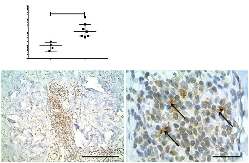
Page 2 of 16                                                                 Abbas et al. Morphea: progress to date and the road ahead

                     A                             B                        C

                                                                            D

Figure 1 Morphea is characterized by periods of activity (A,C) and damage (B,D). (A) Right arm affected with significant inflammation,
evidenced in swelling of digits and forearm. (B) Damage from morphea including joint contractures and hyperpigmented sclerotic plaques. (C)
Active, inflammatory plaque on abdomen with striae. (D) Morphea damage with hyperpigmented, sclerotic plaques, atrophy.

autoantibodies specific to systemic sclerosis, despite having           developments, further work is needed to better define
similar histology (6,7). Extracutaneous manifestations of               clinical subtypes, extracutaneous manifestations, outcome
morphea are uncommon, and include neurological and                      measures and pathogenesis in order to better evaluate and
musculoskeletal findings distinct from those found in                   treat patients with morphea.
systemic sclerosis (3).
   Morphea is characterized by relapsing and remitting
                                                                        Clinical manifestations of morphea
periods of activity, marked by inflammation and fibrosis, and
damage which produces atrophy. Unchecked disease activity               Morphea activity versus damage
in morphea can lead to permanent deformity and functional
impairment, and thus early diagnosis and treatment are                  Morphea is defined by periods of activity (inflammation
imperative to minimize damage (8). Several subtypes of                  and fibrosis) which leads to damage and atrophy. Activity in
morphea exist, each with different clinical manifestations              morphea is characterized histologically by an inflammatory
and degree of involvement of the subcutaneous soft tissues              dermal and subcutaneous lymphocytic infiltrate manifesting
(6,8). The pathogenesis of morphea is incompletely                      clinically as erythema, edema, and lesion extension, with
understood and is an evolving area of research. Studies                 patients reporting symptoms such as pain and pruritis (11).
suggest a multifactorial etiology involving dysregulated                The fibrotic phase often initially overlaps with inflammation
immune and fibrotic pathways, with additional contributing              and is characterized by dense collagen deposition with
factors including genetic predisposition, traumatic or                  admixed inflammatory cells manifesting as hardened yellow
environmental factors, and vascular dysregulation (6,9,10).             to white plaques with an erythematous or violaceous border.
   After many years of neglect, substantial progress has been           These mixed inflammatory and sclerotic lesions ultimately
made in morphea research. The purpose of this review is to              transition into an inactive phase characterized by resolution
summarize new developments in understanding the clinical                of inflammation with sclerosis progressing to atrophy of
manifestations of morphea and their evaluation as well as               the dermis and sometimes underlying soft tissue. Figure 1
management. We will also discuss the current understanding              demonstrates typical appearance of both active and inactive
of the pathogenesis of morphea. Despite these promising                 lesions. The pathological changes of morphea may affect

© Annals of Translational Medicine. All rights reserved.               Ann Transl Med 2021;9(5):437 | http://dx.doi.org/10.21037/atm-20-6222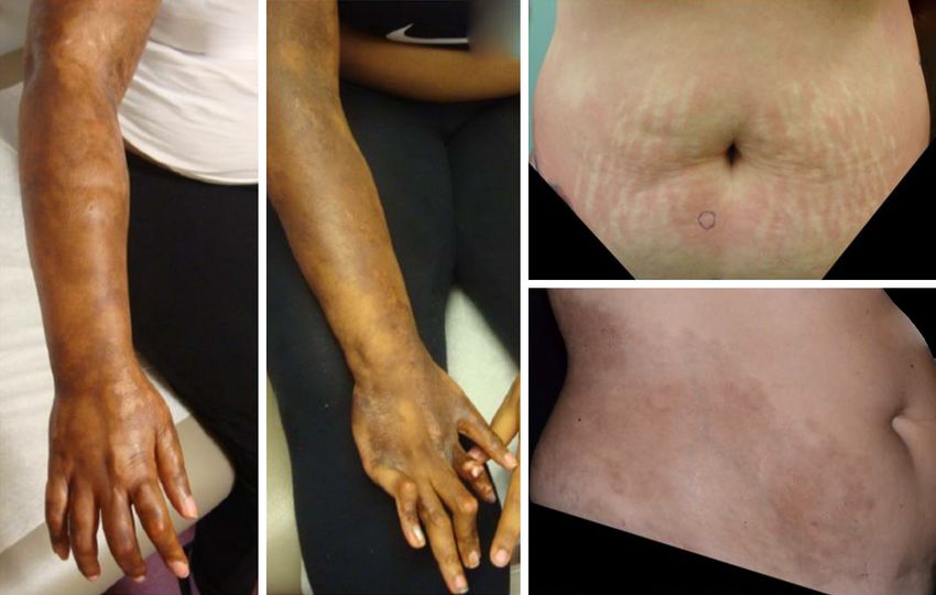
Annals of Translational Medicine, Vol 9, No 5 March 2021                                                                 Page 3 of 16

the dermis, subcutis, soft tissue, and sometimes bone.            subtypes, presenting a substantial barrier to multisite studies
Fibrosis and resultant atrophy of the dermis, soft tissue,        that are crucial in a rare disease like morphea. This also
and bone can cause significant deformity and functional           leads to confusion among clinicians who then are unable to
impairment, such as contractures, limb length discrepancy,        accurately assess their patients.
or limitations in range of motion (8). Current standard of           Of all the different classification schemes, the Padua
care therapies for morphea are immunosuppressive agents           criteria likely performs the best at successfully capturing
that aim to shut down disease activity, and thus early and        the most relevant disease subsets in morphea. In a recent
accurate assessment of activity is crucial in preventing          large prospective cohort study of adult and pediatric-onset
permanent cosmetic and functional sequelae.                       morphea patients, the Padua criteria correctly categorized
                                                                  95% of patients (900/944), in comparison to other
                                                                  classification schemes which only correctly categorized
Subtypes
                                                                  51–54% of patients (14). Furthermore, the groups created
Currently, a number of classification systems for morphea         using the Padua criteria were found to have cohesive clinical
subtypes are in use. The first criteria created to classify       and demographic features. However, there remain some
morphea was the Mayo Clinic Criteria, published by                ambiguities in the Padua criteria, such as how patients with
Peterson et al. in 1995, which classified morphea into five       multiple linear lesions who also meet criteria for generalized
subtypes: plaque, generalized, bullous, linear, and deep (12).    disease should be categorized, when patients with multiple
In 2006, Zulian and Laxer published the Padua criteria,           linear lesions have been shown to be a distinct group with
which outlined five different subtypes: circumscribed,            consistent demographic and clinical characteristics (15,16).
generalized, linear, pansclerotic, and mixed, which does          Additionally, findings like deep involvement occur across
not include bullous morphea and deep morphea. The                 linear, generalized, and circumscribed lesions and may be
Padua criteria however does note that deep involvement            better considered a descriptor and not a separate subtype.
can occur with circumscribed lesions (13). In 2017, the              In order to increase the likelihood of uptake across
European Dermatology Forum proposed a classification              different specialties and centers, refinement of the
system with five subtypes—limited which includes plaque,          existing classification systems should be undertaken
guttate, and superficial morphea, generalized type which          with a multidisciplinary group of relevant stakeholders
includes generalized and pansclerotic subtypes, linear, deep,     using a consensus based process and ultimately validated
and mixed (2). Limitations exist within each classification       by assessing performance in a heterogeneous group of
system. First, authors of the classifications specialize          morphea patients. It is vital that like patients are categorized
either in adult or pediatric medicine and therefore do            consistently in terms of determining associated disease
not see morphea patients across the lifespan. Second, the         outcomes for both patient care as well as for multi-site
criteria were created primarily by either dermatologists          collaborations for both observational and interventional
or rheumatologists, who may have differing perspectives           studies.
and experiences of morphea. This limits the ability to fully
categorize morphea subtypes that may occur outside the
                                                                  Extracutaneous manifestations
authors area of expertise, particularly when it comes to
extracutaneous manifestations or subtypes that occur more         Once believed to be exclusively a skin disorder, newer
commonly in one demographic group. Also of concern,               studies show that morphea is associated with extracutaneous
the existing classification criteria are the result of expert     manifestations distinct from those in systemic sclerosis
opinion, but were not prospectively examined using an             (17-22). These include mucocutaneous, neurological,
unbiased analysis of demographic or clinical features of a        musculoskeletal, and ophthalmologic involvement (8).
large group of adults and children with morphea, making              Mucocutaneous findings in morphea are seen in the
it difficult to determine how well these classification           form of genital and oral lesions. Genital lesions in morphea
systems perform in defining demographically and clinically        occur predominantly in post-menopausal women, and
consistent subsets of morphea patients. The presence of           are associated with more superficial dermal morphea and
different classification systems for morphea, each of which       accompanying extragenital lichen sclerosus lesions (23).
are actively in use, has produced ambiguity among clinicians      Studies have shown that genital lichen sclerosus et
and researchers in the definition and categorization of           atrophicus (LsA) and morphea lesions in extragenital sites

© Annals of Translational Medicine. All rights reserved.         Ann Transl Med 2021;9(5):437 | http://dx.doi.org/10.21037/atm-20-6222
Page 4 of 16 Abbas et al. Morphea: progress to date and the road ahead
may co-exist, thus supporting examination of genitalia in and bone (13). When this occurs, morphea can be associated
those with morphea and the extra genital skin in those with with severe pain, flexion contractures, and functional
LsA, particularly in post menopausal women (24). Oral impairment due to decreased range of motion (39).
involvement has been reported mainly through case reports Deep morphea lesions often have very subtle surface
and case series in the context of facial linear morphea changes, and palpation can be more important than visual
lesions, and can include abnormalities of dentition, loss of inspection to appreciate the extent of these lesions. It also
oral structures, functional impairments from sclerosis of may be difficult to fully evaluate activity in these deeper
tissue (i.e., decreased oral aperture), as well as arthritis and lesions, and given that unchecked morphea activity can
mechanical dysfunction with TMJ pain (25-31). Oral lesions lead to permanent functional sequelae, patients with
tend to directly underlie the cutaneous and soft tissue these deeper manifestations of morphea may benefit from
morphea lesions, and often show abrupt demarcation at the MRI to determine disease activity and damage (40,41).
midline similar to cutaneous lesions (25,27,30,32,33). In Although involvement of joints and areas underlying areas
general, there is a dearth of large, systematic studies on the of morphea is the most common presentation, patients
frequency and type of oral and genital involvement across may also experience sacroiliitis, generalized synovitis,
morphea subtypes, and further work is required in order to and inflammatory arthritis. Although musculoskeletal
better characterize mucocutaneous findings in morphea (24). involvement has been more extensively reported in
Neurologic involvement in morphea can take the form children with linear morphea, limitation of range of
of various manifestations such as migraines, seizures, motion has also been reported in adults with generalized
focal neurologic deficits, and movement disorders (34). symmetric morphea. Little is known however about the
Literature regarding neurologic findings in morphea is association with findings like arthritis and the activity of
also primarily in the form of case reports and case series the cutaneous lesions or their optimal treatment, while
(20,34). Some reviews and reports suggest a role for soft tissue involvement appears to be associated with
neuroimaging in these patients, as MRI and CT which can deep cutaneous lesions and is linked with activity of the
demonstrate findings such as subcortical calcifications and cutaneous disease (16). Thus it is important for patients
brain atrophy associated with cutaneous lesions of morphea with morphea, particularly with linear subtype or deep
involving the head (35). However, the significance of these cutaneous involvement, to undergo a thorough examination
findings is uncertain as some patients present with severe of the musculoskeletal system and prompt evaluation by
symptoms and deficits with no changes on imaging, and rheumatology, orthopedics, or physical medicine and
some patients with imaging findings do not have significant rehabilitation as needed (8).
neuroimaging findings (19,20,34). Additionally, there is Ophthalmologic involvement of morphea is rare, and
uncertainly regarding whether these lesions respond to is generally associated with linear morphea involving the
immunosuppressive agents or are better treated by directly head. Literature regarding this is primarily in the form of
targeting neurological symptoms, particularly in the absence case reports and a large international case series, which
of any sign of central nervous system (CNS) inflammation describe features such as diplopia related to involvement of
on evaluation. Thus, there remains a need for further study periocular muscles and/or inflammatory changes such as
in this area. uveitis and episcleritis (42). CNS involvement leading to
Patients with localizing neurologic clinical manifestations ophthalmologic change has also been reported (43). Patients
such as hemiplegia or visual field deficits would benefit from reporting visual changes or even patients with morphea
prompt multidisciplinary evaluation with consideration of lesions in close proximity to the periorbital region should be
neuroimaging to rule out emergencies such as CNS or optic promptly referred to an ophthalmologist for evaluation (8).
vasculitis (8). Current literature suggests that the severity Overall, the extracutaneous manifestations of morphea
of cutaneous morphea may not correlate with severity of have not yet been systematically studied. Thus, their
nervous system involvement, i.e., there are cases of patients frequency among patients with morphea and their
with subtle skin findings but striking MRI findings and relationship to the activity of skin disease are not
severe neurologic manifestations (19,35-38). well known, and little is known about the response of
Musculoskeletal manifestations of morphea can extracutaneous manifestations to morphea treatment. This
arise when the disease affects not only the skin but also is an important evolving field of research in morphea, and
underlying structures such as fascia, muscle, and even joints there is a need for larger studies describing the frequency,
© Annals of Translational Medicine. All rights reserved. Ann Transl Med 2021;9(5):437 | http://dx.doi.org/10.21037/atm-20-6222Annals of Translational Medicine, Vol 9, No 5 March 2021 Page 5 of 16
clinical findings, and association with morphea activity in In these cases, topical therapies can be used as an adjunct,
the skin. with systemic immunosuppressive therapy held in reserve
in case they cannot tolerate phototherapy. The relatively
favorable side effect profile of phototherapy gives it an
Diagnosis and treatment
advantage over systemic immunosuppressants and thus is
The diagnosis of morphea can typically be made based on the preferred agent in these patients (54). Other procedures
clinical findings, however biopsy of the lesions and imaging that have been reported to be effective in the treatment of
can help confirm the diagnosis or exclude other diagnoses. some morphea patients include photodynamic therapy and
Treatment of morphea depends on clinical activity, depth pulsed dye laser therapy for sclerotic lesions, intralesional
of lesion involvement, and extent of disease, and primarily hyaluronidase injections for morphea-induced microstomia,
centers around limiting disease activity (8,44). Active lesions and extracorporeal photochemotherapy for severe,
that are isolated to a limited surface area can be treated generalized disease (31,54-60). Further research into these
by topical or intralesional steroids, as well as calcineurin agents and procedures is warranted in order to more clearly
inhibitors such as tacrolimus. For patients with more define efficacy and indications for use before they should be
generalized dermal involvement or rapidly developing new used as first line agents.
lesions, ultraviolet (UV) phototherapy can also be used. Damage that results after active lesions progress to
Systemic therapy in morphea is indicated for those with an inactive state include atrophy, pigment changes and
moderate-severe disease, large body surface area involvement, functional impairment. Damage tends to remain stable or
deep involvement, or for lesions that may impact function/ increase after successful management of active disease (61).
cosmesis (facial lesions). The most widely investigated Once lesions are clinically inactive, treatment centers
systemic therapies include combinations of methotrexate around improving quality of life by addressing cosmetic and
and corticosteroids. Mycophenolate mofetil is an emerging functional concerns. Sclerosis and atrophy due to morphea
alternative to methotrexate for those who cannot can lead to limb-length discrepancies, contractures, and
tolerate or have contraindications to methotrexate (45). limited range of motion. It is important to refer these
These medications maybe limited by lack of tolerance or patients to physical therapy, occupational therapy or
toxicity in many patients, underscoring the need for less specialties such as rheumatology or orthopedics early
toxic therapy (46-52). Other systemic therapies used for in order to reduce disability. In addition to functional
morphea include bosentan, infliximab, tofacitinib, and impairment patients also suffer from cosmetic damage.
abatacept (53). While these treatments show promise, Dermal fillers and surgical procedures, such as fat transfer,
current data is insufficient to confirm efficacy of routine can help restore contour to lesions with significant
use, and further study is necessary. atrophy. Recent studies have shown the utility of adipose
There are a wide variety of systemic therapies other tissue as filler for its ability to regenerate soft tissues and
than methotrexate, systemic glucocorticoids, and remodeling capacity provided by its unique cytokine and
mycophenolate used for active morphea. Examples of these growth factor profiles (62). Despite these promising results,
new and emerging treatments include bosentan, infliximab, there is no evidence that fat transfer replaces the use of
tofacitinib, and abatacept (53). While these treatments show immunosuppressives in active facial morphea lesions.
promise, current data is insufficient to confirm efficacy of Therefore, fat transfer should only be used once active
routine use, and further study is necessary. disease is demonstrably controlled with immunosuppressives
Another treatment for morphea is ultraviolet (UV) or quiescent off therapy to avoid recurring tissue loss. Taken
phototherapy, used for a variety of sclerosing and as a whole, treatment efficacy and benefit on life quality of
inflammatory conditions of the skin (54). Phototherapy interventions to mitigate damage are very poorly studied in
options include Ultraviolet A1 (UVA1) and narrowband morphea.
UVB (NBUVB). While both can be used for treatment of
morphea, UVA1 is preferred when available as there is more
Refining outcome measures in morphea
evidence supporting its efficacy in morphea (54). Patients
with extensive dermal morphea are good candidates for Given that the clinical manifestations of morphea are
this treatment, as they have a large amount of body surface dependent on subtype, depth of involvement and phase of
area involved, making topical therapy alone impractical. progression of the lesions, an accurate understanding of the
© Annals of Translational Medicine. All rights reserved. Ann Transl Med 2021;9(5):437 | http://dx.doi.org/10.21037/atm-20-6222Page 6 of 16 Abbas et al. Morphea: progress to date and the road ahead
entire disease picture is essential. Correctly identifying and Patient reported outcomes are another important
quantifying disease activity and damage in different subtypes outcome to consider when designing clinical trials, even
of morphea is key not only to appropriate management more so when evaluating the comparative effectiveness of
but also to conduct well designed studies. Thus, recent new treatments and contrasting their impact on quality of
work has focused on validation of clinical, biomarker, and life. Studies have shown that patients with morphea have
imaging outcomes aimed at accurately quantifying activity significant impairment in quality of life, likely related to
and damage. Validation and acceptance of these measures symptoms such as pain, itch, and fatigue, as well as worry
are important for future large, multisite clinical trials in about progression to systemic disease (15,64,73). However,
morphea, which will be necessary given the rarity of the surprisingly, even patients with clinically severe disease
disease and will ultimately lead to better patient care. report only mild to moderate impact on life quality based
on current patient reported outcome measures (64). This
indicates that current skin based measures may not detect
Clinical outcome measures
aspects of morphea relevant to patients and also may fail
Until recently, morphea was thought to be unresponsive to to capture disease heterogeneity, i.e., one patient may have
therapy largely because existing outcomes did not measure hemifacial atrophy while another suffers from limitation
the effect of treatment on activity. Many previously reported of joint range of motion. Disease specific measures can
outcome measures in morphea exclusively measured overcome these difficulties, and thus the pediatric Localized
damage, and therefore remained unchanged even with Scleroderma Quality of Life Instrument (LoSQI) was
successful immunosuppressive treatment, which exclusively developed as the only disease-specific measure in morphea,
improves activity and stabilizes damage (61). Patient- designed to measure the unique impact of morphea from
reported outcomes (PRO), which are critical to assessing the perspective of the patient (74). While the LoSQI is a
the effect of therapy on patients, are equally challenging to promising step in the right direction for accurately assessing
quantify in morphea. This is because features of morphea, impact of morphea on patients, it thus far only has support
such as tissue loss and sensations of skin tightening, are not for use in the pediatric morphea population. Further work
represented in traditional dermatology and rheumatology needs to be done on adapting this for use in clinical trials
measures. These gaps need to be addressed in order to with adults.
conduct well designed clinical trials (63).
The Localized Scleroderma Cutaneous Assessment Tool
Role of biomarkers
(LoSCAT) is a promising clinical outcome for morphea,
but important aspects of the measure need to be refined. Biomarkers are needed in morphea that identify disease
The LoSCAT is the only clinical score for morphea activity. While some patients present with clearly active or
incorporating both activity and damage, and is the only inactive disease, there are a significant number of patients
clinical measure in morphea developed using the rigor of who present with morphea lesions in which the level of
the Outcome Measures in Rheumatologic Clinical Trials activity is difficult to assess using clincal examination alone,
(OMERACT) principles (39,64-70). To date, the LoSCAT making it difficult to know whether to initiate or escalate
is the most fully validated clinical outcome in morphea and immunosuppressive therapy. Accurate and timely assessment
has strong support for use in measuring disease activity of lesion activity is crucial to successful management of
and damage in a clinical population. However, it has not morphea patients, since the goal of morphea therapy is to
been validated for discrimination in terms of long-term eliminate disease activity in order to prevent permanent
responsiveness to change and minimal clinically important cosmetic and functional sequelae.
differences (MCID) have not yet been defined (68). Thus Studies on the role of biomarkers to indicate disease
further work needs to be done to further validate the use of activity have focused on cytokine levels, as most patients
the LoSCAT in sensitivity to change, as this is absolutely with morphea have normal markers of inflammation
necessary for the planning and conduct of clinical trials in such as erythrocyte sedimentation rate. While increased
order to contextualize change in score beyond statistical concentrations in numerous cytokines have been reported in
significance. This will allow the LoSCAT to be used as an the sera of patients with morphea, such as IL-2, IL-4, IL-6,
outcome in clinical trials to determine treatment response IL-8, IL-13, IP-10, and TNF alpha, current evidence points
(71,72). towards downstream IFN-regulated pathway chemokines
© Annals of Translational Medicine. All rights reserved. Ann Transl Med 2021;9(5):437 | http://dx.doi.org/10.21037/atm-20-6222Annals of Translational Medicine, Vol 9, No 5 March 2021 Page 7 of 16
A 1000 CXCL9
**
100
Fold change
10
1
0.1
Unaffected Inflammatory
B C
Figure 2 CXCL9 is increased in inflammatory morphea skin, supporting its role as a potential biomarker. (A) CXCL9 mRNA expression
is increased in lesional, inflammatory skin when compared to site-matched unaffected skin. (B,C) CXCL9 is present in dermal interstitial
infiltrates and stains dendritic appearing cells. Arrows indicate typical perinuclear cap staining pattern of CXCL9. Scale bar in B =100 mm;
scale bar in C =25 mm. Reprinted from (80).
as the most promising biomarkers in morphea (75-81). be a useful adjunct to clinical examination when it comes
CXCL9, along with other T helper type 1 cytokines, to accurate assessment of disease activity and extent (41).
has been found at increased concentrations in morphea Figure 3 demonstrates MRI findings in morphea. This is
serum, and has been found to correlate with disease activity particularly relevant for deeper morphea lesions extending
as measured by the LOSCAT in multiple independent to the subcutis, fascia, and muscle, which often present
studies (80,81). Further studies also indicate that CXCL10 with subtle cutaneous manifestations. In fact, studies have
may also have similar biomarker capabilities (81). shown that MRI can reveal clinically occult musculoskeletal
CXCL9 gene expression was also found to be increased in involvement (82-84), demonstrating subclinical extension
inflammatory lesional morphea skin, and co-localized with of lesions beyond visible margins (41). MRI has also been
dermal macrophages, implicating the skin as the source of shown to demonstrate active disease that would otherwise
circulating cytokines (80,81). Figure 2 demonstrates the be misclassified as inactive based on using the LoSCAT
elevation of CXCL9 in inflammatory morphea skin. Thus, alone (41). Given how crucial it is to accurately assess
current research indicates that IFN pathway dysregulation activity when managing morphea patients, this further
is associated with activity in morphea but how this underscores the utility of MRI in conjunction with clinical
dysregulation is related to the pathogenesis of morphea evaluation for deep morphea. Future studies in MRI are
remains poorly studied. needed to assess its responsiveness to change and to further
define indications for imaging.
Ultrasound is another tool that can be used for
Imaging measures of morphea
investigation of activity and depth of morphea lesions,
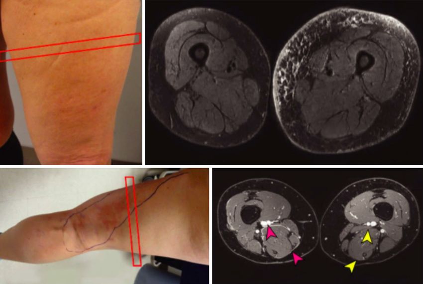
Recent studies have indicated that MRI may have a role as and has promise as an outcome measure in morphea.
an objective outcome measure in morphea, particularly in Ultrasonography is easier to use and more cost-effective than
the case of deep cutaneous or soft tissue involvement. High MRI, and has been found to have high validity and reliability
resolution MRI can demonstrate inflammation, sclerosis, for evaluation of morphea. Ultrasound can differentiate all
and atrophy in morphea lesions, and has been shown to stages of morphea, including active disease, which appears
© Annals of Translational Medicine. All rights reserved. Ann Transl Med 2021;9(5):437 | http://dx.doi.org/10.21037/atm-20-6222Page 8 of 16 Abbas et al. Morphea: progress to date and the road ahead
A B
Right thigh Left thigh
C D
Right thigh (affected)
Left thigh (unaffected)
Figure 3 MRI findings in morphea. Red boxes indicate area imaged. (A) Subtle morphea involving the left thigh. (B) Axial fat-saturated T2-
weighted image of bilateral thighs, with hyperintense areas corresponding to morphea involvement. (C) Morphea of the lower right extremity.
(D) Axial 3-dimentional subtracted postcontrast image of bilateral thighs showing fascial involvement as demonstrated by hyperintense signal
on affected right thigh (red arrows), with unaffected left thigh (yellow arrows) presented for comparison. Reprinted from (80).
hyperechoic (sclerotic) or isoechoic (inflammatory), from palate (88) and for postoperative monitoring to track fat
inactive disease characterized by atrophy and damage, graft retention and assess soft tissue volume of change (89).
which appears hypoechoic (85). Ultrasound can also detect It has great potential for application in morphea affecting
increased cutaneous blood flow, which is another sign of the head and neck, where facial asymmetry can be difficult
lesion activity (86). While results from ultrasonography have to evaluate clinically, and the LoSCAT often falls short in
been correlated to clinical and histopathologic findings, correctly quantifying disease activity and damage. Advanced
further work needs to be done to validate this as an outcome analysis of images can be used in conjunction with clinical
measure. Additionally, it is important to collaborate with a assessment to provide information about vascularity and
musculoskeletal imaging expert with experience in morphea pigmentation to further monitor disease activity (90). 3D
to get optimal information from the scan (including MRI imaging is portable, fast, easy to use, and inexpensive, and
and ultrasound) (8). has many applications in facial morphea. Further studies
Three dimensional stereophotogrammetry (3D imaging) to validate this modality will continue to support its
is a minimally invasive and radiation-free modality rapidly integration into clinical practice.
gaining popularity as the preferred method for quantifying
information about facial soft-tissue, particularly in children,
Histologic markers
where quantifying facial features can be challenging (87). It
has demonstrated a high degree of precision and accuracy Skin biopsy may provide additional information regarding
across different platforms (87), and has been used in a depth of involvement and activity (inflammation) in cases
variety of conditions affecting the face, including cleft where clinical examination is inconclusive or imaging is not
© Annals of Translational Medicine. All rights reserved. Ann Transl Med 2021;9(5):437 | http://dx.doi.org/10.21037/atm-20-6222Annals of Translational Medicine, Vol 9, No 5 March 2021 Page 9 of 16
readily available (91). Pathological findings, particularly further study independently into morphea is warranted.
the pattern of sclerosis and degree of inflammation. For Although it is generally accepted that immune dysfunction
example, studies have shown that a bottom heavy pattern is the principal component in the development of morphea
of sclerosis can be associated with pain and tightness, and (3,99-101), other factors are also thought to contribute to
that severe inflammation may be associated with pain and pathogenesis, including genetic predisposition, traumatic or
functional limitation (91). Histopathologic examination of environmental factors, and vascular dysfunction (3,7,9).
morphea therefore has the potential to be used not only
for diagnosis but also for assessment, assisting in clinical
Immune dysregulation
management by identifying disease activity and damage, and
identifying patients who may require additional monitoring There are several aspects of morphea that point to the role
and treatment (91). of autoimmunity in pathogenesis. Firstly, the natural history
Another current area of study into potential histologic of the disease, with the clinically evident inflammatory
measures of morphea is regarding the altered distribution stage preceding the development of sclerosis, supports
of dermal dendritic cells and vascular abnormalities that the theory of immune dysfunction (8). Additionally,
have been reported to relate the pathogenesis of morphea. histopathologic studies demonstrate an influx of large
Studies of morphea skin indicate that patients with morphea numbers of mononuclear lymphocytes (primarily activated
demonstrate a phenotypic change of CD34+ dendritic cells T lymphocytes), plasma cells, and eosinophils in lesional
into SMA+ myofibroblasts with increasing disease extent morphea skin, also supporting the role of autoimmunity (96).
and fibrosis (92). The degree of loss of CD34+ dendritic Morphea patients have also been found to have elevated
cells has been found to correlate with relative degrees of cytokine levels, such as CXCR3 ligands as well as
inflammation and sclerosis, and thus this has been proposed those associated with Th2 immune responses (80,96).
as a useful marker in predicting morphea severity and extent For example, IL-4, which is produced by CD4+ Th2
(92,93). Further studies have shown that treatments such lymphocytes, can upregulate the production of TGF beta,
as pulsed dye laser and UVA1 phototherapy are effective stimulating fibroblast production of collagen and other
in improving indurated skin and reducing disease activity extracellular matrix proteins, and IL-4 has been detected at
in morphea, and are also associated with an increase in elevated levels in morphea patients (77). Some patients with
the number of CD34+ cells (94,95). Taken together, these morphea also have increased autoantibody levels, further
results underscore the potential use of histologic markers supporting the role of immune dysfunction (3,100-102).
such as CD34+ dendritic cells as measures of disease in To date, most studies examining the autoimmune
morphea, although further studies will be necessary to pathogenesis of morphea have consisted of reports of
validate and refine these measures. circulating chemokine profiles or antibodies, flow cytometry
of peripheral blood, and immunostaining often in a limited
number of samples or without controls (77,103-105). Recent
Pathogenesis of morphea
observations have supported the role of dysregulated immune
The precise pathogenesis of morphea is not completely pathways, particularly IFN gamma, demonstrating that
understood. As with other autoimmune disorders, the CXCL9 and CXCL10 levels are associated with increased
main contributors to long-term morphea damage and clinical measures of disease activity (78-80,106-108).
disability are thought to be the extent and duration of the Figure 4 demonstrates the elevation of these cytokines.
initial active phase, which likely drives subsequent damage- Using microarray and bulk RNA sequencing on human
producing fibrosis (11). However, the dysregulated immune skin samples, clinically early inflammatory morphea lesions
and fibrotic pathways that contribute to these changes have have been found to display an inflammatory morphea
not yet been systematically studied. Current theories are signature, including chemokines CXCL9, CXCL10,
often extrapolated from studies of systemic sclerosis due CXCL11, and their receptor CXCR3, with cell-specific
to a paucity of well-developed studies in morphea (96), transcripts of infiltrating macrophages and T cell subsets
although clinical evidence suggests that morphea has (80,109). There have also been discoveries involving
distinct disease characteristics, encompassing different a fibrotic signature reflecting fibroblast activation and
demographics, clinical features (5,6,13,97) and response collagen production (110), which may enhance retention of
to treatment (7,46-48,98) than systemic sclerosis, and inflammatory cells in the dermis of sclerotic lesions (111).
© Annals of Translational Medicine. All rights reserved. Ann Transl Med 2021;9(5):437 | http://dx.doi.org/10.21037/atm-20-6222Page 10 of 16 Abbas et al. Morphea: progress to date and the road ahead
A CXCL9 CXCL10 B CXCL9 CXCL10
10000 1000
Concentration (pg/mL)
10000 1000
Concentration (pg/mL)
Concentration (pg/mL)
Concentration (pg/mL)
1000 100
1000 100
100 10
r=0.16
E CXCL9 and CXCL10
r=0.44
CXCL10 concentration (pg/mL)
100 10 P=0.2 1000
P=0.0001
10 1
0 60 100 160 0 60 100 160 100
10 1
Morphea Controls Morphea Controls mLoSSI score mLoSSI score
C 100 37%
CXCL9
9% 100 48%
CXCL10
35% D10000 CXCL9
1000
CXCL10 10
r=0.74
PAnnals of Translational Medicine, Vol 9, No 5 March 2021 Page 11 of 16
pathogenesis using transcriptional profiling, animal or ex remains to thoroughly validate these measures before use in
vivo disease models, or molecular approaches. Further study the clinical setting as well as for research purposes.
using these state of the art approaches are necessary to gain The present understanding of morphea pathogenesis
a detailed, unbiased picture of upstream and downstream is incomplete but points primarily towards the role
pathways in human skin that are likely implicated in of dysregulated immune and fibrotic pathways, with
morphea pathogenesis, particularly dysregulated IFN environmental triggers, genetic predisposition, and
gamma mediated pathways, which appear particularly vascular dysfunction also playing a role. There remains a
promising. Furthermore, many of the gene signatures being gap in knowledge in clearly elucidating the pathogenesis
currently studied in morphea can be coupled to clinical of the disease, and further study is necessary to provide a
measures of disease and have the potential to be used as full understanding of the environmental, systemic, local,
biomarkers, allowing for prediction of disease course and genetic and immunopathological factors underpinning
therapeutic response. morphea pathogenesis. This is particularly important
Studying the mechanism of action behind the given that current treatments for morphea revolve around
dysregulated pathways in morphea poses a clinical and the use of immunosuppressives such as corticosteroids
pathologic challenge due to the clinical heterogeneity of and methotrexate to target activity, which are limited
the disease, as it often has variable anatomic patterning, by significant adverse effects. A better understanding of
morphology, and depth of tissue involvement (99). disease pathogenesis will allow for refinement of outcome
Moreover, the relative rarity of the disease makes it difficult measures and development of therapeutic targets and novel
to execute large studies correlating biological samples with biomarkers.
accompanying clinical outcomes. Thus, there remains
an important gap in knowledge that must be filled, as an
Acknowledgments
improved understanding of the underlying molecular
mechanisms of morphea is likely to allow for refining Funding: This manuscript was funded by the James Gilliam
of outcome measures and the advent of novel targeted Distinguished Chair in Dermatology.
therapies, especially given that current, non-targeted
treatments for morphea are often limited by substantial
Footnote
toxicity.
Provenance and Peer Review: This article was commissioned
by the Guest Editors (Drs. Richard D. Sontheimer, M.
Conclusions
Kari Connolly, David F. Fiorentino, and Victoria P. Werth)
Morphea is an inflammatory skin condition characterized for the series “Rheumatologic Skin Disease” published in
by activity (inflammation) presenting as erythematous and Annals of Translational Medicine. The article has undergone
violaceous indurated plaques evolving to hyperpigmented external peer review.
lesions with central sclerosis and atrophy. There are several
subtypes of morphea, and while a number of different Conflicts of Interest: All authors have completed the ICMJE
classification systems exist, they are hindered by significant uniform disclosure form (available at http://dx.doi.
limitations and further work must be done to refine and org/10.21037/atm-20-6222). The series “Rheumatologic
clarify these schemes. For example, these schemes do not Skin Disease” was commissioned by the editorial office
mention extracutaneous manifestations of morphea, a without any funding or sponsorship. The authors have no
current evolving area of research. other conflicts of interest to declare.
Given that unchecked disease activity in morphea can
lead to severe cosmetic and functional sequelae, it is crucial Ethical Statement: The authors are accountable for all
to identify activity and initiate treatment early. Thus, recent aspects of the work in ensuring that questions related
progress has been made in developing and refining outcome to the accuracy or integrity of any part of the work are
measures in morphea. Clinical, biomarker, imaging, and appropriately investigated and resolved.
histologic outcomes have all been studied in order to allow
for more accurate assessment of disease activity and severity. Open Access Statement: This is an Open Access article
Despite promising results in this arena, further work distributed in accordance with the Creative Commons
© Annals of Translational Medicine. All rights reserved. Ann Transl Med 2021;9(5):437 | http://dx.doi.org/10.21037/atm-20-6222Page 12 of 16 Abbas et al. Morphea: progress to date and the road ahead
Attribution-NonCommercial-NoDerivs 4.0 International 1995;70:1068-76.
License (CC BY-NC-ND 4.0), which permits the non- 13. Laxer RM, Zulian F. Localized scleroderma. Curr Opin
commercial replication and distribution of the article with Rheumatol 2006;18:606-13.
the strict proviso that no changes or edits are made and the 14. Prasad S, Zhu JL, Schollaert-Fitch K, et al. Characterizing
original work is properly cited (including links to both the morphea subsets using a multi-center, prospective, cross-
formal publication through the relevant DOI and the license). sectional analysis of morphea in adults and children. J
See: https://creativecommons.org/licenses/by-nc-nd/4.0/. Investig Dermatol 2020;140:S73.
15. Kunzler E, Florez-Pollack S, Teske N, et al. Linear
morphea: Clinical characteristics, disease course, and
References
treatment of the Morphea in Adults and Children cohort. J
1. Peterson LS, Nelson AM, Su WP, et al. The epidemiology Am Acad Dermatol 2019;80:1664-70.e1.
of morphea (localized scleroderma) in Olmsted County 16. Teske N, Welser J, Jacobe H. Skin mapping for the
1960-1993. J Rheumatol 1997;24:73-80. classification of generalized morphea. J Am Acad Dermatol
2. Knobler R, Moinzadeh P, Hunzelmann N, et al. 2018;78:351-7.
European Dermatology Forum S1-guideline on the 17. Blaszczyk M, Krolicki L, Krasu M, et al. Progressive
diagnosis and treatment of sclerosing diseases of the facial hemiatrophy: central nervous system involvement
skin, Part 1: localized scleroderma, systemic sclerosis and relationship with scleroderma en coup de sabre. J
and overlap syndromes. J Eur Acad Dermatol Venereol Rheumatol 2003;30:1997-2004.
2017;31:1401-24. 18. Zulian F, Vallongo C, Woo P, et al. Localized scleroderma
3. Leitenberger JJ, Cayce RL, Haley RW, et al. Distinct in childhood is not just a skin disease. Arthritis Rheum
autoimmune syndromes in morphea: a review of 245 adult 2005;52:2873-81.
and pediatric cases. Arch Dermatol 2009;145:545-50. 19. Amaral TN, Peres FA, Lapa AT, et al. Neurologic
4. Sehgal VN, Srivastava G, Aggarwal AK, et al. Localized involvement in scleroderma: a systematic review. Semin
scleroderma/morphea. Int J Dermatol 2002;41:467-75. Arthritis Rheum 2013;43:335-47.
5. Christen-Zaech S, Hakim MD, Afsar FS, et al. Pediatric 20. Kister I, Inglese M, Laxer RM, et al. Neurologic
morphea (localized scleroderma): review of 136 patients. J manifestations of localized scleroderma: a case report and
Am Acad Dermatol 2008;59:385-96. literature review. Neurology 2008;71:1538-45.
6. Careta MF, Romiti R. Localized scleroderma: clinical 21. Stone J, Franks AJ, Guthrie JA, et al. Scleroderma "en
spectrum and therapeutic update. An Bras Dermatol coup de sabre": pathological evidence of intracerebral
2015;90:62-73. inflammation. J Neurol Neurosurg Psychiatry
7. Fett N, Werth VP. Update on morphea: part I. 2001;70:382-5.
Epidemiology, clinical presentation, and pathogenesis. J 22. Marzano AV, Menni S, Parodi A, et al. Localized
Am Acad Dermatol 2011;64:217-28; quiz 29-30. scleroderma in adults and children. Clinical and
8. Florez-Pollack S, Kunzler E, Jacobe HT. Morphea: laboratory investigations on 239 cases. Eur J Dermatol
Current concepts. Clin Dermatol 2018;36:475-86. 2003;13:171-6.
9. Grabell D, Hsieh C, Andrew R, et al. The role of skin 23. Jacobe HT, Prasad S, Black SM, et al. 543 Clinical
trauma in the distribution of morphea lesions: a cross- and demographic features of morphea patients with
sectional survey of the Morphea in Adults and Children mucocutaneous involvement: A cross sectional study from
cohort IV. J Am Acad Dermatol 2014;71:493-8. The Morphea of Adults and Children (MAC Cohort). J
10. Li SC. Scleroderma in Children and Adolescents: Investig Dermatol 2020;140:S74.
Localized Scleroderma and Systemic Sclerosis. Pediatr 24. Lutz V, Frances C, Bessis D, et al. High frequency of
Clin North Am 2018;65:757-81. genital lichen sclerosus in a prospective series of 76
11. Martini G, Fadanelli G, Agazzi A, et al. Disease course patients with morphea: toward a better understanding of
and long-term outcome of juvenile localized scleroderma: the spectrum of morphea. Arch Dermatol 2012;148:24-8.
Experience from a single pediatric rheumatology Centre 25. Tang MM, Bornstein MM, Irla N, et al. Oral mucosal
and literature review. Autoimmun Rev 2018;17:727-34. morphea: a new variant. Dermatology 2012;224:215-20.
12. Peterson LS, Nelson AM, Su WP. Classification of 26. Barton DH, Henderson HZ. Oral-facial characteristics
morphea (localized scleroderma). Mayo Clin Proc of circumscribed scleroderma: case report. J Clin Pediatr
© Annals of Translational Medicine. All rights reserved. Ann Transl Med 2021;9(5):437 | http://dx.doi.org/10.21037/atm-20-6222Annals of Translational Medicine, Vol 9, No 5 March 2021 Page 13 of 16
Dent 1993;17:239-42. Rheumatology (Oxford) 2009;48:1410-3.
27. Hørberg M, Lauesen SR, Daugaard-Jensen J, et al. Linear 40. Schanz S, Henes J, Ulmer A, et al. Response Evaluation
scleroderma en coup de sabre including abnormal dental of Musculoskeletal Involvement in Patients With Deep
development. Eur Arch Paediatr Dent 2015;16:227-31. Morphea Treated With Methotrexate and Prednisolone:
28. McNamara PH, Toner M, Kearns G, et al. Focal seizures A Combined MRI and Clinical Approach. AJR Am J
secondary to cortical dysplasia associated with isolated Roentgenol 2013;200:W376-82.
oral morphea and odontogenic carcinoma. Seizure 41. Abbas LF, O’Brien JC, Goldman S, et al. A Cross-sectional
2013;22:159-61. Comparison of Magnetic Resonance Imaging Findings
29. Wang P, Guo W, Liu S. A rare case of juvenile localised and Clinical Assessment in Patients With Morphea. JAMA
scleroderma with intra-oral and dental involvement. Exp Dermatol 2020;156:590-2.
Ther Med 2015;10:2213-5. 42. Zannin ME, Martini G, Athreya BH, et al. Ocular
30. Van der Veken D, De Haes P, Hauben E, et al. A rare involvement in children with localised scleroderma: a
cause of gingival recession: morphea with intra-oral multi-centre study. Br J Ophthalmol 2007;91:1311-4.
involvement. Oral Surg Oral Med Oral Pathol Oral Radiol 43. Fledelius HC, Danielsen PL, Ullman S. Ophthalmic
2015;119:e257-64. findings in linear scleroderma manifesting as facial en coup
31. Abbas LF, Coias J, Jacobe HT, et al. Hyaluronidase de sabre. Eye (London, England) 2018;32:1688-96.
injections for treatment of symptomatic pansclerotic 44. Zwischenberger BA, Jacobe HT. A systematic review of
morphea-induced microstomia. JAAD Case Rep morphea treatments and therapeutic algorithm. J Am Acad
2019;5:871-3. Dermatol 2011;65:925-41.
32. Pace C, Ward SE, Pace A. A rare case of frontal linear 45. Arthur M, Fett NM, Latour E, et al. Evaluation of the
scleroderma (en coup de sabre) with intra-oral and dental Effectiveness and Tolerability of Mycophenolate Mofetil
involvement. Br Dent J 2010;208:249-50. and Mycophenolic Acid for the Treatment of Morphea.
33. Seow WK, Young W. Localized scleroderma in childhood: JAMA Dermatol 2020;156:521-8.
review of the literature and case report. Pediatr Dent 46. Zulian F, Martini G, Vallongo C, et al. Methotrexate
1987;9:240-4. treatment in juvenile localized scleroderma: a randomized,
34. Amaral TN, Marques Neto JF, Lapa AT, et al. Neurologic double-blind, placebo-controlled trial. Arthritis Rheum
involvement in scleroderma en coup de sabre. Autoimmune 2011;63:1998-2006.
Dis 2012;2012:719685. 47. Zulian F, Vallongo C, Patrizi A, et al. A long-term follow-
35. Chiu YE, Vora S, Kwon EKM, et al. A significant up study of methotrexate in juvenile localized scleroderma
proportion of children with morphea en coup de sabre and (morphea). J Am Acad Dermatol 2012;67:1151-6.
Parry-Romberg syndrome have neuroimaging findings. 48. Torok KS, Arkachaisri T. Methotrexate and corticosteroids
Pediatr Dermatol 2012;29:738-48. in the treatment of localized scleroderma: a standardized
36. Doolittle DA, Lehman VT, Schwartz KM, et al. CNS prospective longitudinal single-center study. J Rheumatol
imaging findings associated with Parry-Romberg syndrome 2012;39:286-94.
and en coup de sabre: correlation to dermatologic and 49. Maybury CM, Jabbar-Lopez ZK, Wong T, et al.
neurologic abnormalities. Neuroradiology 2015;57:21-34. Methotrexate and liver fibrosis in people with psoriasis: a
37. Menni S, Marzano AV, Passoni E. Neurologic systematic review of observational studies. Br J Dermatol
abnormalities in two patients with facial hemiatrophy 2014;171:17-29.
and sclerosis coexisting with morphea. Pediatr Dermatol 50. Bulatović M, Heijstek MW, Verkaaik M, et al. High
1997;14:113-6. prevalence of methotrexate intolerance in juvenile
38. Lis-Święty A, Brzezińska-Wcisło L, Arasiewicz H. idiopathic arthritis: development and validation of a
Neurological abnormalities in localized scleroderma methotrexate intolerance severity score. Arthritis Rheum
of the face and head: a case series study for evaluation 2011;63:2007-13.
of imaging findings and clinical course. Int J Neurosci 51. Fatimah N, Salim B, Nasim A, et al. Frequency of
2017;127:835-9. methotrexate intolerance in rheumatoid arthritis patients
39. Martini G, Ramanan AV, Falcini F, et al. Successful using methotrexate intolerance severity score (MISS
treatment of severe or methotrexate-resistant juvenile questionnaire). Clin Rheumatol 2016;35:1341-5.
localized scleroderma with mycophenolate mofetil. 52. van Dijkhuizen EH, Bulatović Ćalasan M, Pluijm SM,
© Annals of Translational Medicine. All rights reserved. Ann Transl Med 2021;9(5):437 | http://dx.doi.org/10.21037/atm-20-6222Page 14 of 16 Abbas et al. Morphea: progress to date and the road ahead
et al. Prediction of methotrexate intolerance in juvenile Rheumatol 2009;36:2819-29.
idiopathic arthritis: a prospective, observational cohort 68. Arkachaisri T, Vilaiyuk S, Torok KS, et al. Development
study. Pediatr Rheumatol Online J 2015;13:5. and initial validation of the localized scleroderma skin
53. . !!! INVALID CITATION !!! (5, 54-64). damage index and physician global assessment of disease
54. Prasad S, Coias J, Chen HW, et al. Utilizing UVA-1 damage: a proof-of-concept study. Rheumatology (Oxford)
Phototherapy. Dermatol Clin 2020;38:79-90. 2010;49:373-81.
55. Karrer S, Abels C, Landthaler M, et al. Topical 69. Kelsey CE, Torok KS. The Localized Scleroderma
photodynamic therapy for localized scleroderma. Acta Cutaneous Assessment Tool: responsiveness to change
Derm Venereol 2000;80:26-7. in a pediatric clinical population. J Am Acad Dermatol
56. Eisen D, Alster TS. Use of a 585 nm pulsed dye laser for 2013;69:214-20.
the treatment of morphea. Dermatol Surg 2002;28:615-6. 70. Condie D, Grabell D, Jacobe H. Morphea in Adults
57. Kineston D, Kwan JM, Uebelhoer NS, et al. Use of and Children Cohort VI: A cross-sectional omparison
a fractional ablative 10.6-μm carbon dioxide laser in of outcomes in adults with pediatric-onset morphea and
the treatment of a morphea-related contracture. Arch those with adult-onset morphea. Arthritis Rheumatol
Dermatol 2011;147:1148-50. 2014;66:3496-504.
58. Neustadter JH, Samarin F, Carlson KR, et al. 71. Klimas NK, Shedd AD, Bernstein IH, et al. Health-
Extracorporeal photochemotherapy for generalized deep Related Quality of Life in Morphea. Br J Dermatol
morphea. Arch Dermatol 2009;145:127-30. 2015;172:1329-37.
59. Cribier B, Faradji T, Le Coz C, et al. Extracorporeal 72. Vasquez R, Jabbar A, Khan F, et al. Recurrence of morphea
photochemotherapy in systemic sclerosis and severe after successful ultraviolet A1 phototherapy: A cohort
morphea. Dermatology 1995;191:25-31. study. J Am Acad Dermatol 2014;70:481-8.
60. Pileri A, Raone B, Raboni R, et al. Generalized 73. Szczęch J, Samotij D, Jaworecka K, et al. Quality of Life
morphea successfully treated with extracorporeal in Patients with Morphea: A Cross-Sectional Study and
photochemotherapy (ECP). Dermatol Online J a Review of the Current Literature. Biomed Res Int
2014;20:21258. 2020;2020:9186274.
61. O'Brien JC, Nymeyer H, Green A, et al. Changes in 74. Zigler C, Ardalan K, Schollaert-Fitch K, et al. The
Disease Activity and Damage Over Time in Patients With Localized Scleroderma Quality of Life instrument
Morphea. JAMA Dermatol 2020;156:513-20. (LoSQI): Initial validation in pediatric localized
62. Bellini E, Grieco MP, Raposio E. The science behind scleroderma (Abstract). Arthritis Rheumatol 2017;69. doi:
autologous fat grafting. Ann Med Surg (Lond) 10.1002/art.40321.
2017;24:65-73. 75. Hasegawa M, Sato S, Nagaoka T, et al. Serum levels of
63. Zigler C, Ardalan K, Torok K. Establishing quality of tumor necrosis factor and interleukin-13 are elevated
life content domains in pediatric localized scleroderma. in patients with localized scleroderma. Dermatology
Arthritis Rheumatol 2016;68, Suppl 10. 2003;207:141-7.
64. Das S, Bernstein I, Jacobe H. Correlates of self-reported 76. Ihn H, Sato S, Fujimoto M, et al. Demonstration of
quality of life in adults and children with morphea. J Am interleukin 8 in serum samples of patients with localized
Acad Dermatol 2014;70:904-10. scleroderma. Arch Dermatol 1994;130:1327-8.
65. Kurzinski K, Kelsey C, Torok K. Prediction of disease 77. Ihn H, Sato S, Fujimoto M, et al. Demonstration of
relapse in juvenile localized scleroderma. Arthritis Rheum interleukin-2, interleukin-4 and interleukin-6 in sera from
2014;66:S1324. patients with localized scleroderma. Arch Dermatol Res
66. Mertens JS, Marsman D, van de Kerkhof PC, et al. Use of 1995;287:193-7.
Mycophenolate Mofetil in Patients with Severe Localized 78. Magee KE, Kelsey CE, Kurzinski KL, et al. Interferon-
Scleroderma Resistant or Intolerant to Methotrexate. Acta gamma inducible protein-10 as a potential biomarker in
Derm Venereol 2016;96:510-3. localized scleroderma. Arthritis Res Ther 2013;15:R188.
67. Arkachaisri T, Vilaiyuk S, Li S, et al. The localized 79. Torok KS, Kurzinski K, Kelsey C, et al. Peripheral blood
scleroderma skin severity index and physician global cytokine and chemokine profiles in juvenile localized
assessment of disease activity: a work in progress toward scleroderma: T-helper cell-associated cytokine profiles.
development of localized scleroderma outcome measures. J Semin Arthritis Rheum 2015;45:284-93.
© Annals of Translational Medicine. All rights reserved. Ann Transl Med 2021;9(5):437 | http://dx.doi.org/10.21037/atm-20-6222You can also read