Identification and functional characterization of two bamboo FD gene homologs having contrasting effects on shoot growth and flowering
←
→
Page content transcription
If your browser does not render page correctly, please read the page content below
www.nature.com/scientificreports
OPEN Identification and functional
characterization of two bamboo FD
gene homologs having contrasting
effects on shoot growth
and flowering
Smritikana Dutta1, Anwesha Deb1, Prasun Biswas1,2, Sukanya Chakraborty1, Suman Guha3,
Devrani Mitra1, Birgit Geist4, Anton R. Schäffner4 & Malay Das1*
Bamboos, member of the family Poaceae, represent many interesting features with respect to
their fast and extended vegetative growth, unusual, yet divergent flowering time across species,
and impact of sudden, large scale flowering on forest ecology. However, not many studies have
been conducted at the molecular level to characterize important genes that regulate vegetative
and flowering habit in bamboo. In this study, two bamboo FD genes, BtFD1 and BtFD2, which are
members of the florigen activation complex (FAC) have been identified by sequence and phylogenetic
analyses. Sequence comparisons identified one important amino acid, which was located in the
DNA-binding basic region and was altered between BtFD1 and BtFD2 (Ala146 of BtFD1 vs. Leu100 of
BtFD2). Electrophoretic mobility shift assay revealed that this alteration had resulted into ten times
higher binding efficiency of BtFD1 than BtFD2 to its target ACGT motif present at the promoter of
the APETALA1 gene. Expression analyses in different tissues and seasons indicated the involvement
of BtFD1 in flower and vegetative development, while BtFD2 was very lowly expressed throughout
all the tissues and conditions studied. Finally, a tenfold increase of the AtAP1 transcript level by
p35S::BtFD1 Arabidopsis plants compared to wild type confirms a positively regulatory role of BtFD1
towards flowering. However, constitutive expression of BtFD1 had led to dwarfisms and apparent
reduction in the length of flowering stalk and numbers of flowers/plant, whereas no visible phenotype
was observed for BtFD2 overexpression. This signifies that timely expression of BtFD1 may be critical
to perform its programmed developmental role in planta.
Bamboos belong to the subfamily Bambusoideae, family Poaceae and are widely distributed in Asia, Africa and
America1,2. The plant group displays a wide range of variation across species with respect to flowering time and
nature. Here flowering takes place after a prolonged vegetative phase, which may be extend up to 120 years3.
When flowering occurs in a few culms of a population it is called sporadic flowering4, while in gregarious flow-
ering a long stretch of geographical area is influenced for blooming5. Usually bamboo flowering is followed by
death of each and individual culm and is known as monocarpy or semelparity.
Onset of flowering under favourable environment is decided by a complex regulatory crosstalk at molecular
level and several mechanisms such as photoperiod, vernalization, autonomous, hormonal and age pathways
have been characterized in p lants6–10. In silico studies indicate that the majority of these pathways also exist
11
in bamboo . Non targeted transcriptome sequencing has been undertaken to identify floral tissue specifically
expressed sequence tags (ESTs) of short lengths from many temperate/tropical, woody/herbaceous bamboo spe-
cies such as Dendrocalamus latiflorus12,13, Phyllostachys edulis14–16, P. violascens17, P. aurea, Guadua inermis, Otatea
acuminata, Lithachne pauciflora18 and Fargesia macclureana19. In addition, identification and expression analyses
of a group of floral pathway genes or gene families have been undertaken. For example, ten genes related to floral
1
Department of Life Sciences, Presidency University, Kolkata, India. 2Department of Botany, Kalna College, Kalna,
West Bengal, India. 3Department of Statistics, Presidency University, Kolkata, India. 4Institute of Biochemical Plant
Pathology, Department of Environmental Sciences, Helmholtz Zentrum München, München, Germany. *email:
malay.dbs@presiuniv.ac.in
Scientific Reports | (2021) 11:7849 | https://doi.org/10.1038/s41598-021-87491-6 1
Vol.:(0123456789)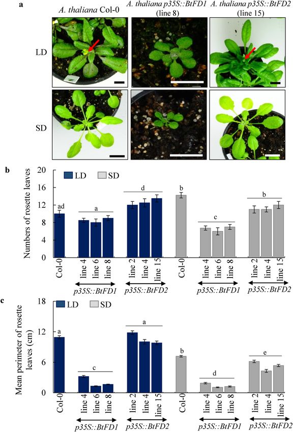
www.nature.com/scientificreports/
transition and meristem identity were identified in D. latiflorus20, whereas sixteen MADS box genes were reported
from Bambusa edulis21. A few studies have been conducted to functionally characterize important flowering genes
such as MADS18 from D. latiflorus22, FLOWERING LOCUS T (FT) from P. meyeri23,24, TERMINAL FLOWER
1 (TFL1) like gene from B. oldhamii25, FRIGIDA (FRI) from P. violascens26, and MADS1, 2 from P. praecox27.
FD genes encoding transcription factors are members of the group A basic leucine zipper (bZIP) family28.
They are ubiquitously found in angiosperms, but not in any other plant lineages29. Studies conducted on Arabi-
dopsis (A. thaliana) and rice (O. sativa) suggest that the transition of shoot apical meristem (SAM) to inflores-
cence meristem (IM) is primarily governed by interaction among AtFT/OsHd3a, At14-3–3/OsGf14 and AtFD/
OsFD1 proteins to form the florigen activation complex (FAC) preceding flowering30–34. Subsequently, FD binds
to the CRE binding element (ACGT) present in the promoter of floral meristem identity gene APETALA133,35,36
(AP1). Two paralogous copies of FD genes (AtFD and AtFDP) have been identified in Arabidopsis37, whereas
three copies are present in r ice29. Other than these reference plants, FD homologs have been discovered from
many other plants.
The loss-of-function mutation of either AtFD or AtFDP resulted in late flowering in Arabidopsis, while their
overexpression demonstrated early flowering indicating their possible involvement in fl owering37,38. Similarly,
the RNAi lines of OsFD1 demonstrated a late flowering phenotype, while overexpression of OsFD1 resulted into
early flowering29,33. In addition to flower induction, other pleiotropic roles of FD genes such as inflorescence
development39,40, leaf d evelopment29 and alternative growth c essation41,42 have also been observed. This clearly
indicates that FD performs diverse important roles in the vegetative and reproductive developments of plants.
Therefore, in order to understand the diverse functions of FD genes in plant growth and development, new
studies need to be conducted on yet unexplored, non-reference plants demonstrating remarkable vegetative or
flowering habit. Bamboos represent a particularly interesting plant group due to their semelparous life cycle and
transition to flowering after decades of vegetative growth. Therefore, the main aim was to identify and character-
ize bamboo FD genes. This study addressed the sequence diversity and differential DNA binding properties of
two FD genes isolated from Bambusa tulda in conjunction with their functional diversity based on expression
patterns and impact on vegetative and flowering development in a heterologous system.
Results
Identification and sequence characterization of BtFD1 and BtFD2 genes. To study the role and
diversity of FD genes (Table 1) in bamboo, B. tulda was selected, because its floral developmental stages have
relatively been better characterized than any other bamboo species11,43, occurrence of sporadic flowering events
in the species from time to time4,44 and its enormous economic importance in Asia. Two copies of BtFD genes
have been identified by designing primers from the conserved regions of homologous FD genes, PCR and multi-
ple sequencing (Supplementary Fig. S1, Fig. 1, BtFD1: MF983712 and BtFD2: MH142577). Homology search of
the BtFD1 sequence identified Sasa veitchii (Bambusoideae) FD (SvFDL1: BAS04368.1, SvFDL2: BAS04369.1) as
its closest homolog having highest similarity (77%), while the BtFD2 sequence revealed highest similarity (92%)
against Panicum hallii FD homolog (XP_025812603.1). Predicted lengths of BtFD1 and BtFD2 proteins were 202
and 159 amino acids, respectively, and both of them contain the characteristic bZIP domain. However, differ-
ences between BtFD1 and BtFD2 proteins were observed with reference to other domains. BtFD1 contains motif
1 [MEDD(E/D)DMW(A/G)XTSSPSASPP], the LSL motif [T(A/V)LSLN] and the SAP motif [(S/T)LXRX(S/T)
(A/T)(P/Q)F], while BtFD2 contains motif 2 [NYHHYQMAV(A/H)AA], motif 3 [(L/M/V)SGCSSLFSIS(S/T)]
and a modified SAP motif (Supplementary Fig. S2). A detailed sequence comparison of the bZIP domains pre-
sent in BtFD1 and BtFD2 proteins identified five amino acids changes (Supplementary Fig. S3). Out of these,
only the change of Ala146 (BtFD1) > Leu100 (BtFD2) was located in the DNA binding basic region. Therefore,
it was investigated whether this amino acid change may or may not impact the binding efficiency of BtFD1 and
BtFD2.
Phylogenetic relationship of BtFD1 and BtFD2 genes with homologs obtained from other
Poaceae and non‑Poaceae members. The phylogenetic analysis of BtFD1 and BtFD2 genes with
homologs obtained from Poaceae and non-Poaceae members identified three major clusters. The cluster 1 was
comprised of FD1 homologs obtained from all the Poaceae plants along with three bamboos (B. tulda, S. veitchii,
P. heterocycla). The cluster 2 was comprised of FD1 homologs obtained from all the non-Poaceae members, while
the cluster 3 was comprised of all FD2 homologs (Fig. 1). Cluster 1 specific for Poaceae FD1s was subdivided
into two major sub-clusters. The sub-cluster 1 hosted FD1 sequences obtained from annual (Z. mays, S. bicolor,
S. italica, S. viridis, O. sativa, O. brachyantha) and perennial (Z. japonica, D. oligosanthes, P. hallii, P. virgatum)
plants, whereas sub-cluster 2 hosted only annual plants such as H. vulgare, T. aestivum, A. tauschii, B. distachyon,
B. stacei. The B. tulda FD1 was placed in sub-cluster 1 along with two other bamboos P. heterocycla and S. veitchii
FD1 (Fig. 1). Similarly, the FD2 specific cluster 3 was also subdivided into two sub-clusters. Here, B. tulda FD2
was clustered with P. heterocycla along with other annuals and perennial plants (Fig. 1).
Expression analyses of BtFD1 and BtFD2 genes in different tissues, diurnal conditions and sea-
sons. Transcriptional expression patterns of BtFD1 and BtFD2 genes were investigated in diverse vegetative
as well as reproductive tissues, diurnal conditions and seasons to understand the functions of these genes in
bamboo vegetative as well as reproductive development. Among nine different tissues studied, expression of
BtFD1 was highest in shoot apex, followed by YLF and culm-sheath in comparison to rhizome. In contrary, the
expression level of BtFD2 was consistently very low in majority tissues studied (Fig. 2a). When diurnal expres-
sion patterns were analysed, expression level of BtFD1 in YLF was highest in the afternoon (4 pm), which was
Scientific Reports | (2021) 11:7849 | https://doi.org/10.1038/s41598-021-87491-6 2
Vol:.(1234567890)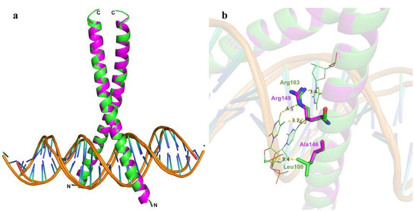
www.nature.com/scientificreports/
FD1 homologs identified FD2 homologs identified
Locus ID/ Locus ID/
Monocot plant Flowering Accession number Query cover Accession number Query cover
groups Plant species habit of best BLAST hit Identity (%) (%) E value of best BLAST hit Identity (%) (%) E value
Oryza sativa Annual OS09G36910 100 100 0.0 OS06G50830 100 100 0.0
O. brachyantha Annual OB09G24570 74 76 2e-72 OB02G45490 76 41 4e-25
Zea mays Annual ZM00001D022613 45 55 9e-37 ZM00001D036392 76 80 1e-74
Hordeum
Annual HVU0041G3471 70 45 3e-18 HVU0045G2520 74 80 1e-74
vulgare
Sorghum
Annual SOBIC002G280800 45 64 8e-34 SOBIC010G269000 77 82 2e-78
bicolor
Triticum
Annual TAE32871G001 50 62 7e-39 TAE36408G001 73 80 9e-75
aestivum
Aegilops
Annual XP020151150 50 78 3e-29 XP020177215 74 82 6e-72
tauschii
Brachypodium
Annual BRADI4G36587 52 62 4e-35 BRADI1G29920 75 81 1e-68
distachyon
B. stacei Annual BRAST05G197700 45 47 3.7e-18 BRAST07G238100 77 75 5.2e-51
Poaceae
Setaria italica Annual SEITA2G291300 49 57 1e-43 SEITA4G282900 81 86 6e-84
S. viridis Annual SEVIR2G302300.1 43 54 7.9e-25 SEVIR4G295100 78 84 1.1e-59
Panicum hallii Perennial PAHALB03671.1 45 59 5.4e-20 PAHALD00176 77 81 1.8e-58
PAVIRJ04759CON-
P. virgatum Perennial PAVIRJ04490.1 42 59 5.4 e-21
78 81 2.3e-59
TIG 06,819
Oropetium OROPETIUM_ OROPETIUM_
Perennial 66 42 5e-17 77 84 3e-82
thomaeum 20150105_06520 20150105_16409
ZJNSC00122.1. ZJNSC00008.1.
Zoysia japonica Perennial 50 58 2e-27 77 81 1e-76
G00160.1 G00500.1
Dicanthelium
Perennial OEL23045 43 94 1e-29 OEL37294 76 79 6e-64
oligosanthes
Sasa veitchii Perennial BAS04368 52 98 2e-44 NHF – – –
Phyllostachys
Perennial PH01000511G0500 50 59 6e-36 PH01001986G0070 79 82 3e-68
heterocycla
Zostera marina Perennial ZOSMA70G00700 60 43 1 e-12
NHF – – –
Spirodela e-12
Perennial SPIPO3G0017700 55 41 5 NHF – – –
polyrhiza
Musa acumi-
Annual MAC12G0975 59 42 1e-17 NHF – – –
nata
Ananas
Perennial ACO009346 59 42 8.8e-10 NHF – – –
comosus
Non-Poaceae Phalenopsis
Perennial PEQU26966 54 41 7e-15 NHF – – –
equestris
Dendrobium
Perennial XP020692523.1 55 43 7e-16 NHF – – –
catenatum
Elaeis guineen-
Perennial EGU0206G0533 47 42 5e-19 NHF – – –
sis
Phoenix dac-
Perennial XP008780307 50 51 2e-17 NHF – – –
tylifera
Table 1. In silico identification of FD gene homologs identified from the Poaceae and non-Poaceae members
of monocotyledonous plants. The BLASTP analyses were performed using O. sativa amino acid sequences as
queries. The criteria used for BLAST analyses and subsequent selection of homologs were: identities (≥ 50%), E
values and coverage of the query sequences against the obtained hit sequences (≥ 60%). When multiple BLAST
hits were obtained, only the top hit sequences were considered for further analyses. NHF no hit found.
not the case for YLN. However, the expression level of BtFD2 was consistently very low except in a single time
point i.e. afternoon (4 pm) in YLF (Fig. 2b).
Close observation of B. tulda flowering habit from 2015 to 2018 revealed that sporadic flowering events
usually recurred in spring every year. Therefore, to get further insight into the functions of BtFD1 and BtFD2
genes, their expression in young leaves were studied at three time points before onset of flowering, i.e., sum-
mer (April-June), monsoon (July–August), autumn (September–October), during onset of flowering i.e., winter
(November-January) and after i.e., spring (February–March, Fig. 3). The expression level of BtFD1 was notably
higher in winter compared to other seasons (Fig. 3). In contrary, no such pattern was found for BtFD2 expres-
sion. It was also barely detectable and quite comparable in YLF and YLN in all the seasons except a little increase
in YLN in spring.
Scientific Reports | (2021) 11:7849 | https://doi.org/10.1038/s41598-021-87491-6 3
Vol.:(0123456789)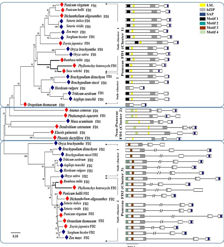
www.nature.com/scientificreports/
Figure 1. Phylogenetic analysis and predicted gene models of FD homologs identified in monocotyledonous
plants. The Neighbour Joining (NJ) tree of the FD homologs was constructed with the full-length protein
sequences by Mega 7.0 using default parameters and bootstrap value 2000. In the gene structures exons
were marked as rectangles having conserved motifs marked in solid boxes and introns as solid lines. Annual
species were marked in blue and perennials in red rhomboids. The motifs conserved are M 1 [MEDD(E/D)
DMW(A/G)XTSSPSASPP], M 2 [NYHHYQMAV(A/H)AA], M 3 [(L/M/V)SGCSSLFSIS(S/T)], M 4 [(M/V)
EEVWKDINLSSLHD], LSL [T(A/V)LSLN], Bzip and SAP [(S/T)LXRX(S/T)(A/T)(P/Q)F].
In silico and EMSA analyses to study interaction between bZIP domains of BtFD proteins and
ACGT motif. The bZIP domain of FD proteins needs to interact with the conserved CRE binding element
(ACGT) present in the promoter of AP1 in order to perform DNA binding activity, Therefore, the overall poten-
tial of bZIP domains present in BtFD1/BtFD2 to bind to the ACGT motif was analysed. Comparison of the bZIP
domains of BtFD1, BtFD2 and their homologous sequences revealed a striking difference, i.e. Ala146 of BtFD1
was replaced by Leu100 in BtFD2 (Supplementary Fig. S3). In order to assess the impact of such an amino acid
change, a two-pronged approach was adopted—(1) in silico prediction of overall DNA binding ability of BtFD1
and BtFD2, and (2) validation of the in silico prediction using EMSA analyses.
Docked structures of both BtFD1 and BtFD2 bZIP models predicted positive interactions with CRE DNA
containing ACGT motif. Superimposed docked structures also revealed that the interactions of both BtFD1 and
BtFD2 could take place in a similar manner (Fig. 4a). Several amino acid residues located at the basic region,
Scientific Reports | (2021) 11:7849 | https://doi.org/10.1038/s41598-021-87491-6 4
Vol:.(1234567890)www.nature.com/scientificreports/
Figure 2. Comparison of tissue specific and diurnal expression pattern of BtFD1 and BtFD2 genes. (a) Tissue
specific expressions of BtFD1 and BtFD2 in nine different tissue stages of B. tulda. Each bar represents mean of
three biological replicates ± SE. (b) Diurnal expressions of BtFD1 and BtFD2 in YLF and YLN in SD and LD.
Each data point represents mean of three biological replicates ± SE. Transcript expression of eIF4α was used to
normalize expression data. The relative fold change was calculated by 2 −ΔΔCT method using the expression data
in rhizome as calibrator and is plotted using Y axis. CS culm sheath, YLF young leaf from flowering culm, YLN
young leaf from non-flowering culm, I inter node, SA shoot apex, IFB immature floral bud, MFB mature floral
bud, R root.
spanning from His133 to Gln153 in BtFD1 and Arg87 to Arg107 in BtFD2 were found to interact with the CRE
consensus sequence. In silico docking analysis suggested that Arg142, Ser144, Arg147, Ser148 and Arg149 of
BtFD1 and Arg96, Leu100, Arg101, Ser102 and Arg103 of BtFD2 were particularly found to be directly interact-
ing with TGACGTCA consensus CRE DNA. Additionally, in silico analysis predicted direct contact for Leu100
in BTFD2 with a conserved dT residue of ACGT motif, whereas the corresponding Ala146 in BtFD1 had no
interfering interactions with DNA (Fig. 4b). Even though BtFD2 gained additional interaction in this way, this
non-polar-polar interaction was unfavourable in nature and therefore, could interfere with its DNA binding
specificity. Ala146 on the other hand, though also non-polar, might be advantageous in this position because of
its smaller size. To validate this result further, EMSA studies were conducted using mimics of BtFD1 and BtFD2
bZIPs, which only differed by a single amino acid (Ala146 of BtFD1 vs. Leu100 of BtFD2, Fig. 5a). Varying
Scientific Reports | (2021) 11:7849 | https://doi.org/10.1038/s41598-021-87491-6 5
Vol.:(0123456789)www.nature.com/scientificreports/
Figure 3. Expression analyses of BtFD1 and BtFD2 genes in YLF and YLN of B. tulda in five different seasons.
Each bar represents mean expression of three biological replicates ± SE. The eIF4α was used to normalize
expression data of the targeted flowering genes. The relative fold change was calculated by 2 −ΔΔCT method using
the expression level observed in rhizome as the calibrator. YLF young leaf from flowering culm, YLN young leaf
from non-flowering culm.
Figure 4. In silico interactions of bZIP domains of BtFD1 and BtFD2 with ACGT motif. (a) Superimposed
structures of bZIP domains of BtFD1 (magenta) and BtFD2 (green) interacting with ACGT motif in a similar
manner. (b) Arg149/Arg103 of BtFD1/BtFD2 interact with cognate DNA sequence containing ACGT motif and
Leu100 of BtFD2 making additional contact with DNA (dT residue).
Scientific Reports | (2021) 11:7849 | https://doi.org/10.1038/s41598-021-87491-6 6
Vol:.(1234567890)www.nature.com/scientificreports/
Figure 5. EMSA study to compare the DNA binding efficiency of bZIP domains of BtFD1 and BtFD2 proteins.
(a) Sequence alignment of bZIP domains of TobZL (experimental template) vs. BtFD1 and BtFD2 reveals
changes at two amino acids residues (His to Ser and Lys to Ala/Leu). (b) EMSA studies of BtFD1 and (c) BtFD2
mimics show overall ability to bind with CRE DNA. Lanes 1–7 in both the gels contain twofold serially diluted
proteins (0.080 to 5.000 μM for BtFD1 and 0.800 to 50.00 μM for BtFD2 bZIP mimics), lane 8 contains free
DNA.
concentrations of bZIP mimics of BtFD1 and BtFD2 proteins were used for EMSA analysis, which showed that
reappearance of free DNA begins after 0.30 µM in case of BtFD1 and 3.12 µM in case of BtFD2 (Fig. 5b,c). Free
DNA is observed at 0.28 µM in case of BtFD1, which is 2.81 µM for BtFD2 (Supplementary Figs. S5a, S5b). There-
fore, the concentration ranges of ‘binding to no-binding’ for bZIP mimic of BtFD1 was 0.31–0.28 µM, whereas it
was 3.12–2.81 µM for BtFD2. Taken together, the finding clearly demonstrated a tenfold enhanced DNA binding
specificity for BtFD1 bZIP mimic compared to its BtFD2 analogue. This means the required threshold value
for BtFD2-CRE DNA interaction is much higher than that of BtFD1 (Fig. 5b,c). This further consolidated the
consequence of the single amino acid substitution (Supplementary Figs. S5a–d).
Scientific Reports | (2021) 11:7849 | https://doi.org/10.1038/s41598-021-87491-6 7
Vol.:(0123456789)www.nature.com/scientificreports/
Constitutive expression of BtFD1 and BtFD2 genes in Arabidopsis. In order to study roles of BtFD1
and BtFD2 genes on the vegetative as well as reproductive development of plants, these homologs were constitu-
tively expressed in Arabidopsis (Columbia) plants. The phenotypes of transgenic p35S::BtFD1 Arabidopsis plants
revealed drastic suppression of vegetative and floral growth in short day (SD) and long day (LD) conditions
(Fig. 6a). Leaf numbers observed in three independent p35S::BtFD1 transgenic lines after four weeks of growth
were 8 to 9 in LD and 6 to 7 in SD, which were 10 and 14 in wild-type plants, respectively (Fig. 6b). The reduc-
tion in leaf number in p35S::BtFD1 plants in comparison to WT was statistically significant in SD (p.adj = 0.000),
but not in LD (p.adj = 0.193), when one-way ANOVA was conducted. In contrary, change in leaf numbers of
p35S::BtFD2 transgenic plants in comparison to WT were statistically insignificant in SD (p.adj = 0.007) as well
as LD (p.adj = 0.040). Apart from the numbers, size of leaves were also significantly reduced in p35S::BtFD1
plants in SD (p.adj = 0.000) and LD (p.adj = 0.000) compared to WT (Fig. 6c). In contrary, the difference in
leaf size between p35S::BtFD2 and WT plants were statistically significant in SD (p.adj = 0.000), but not in LD
(p.adj = 0.725) (Fig. 6c). In order to simultaneously consider the effects of genetic background (WT, p35S::BtFD1,
p35S::BtFD2) as well as duration of light (SD, LD), two-way ANOVA was also conducted. The genetic back-
ground had significant effect on leaf numbers (p = 0.000), whereas the light duration did not (p = 0.356). Number
of leaves were significantly reduced in p35S::BtFD1 plants compared to the WT (p.adj = 0.000). In contrary, no
significant difference was obtained for leaf numbers of p35S::BtFD2 plants compared to WT (p.adj = 0.952).
However, both the genetic background (p = 0.000) as well as the light duration have significant effects (p = 0.000)
on leaf perimeter. Also, change in perimeter was significant in both the cases for p35S::BtFD1 plants compared
to the WT (p.adj = 0.000), which was not the case for the p35S::BtFD2 plants compared to the WT (p.adj = 0.022).
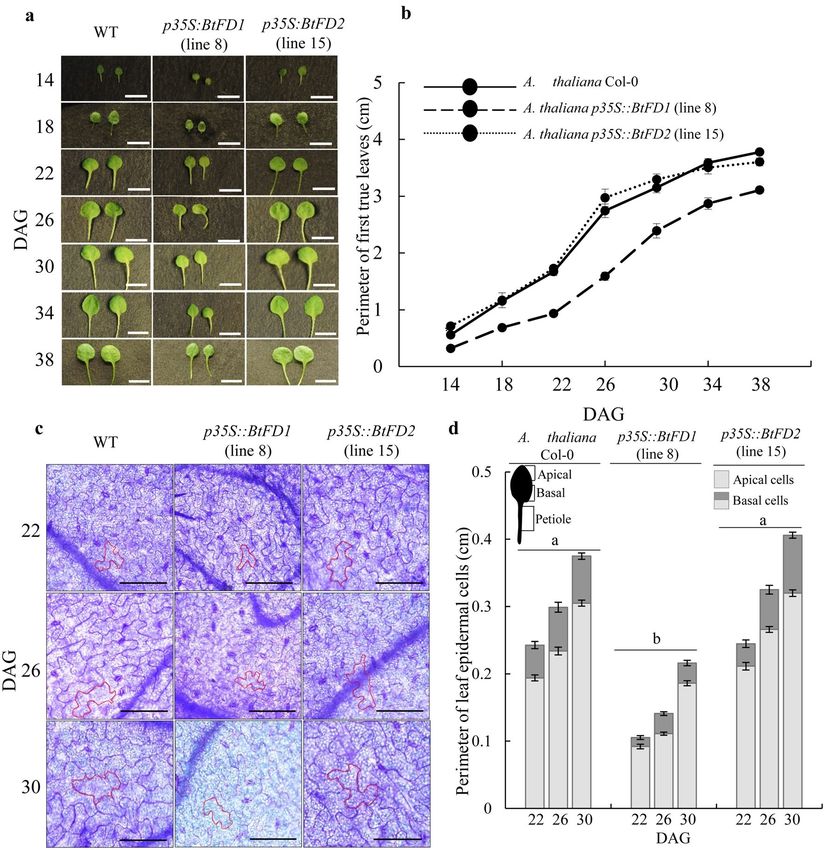
In order to obtain kinetic differences in leaf growth, perimeter of first true leaves were measured in four-
day intervals in SD (Fig. 7a). Consistently, the leaf perimeter of p35S::BtFD1 trasgenic plants were significantly
lower than WT and p35S::BtFD2 (Fig. 7b). Further, histological observation on leaf epidermal cells of first true
leaves of these plants revealed that the perimeter were significantly lower (0.234 ± 0.005 cm to 0.299 ± 0.007 cm)
in p35S::BtFD1 (p.adj = 0.000) plants compared to WT, but not in case of p35S::BtFD2 (p.adj = 0.066, Fig. 7c,d).
Like vegetative growth, the flowering time was extremely delayed in p35S::BtFD1 Arabidopsis plants compared to
p35S::BtFD2 and WT (Supplementary Figs. S4a, S4b). This was apparent by the significant increase of leaf number
in p35S::BtFD1 plants compared to WT (p.adj = 0.000), but not in case of p35S::BtFD2 (p.adj = 0.558). Addition-
ally, the length of the flowering stalk and the numbers of flowers/plant were strongly reduced in p35S::BtFD1
transgenic plants, while no obvious difference was noticed for p35S::BtFD2 and WT plants in LD (Supplementary
Fig. S4a). In order to promote flowering, FD binds to AP1 to induce it at the transcriptional level. Therefore, the
expression of AtAP1 was measured in the wild type, p35S::BtFD1, and p35S::BtFD2 Arabidopsis plants. Indeed, the
expression of the AtAP1 gene in the four-week-old leaves of p35S::BtFD1 Arabidopsis plants grown under LD was
tenfold higher compared to WT, which was only twofold in case of p35S::BtFD2 plants (Supplementary Fig. S4c).
Discussion
Bamboo FD genes are similar to other Poaceae FD homologs in terms of sequence similarity
and phylogenetic relationships. FD is a bZIP family protein and plays important roles in controlling
the timing of reproductive phase transition in angiosperms29,33,45. In addition, its role in vegetative development
has also been observed29. In this study two bamboo FD genes (BtFD1 and BtFD2) were identified and their
sequences were characterized to study phylogenetic relationships of these genes to other homologous monocot
genes. Characterization of the amino acid sequences inferred that like other Poaceae FD1s, bamboo BtFD1
possessed motifs 1, LSL, bZIP and SAP, but not motif 4, which is usually characteristic of non-Poaceae FD1s29
(Supplementary Fig. S2). Among these motifs, bZIP and SAP were found absolutely necessary for the interaction
with AP136 and 14–3-3, respectively. However, the functional significance of other motifs in flowering needs fur-
ther investigation33. In contrary, the LSL motif is absent in all FD2 homologs including bamboo that have been
identified so far suggesting their less likely involvement in flowering (Supplementary Fig. S2).
Phylogenetic analyses of the FD homologs obtained from monocotyledonous plants revealed the presence of
three major clusters. Cluster 1 and 2 were comprised of FD1 homologs of Poaceae and non-Poaceae members
respectively, while all FD2s from the Poaceae species were placed in cluster 3 (Fig. 1). All the bamboo FD1 and
FD2 homologs (P. heterocycla, B. tulda and S. veteichii) were found in the Poaceae specific clades of FD1 and
FD2 respectively (Fig. 1). It had previously been found that the FD gene clade could be broadly classified into
four subgroups, which were Poaceae specific FD1, Poaceae specific FD2, Poaceae specific FD3 and FDs obtained
from eudicots as well as non-Poaceae members of monocots29.
Bamboo FD1 and FD2 genes are divergent in expression patterns with respect to tissues, diur-
nal conditions and seasons. The detailed expression analyses of BtFD1 and BtFD2 genes in diverse tis-
sues, diurnal conditions and seasons may provide clues about their possible functionality. It is well established
that in SAM, FT interacts with FD to form FAC and consequently floral meristem identity genes are activated to
induce flowering45. Therefore, FD expression has primarily been observed in SAM tissues of Arabidopsis, rice, P.
sativum, P. tremula x P. alba and P. aphrodite plants29,30,33,40,42,46. In addition, expression of FD1 was also detected
in leaves of O. sativa29, T. aestivum36 and A. chinensis41 plants. In bamboo, the expression level of BtFD1 gene was
highest in shoot apex. However, the expression of BtFD2 was very low in all the tissues. This is unlike rice, where
FD2 was primarily expressed in leaves29. Like many other flowering genes, FD also was found diurnally regulated
in rice34 and P. tremula x P. alba42. In bamboo, expression of BtFD1 in YLF was highest in the afternoon (4 pm),
but in YLN it was in the morning (8am). In poplar, similar diurnal regulation of FD was observed in SD, where
it attained its maximum expression at mid night, whereas no such pattern was observed in L D42. In contrary, the
diurnal expression of BtFD2 remained consistently low throughout the day. Expression analyses across seasons
Scientific Reports | (2021) 11:7849 | https://doi.org/10.1038/s41598-021-87491-6 8
Vol:.(1234567890)www.nature.com/scientificreports/
Figure 6. Phenotypic comparisons of wild type (WT) and transgenic p35S::BtFD1 and p35S::BtFD2 A. thaliana
plants. (a) Plants were grown in LD (16-h light and 8-h dark) and SD (10-h light and 14-h dark) for four weeks.
Arrow indicates emerged inflorescence axis. The scale bar represents 1 cm. (b) Comparisons of rosette leaf
numbers of four-week-old transgenic plants in (c) Comparisons of perimeters of rosette leaves of transgenic vs.
WT plants. Each bar represents mean perimeter of eight individual mature leaves ± SE. comparison to WT in SD
and LD. Each bar represents mean leaf numbers obtained from four individual plants ± SE. One-way ANOVA
analyses were performed to test statistical significance at p.adj ≤ 0.0001. LD long day, SD short day.
also point towards a role of BtFD1in flower induction. Transcript accumulation of BtFD1 in the floral inductive
tissue YLF began in autumn and reached the maximum level in winter, i.e. just before sprouting (Fig. 3). This
observation was comparable to perennial dicots poplar and kiwifruit, where FD was transiently expressed just
before flowering every year41,42. In contrary, expression of BtFD2 was almost negligible throughout the year.
Taken together, the analysed expression data suggest that BtFD1 may perform important roles in flower and
vegetative development of bamboo, whereas the function of BtFD2 is yet to be discovered.
Scientific Reports | (2021) 11:7849 | https://doi.org/10.1038/s41598-021-87491-6 9
Vol.:(0123456789)www.nature.com/scientificreports/
Figure 7. Comparison of leaf sizes of wild type A. thaliana (WT) with that of transgenic p35S::BtFD1 and
p35S::BtFD2 plants. (a) Representative first true leaves of WT, p35S::BtFD1 and p35S::BtFD2 plants in 14, 18, 22,
26, 30, 34, 38 days after germination (DAG). (b) Perimeter of first true leaves measured in every four DAG in
SD (10-h light and 14-h dark). Each data represents mean of ten individual data ± SE. The scale bar represents
5 mm. (c) Leaf epidermal cells obtained from the first true leaves of 22, 26 and 30 DAGs in SD. In each picture,
the cell wall of a representative cell is marked by red outline. The scale bar represents 0.1 mm. (d) Perimeter of
leaf epidermal cells measured in 22, 26 and 30 DAGs in. SD. Each data represents mean of thirty (fifteen apical
and fifteen basal) individual cells ± SE. Mixed three-way ANOVA were performed to test statistical significance
at p.adj ≤ 0.0001.
A single amino acid change resulting into differential binding efficiency between bZIP domains
of BtFD1/BtFD2 and CRE DNA. Sequence analyses and in silico characterization of the two BtFD proteins
confirmed that they belong to bZIP transcription factor family. Among several different subfamilies of bZIPs,
BtFDs were found to be homologous to CREB. Co-crystal structure of CRE DNA—CREB bZIP of Mus muscu-
lus35 (PDB ID IDH3) was chosen for homology modelling purpose. Generally, the CREB family bZIP members
are capable to interact with A box (TACGTA), G box (CACGTG) or C box (GACGTC) elements present in the
promoter region of their target genes causing transcriptional upregulation47,48. In plants, the FD1 members of
CREB family are involved in the establishment of floral meristem identity30,36,39. Overall, the bZIP regions of
BtFD1 and BtFD2 proteins differ in five different amino acid positions. Particularly one position (Ala146 is
BtFD1 vs. Leu100 in BtFD2) at the crucial DNA binding site (NXXAAXXSR) was interesting. Therefore, it was
asked whether any of these amino acid changes, in particular this single amino acid substitution, could have any
impact on their DNA binding activity. Our in silico analyses revealed that the bZIP domains identified in BtFD1
Scientific Reports | (2021) 11:7849 | https://doi.org/10.1038/s41598-021-87491-6 10
Vol:.(1234567890)www.nature.com/scientificreports/
and BtFD2 were capable to dimerise and form a bZIP structure. They also demonstrated specific interaction with
the TGACGTCA sequence. In particular, Asn141, Arg149 of BtFD1 and Asn95, Arg103 of BtFD2 can directly
interact with cognate DNA substrate. Similar interaction has also been found in m aize39 and w heat36. EMSA
analysis highlighted that the change of Ala146 in BtFD1 vs. Leu100 in BTFD2 resulted into ten times enhanced
binding of BtFD1 than BtFD2. This may be the result of an additional, yet unfavourable contact between Leu100
of BtFD2 and dT residue of ACGT motif, apparent in the docked structure. It might be possible that Leu100
interfered with the interaction of target DNA by making a polar vs. non-polar interaction. In contrary, the
shorter Ala146 residue, which was present in BtFD1 could not interfere and thus enabling higher DNA binding
efficiency of BtFD1 (Fig. 5b,c).
Ectopic expression of BtFD1 severely suppressed vegetative growth and flowering in Arabi-
dopsis, but BtFD2 did not. In order to study the functions of BtFD1 and BtFD2 genes in planta, transgenic
alterations of these genes needed to be carried out. Altering activities of these genes in bamboo itself were dif-
ficult due to many reasons such as long-life cycle, difficulty with in vitro regeneration and unavailability of effi-
cient transformation methods49,50. Therefore, BtFD1 and BtFD2 genes were ectopically expressed in Arabidopsis
plants and their phenotypes were compared.
The vegetative growth of transgenic Arabidopsis plants overexpressing BtFD1 gene was severely suppressed
with respect to the number and size of the rosette leaves (Fig. 6a–c). Similar phenotypes had been noticed when
AtFD and AtFDP together were overexpressed in rice45 and also when poplar FDL1 was overexpressed in Populus
tremula × tremuloides42,51. The involvement of FD in controlling vegetative growth has been observed in a pea
loss-of-function mutant, demonstrating severe branching even after flower induction40. A few molecular players
in connection to the growth retardation due to FD1 overexpression have been identified. For instance, in poplar,
BRANCHED1 and 2 genes, which promote shoot growth by maintaining proper auxin and cytokinin levels were
downregulated52,53. Overexpression of Arabidopsis FD and FDP in rice resulted in the down-regulation of many
cell wall growth responsive genes such as EXTENSIN, EXPANSIN and XTH145. Similar to all these previous
observations, in this study the p35S::BtFD1 Arabidopsis plants revealed reduced leaf and leaf epidermal cell sizes
compared to p35S::BtFD2 and wild-type plants.
The role of FD1 in flower induction has already been established by a large body of literature and a variety of
mutant phenotypes have been observed: (a) Delay in flowering was observed in the loss-of-function mutants of
Arabidopsis30,37,38, pea40 and maize39, while early flowering was observed in the FD1 overexpressing lines of r ice29,33
and Phalaenopsis46. (b) However, exceptions to this line of observation have also been noticed42,45,51. When AtFD
and AtFDP together were overexpressed in rice, flowering and vegetative growth has been retarded45. Similarly,
overexpression of poplar FDL1 in Populus tremula × tremuloides resulted into delayed flowering in S D42,51. Our
results demonstrated that transgenic Arabidopsis plants overexpressing BtFD1 exhibited a delay in flowering time
and numbers of flowers/plant compared to p35S::BtFD2 and WT plants (Supplementary Fig. S4a). However,
expression of AtAP1 was remarkably higher in p35S::BtFD1 Arabidopsis than p35S::BtFD2 and WT plants. Similar
observations were also reported in AtFD and AtFDP overexpressing lines of rice45 and FDL overexpressing lines of
poplar51, which, nevertheless, led to late flowering phenotypes. Together, it can be concluded that timely expres-
sion of BtFD1 may be critical to perform its programmed flower specific role in planta, which was altered in the
transgenic Arabidopsis plants constitutively overexpressing BtFD1 in a spatially and timely improper manner.
Therefore, the apparent delay in flowering time could be an indirect effect of extensive suppression of vegetative
growth, while in contrast, the flowering program is still enhanced based on the marker gene AtAP1 induction.
It is already well accepted that flowering can only be induced after plants attain sufficient vegetative g rowth54.
The evolution of gene function within the FD family revealed the existence of functional redundancy in
Arabidopsis45. In contrary, a clear functional diversification was noticed between the two rice FD genes OsFD1
vs. OsFD229. Our study revealed that the two bamboo FD genes imposed contrasting effects on shoot growth
and flowering time, which may be mediated by two ways: (a) by acquiring expression divergence where BtFD1
maintained a flower associated expression pattern whereas expression level of BtFD2 was consistently low and
(b) by adapting a single amino acid change (Ala146 vs. Leu 100) located in their DNA binding region which may
cause a differential binding to their target protein AP1. Future studies are required to investigate the impact of
residue alterations in the other four positions. Such single residue swapping was found sufficient to convert the
flowering repressor TFL1 to an activator FT and vice versa by altered interaction with their interactor proteins55.
Taken together, it can be concluded that regions involved in protein–protein or DNA–protein interactions can
be potential targets to study the functional evolution of closely related homologous genes. Further studies are
required to uncover whether BtFD1 is anyhow involved in long perennialism of bamboo and whereas its homolog
BtFD2 evolved any additional function or required other interacting partner to be functional.
Materials and methods
Collection of Bambusa tulda tissues for gene expression analyses. Flowering tissues were
obtained from three populations of B. tulda located in Shyamnagar, W.B. (SHYM7, SHYM16, 22.38° N. 88.40°
E) and B andel11 (BNDL22, 22.93° N. 88.38° E). Recurrent incidence of sporadic flowering was noticed every year
in spring from 2015 to 2018. Corresponding voucher specimen were submitted to the Botanical Survey of India
(B.S.I), Shibpur (deposition nos. 56A, 56B, 57A, 57B, 58A. 58B, 59A, 59B, 59C on 05.06.2015). To perform tissue
specific gene expression analysis, six vegetative tissues such as young leaf from flowering (YLF) and nonflower-
ing culm (YLN), culm sheath (CS), root (R), internode (I), shoot apex (SA) and two flowering tissues such as
immature and mature floral buds (IFB, MFB) were collected. In order to perform diurnal expression analyses,
YLF and YLN were collected at four different time points of a day- morning (8 am), noon (12 pm), afternoon
(4 pm) and night (8 pm) for both long day (LD, 14 h light exposure, sunrise at 4:30 am and sunset at 6:30 pm) and
Scientific Reports | (2021) 11:7849 | https://doi.org/10.1038/s41598-021-87491-6 11
Vol.:(0123456789)www.nature.com/scientificreports/
short-day (SD, 11 h light exposure, sunrise at 6 am and sunset at 5 pm). Tissues were also collected in five dif-
ferent seasons: summer (April–June, 2017), monsoon (July–August, 2017) autumn (September–October, 2017),
winter (November–January, 2017) and spring (February–March, 2018). At least three, independent biological
replicates were used for each tissue stage/diurnal condition/season.
Isolation of nucleic acids and preparation of cDNA libraries. Isolation of genomic DNA was carried
out from young, healthy leaves by using DNeasy Plant Mini Kit (QIAGEN, Germany). Total RNA was isolated
by a combination of Trizol (INVITROGEN, USA) and RNAeasy Plant Mini Kit (QIAGEN, Germany) as per the
procedure described b efore43,56. Samples were treated with DNase I enzyme (THERMO SCIENTIFIC, USA) to
avoid genomic DNA contamination, if any. Quality and quantity of the samples were checked in a BioSpectrom-
eter (EPPENDORF, Germany) and agarose-formamide gel elctrophoresis. Approximately 1 μg of total RNA was
used for cDNA synthesis using verso cDNA synthesis kit (THERMO SCIENTIFIC, USA) following manufactur-
er’s protocol. For real time RT-qPCR analyses, 2 μl of tenfolds diluted stock solution of cDNA samples was used.
Analysing FD gene and amino acid sequences obtained from various genome databases. Rice
gene sequences (OsFD1: OS09G36910 and OsFD2: OS06G50830) were used as queries to retrieve genomic as
well as amino acid sequences of FD1 and FD2 genes available in various genome databases. BLASTP analy-
ses were performed in Phytozome (https://phytozome.jgi.doe.gov/pz/portal.html), Plaza_monocot_v4 (https://
bioinformatics.psb.ugent.be/plaza/versions/plaza_v4_monocots/) and NCBI (https://www.ncbi.nlm.nih.gov)
databases. All BLASTP hits were obtained using the set criteria of an E-value threshold ≤ e−10, identity ≥ 40% and
length coverage with respect to the query sequence ≥ 40%. However, when multiple hits were obtained, only the
best BLASTP hit was selected for further analyses. If no homologous genes were found using the set criteria, it is
mentioned as ‘no hit found’ (NHF, Table 1).
Primer designing, PCR amplification and sequencing of B. tulda BtFD1 and BtFD2 genes. In
order to obtain B. tulda genes, homologous sequences obtained from closely related monocot species were
aligned and degenerate primers were designed from the conserved regions by using Primer3 program (http://
bioinfo.ut.ee/primer3-0.4.0/, Supplementary Table S1). PCR amplification was conducted using high fidelity
Phusion Taq DNA polymerase (THERMO SCIENTIFIC, USA). Amplified PCR products of desired molecular
weight were gel purified by using GeneJET gel elution kit (THERMO SCIENTIFIC, USA) and cloned into TA
vector (pGEM-T Easy Vector Systems, PROMEGA, USA) or blunt end vector (pJET PCR cloning kit, THERMO
SCIENTIFIC, USA). Selection of bacterial colonies were done based on the blue-white screening and/or ampi-
cillin sensitivity (100 µg/ml). Plasmids were isolated by GeneJET plasmid miniprep kit (THERMO SCIEN-
TIFIC, USA). Sanger’s sequencing was undertaken and contigs were assembled by CAP3 (www. http://doua.
prabi.fr/software/cap3) prior to submission to NCBI (MF983712, MH142577). The full length genomic and cod-
ing sequences (CDS) were analysed in the Gene Structure Display Server (GSDS, http://gsds.cbi.pku.edu.cn/
index.php) to predict the gene models.
Sequence data and phylogenetic analyses. The FD gene sequences identified from B. tulda were used
as query and BLASTP analyses were performed in NCBI (https://www.ncbi.nlm.nih.gov) database to identify its
homologous sequences in related species. Amino acid sequences of BtFD1 and 2 genes were aligned with other
homologous sequences using the Clustal Omega program (https: //www.ebi.ac.uk/Tools/msa/clustalo/). A phy-
logenetic tree was constructed by using the Neighbour Joining (NJ) method in MEGA7 tool57. Bootstrap analysis
with values for 2000 replicates was conducted to estimate nodal support.
In silico modelling and docking studies. In silico analysis was performed to predict the possibility of
binding between of bZIP domains of BtFD1 or BtFD2 to the ACGT motif. Due to unavailability of BtFD1 and
BtFD2 crystal structures, amino acid sequences corresponding to their bZIP domains were first subjected to
homology modelling by SwissModel (https://swissmodel.expasy.org). Both of them demonstrated significant
sequence homology (36% identity) with their nearest structural homologue CRE binding protein known from
Mus musculus35 (PDB ID IDH3). Therefore, it was chosen as template for modelling bZIPs of BtFD1 and BtFD2.
Ramachandran analysis using Molprobity option from Swissmodel revealed 98.96% residues to be in favour-
able region for BtFD1 and 95% for BtFD2 and both were modelled in their dimeric state. The two bZIP models
were then subjected to energy minimization using Chimera (https://www.cgl.ucsf.edu/chimera/). Ramachan-
dran analysis through Procheck (https://servicesn.mbi.ucla.edu/PROCHECK/) and post energy minimization
revealed complete inclusion of residues in favourable region for both the models. The energy minimized struc-
tures were then docked with 21 bp CRE DNA sequence (obtained from 1DH3 crystal structure) using NPdock
(http://genesilico.pl/NPDock).
Site‑directed mutagenesis for electrophoretic mobility shift assay (EMSA). In order to validate
the prediction of binding between bZIP domains of BtFD1 and BtFD2 with the ACGT motif, electrophoretic
mobility shift assay was performed. The bZIP sequence obtained from Thalassiosira oceanica LOV photorecep-
tor (To_bZIP + LOV − TobZL) protein, was used for site directed mutagenesis to obtain bZIP mimics of BtFD1
and BtFD2 proteins. Pairwise sequence alignment between among bZIP regions of BtFD1, BtFD2 and TobZL
revealed two amino acid differences at the DNA binding basic region (Fig. 5a). Double mutations leading to
conversion of His > Ser144 and Lys > Ala146 were introduced in TobZL to mimic BtFD1 bZIP and His310 > Ser98
Scientific Reports | (2021) 11:7849 | https://doi.org/10.1038/s41598-021-87491-6 12
Vol:.(1234567890)www.nature.com/scientificreports/
and Lys312 > Leu100 to mimic BtFD2 bZIP. Mutations were done using standard procedures to induce site
directed mutagenesis and were verified by DNA sequencing (EUROFINS GENOMICS INDIA PVT. LTD).
Over‑expression and purification of BtFD1 and BtFD2 bZIP mimics. The BtFD1 and BtFD2 bZIP
mimics were cloned in pET28a expression vector, transformed in E. coli [BL21(DE3)C43] and grown at 37 °C.
After isopropyl β-D-1-thiogalactopyranoside induction, bacterial cells were grown at 20 °C for overnight. Cells
were then centrifuged and pellets were re-suspended in buffer containing 20 mM Tris (pH 8.0), 10 mM NaCl,
10% glycerol in presence of the protease inhibitor. Following sonication on ice and centrifugation, the superna-
tant was incubated with Ni–NTA agarose (QIAGEN, Germany) for 2 h. After washing in 10 mM imidazole con-
taining re-suspension buffer, proteins were finally eluted with 250 mM imidazole. The eluted fractions were next
pooled and excess imidazole was removed using PD10 desalting column (SIGMA). Proteins were concentrated
and stored at − 20 °C in aliquots for future use.
Electrophoretic mobility shift assay. Electrophoretic mobility shift assay was carried out to study DNA-
binding activity of BtFD1 and BtFD2 proteins. A 24 bp DNA fragment [5′ d(TGTAGCGTCTGACGTGGTTCC
CAC) 3′ and complementary sequence] containing the consensus CREB binding site, TGACGT, were synthe-
sized (INTEGRATED DNA TECHNOLOGIES). Lyophilized DNA strands (labeled and un-labelled) were sus-
pended in nuclease free water and 10 μM of it was annealed by rapid heating at 95 °C followed by gradual cool-
ing in annealing buffer (12 mM Tris; pH 8.0, 30 mM NaCl). A final concentration of 0.5 μM double stranded
DNA was used in the protein DNA binding assay buffer (50 mM Tris–HCl pH 8.0, 20 mM NaCl, 0.5 mM DNA,
1.25 mM MgCl2, 20% glycerol). Serially diluted protein was added to the solution followed by an incubation at
22 °C for 1 h. The protein DNA complex along with the control set (only DNA) was resolved in 10% polyacryla-
mide gels at 180 V for 35 min. The gel was then stained with SyBr Gold (THERMO SCIENTIFIC, USA) in 0.5X
TBE buffer for 40 min and imaged using a gel documentation system (BIORAD, USA).
Gene expression analyses by real time RT‑qPCR. To perform real time RT-qPCR analyses, gene spe-
cific primers were designed from the coding sequences of the BtFD1 and BtFD2 genes using Primer3 program
(http://bioinfo.ut.ee/primer3-0.4.0/, Supplementary Table S1). The real time RT-qPCR analyses were performed
by using SsoAdvanced Universal SYBR Green Supermix (BIO-RAD, USA) and CFX connect real-time PCR
detection system (BIO-RAD, USA). To confirm the absence of any primer dimers in the amplified products,
a standard melt curve analysis was conducted. The BteIF4α and AtACT2 genes were previously identified as
ideal reference gene for normalizing expression data obtained from Bambusa58 and Arabidopsis56, respectively.
The relative fold change in gene expression level was calculated by the 2−ΔΔCt method59. The PCR amplification
efficiency were measured for the five pairs of primers used in RT-qPCR. Two fold serial dilutions of the pooled
cDNA templates were used to obtain standard curves for each primer pair. The amplification efficiency was ana-
lyzed using the formula60 10(−1/slope) − 1 × 100. The obtained percentage of efficiency was 95%-98%.
Gateway cloning of BtFD1 and BtFD2 genes. Gateway recombination sequences were tagged to the 5′
end of the primers to PCR amplify BtFD1 and 2 genes using Phusion Taq DNA polymerase enzyme (THERMO
SCIENTIFIC, USA, Supplemental Table S1). Approximately 100 ng of gel-purified PCR fragments were recom-
bined with 100 ng of pDONR221 donor vector using BP Clonase enzyme (INVITROGEN, USA). Reactions
were transformed into E. coli (DH5α) and isolated plasmids were verified by DNA sequencing before recom-
bination into the binary pAlligator2 vector providing The CaMV 35S promoter for expression61. Finally, the
expression clones were mobilized to competent Agrobacterium tumefaciens (pGV3101/pMP90) by electropora-
tion using a BIO-RAD Gene Pulser.
In planta transformation, selection, phenotypic characterization and statistical analy-
sis. Approximately six-week-old A. thaliana (Col-0) plants were transformed by the floral dipping m
ethod62.
Transformed T1 seeds were selected on the basis of green fluorescence of the GFP reporter gene61. The number
and perimeter of the rosette leaves were measured from three independent T 3 plants having single insertions
in order to perform phenotypic comparisons with wild-type A. thaliana Col-0 plants grown in both long day
(LD, 16-h light and 8-h dark) and short day (SD, 10-h light and 14-h dark) conditions for four weeks. One-way
ANOVA was carried out in R software (version 3.4.4) to find the degree of significance with respect to the differ-
ence in leaf numbers and sizes among p35S::BtFD1, p35S::BtFD2 and WT plants. For the analyses of leaf number,
twelve replicates were considered for each of p35S::BtFD1 and p35S::BtFD2 transgenic plants (3 transgenic lines
and 4 individual plants), whereas for WT it was 4. For the analyses of leaf perimeter, 24 replicates were consid-
ered for each of the p35S::BtFD1 and p35S::BtFD2 transgenic plants (3 transgenic lines and 8 individual leaves),
whereas for WT it was 8. Since comparisons among three genetic backgrounds of plants (WT, p35S::BtFD1 and
p35S::BtFD2) were performed in pairs (3 pairs), adjusted p-values (Tukey’s HSD) were considered and expressed
as p.adj. In case of two-way ANOVA, one factor was considered as the genetic background (WT, p35S::BtFD1
and p35S::BtFD2), whereas the other factor was the duration of light (LD vs. SD). Here also, adjusted p-values
(Tukey’s HSD) were used for conducting pairwise comparisons among three genetic backgrounds of plants (WT,
p35S::BtFD1 and p35S::BtFD2). In order to study if there is any significant change in flowering time among WT,
p35S::BtFD1 and p35S::BtFD2 plants, total number of rosette leaves were counted during the time of flowering
from 6 independent plants/genetic background and one-way ANOVA was carried out to test significance in dif-
ference. The adjusted p values were obtained via Tukey’s HSD.
In order to obtain kinetic pattern of the differences in leaf growth, the perimeter of the first true leaves were
measured in four day intervals in SD by using photographs and ImageJ software63. In addition, histological
Scientific Reports | (2021) 11:7849 | https://doi.org/10.1038/s41598-021-87491-6 13
Vol.:(0123456789)www.nature.com/scientificreports/
observation was performed on leaf epidermal cells since it had previously been observed that a positive correla-
tion exists between expansion of leaf lamina and size of epidermal c ells64. It was observed in the light microscope
using NIS elements software and DS-Qi2 NIKON camera and the perimeter of epidermal cells were obtained
from the apical and basal parts of the first true leaves of 22-, 26-, and 30-day-old plants grown in SD. Ten epider-
mal cells obtained from leaves of three independent plants of WT, p35S::BtFD1 and p35S::BtFD2 were subjected
to mixed three-way ANOVA to test significance in difference of epidermal cell sizes. The adjusted p values were
obtained via Bonferroni correction. In order to observe epidermal cells in the light microscope, first true leaves
were preserved in 10% formaldehyde: 50% ethanol: 5% acetic acid solution. Leaves were dipped in absolute
ethanol and boiled for 30–45 s to remove chlorophylls and were subsequently stained with 0.01% toluidine blue.
Received: 30 March 2020; Accepted: 26 March 2021
References
1. Clark L. G., Londoño, X., Ruiz-Sanchez, E. Bamboo taxonomy and habitat. Trop. For. 10, 1–3 (2015).
2. Kellogg, E. A. Flowering Plants · Monocotyledons (Springer XIII, 1998).
3. Janzen, D. Why Bamboos Wait So Long to Flower. Ann. Rev. Ecol. Syst. 7, 347–391 (1976).
4. Bhattacharya, S., Das, M. & Pal, A. Morphological and molecular characterization of Bambusa tulda with a note on flowering. Ann
Bot. 98, 529–535 (2006).
5. Bhattacharya, S., Ghosh, J., Das, M. & Pal, A. Morphological and molecular characterization of Thamnocalamus spathiflorus subsp.
spathiflorus at population level. Plant Syst Evol. 282, 13–20 (2009).
6. Putterill, J., Laurie, R. & Macknight, R. It’s time to flower: the genetic control of flowering time. Bioassays 26, 363–373 (2004).
7. Song, Y. H., Shim, J. S., Kinmonth-Schultz, H. A. & Imaizumi, T. Photoperiodic flowering: Time measurement mechanisms in
leaves. Annu. Rev. Plant Biol. 66, 441–464 (2015).
8. Hung, C., Qiu, J., Sun, Y., Chen, J. & Kittur, F. S. Gibberellin deficiency is responsible for shy-flowering nature of Epipremnum
aureum. Sci Rep. 6, 1–11 (2016).
9. Bouché, F., Woods, D. P., Amasino, R. M. & Wisconsin, F. B. Winter memory throughout the plant kingdom: Different paths to
flowering. Plant Physiol. 173, 27–35 (2017).
10. Zhou, J. C. Y. & Xie, T. L. C. Research progress on the autonomous flowering time pathway in Arabidopsis. Physiol. Mol. Biol. Plants
23, 477–485 (2017).
11. Biswas, P., Chakraborty, S., Dutta, S., Pal, A. & Das, M. Bamboo flowering from the perspective of comparative genomics and
transcriptomics. Front. Plant Sci. 7, 1–10 (2016).
12. Liu, M. et al. Transcriptome Sequencing and De Novo Analysis for Ma Bamboo (Dendrocalamus latiflorus Munro) Using the
Illumina Platform. PLoS ONE 7, 1–11 (2012).
13. Zhang, X., Zhao, L., Larson-rabin, Z., Li, D. & Guo, Z. D. Novo sequencing and characterization of the floral transcriptome of
Dendrocalamus latiflorus (Poaceae: Bambusoideae). PLoS ONE 7, e42082 (2012).
14. Gao, J. et al. Characterization of the floral transcriptome of Moso bamboo (Phyllostachys edulis) at different flowering developmental
stages by transcriptome sequencing and RNA-seq analysis. PLoS ONE 9, e98910 (2014).
15. Ge, W. et al. Main regulatory pathways, key genes and microRNAs involved in flower formation and development of moso bamboo
(Phyllostachys edulis). Plant Biotechnol J. 15, 82–96 (2017).
16. Zhao, H. et al. Comprehensive analysis of multi-tissue transcriptome data and the genome-wide investigation of GRAS family in
Phyllostachys edulis. Sci. Rep. 6, 1–15 (2016).
17. Jiao, Y. et al. Comparative transcriptomic analysis of the flower induction and development of the Lei bamboo (Phyllostachys
violascens). BMC Bioinform. 20, 687 (2019).
18. Wysocki, W. P., Ruiz-Sanchez, E., Yin, Y. & Duvall, M. R. The floral transcriptomes of four bamboo species (Bambusoideae;
Poaceae): Support for common ancestry among woody bamboos. BMC Genom. 17, 1–14 (2016).
19. Li, Y. et al. De novo sequencing of the transcriptome reveals regulators of the floral transition in Fargesia macclureana (Poaceae).
BMC Genom. 20, 1035 (2019).
20. Wang, X., Zhang, X., Zhao, L. & Guo, Z. Morphology and quantitative monitoring of gene expression patterns during floral induc-
tion and early flower development in Dendrocalamus latiflorus. Int. J. Mol. Sci. 15, 12074–12093 (2014).
21. Shih, M. et al. BeMADS1 is a key to delivery MADSs into nucleus in reproductive tissues: De novo characterization of Bambusa
edulis transcriptome and study of MADS genes in bamboo floral development. BMC Plant Biol. 14, 179 (2014).
22. Tian, B., Chen, Y., Yan, Y. & Li, D. Isolation and ectopic expression of a bamboo MADS-box gene. Chin. Sci. Bull. 50, 217–224
(2005).
23. Hisamoto, Y., Kashiwagi, H. & Kobayashi, M. Use of flowering gene FLOWERING LOCUS T (FT) homologs in the phylogenetic
analysis of bambusoid and early diverging grasses. J. Plant Res. 121, 451–461 (2008).
24. Yang, Z. et al. Identifcation and Characterization of the PEBP Family Genes in Moso Bamboo (Phyllostachys heterocycla). Sci. Rep.
9, 14998 (2019).
25. Zeng, H. Y., Lu, Y. T., Yang, X. M., Xu, Y. H. & Lin, X. C. Ectopic expression of the BoTFL1-like gene of Bambusa oldhamii delays
blossoming in Arabidopsis thaliana and rescues the tfl1 mutant phenotype. Genet. Mol. Res. 14, 9306–9317 (2015).
26. Liu, S. N., Zhu, L. F., Lin, X. C. & Ma, L. Y. Overexpression of the repressor gene PvFRI-L from Phyllostachys violascens delays
flowering time in transgenic Arabidopsis thaliana. Biol. Plant. 60, 401–409 (2016).
27. Lin, E. P. et al. Identification and characterization of two Bamboo (Phyllostachys praecox) AP1/SQUA-like MADS-box genes during
floral transition. Planta 231, 109–120 (2009).
28. Jakoby, M. et al. bZIP transcription factors in Arabidopsis. Trend. Plant Sci. 7, 106–111 (2002).
29. Tsuji, H., Nakamura, H., Taoka, K. I. & Shimamoto, K. Functional diversification of FD transcription factors in rice, components
of florigen activation complexes. Plant Cell Physiol. 54, 385–397 (2013).
30. Wigge, P. A. et al. Integration of spatial and temporal information during floral induction in Arabidopsis. Science 309, 1056–1059
(2005).
31. Tamaki, S., Matsuo, S., Wong, H. L., Yokoi, S. & Shimamoto, K. Hd3a protein is a mobile flowering signal in rice. Science 316,
1033–1036 (2014).
32. Komiya, R., Yokoi, S. & Shimamoto, K. A gene network for long-day flowering activates RFT1 encoding a mobile flowering signal
in rice. Development 3450, 3443–3450 (2009).
33. Taoka, K. I. et al. 14-3-3 proteins act as intracellular receptors for rice Hd3a florigen. Nature 476, 332–335 (2011).
34. Brambilla, V. et al. Antagonistic transcription factor complexes modulate the floral transition in rice. Plant Cell 29, 2801–2816
(2017).
Scientific Reports | (2021) 11:7849 | https://doi.org/10.1038/s41598-021-87491-6 14
Vol:.(1234567890)You can also read