Messenger RNA processing is altered in autosomal dominant leukodystrophy
←
→
Page content transcription
If your browser does not render page correctly, please read the page content below
HMG Advance Access published February 11, 2015

                                                                              Human Molecular Genetics, 2015, 1–11

                                                                              doi: 10.1093/hmg/ddv034
                                                                              Original Article

ORIGINAL ARTICLE

Messenger RNA processing is altered in autosomal
dominant leukodystrophy†
Anna Bartoletti-Stella1, Laura Gasparini2, Caterina Giacomini2, Patrizia
Corrado1, Rossana Terlizzi1,3, Elisa Giorgio4, Pamela Magini5, Marco Seri5,
Agostino Baruzzi3, Piero Parchi1,3, Alfredo Brusco4,6, Pietro Cortelli1,3
and Sabina Capellari1,3, *
1
 Department of Biomedical and Neuromotor Sciences, University of Bologna, Bologna 40123, Italy, 2Department
of Neuroscience and Brain Techonologies, Istituto Italiano di Tecnologia, Genova 16163, Italy, 3IRCCS Istituto delle
Scienze Neurologiche di Bologna, UOC Clinica Neurologica, Ospedale Bellaria, Bologna 40139, Italy, 4Department
of Medical Sciences, University of Torino, Torino 10126, Italy, 5Medical Genetics Unit, Department of Medical and
Surgical Sciences, University of Bologna 40138, Italy and 6Città della Salute e della Scienza, University Hospital,
Medical Genetics Unit, Torino 10126, Italy
*To whom correspondence should be addressed at: IRCCS Istituto delle Scienze Neurologiche di Bologna, Department of Biomedical and Neuromotor
Sciences, University of Bologna, Via Altura 1/8, 40139, Bologna, Italy. Tel: +39 0514966115; Fax: +39 0514966208; Email: sabina.capellari@unibo.it

Abstract
Adult-onset autosomal dominant leukodystrophy (ADLD) is a slowly progressive neurological disorder characterized by
autonomic dysfunction, followed by cerebellar and pyramidal features. ADLD is caused by duplication of the lamin B1 gene
(LMNB1), which leads to its increased expression. The molecular pathways involved in the disease are still poorly understood.
Hence, we analyzed global gene expression in fibroblasts and whole blood of LMNB1 duplication carriers and used Gene Set
Enrichment Analysis to explore their gene signatures. We found that LMNB1 duplication is associated with dysregulation of
genes involved in the immune system, neuronal and skeletal development. Genes with an altered transcriptional profile
clustered in specific genomic regions. Among the dysregulated genes, we further studied the role of RAVER2, which we found to
be overexpressed at mRNA and protein level. RAVER2 encodes a putative trans regulator of the splicing repressor polypyrimidine
tract binding protein (PTB) and is likely implicated in alternative splicing regulation. Functional studies demonstrated an
abnormal splicing pattern of several PTB-target genes and of the myelin protein gene PLP1, previously demonstrated to be
involved in ADLD. Mutant mice with different lamin B1 expression levels confirmed that Raver2 expression is dependent on
lamin B1 in neural tissue and determines an altered splicing pattern of PTB-target genes and Plp1. Overall our results
demonstrate that deregulation of lamin B1 expression induces modified splicing of several genes, likely driven by raver-2
overexpression, and suggest that an alteration of mRNA processing could be a pathogenic mechanism in ADLD.

†
 Gene and protein names have been reported as indicated by HGNC. Human gene symbols are italicized, all letters are in upper case; e.g. LMNB1. Also mRNA
 uses the gene symbol and formatting conventions. We added a suffix, mRNA, after the gene symbol to distinguish it from genomic DNA. Mouse gene
 symbols are italicized, first letter upper case all the rest lower case. Protein designation is the same of gene symbol, but not italicized.
Received: November 15, 2014. Revised and Accepted: January 27, 2015
© The Author 2015. Published by Oxford University Press.
This is an Open Access article distributed under the terms of the Creative Commons Attribution Non-Commercial License (http://creativecommons.org/
licenses/by-nc/4.0/), which permits non-commercial re-use, distribution, and reproduction in any medium, provided the original work is properly cited.
For commercial re-use, please contact journals.permissions@oup.com

                                                                                                                                                  1
2 | Human Molecular Genetics
Introduction whole-genome RNA profiling on human skin fibroblasts and
whole blood of subjects with LMNB1 duplications versus controls.
Adult-onset autosomal dominant leukodystrophy (ADLD) is a
In mutated subjects, LMNB1 overexpression was previously
slowly progressive demyelinating disease. Symptoms appear be-
verified by reverse transcriptase-quantitative polymerase chain
tween the fourth and sixth decades of life and include autonomic
reaction (RT-qPCR; Fig. 1A and B). We found 169 differentially ex-
dysfunction (bowel/bladder dysfunction, impotence, orthostatic
pressed genes (DEGs) in ADLD fibroblasts (94 upregulated and 75
hypotension and decreased sweating), followed by cerebellar
downregulated) and 485 DEGs in whole blood (256 upregulated
(ataxia, dysmetria, nystagmus and action tremors) and pyram-
and 229 downregulated; Supplementary Material, Tables S1 and
idal (spasticity and weakness in extremities) features. Mild cogni-
S2). Microarray results were validated by RT-qPCR (Fig. 1C and D).
tive, visual and auditory impairments have also been observed in
To identify common molecular changes in the two cell types,
some cases (1). ADLD is caused by the duplication of the lamin B1
we compared fibroblasts and blood to identify shared DEGs.
(LMNB1) gene, resulting in increased expression of LMNB1 mRNA
Three genes were deregulated in both tissues: the ribonucleopro-
and protein (2,3). In one family with ADLD, there was no duplica-
tein PTB-binding 2 (RAVER2) and the solute carrier family 39,
tion of LMNB1, but its overexpression may be associated with a
member 11 (SLC39A11) genes were upregulated in both tissues,
mutation in a regulatory region of the gene (4).
while AF4/FMR2 family, member 3 (AFF3) was downregulated in
Lamin B1 is an essential protein during development in Dros-
ophila melanogaster, Caenorhabditis elegans and mammalian cells
(5–7). Lmnb1-deficient (Lmnb1Δ/Δ) mice display impaired embry-
onic development and die at birth due to lung and heart failure
(7). Proper expression of Lmnb1 is crucial for brain development,
and its deficiency results in a reduced brain size and impaired
corticogenesis (7–9). Indeed, in the mouse brain, the level of
lamin B1 protein varies during neurogenesis and neuronal differ-
entiation (10) and peaks at birth (11). Lamin B1 levels are also im-
portant for maintaining normal cerebral function during
adulthood. Overexpression of lamin B1 reduces myelination
and induces neuronal axon degeneration in adult mice (12), con-
sistent with the adult-onset phenotype of ADLD in humans.
Lamin B1 is a component of the nuclear lamina, a protein
meshwork underlying the inner nuclear membrane, which con-
tributes to the size, shape and stability of the nucleus (13). Its
overexpression, for example, in ADLD cells, alters nuclear me-
chanics and ionic permeability (14). The nuclear lamina also reg-
ulates gene expression by acting as a platform that tethers
chromatin at lamina-associated domains (LADs) and contribut-
ing to the spatial organization of chromosomes within the nu-
cleus (15). In Lmnb1Δ/Δ mouse embryonic fibroblasts (MEFs), the
lamin B1 deficiency induces differential expression of genes clus-
tered in specific chromosomal locations unrelated to their func-
tion, due to a reorganization of chromosome positioning relative
to the nuclear lamina (16). In oligodendrocytes purified from a
transgenic mouse model overexpressing Lmnb1, the transcrip-
tome is characterized by dysregulation of several genes, most of
which are involved in myelin formation and maintenance (12).
Among these genes, the proteolipid protein 1 gene (Plp1) shows
downregulated expression, due to reduced occupancy of the tran-
scription factor Yin Yang 1 (Yy1) at the promoter region (12). None-
theless, how the pathological overexpression of lamin B1 influences
gene expression in ADLD, and to what extent, remains unclear.
To address this fundamental question, we investigated how
lamin B1 levels impact the whole-genome expression profile in tis-
sues derived from ADLD patients. Overall, our results suggest that
deregulation of lamin B1 induces modified splicing of several
genes, which is at least partially driven by the enhanced expression
of raver-2, a RNA-binding protein (RBP) that modulates the splicing
repressor polypyrimidine tract binding protein (PTB) (17–20).
Figure 1. Transcriptional signature of ADLD. (A and B) LMNB1 mRNA
quantification in ADLD fibroblasts (A) and whole blood (B) relative to control
samples (CTR). LMNB1 mRNA levels were normalized to HMBS mRNA
Results (hydroxymethylbilane synthase gene) expression. Bars represent the mean ± SD
of values obtained from fibroblasts (ADLD, n = 4; CTR, n = 6) or blood (ADLD,
Lamin B1 overexpression is associated with an altered n = 4; CTR, n = 8); *P < 0.05, Student’s t-test. (C and D) Validation of gene
transcriptional profile in whole blood and skin fibroblasts expression obtained from microarray data by RT-qPCR. Gene expression in
ADLD (n = 4) fibroblasts (C) or blood (D). Results from microarray and RT-qPCR
To investigate non-cell-type-specific, pathologically relevant are shown. Bars represent the average fold change over CTR ± SD. Six/eight
pathways altered by LMNB1 overexpression, we performed CTRs were used for analyses on fibroblasts and blood, respectively.Human Molecular Genetics | 3
whole blood and upregulated in fibroblasts (Supplementary of alternative splicing (19,20,24), suggesting that lamin B1 affects
Material, Tables S3). RNA processing.
Overall, these results indicate that lamin B1 overexpression is
associated with dysregulation of several genes in whole blood or
fibroblasts of ADLD patients, which may be relevant to pathology. LMNB1 duplication affects regulation of gene expression
in specific genomic regions
Lamin B1 interacts with chromatin and determines chromosomal
LMNB1 duplication alters expression of genes involved
positioning in the mitotic spindle, regulating the repression of
in different biological pathways
selected genes (15,16). Accordingly we found that chromosome,
To gain insights into biological processes affected in ADLD, we spindle and chromatin binding gene sets are enriched in ADLD
performed an enrichment analysis of Gene Ontology categories tissues (Table 1). It was previously demonstrated that lamin
using Gene Set Enrichment Analysis (GSEA) on pooled transcrip- B1 deficiency induces the upregulation of clusters of genes on
tomes of ADLD versus CTR tissues (21). Consistent with previous specific chromosomes through their mislocalization (16). There-
findings, we identified the following lamin B1-dependent bio- fore, to investigate if the overexpression of lamin B1 affects gene
logical processes: cell cycle, chromosome segregation, response expression in specific genomic regions, we performed a position-
to oxidative stress and nervous system development (Table 1 al gene set analysis using GSEA (21). We found that the deregu-
and Supplementary Material, Tables S4–S6) (8,16,22,23). More- lated genes statistically clustered in 19 cytobands (FDR4 | Human Molecular Genetics
Table 2. Chromosome enrichment identified regions that contain groups of genes that preferentially are increased or decreased in ADLD patients
ADLD increased ADLD decreased
CHR Cytoband NES NOM p-val FDR q-val CHR Cytoband NES NOM p-val FDR q-val
1 1p31 1.74 0.002 0.099 1 1q44 −1.61 0.006 0.137
1p32 1.59 0.002 0.220 4 4p12 −1.58 0.026 0.160
1p34 1.60 0.000 0.230 4p14 −1.51 0.035 0.245
2 2q13 1.81 0.002 0.223 4q13 −1.65 0.000 0.109
2q34 1.78 0.002 0.111 6 6p22 −1.77 0.004 0.007
2p23 1.53 0.015 0.247 6p23 −1.80 0.004 0.152
6 6p25 1.75 0.002 0.118 7 7q36 −1.67 0.000 0.109
9 9p21 1.80 0.000 0.136
11 11p13 1.56 0.028 0.200
11q13 1.59 0.000 0.200
15 15q22 1.59 0.008 0.190
15q23 1.59 0.019 0.175
NES, normalized enrichment score (analyzed using GO gene sets in the GSEA software); NOM p-val, nominal P-value; FDR q-val, false discovery rate, nominal P-value
corrected for multiple hypotheses testing. Only gene sets with FDR q-valHuman Molecular Genetics | 5
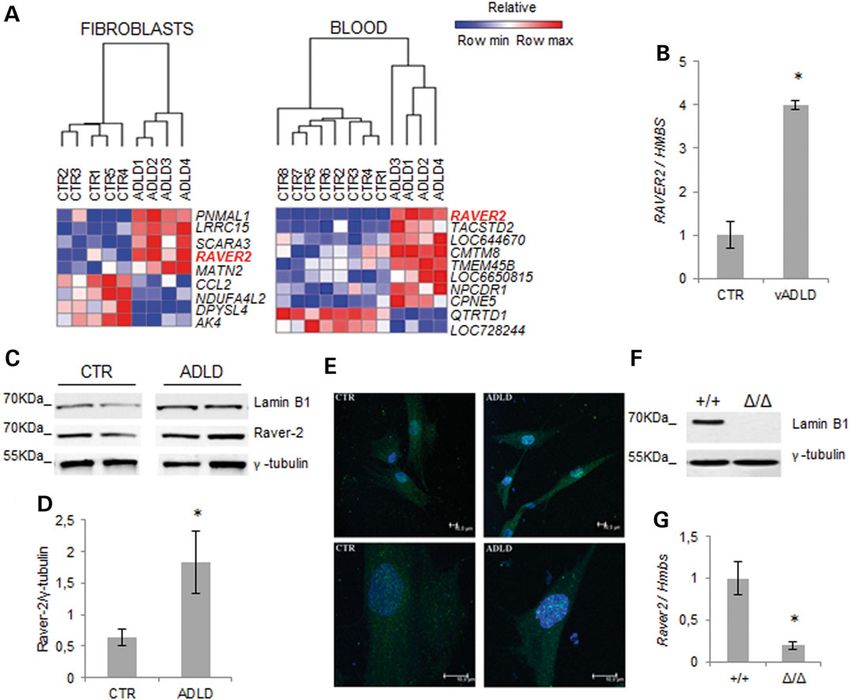
Figure 2. RAVER2 expression varies as a function of lamin B1 levels. (A) HeatMap of DEGs with statistically significant changes (FDR-adjusted P-value smaller than 0.05) in
fibroblasts (ADLD, n = 4; CTR, n = 5) and blood (ADLD, n = 4; CTR, n = 8). The GENE-E software (http://www.broadinstitute.org/cancer/software/GENE-E/) was used to generate
the heatmaps. (B) Quantitative analysis of RAVER2 mRNA in vADLD and CTR fibroblast samples. The RAVER2 mRNA levels were normalized to HMBS mRNA expression. Bars
represent the mean relative expression ± SD of one vADLD (two different batches of cells from the same donor) versus fibroblasts from six CTRs. *P < 0.05, Student’s t-test. (C)
Representative western blot of lamin B1 and raver-2 in fibroblasts from two ADLD patients and two CTRs. γ-Tubulin was used as loading control. (D) Quantitative analysis of
raver-2 protein levels in fibroblasts from ADLD patients (n = 4) and age-matched CTR subjects (n = 4). The data are normalized to γ-tubulin and expressed as percentages of
CTR levels. Bars represent average ± SEM. *P < 0.05, Mann–Whitney Rank Sum test. (E) Representative confocal images of primary fibroblasts from one ADLD patient and one
age-matched CTR immunodecorated against raver-2 (green). Nuclei are counterstained with 2-(4-amidinophenyl)-6-indolecarbamidine dihydrochloride. Scale bars: 10 µm. (F)
Representative western blot of lamin B1 protein expression in brain of Lmnb1+/+ and Lmnb1Δ/Δ E18.5 embryos. (G) Raver2 mRNA expression in brain of E18.5 mouse embryos
analyzed by RT-qPCR. *P < 0.05, Student’s t-test.
Table 3. Top 10 ontology terms associated with cell types expressing analyzed the alternative splicing of PLP1 in ADLD fibroblasts by
RAVER2 promoter reverse transcriptase-polymerase chain reaction (RT-PCR) and
dHPLC. Fibroblasts of ADLD subjects showed a 20% increase
Ontology-based sample term enrichment analysis P-value
in the embryonic PLP1 isoform (Fig. 4A). Conversely, in the brain
Organ system subdivision 5.13e−16 of Lmnb1Δ/Δ embryos, the adult Plp1 isoform was increased rela-
Ectoderm-derived structure 6.92e−14 tively to the embryonic isoforms (Fig. 4B). Similarly to PTB-target
Presumptive ectoderm 6.92e−14 genes, in Lmnb1Δ/Δ MEFs, we did not observe any differences of
Neuroectoderm 1.13e−11 Plp1 splicing pattern (Supplementary Material, Fig. S5). These re-
Adult organism 1.36e−10 sults indicate that lamin B1 plays a crucial role in the regulation
Nervous system 4.07e−10 of PLP1 expression, shifting the balance between adult and em-
Presumptive neural plate 7.14e−10 bryonic isoforms as a function of its levels.
CNS 9.34e−10
Structure with developmental contribution 7.30e−09
from neural crest
Discussion
Digestive system 1.88e−08
Changes in expression levels of lamin B1 are deleterious; duplica-
HUMAN p1@RAVER2 promoter expression identified by FANTOM5 interrogation. tion of the LMNB1 gene causes ADLD in humans (2), while Lmnb16 | Human Molecular Genetics
gene silencing has been demonstrated to be lethal in mice (7). most similar expression profile to different areas of the CNS
However, the mechanisms whereby the pathological expression (37), while skin fibroblasts have been used to model neurodegen-
of lamin B1 causes its damaging effects are still unclear. To inves- erative diseases in vitro, given their availability and robustness
tigate this crucial issue we have demonstrated, by using different (38). Therefore, to investigate global pathways altered by lamin
approaches, that lamin B1 dysregulation affects different steps of B1 overexpression, we analyzed the transcriptome of whole
gene expression, namely transcription and alternative splicing. blood and skin fibroblasts in ADLD patients and controls. ADLD
First, to gain insights into pathologically relevant pathways cells show a significant number of deregulated genes that are in-
that are altered in ADLD, we performed whole-genome expres- volved in development of the nervous system. This is consistent
sion profiling in two peripheral tissues. Because of the difficulty with the notion that Lmnb1Δ/Δ embryos display reduced brain
in obtaining human pathological tissues, other cell types, such size and impaired corticogenesis (7–9). Moreover, since lamin
as fibroblasts and lymphocytes, have been widely used (34–36). B1 is broadly expressed (39), it is not surprising that enrichment
Whole blood samples have been demonstrated to have the analysis of gene ontology categories also indicates that lamin B1
overexpression affects other tissues/organs, such as the immune
system. The fact that genes of the immune system are also af-
Table 4. PTB-regulated splicing events considered in this study
fected is in line with increasing evidence that various immune
Gene Exon Type of AS Regulated Publication system components are involved in neurodegenerative diseases,
eventa such as Alzheimer’s (AD), Parkinson’s (PD), Huntington’s (HD)
and Amyotrophic lateral sclerosis (ALS) (40,41). In addition, a
TPM1 2/3
Mutually 2 Gromak et al. (28) cluster of genes involved in muscle contraction are deregulated
exclusive in LMNB1 duplication carriers, consistent with the nuclear abnor-
PKM2 9/10 Mutually 10 Xue et al. (29)
malities and defects in actin turnover, which were described in
exclusive
one ADLD patient, where a myopathy is clinically evident (42).
ACTN1 NM/SM Mutually NM Matlin et al. (30)
Lamin B1 regulates gene expression by anchoring chromatin
exclusive
to the nuclear lamina, which acts as a scaffold for chromatin re-
GANAB 6 Cassette SK Spellman et al. (31)
modeling, and is thus critical for the spatial organization of chro-
PHF21A 14 Cassette IN Xue et al. (32)
mosomes in the nucleus (15). Accordingly, in Lmnb1Δ/Δ MEFs,
SPAG9 4 Cassette SK Xue et al. (29)
EIF4G2 9 Cassette IN Xue et al. (29) DEGs cluster in specific chromosomal locations due to changes
FAM38A 8 Cassette IN Xue et al. (29) in chromosome positions relative to the nuclear lamina (16).
Similarly, in LMNB1 duplication carriers, we found that DEGs
AS, alternative splicing. cluster in specific genomic regions, suggesting that changes in
a
Regulated exon number (for mutually exclusive type) or name (NM/SM). Exon lamin B1 levels invariably induce the delocalization of chromo-
inclusion (IN) or exon skipping (SK). somes at the nuclear lamina. The reorganization of chromosome
Figure 3. Abnormal lamin B1 expression is associated with altered splicing of PTB-target genes. Top significantly altered PTB-regulated alternative splicing identified in
ADLD blood (A) and fibroblasts (B). Alternative splicing was analyzed by RT-PCR and dHPLC. For each gene, a representative RT-PCR is shown above the dHPLC data graph.
Bars represent the average percentage of each splicing event ± SD. *P < 0.05, Student’s t test, n = 4. EX, exon; IN, exon inclusion; SK, exon skipping; F, fibroblasts; B, blood. The
splicing pattern of other PTB-regulated genes analyzed in blood and fibroblasts are shown, respectively, in Supplementary Material, Figures S1 and S2. (C) Significantly
altered PTB-regulated alternative splicing events identified in brains of Lmnb1+/+ (n = 4) and Lmnb1Δ/Δ (n = 3) E18.5 embryos. Bars represent the average percentage of each
splicing event ± SD. *P < 0.05, Student’s t test. The splicing pattern of Fam38a, that was not significantly altered, is shown in Supplementary Material, Figure S3.Human Molecular Genetics | 7
blood and fibroblasts, where we confirmed an increased protein
level in the nucleus. In human fibroblasts, the RAVER2 locus lo-
cated on cytoband 1p31 is physiologically bound by lamin B1 in
a LAD (15,46), which typically represents a repressive location.
Thus, RAVER2 overexpression in ADLD suggests that its locus is
displaced as a result of chromosomal repositioning caused by in-
creased lamin B1 levels. We were able to confirm these data by
using positional gene set analysis, demonstrating that cytoband
1p31 contains a cluster of genes that are overexpressed in ADLD.
A recent computational study has revealed a binding site for the
transcription factor OCT-1 in the RAVER2 5′-flanking region (26).
This evidence, along with the previous observation that OCT-1
recruitment to the nuclear periphery is increased in ADLD cells
(42), further supports the view that lamin B1 levels modulate dis-
tinct pathways through repositioning of specific genomic loci.
To date, functions ascribed to raver-2, similarly to its homolo-
gous raver-1 (47), include the modulation of alternative splicing
through the splicing repressor PTB (20).
Here, we demonstrated the increase of raver-2 expression in
ADLD tissues associated with modified splicing of PTB-target
genes. Likewise, the brains of Lmnb1Δ/Δ embryos are also charac-
terized by an altered raver-2/PTB pathway, which could explain
their abnormal development, with reduced brain size and im-
paired corticogenesis (7–9). Interestingly, our results are in line
with recent findings demonstrating that the silencing of LMNB1
results in a high number of enlarged nuclear speckles and exten-
sive changes in alternative splicing of multiple genes (48). Alter-
native splicing has an important role in defining tissue
specificity, and this process is highly regulated during organo-
genesis, with different tissues (e.g. the placenta and embryo) ex-
pressing distinct isoforms during different developmental stages
(49). Recent high-throughput studies have shown that more than
50% of alternative splicing isoforms are differentially expressed
among tissues (50). Accordingly, we found that PTB-target
genes altered by lamin B1/raver-2 overexpression are mainly tis-
sue-specific. Raver-2 is probably not the only splicing regulator
altered by lamin B1 overexpression, and possibly, other players
are necessary to define the disease, but their number and identity
Figure 4. Abnormal levels of lamin B1 alter PLP1 splicing. (A) Analysis of is extremely difficult to be found given that the regulation of al-
embryonic (DM20) and adult (PLP) isoforms of PLP1 gene in ADLD (n = 4) and CTR ternative splicing is highly tissue-specific and is likely that in
(n = 5) fibroblasts analyzed through RT-PCR and dHPLC run. Bars represent the
each tissue one or more of them could regulate PTB.
mean ± SD. *P < 0.05, Student’s t-test, n = 5. (B) Analysis of Plp1 splicing pattern in
brain of Lmnb1+/+ (n = 4) and Lmnb1Δ/Δ (n = 3) embryos. *P < 0.05, Student’s t-test. The primary target tissue of ADLD is the CNS, specifically
Representative gels of mRNA splicing isoforms are shown above dHPLC data myelin. In ADLD patients, white matter defects are observed, par-
graphs. ticularly in the cerebellum, corticospinal tracts and corpus callo-
sum, which lead to brain and spinal cord atrophy (2). Previous
positioning was also found in skin fibroblasts from patients with studies in ADLD patients have shown the decreased transcription
cardiomyopathy and Hutchinson–Gilford progeria syndrome of myelin proteins (mostly PLP1) in oligodendrocytes (12). Here,
bearing mutations in the cognate lamin A/C gene (43,44). To- we have demonstrated an altered splicing pattern of PLP1 in
gether, these findings indicate that alterations to the levels of ADLD fibroblasts and Lmnb1Δ/Δ embryos. It is worth noting that
lamins are linked with changes in chromosome positioning. an abnormal balance of the two PLP1 isoforms was also observed
Second, in order to identify the principal genes that result in in fibroblasts derived from patients with a different leukodystro-
the phenotype caused by LMNB1 overexpression, we compared phy, namely Pelizaeus–Merzbacher disease, although in the re-
the DEGs identified in whole blood and skin fibroblasts. During verse direction compared with ADLD (33). Because a direct link
differentiation, changes in temporal and spatial expression of la- between PTB and myelin genes has never been demonstrated,
mins cause distinctive chromatin patterns conferring unique this finding suggests that other splicing factors could be affected
transcriptomes in different cell lineages and developmental by lamin B1/raver-2 dysregulation. Consistent with this hypoth-
stages (45). Indeed, in a conditional transgenic mouse model esis, in an ADLD transgenic mouse model, Hnrnpr protein is up-
overexpressing Lmnb1 in oligodendroglia, the majority of the de- regulated in the oligodendrocytes (12). Like raver-2, Hnrnpr
regulated genes are involved in typical oligodendrocyte func- protein belongs to the subfamily of ubiquitously expressed het-
tions, such as the formation and maintenance of myelin (12). erogeneous nuclear ribonucleoproteins and interacts with the
Consistent with this cell-specificity of the transcriptome, we de- telomeric protein, survival of motor neuron 1 (SMN1), which is
monstrated that only three DEGs are common to both human a regulator of RNA processing (51).
fibroblasts and blood, the most significant being RAVER2. Recently, transcriptome profiling and computational predic-
RAVER2 gene expression was up-regulated in both ADLD whole tions have indicated that an unexpectedly high proportion of8 | Human Molecular Genetics
diseases, including Fragile X-associated tremor/ataxia syn- Microarray and bioinformatics analysis
drome, spinal muscular atrophy and frontotemporal dementia,
RNA from fibroblasts and whole blood was extracted using the
are characterized by aberrant alternative RNA splicing (52). Myo-
RNeasy Plus Kit and PAXgene™ Blood RNA System Kit (Qiagen).
tonic dystrophy 1, which is caused by dynamic and unstable
RNA quality was tested by Bioanalyzer 2100 (Agilent Technologies,
expanded microsatellite sequences in DMPK gene, is the proto-
Santa Clara, CA, USA) and used when the integrity quality number
type of this group of diseases, also called spliceopathies. The nu-
(RIN) scored above 8. Four-hundred nanograms of RNA for each
clear export of the mutated RNAs is defective, and they aggregate
sample were amplified using the Illumina® TotalPrep™ RNA Amp-
to form nuclear foci, which then recruit and sequester RBP while
lification Kit (Ambion, Thermo Fisher Scientific, Waltham, MA,
stabilizing other splicing regulators (53). The unbalanced action
USA). Samples were analyzed with the Illumina whole-genome
of these regulatory proteins disrupts RNA metabolism, with al-
HumanHT-12 v4 (Illumina, San Diego, CA, USA) using the Illumi-
terations in the alternative splicing of numerous pre-messenger
na’s Beadarray system 500 G Scanner. Image signal intensity was
RNAs in several tissues (53). Other mechanisms causing aberrant
extracted; the background subtracted and normalized using Illumi-
splicing are disruption of cis-splicing sites or trans-acting factors;
na Inc. BeadStudio software version 3.4.0 (Consorzio per il Centro di
dysfunction of RBP required for the efficient assembly of small
Biomedicina Molecolare, Trieste, Italy). All statistical analyzes were
nuclear ribonucleoprotein (snRNP) complexes and spliceosomal
performed in the R environment using specific packages of Biocon-
snRNP biogenesis and mutations in proteins that regulate the
ductor (56). The raw signal intensities were processed with Lumi
splicing of several human transcripts or protect extra-long in-
package applying variance-stabilizing transformation (VST) and
trons from incorrect splicing at cryptic sites (54). We suggest
quantile normalization (57). The Limma package was used to iden-
that also the deregulation of lamin B1 levels, possibly through
tify DEGs (58). The data discussed in this publication have been de-
modulation of RAVER2 expression, that was demonstrated to
posited in NCBI’s Gene Expression Omnibus (59) and are accessible
modify PTB pathway (20) could be an additional mechanism
through GEO Series accession number GSE65368 (http://www.ncbi.
inducing disruption of RNA metabolism.
nlm.nih.gov/geo/query/acc.cgi?acc=GSE95368).
Based on the present findings, we confirm previous studies
Genes with a P-value |0.5| were considered DEGs. This selection method was demon-
gest that this biological function is mediated by raver-2 expres-
strated having an higher concordance degree of DEGs between
sion levels. Changes in lamin B1 levels induce through
different platforms when compared with DEGs selected only on
chromosome repositioning, modifications of the expression of
P-value ranking (60). Significance levels ( p-val) were corrected
multiple genes, including RAVER2, which is a modulator of PTB
using Benjamini and Hochberg (BH) false discovery rate (FDR)
(20), and possibly other splicing regulators.
method ( p-adj) for multiple hypotheses testing (61).
Consequently, we propose that ADLD could be partly re-
GSEA was conducted on data from the full chip with no prior
garded as a spliceopathy caused by increased levels of lamin
filtering (21). The GO gene sets databases (C5.bp.v4.0, C5.cc.v4.0,
B1/raver-2. Thus, adult-onset CNS demyelination could be the
C5.mf.v4.0) and positional gene sets (C1.all.v4.0) from the Mo-
result of increased expression, during adulthood, of the embry-
lecular Signatures Database (www.broadinstitute.org/gsea/
onic isoform of the PLP1 protein, which has a crucial role in
msigdb/index.jsp) were used for enrichment analysis. A total of
myelin maintenance. Accordingly, in ADLD, in addition to the
1000 permutations were used to obtain the nominal P-value
already-demonstrated altered expression of myelin genes and
(NOM p-val). Normalized enrichment score (NES) was measured.
increased nuclear rigidity (12,14), we propose that the dysregu-
Gene sets with nominal P-valueHuman Molecular Genetics | 9
containing 1 μl of cDNA, 1 U FastStart High Fidelity PCR System Acknowledgements
(Roche), 120 μ dNTPs (Roche) and 20 p of each primer
We are gratefully indebted with all family members who partici-
(Sigma-Aldrich, St. Louis, MO, USA). Fragments obtained within
pated in the study. We thank Dr Silvia Piras for excellent technical
the log-linear amplification range were analyzed without further
assistance.
purification on dHPLC. We calculated the ratio between the areas
of the peaks representing the two isoforms, using WAVEMAKER
Conflict of Interest statement. None declared.
Software (Transgenomic) in duplication carriers and control
samples. Differences in relative expression of isoforms were
statistically assessed by Student’s t-test for three independent
experiments. Funding
This work was supported by TELETHON (grant number
Animals GGP10184) and the Gino Galletti Foundation. Funding to pay
the Open Access publication charges for this article was provided
Lmnb1-null (Lmnb1Δ/Δ) mice (7) were obtained from the Mutant
by Fondazione Telethon.
Mouse Regional Resource Center (University of California,
Davis, CA, USA). Animal health and comfort were veterinary-
controlled. The mice were housed in filtered cages in a temp-
erature-controlled room with a 12:12 h dark/light cycle with ad References
libitum access to water and food. All of the animal experiments 1. Cortelli, P., Terlizzi, R., Capellari, S. and Benarroch, E. (2012)
were performed in accordance with the European Community Nuclear lamins: functions and clinical implications. Neur-
Council Directive dated 24 November 1986 (86/609/EEC) and ology, 79, 1726–1731.
were approved by the Italian Ministry of Health and the IIT Ethical 2. Padiath, Q.S., Saigoh, K., Schiffmann, R., Asahara, H., Yama-
Committee. da, T., Koeppen, A., Hogan, K., Ptácek, L.J. and Fu, Y.H. (2006)
Embryonic brains were collected from Lmnb1Δ/Δ embryos and Lamin B1 duplications cause autosomal dominant leukody-
wild-type (Lmnb1+/+) littermates at 18.5 days of gestation and strophy. Nat. Genet., 38, 1114–1123.
processed using the RNeasy Lipid Tissue Mini Kit (Qiagen). 3. Giorgio, E., Rolyan, H., Kropp, L., Chakka, A.B., Yatsenko, S., Di
MEFs were isolated from wild-type Lmnb1+/+ and Lmnb1Δ/Δ Gregorio, E., Lacerenza, D., Vaula, G., Talarico, F., Mandich, P.
embryos at 13.5 days of gestation (14). et al. (2013) Analysis of LMNB1 duplications in autosomal
dominant leukodystrophy provides insights into duplication
Western blot analysis mechanisms and allele-specific expression. Hum. Mutat., 34,
1160–1171.
Fibroblasts were lysed by boiling for 5 min in 1% sodium dodecyl
4. Brussino, A., Vaula, G., Cagnoli, C., Panza, E., Seri, M., Di Gre-
sulfate (SDS), 1 m ethylenediaminetetraacetic acid, 5 m 4-(2-
gorio, E., Scappaticci, S., Camanini, S., Daniele, D., Bradac, G.B.
Hydroxyethyl)piperazine-1-ethanesulfonic acid ( pH 7.4), mouse
et al. (2010) A family with autosomal dominant leukodystro-
embryonic brains in 50 m tris(hydroxymethyl)aminomethane,
phy linked to 5q23.2-q23.3 without lamin B1 mutations.
0.5 M NaCl, 1% SDS and 1% Tx-100. Equal amounts of proteins
Eur. J. Neurol., 17, 541–549.
were separated on NuPage 10% bis-tris polyacrylamide gels (Invi-
5. Lenz-Böhme, B., Wismar, J., Fuchs, S., Reifegerste, R.,
trogen, Thermo Scientific) and analyzed (14) using primary rabbit
Buchner, E., Betz, H. and Schmitt, B. (1997) Insertional muta-
polyclonal antibodies against lamin B1 (Abcam, Cambridge, Eng-
tion of the Drosophila nuclear lamin Dm0 gene results in
land, UK), γ-tubulin (Sigma), actin (Sigma) and raver-2 (Abcam).
defective nuclear envelopes, clustering of nuclear pore com-
Densitometric analysis was performed using the NIH ImageJ pro-
plexes, and accumulation of annulate lamellae. J. Cell Biol.,
gram (65). The protein levels were normalized on γ-tubulin or
137, 1001–1016.
actin content.
6. Liu, J., Rolef Ben-Shahar, T., Riemer, D., Treinin, M., Spann, P.,
Weber, K., Fire, A. and Gruenbaum, Y. (2000) Essential roles for
Immunofluorescence Caenorhabditis elegans lamin gene in nuclear organization, cell
Fibroblasts were plated onto poly--lysine-coated coverslips, cycle progression, and spatial organization of nuclear pore
serum-deprived for 24 h, fixed in 4% PFA and immuno-labeled complexes. Mol. Biol. Cell, 11, 3937–3947.
as previously described (14), using the anti-raver-2 antibody 7. Vergnes, L., Péterfy, M., Bergo, M.O., Young, S.G. and Reue, K.
(Abcam). The confocal optical sectioning was performed at (2004) Lamin B1 is required for mouse development and nu-
room temperature using a Leica TCS SP5 AOBS TANDEM inverted clear integrity. Proc. Natl. Acad. Sci. USA, 101, 10428–10433.
confocal microscope that was equipped with a HCX PL APO 40× 8. Coffinier, C., Jung, H.J., Nobumori, C., Chang, S., Tu, Y., Barnes,
1.25 Oil and a HCX PL APO blue 63× 1.4NA Oil objective lenses R.H. 2nd, Yoshinaga, Y., de Jong, P.J., Vergnes, L., Reue, K. et al.
(Leica Microsystems, Wetzlar, Germany). (2011) Deficiencies in lamin B1 and lamin B2 cause neurode-
velopmental defects and distinct nuclear shape abnormal-
ities in neurons. Mol. Biol. Cell, 22, 4683–4693.
Statistical analysis 9. Kim, Y., Sharov, A.A., McDole, K., Cheng, M., Hao, H., Fan, C.
Student’s t-test was used for data comparison; Mann–Whitney M., Gaiano, N., Ko, M.S. and Zheng, Y. (2011) Mouse B-type la-
Rank Sum test was used for comparing raver-2 protein expres- mins are required for proper organogenesis but not by embry-
sion in fibroblasts. The differences were considered to be statis- onic stem cells. Science, 334, 1706–1710.
tically significant when P < 0.05. 10. Takamori, Y., Tamura, Y., Kataoka, Y., Cui, Y., Seo, S., Kanaza-
wa, T., Kurokawa, K. and Yamada, H. (2007) Differential ex-
pression of nuclear lamin, the major component of nuclear
Supplementary Material lamina, during neurogenesis in two germinal regions of
Supplementary Material is available at HMG online. adult rat brain. Eur. J. Neurosci., 25, 1653–1662.10 | Human Molecular Genetics
11. Lin, S.T. and Fu, Y.H. (2009) miR-23 regulation of lamin B1 is 26. Romanelli, M.G., Lorenzi, P., Diani, E., Filippello, A., Avesani, F.
crucial for oligodendrocyte development and myelination. and Morandi, C. (2012) Transcriptional regulation of the
Dis. Model. Mech., 2, 178–188. human Raver2 ribonucleoprotein gene. Gene, 493, 243–252.
12. Heng, M.Y., Lin, S.T., Verret, L., Huang, Y., Kamiya, S., Padiath, 27. Spellman, R., Rideau, A., Matlin, A., Gooding, C., Robinson, F.,
Q.S., Tong, Y., Palop, J.J., Huang, E.J., Ptáček, L.J. and Fu, Y.H. McGlincy, N., Grellscheid, S.N., Southby, J., Wollerton, M. and
(2013) Lamin B1 mediates cell-autonomous neuropathology Smith, C.W. (2005) Regulation of alternative splicing by PTB
in a leukodystrophy mouse model. J. Clin. Invest., 123, 2719– and associated factors. Biochem. Soc. Trans., 33, 457–460.
2729. 28. Gromak, N., Rideau, A., Southby, J., Scadden, A.D., Gooding,
13. Dechat, T., Pfleghaar, K., Sengupta, K., Shimi, T., Shumaker, D. C., Hüttelmaier, S., Singer, R.H. and Smith, C.W. (2003) The
K., Solimando, L. and Goldman, R.D. (2008) Nuclear lamins: PTB interacting protein raver1 regulates alpha-tropomyosin
major factors in the structural organization and function of alternative splicing. EMBO J., 22, 6356–6364.
the nucleus and chromatin. Genes Dev., 22, 832–853. 29. Xue, Y., Zhou, Y., Wu, T., Zhu, T., Ji, X., Kwon, Y.S., Zhang, C.,
14. Ferrera, D., Canale, C., Marotta, R., Mazzaro, N., Gritti, M., Yeo, G., Black, D.L., Sun, H. et al. (2009) Genome-wide analysis
Mazzanti, M., Capellari, S., Cortelli, P. and Gasparini, L. of PTB–RNA interactions reveals a strategy used by the gen-
(2014) Lamin B1 overexpression increases nuclear rigidity in eral splicing repressor to modulate exon inclusion or skip-
autosomal dominant leukodystrophy fibroblasts. FASEB J., ping. Mol. Cell, 36, 996–1006.
28, 3906–3918. 30. Matlin, A.J., Southby, J., Gooding, C. and Smith, C.W. (2007) Re-
15. Guelen, L., Pagie, L., Brasset, E., Meuleman, W., Faza, M.B., pression of alpha-actinin SM exon splicing by assisted bind-
Talhout, W., Eussen, B.H., de Klein, A., Wessels, L., de Laat, ing of PTB to the polypyrimidine tract. RNA, 13, 1214–1223.
W. and van Steensel, B. (2008) Domain organization of 31. Spellman, R., Llorian, M. and Smith, C.W. (2007) Crossregula-
human chromosomes revealed by mapping of nuclear lam- tion and functional redundancy between the splicing regula-
ina interactions. Nature, 453, 948–951. tor PTB and its paralogs nPTB and ROD1. Mol. Cell, 27, 420–434.
16. Malhas, A., Lee, C.F., Sanders, R., Saunders, N.J. and Vaux, D.J. 32. Xue, Y., Ouyang, K., Huang, J., Zhou, Y., Ouyang, H., Li, H.,
(2007) Defects in lamin B1 expression or processing affect Wang, G., Wu, Q., Wei, C., Bi, Y. et al. (2013) Direct conversion
interphase chromosome position and gene expression. of fibroblasts to neurons by reprogramming PTB-regulated
J. Cell Biol., 176, 593–603. microRNA circuits. Cell, 152, 82–96.
17. Castello, A., Fischer, B., Eichelbaum, K., Horos, R., Beckmann, 33. Regis, S., Grossi, S., Corsolini, F., Biancheri, R. and Filocamo,
B.M., Strein, C., Davey, N.E., Humphreys, D.T., Preiss, T., Stein- M. (2009) PLP1 gene duplication causes overexpression and
metz, L.M., Krijgsveld, J. and Hentze, M.W. (2012) Insights into alteration of the PLP/DM20 splicing balance in fibroblasts
RNA biology from an atlas of mammalian mRNA-binding from Pelizaeus–Merzbacher disease patients. Biochim. Bio-
proteins. Cell, 149, 1393–1406. phys. Acta, 1792, 548–554.
18. Kleinhenz, B., Fabienke, M., Swiniarski, S., Wittenmayer, N., 34. Cooper-Knock, J., Kirby, J., Ferraiuolo, L., Heath, P.R., Rattray,
Kirsch, J., Jockusch, B.M., Arnold, H.H. and Illenberger, S. M. and Shaw, P.J. (2012) Gene expression profiling in human
(2005) Raver2, a new member of the hnRNP family. FEBS neurodegenerative disease. Nat. Rev. Neurol., 8, 518–530.
Lett., 579, 4254–4258. 35. Fogel, B.L., Cho, E., Wahnich, A., Gao, F., Becherel, O.J., Wang,
19. Henneberg, B., Swiniarski, S., Becke, S. and Illenberger, S. X., Fike, F., Chen, L., Criscuolo, C., De Michele, G. et al. (2014)
(2010) A conserved peptide motif in Raver2 mediates its inter- Mutation of senataxin alters disease-specific transcriptional
action with the polypyrimidine tract-binding protein. Exp. networks in patients with ataxia with oculomotor apraxia
Cell Res., 316, 966–979. type 2. Hum. Mol. Genet., 23, 4758–4769.
20. Das, S.K., Holt, D.G., Uehara, H., Zhang, X., Archer, B. and 36. Bernardini, C., Lattanzi, W., Bosco, P., Franceschini, C., Plazzi,
Ambati, B.K. (2014) Raver2 preserves corneal avascularity by G., Michetti, F. and Ferri, R. (2012) Genome-wide gene expres-
increasing sFlt1 production [ARVO E-abstract]. Invest. Ophthal- sion profiling of human narcolepsy. Gene Expr., 15, 171–181.
mol. Vis. Sci., 55, 3249. 37. Sullivan, P.F., Fan, C. and Perou, C.M. (2006) Evaluating the
21. Subramanian, A., Tamayo, P., Mootha, V.K., Mukherjee, S., comparability of gene expression in blood and brain.
Ebert, B.L., Gillette, M.A., Paulovich, A., Pomeroy, S.L., Golub, Am. J. Med. Genet. B. Neuropsychiatr. Genet., 141B, 261–268.
T.R., Lander, E.S. and Mesirov, J.P. (2005) Gene Set Enrichment 38. Auburger, G., Klinkenberg, M., Drost, J., Marcus, K., Morales-
Analysis: a knowledge-based approach for interpreting gen- Gordo, B., Kunz, W.S., Brandt, U., Broccoli, V., Reichmann,
ome-wide expression profiles. Proc. Natl. Acad. Sci. USA, 102, H., Gispert, S. and Jendrach, M. (2012) Primary skin fibroblasts
15545–15550. as a model of Parkinson’s disease. Mol. Neurobiol., 46, 20–27.
22. Hutchison, C.J. (2014) B-type lamins in health and disease. 39. Schreiber, K.H. and Kennedy, B.K. (2013) When lamins go bad:
Semin. Cell Dev. Biol., 29, 158–163. nuclear structure and disease. Cell, 152, 1365–1375.
23. Malhas, A.N., Lee, C.F. and Vaux, D.J. (2009) Lamin B1 controls 40. Björkqvist, M., Wild, E.J. and Tabrizi, S.J. (2009) Harnessing im-
oxidative stress responses via Oct-1. J. Cell Biol., 184, 45–55. mune alterations in neurodegenerative diseases. Neuron, 64,
24. Melko, M., Douguet, D., Bensaid, M., Zongaro, S., Verheggen, 21–24.
C., Gecz, J. and Bardoni, B. (2011) Functional characterization 41. Rodrigues, M.C., Sanberg, P.R., Cruz, L.E. and Garbuzova-
of the AFF (AF4/FMR2) family of RNA-binding proteins: in- Davis, S. (2014) The innate and adaptive immunological as-
sights into the molecular pathology of FRAXE intellectual dis- pects in neurodegenerative diseases. J. Neuroimmunol., 269,
ability. Hum. Mol. Genet., 20, 1873–1885. 1–8.
25. FANTOM Consortium and the RIKEN PMI and CLST (DGT)For- 42. Columbaro, M., Mattioli, E., Maraldi, N.M., Ortolani, M., Gas-
rest, A.R., Kawaji, H., Rehli, M., Baillie, J.K., de Hoon, M.J., Lass- parini, L., D’Apice, M.R., Postorivo, D., Nardone, A.M., Avnet,
mann, T., Itoh, M., Summers, K.M., Suzuki, H., Daub, C.O. et al. S., Cortelli, P. et al. (2013) Oct-1 recruitment to the nuclear en-
(2014) A promoter-level mammalian expression atlas. Nature, velope in adult-onset autosomal dominant leukodystrophy.
507, 462–470. Biochim. Biophys. Acta, 1832, 411–420.Human Molecular Genetics | 11
43. Mewborn, S.K., Puckelwartz, M.J., Abuisneineh, F., Fahren- 54. Caillet-Boudin, M.L., Fernandez-Gomez, F.J., Tran, H., Dhae-
bach, J.P., Zhang, Y., MacLeod, H., Dellefave, L., Pytel, P., nens, C.M., Buee, L. and Sergeant, N. (2014) Brain pathology
Selig, S., Labno, C.M. et al. (2010) Altered chromosomal posi- in myotonic dystrophy: when tauopathy meets spliceopathy
tioning, compaction, and gene expression with a lamin A/C and RNAopathy. Front. Mol. Neurosci., 6, 57.
gene mutation. PLoS ONE, 5, e14342. 55. Guaraldi, P., Donadio, V., Capellari, S., Contin, M., Casadio, M.
44. McCord, R.P., Nazario-Toole, A., Zhang, H., Chines, P.S., Zhan, C., Montagna, P., Liguori, R. and Cortelli, P. (2010) Isolated nor-
Y., Erdos, M.R., Collins, F.S., Dekker, J. and Cao, K. (2013) Cor- adrenergic failure in adult-onset autosomal dominant leuko-
related alterations in genome organization, histone methyla- dystrophy. Auton. Neurosci., 159, 123–126.
tion, and DNA-lamin A/C interactions in Hutchinson–Gilford 56. Gentleman, R.C., Carey, V.J., Bates, D.M., Bolstad, B., Dettling,
progeria syndrome. Genome Res., 23, 260–269. M., Dudoit, S., Ellis, B., Gautier, L., Ge, Y., Gentry, J. et al. (2004)
45. Peric-Hupkes, D., Meuleman, W., Pagie, L., Bruggeman, S.W., Bioconductor: open software development for computational
Solovei, I., Brugman, W., Gräf, S., Flicek, P., Kerkhoven, R.M., biology and bioinformatics. Genome Biol., 5, R80.
van Lohuizen, M. et al. (2010) Molecular maps of the reorgan- 57. Du, P., Kibbe, W.A. and Lin, S.M. (2008) lumi: a pipeline for pro-
ization of genome-nuclear lamina interactions during differ- cessing Illumina microarray. Bioinformatics, 24, 1547–1548.
entiation. Mol. Cell, 38, 603–613. 58. Wettenhall, J.M. and Smyth, G.K. (2004) limmaGUI: a graphic-
46. Sadaie, M., Salama, R., Carroll, T., Tomimatsu, K., Chandra, T., al user interface for linear modeling of microarray data. Bio-
Young, A.R., Narita, M., Pérez-Mancera, P.A., Bennett, D.C., informatics, 20, 3705–3706.
Chong, H. et al. (2013) Redistribution of the Lamin B1 genomic 59. Edgar, R., Domrachev, M. and Lash, A.E. (2002) Gene Expres-
binding profile affects rearrangement of heterochromatic do- sion Omnibus: NCBI gene expression and hybridization
mains and SAHF formation during senescence. Genes Dev., 27, array data repository. Nucleic Acids Res., 30, 207–210.
1800–1808. 60. Shi, L., Jones, W.D., Jensen, R.V., Harris, S.C., Perkins, R.G.,
47. Romanelli, M.G., Diani, E. and Lievens, P.M. (2013) New in- Goodsaid, F.M., Guo, L., Croner, L.J., Boysen, C., Fang, H. et al.
sights into functional roles of the polypyrimidine tract-bind- (2008) The balance of reproducibility, sensitivity, and specifi-
ing protein. Int. J. Mol. Sci., 14, 22906–22932. city of lists of differentially expressed genes in microarray
48. Camps, J., Wangsa, D., Falke, M., Brown, M., Case, C.M., Erdos, studies. BMC Bioinformatics, 9, S10.
M.R. and Ried, T. (2014) Loss of lamin B1 results in prolonga- 61. Benjamini, Y., Drai, D., Elmer, G., Kafkafi, N. and Golani, I.
tion of S phase and decondensation of chromosome territor- (2001) Controlling the false discovery rate in behavior genet-
ies. FASEB J., 28, 3423–3434. ics research. Behav. Brain Res., 125, 279–284.
49. Revil, T., Gaffney, D., Dias, C., Majewski, J. and Jerome-Ma- 62. Supek, F., Bošnjak, M., Škunca, N. and Šmuc, T. (2011) REVIGO
jewska, L.A. (2010) Alternative splicing is frequent during summarizes and visualizes long lists of gene ontology terms.
early embryonic development in mouse. BMC Genomics, 11, PLoS ONE, 6, e21800.
399. 63. Durrenberger, P.F., Fernando, F.S., Magliozzi, R., Kashefi, S.N.,
50. Chen, M. and Manley, J.L. (2009) Mechanisms of alternative Bonnert, T.P., Ferrer, I., Seilhean, D., Nait-Oumesmar, B.,
splicing regulation: insights from molecular and genomics Schmitt, A., Gebicke-Haerter, P.J. et al. (2012) Selection of novel
approaches. Nat. Rev. Mol. Cell Biol., 10, 741–754. reference genes for use in the human central nervous system: a
51. Mourelatos, Z., Abel, L., Yong, J., Kataoka, N. and Dreyfuss, G. BrainNet Europe Study. Acta Neuropathol., 124, 893–903.
(2001) SMN interacts with a novel family of hnRNP and spli- 64. Livak, K.J. and Schmittgen, T.D. (2001) Analysis of relative
ceosomal proteins. EMBO J., 20, 5443–5452. gene expression data using real-time quantitative PCR and
52. Singh, R.K. and Cooper, T.A. (2012) Pre-mRNA splicing in dis- the 2(-Delta Delta C(T)) method. Methods, 25, 402–408.
ease and therapeutics. Trends Mol. Med., 18, 472–482. 65. Schneider, C.A., Rasband, W.S. and Eliceiri, K.W. (2012) NIH
53. Osborne, R.J. and Thornton, C.A. (2006) RNA-dominant dis- Image to ImageJ: 25 years of image analysis. Nat. Methods, 9,
eases. Hum. Mol. Genet., 15, R162–R169. 671–675.You can also read