SNP rs10420324 in the AMPA receptor auxiliary subunit TARP γ 8 regulates the susceptibility to antisocial personality disorder
←
→
Page content transcription
If your browser does not render page correctly, please read the page content below
www.nature.com/scientificreports
OPEN SNP rs10420324 in the AMPA
receptor auxiliary subunit TARP
γ‑8 regulates the susceptibility
to antisocial personality disorder
Shi‑Xiao Peng1,2, Yue‑Ying Wang1,2, Min Zhang3, Yan‑Yu Zang1,2, Dan Wu1, Jingwen Pei1,2,
Yansong Li4, Jiapei Dai5, Xiaoyun Guo6, Xingguang Luo7, Ning Zhang3,8, Jian‑Jun Yang9,
Chen Zhang10, Xiang Gao1,2, Na Liu8* & Yun Stone Shi1,2,11,12*
In the brain, AMPA receptors mediate fast excitatory neurotransmission, the dysfunction of which
leads to neuropsychiatric disorders. Synaptic function of AMPA receptors is tightly controlled by a
protein group called transmembrane AMPAR regulatory proteins (TARPs). TARP γ-8 (also known as
CACNG8) preferentially expresses in the hippocampus, cortex and subcortical regions that are critical
for emotion generation indicating its association with psychiatric disorders. Here, we identified
rs10420324 (T/G), a SNP located in the human CACNG8 gene, regulated reporter gene expression
in vitro and TARP γ-8 expression in the human brain. A guanine at the locus (rs10420324G) suppressed
transcription likely through modulation of a local G-quadruplex DNA structure. Consistent with
these observations, the frequency of rs10420324G was higher in patients with anti-social personality
disorder (ASPD) than in controls, indicating that rs10420324G in CACNG8 is more voluntary for ASPD.
We then characterized the behavior of TARP γ-8 knockout and heterozygous mice and found that
consistent with ASPD patients who often exhibit impulsivity, aggression, risk taking, irresponsibility
and callousness, a decreased γ-8 expression in mice displayed similar behaviors. Furthermore,
we found that a decrease in TARP γ-8 expression impaired synaptic AMPAR functions in layer 2–3
pyramidal neurons of the prefrontal cortex, a brain region that inhibition leads to aggression, thus
explaining, at least partially, the neuronal basis for the behavioral abnormality. Taken together,
our study indicates that TARP γ-8 expression level is associated with ASPD, and that the TARP γ-8
knockout mouse is a valuable animal model for studying this psychiatric disease.
Human emotion arises from interactions between brain regions within the limbic system, including the amyg-
dala, hippocampus, insula, and cingulate c ortex1–3. Neurons from different brain areas form neural circuits
through connection at highly organized intercellular structures, the synapses. The strength of synaptic transmis-
sion is dynamically regulated by neuronal activity, a process called synaptic plasticity. In the last two decades,
it has been well established that the synaptic plasticity in specific neuronal circuits are related to emotional
1
MOE Key Laboratory of Model Animal for Disease Study, Department of Neurology, Drum Tower Hospital,
Medical School, Nanjing University, Nanjing 210032, China. 2State Key Laboratory of Pharmaceutical
Biotechnology, Model Animal Research Center, National Resource Center for Mutant Mice, Medical School,
Nanjing University, Nanjing 210032, China. 3School of Psychology, Nanjing Normal University, Nanjing 210029,
China. 4Reward, Competition and Social Neuroscience Lab, Department of Psychology, School of Social and
Behavioral Sciences, Nanjing University, Nanjing 210023, China. 5Chinese Brain Bank Center, Wuhan 430074,
China. 6Shanghai Mental Health Center, Shanghai Jiao Tong University School of Medicine, Shanghai 200030,
China. 7Division of Human Genetics, Department of Psychiatry, Yale University School of Medicine, New Haven,
CT 06510, USA. 8Department of Medical Psychology, Nanjing Medical University Affiliated Nanjing Brain Hospital,
Nanjing 210029, China. 9Department of Anesthesiology and Perioperative Medicine, First Affiliated Hospital of
Zhengzhou University, Zhengzhou 450052, Henan Province, China. 10School of Basic Medical Sciences, Beijing Key
Laboratory of Neural Regeneration and Repair, Advanced Innovation Center for Human Brain Protection, Capital
Medical University, Beijing 100069, China. 11Institute for Brain Sciences, Nanjing University, Nanjing 210032,
China. 12Chemistry and Biomedicine Innovation Center, Nanjing University, Nanjing 210032, China. *email: naliu_
nbh@njmu.edu.cn; yunshi@nju.edu.cn
Scientific Reports | (2021) 11:11997 | https://doi.org/10.1038/s41598-021-91415-9 1
Vol.:(0123456789)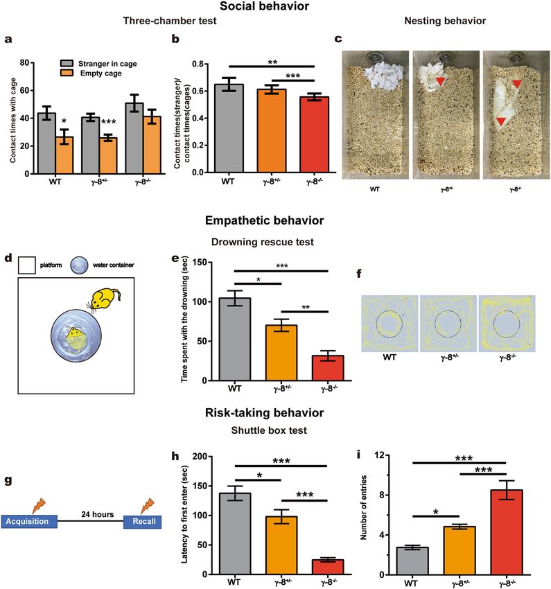
www.nature.com/scientificreports/
regulation4,5. Glutamate is the major excitatory neurotransmitter in the brain, and AMPA-type glutamate recep-
tors (AMPARs) on the postsynaptic membrane rapidly transmit excitatory signals by opening to allow cations
to enter the neuron. The trafficking of AMPARs to and from the synaptic membrane also mediates long-term
plasticity of excitatory s ynapses6,7. Dysregulation of AMPAR trafficking results in impaired synaptic plasticity,
abnormal neuronal networks, altered emotions or behaviors, and neuronal d iseases8–11.
In the brain, AMPARs are homomeric or heteromeric tetramers composed of the pore-forming subunits of
GluA1–4. Recently, an expanding list of transmembrane auxiliary subunits are identified to associate with the
AMPAR complex, including transmembrane AMPAR regulatory protein (TARP), cornichon-like (CNIH), germ
cell-specific gene 1-like(Gsg1l) and the cysteine-knot AMPAR-modulating protein (CKAMP)/Shisa protein
family12–15, which modulate AMPAR biophysics, intracellular trafficking and synaptic expression16–18.
TARPs are the earliest-identified and best-characterized AMPAR auxiliary subunits. They are officially named
as the calcium channel gamma subunits (CACNGs) for their homology to CACNG1. Six TARPs have been clas-
sified as type 1 (γ-2, γ-3, γ-4, and γ-8) or type 2 (γ-5 and γ-7) based on their sequence similarities and functional
properties17. In the brain, these six proteins have different expression p atterns19. Key insights regarding their
physiological roles have been derived from studies on mutant mice. For example, the absence of the prototypical
auxiliary subunit γ-2 (stargazin) diminishes excitatory synaptic transmission from cerebellar mossy fibers to
granule cells20–22 and causes behavioral abnormalities including ataxia, head elevation23. Selective deletion of γ-2
and γ-7 in the cerebellar Purkinje neurons of mice results in severe disruption of motor b ehaviors24.
25
TARP γ-8 (CACNG8) was first reported in 2001 for its homology to TARP γ-2 . In γ-8 knockout mice,
hippocampal AMPA receptors do not progress through the secretory pathway and are not efficiently trafficked
to dendrites26,27, and the mice show hyperactivity and marked reductions in digging and burying behaviors28.
High TARP γ-8 expression was detected in the cortex, hippocampus and subcortical regions19, brain areas that
are critical for emotion generation. This expression pattern suggests that TARP γ-8 may affect emotion-related
behaviors. In deeds, a large-scale association study suggests that a two-SNP haplotype (rs10420331-rs11084307)
in the intronic region of CACNG8 is associated with schizophrenia in a Chinese Han population29. However,
whether and how these two SNPs cause emotional disorders are unclear. The exact role and the molecular
mechanisms of TARP γ-8 in emotion remain elusive.
In this study, to understand whether and how TARP γ-8 regulates human emotion, we first analyzed SNPs in
human CACNG8 gene and identified SNP rs10420324 located in the first intron which controlled the expression
of a reporter gene in vitro and the expression of TARP γ-8 in human brains. Then we found that rs10420324G was
associated with antisocial personality disorder (ASPD) in a Chinese Han sample set. Systematic neurobehavioral
tests of TARP γ-8−/− and γ-8+/− mice indicated that they have impaired social behaviors, including impulsion,
aggression, risk ignoring, irresponsibility-like, and callousness-like, which closely mimic antisocial behaviors in
humans. Therefore, our study suggests that SNP rs10420324 in CACNG8 regulates the susceptibility to ASPD.
We propose that the TARP γ-8 knockout mouse is a valuable animal model for studying ASPD.
Results
SNP rs10420324 in the human CACNG8 gene regulates gene expression. It was reported
that a two-SNP haplotype (rs10420331-rs11084307) in the intronic region of CACNG8 was associated with
schizophrenia29. Since these two SNPs do not alter the γ-8 protein sequence, we suspected that they might regu-
late the gene expression level. To test this possibility, we created a GFP reporter system by fusing a γ-8 genome
sequence containing partial exons and an intron region including different SNPs to GFP cDNA controlled by
the EF1α promoter (Fig. 1a). The reporter vectors were transfected into HEK293T cells, and GFP expression was
analyzed by western blot. Neither rs10420331(G/A) nor rs1084307(T/C) affected the expression level of the GFP
reporter protein (Fig. 1b,c), indicating they may not affect the expression of TARP γ-8.
Since closely localized SNPs, such as those within a single intron, often exhibit genetic linkage, we wondered
whether the TARP γ-8 expression levels could be controlled by other CACNG8 SNPs, rather than the previously
reported ones. There are 6 high-frequency (> 0.05) SNPs in CACNG8 (rs10420324, rs11084307, rs8111479,
rs4806677, rs34012356, and rs10420331). rs10420324, rs11084307, rs8111479 and rs10420331 are in intron 1,
while rs34012356 is in intron 2 and rs4806677 is in intron 3. Their variation in the Chinese Han Beijing (CHB)
population was identified according to the SNPinfo web server30 (Fig. 1d). The results show that some SNPs have
high linkage disequilibrium (LD) values, indicating that these SNPs are genetically linked directly or indirectly.
To determine the effects of the six SNPs on gene expression, reporters for each SNP were constructed using
the same strategy shown in Fig. 1a. The expression of the GFP reporter protein was approximately 50% lower
in the cells transfected with rs10420324G than in those transfected with rs10420324T, while the other 5 pairs
of SNP variations had no effect on GFP expression (Fig. 1b,c). To further determine the effects on expression,
the pre-mRNA and mRNA expression levels of the GFP fusion construct were determined by RT-PCR using
specifically designed primers (Fig. 1a, bottom panel). The pre-mRNA and mRNA expression levels were lower
in the cells transfected with rs10420324G than in those transfected with rs10420324T (Fig. 1e), indicating that
the transcription of the reporter fusion constructs was regulated by rs10420324. Together, these results indicate
that TARP γ-8 expression levels could be controlled by rs10420324, rather than the previously reported ones. It
should be noted that rs10420324 is highly linked (LD = 0.7529) to the reported r s1042033129.
SNP rs10420324 regulates G‑quadruplex conformation and stability. The SNP rs10420324
is located in a guanine-rich region, and the local sequence likely forms a G-quadruplex secondary structure,
a stack of planar four-guanine units called G-quartets, that affects gene expression by blocking the progres-
sion of RNA polymerase31,32. To analyze G-quadruplex formation and stability, two oligonucleotides contain-
ing rs10420324(G/T) and one control oligonucleotide with guanine-to-adenine substitutions were synthesized
Scientific Reports | (2021) 11:11997 | https://doi.org/10.1038/s41598-021-91415-9 2
Vol:.(1234567890)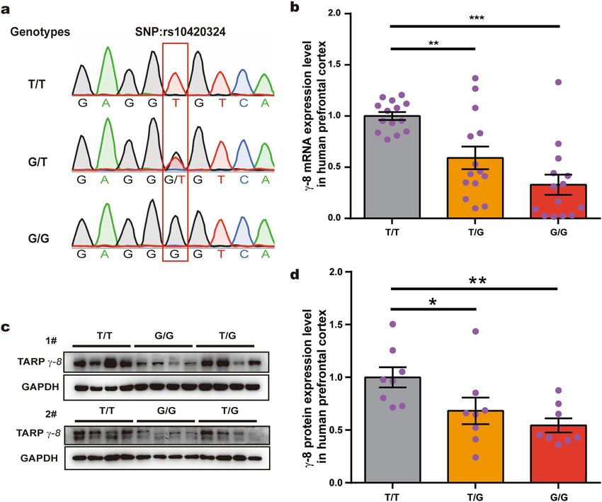
www.nature.com/scientificreports/

                                  Figure 1.  The rs10420324 SNPs regulate reporter gene expression by regulating the conformation and stability of a
                                  local G-quadruplex. (a) Schematic of the SNP fragment constructs inserted in the GFP reporter vector used in (b, c, e).
                                  The primers F1 and R1 were designed to examine pre-mRNA, while primers F2 and R2 were for mRNA. (b) Western
                                  blot analysis of GFP and β-tubulin protein in HEK 293 T cells transfected with the reporter plasmid. (c) Quantification
                                  of GFP expression levels. GFP expression was normalized using β-tubulin. (d) A linkage disequilibrium (LD) map for
                                  the TARP γ-8 gene was obtained from the SNPinfo web server, demonstrating a high LD value between the indicated
                                  SNPs. MAF: minor allele frequency. Red lines: Tag SNP location. Blue lines: SNP linked with Tag SNP location. (e)
                                  Fold change in GFP pre-mRNA and mRNA in HEK 293 T cells transfected with the reporter plasmid. GFP pre-
                                  mRNA and mRNA expression was normalized with β-actin. (f) Oligonucleotides containing the SNP rs10420324 were
                                  synthesized for CD spectral analysis. Underlined sequences indicate the guanine tandem repeats. An oligonucleotide
                                  with a G-rich sequence fragmented by guanine-to-adenine substitutions (in red) was used as a negative control. (g) CD
                                  spectra were measured in the presence of 100 mM KCl at 25 °C and 65 °C. The CD spectrum of the negative control
                                  was measured at 25 °C. Data represent the mean ± s.e.m. independent-sample t-test, *p < 0.05, **p < 0.01, ***p < 0.001.

Scientific Reports |   (2021) 11:11997 |               https://doi.org/10.1038/s41598-021-91415-9                                                      3

                                                                                                                                                  Vol.:(0123456789)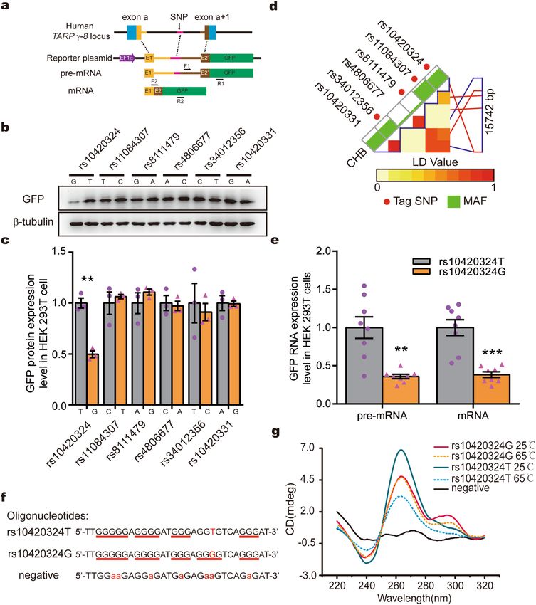
www.nature.com/scientificreports/

                                            Figure 2.  SNP rs10420324 determines TARP γ-8 expression in human brains. (a) Sanger sequencing results
                                            showing the T/T, G/T, and G/G genotypes at the CACNG8 rs10420324 locus in human prefrontal cortex tissues
                                            (outlined with a red box). 101 human prefrontal cortex tissues of male adults (25–45 years old) were obtained
                                            from a Chinese brain bank in Wuhan. (b) Fold change in the TARP γ-8 mRNA expression level in the human
                                            prefrontal cortex tissues with different rs10420324 genotypes (n = 14 for each). TARP γ-8 mRNA expression
                                            was normalized using β-actin mRNA. (c) Western blot analysis of the TARP γ-8 proteins in human prefrontal
                                            cortex tissues with different rs10420324 genotypes (n = 8 for each genotype). GAPDH was an internal reference.
                                            (d) Quantification of the TARP γ-8 protein expression levels in (c). TARP γ-8 expression was normalized using
                                            GAPDH. Data represent the mean ± s.e.m. one-way ANOVA, *p < 0.05, **p < 0.01, ***p < 0.001.

                                            (Fig. 1f) and analyzed by circular dichroism (CD). The CD wavelength of rs10420324T at 25 °C showed a posi-
                                            tive Cotton effect near 265 nm and a negative one at approximately 240 nm (Fig. 1g), suggestive of a parallel-
                                            form G-quadruplex. The CD spectra of rs10420324G showed positive ellipticity near 265 nm, a strong shoul-
                                            der at approximately 295 nm, and a negative one close to 240 nm, suggestive of a hybrid-form G-quadruplex,
                                            while the control oligonucleotide displayed no typical G-quadruplex waveforms (Fig. 1g). We then measured the
                                            G-quadruplex stability by analyzing the thermostability and heated each to 65 °C. The G-quadruplex waveform
                                            of rs10420324G was thermostable, with little change in the spectra, while the positive and negative ellipticities in
                                            the CD spectra of rs10420324T were significantly reduced by heating (Fig. 1g), suggesting that the G-quadruplex
                                            formed by rs10420324T was less stable. Taken together, these results demonstrate that rs10420324 affects the
                                            conformation and stability of a local G-quadruplex in CACNG8.

                                            SNP rs10420324G suppresses TARP γ‑8 expression in the human brain. Next, we aimed to
                                            understand the effect of rs10420324 on TARP γ-8 expression. As the SNP rs10420324 has been found only in
                                            humans, we analyzed human prefrontal cortex tissues of 101 male adults (25–45 years old) from a Chinese brain
                                            bank in Wuhan. The samples were genotyped by Sanger sequencing, which identified 35, 52, and 14 samples with
                                            the T/T, G/T, and G/G genotypes at the rs10420324 site, respectively (Fig. 2a). Quantitative RT-PCR revealed

          Scientific Reports |   (2021) 11:11997 |               https://doi.org/10.1038/s41598-021-91415-9                                                   4

Vol:.(1234567890)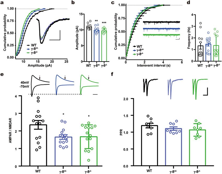
www.nature.com/scientificreports/
Group Genotype frequency (%)
rs10420324 n GG TG TT χ2 p
ASPD 135 41 (30.4)↑ 62 (45.9) 32 (23.7)↓ 8.471* 0.014
Controls 487 96 (19.7) 230 (47.2) 161 (33.1)
Table 1. Genotype frequency difference between the ASPD and control groups. ASPD antisocial personality
disorder. *Intergroup comparisons were tested by chi-square test.
Group Allele frequency (%)
rs10420324 n G T χ2 p
ASPD 270 144 (53.3)↑ 126 (46.7)↓ 8.537** 0.003
Controls 974 422 (43.3) 552 (56.7)
Table 2. Allele frequency difference between the ASPD and control groups. ASPD antisocial personality
disorder. **Intergroup comparisons were tested by chi-square test.
that the mRNA expression level of TARP γ-8 was negatively correlated with the number of Gs at the rs10420324
site. Specifically, relative to that in T/T samples, the mRNA expression of TARP γ-8 was 59.2% in the G/T and
32.9% in the G/G samples (Fig. 2b, n = 14 for each genotype). Consistent with the mRNA expression levels, the
protein levels of TARP γ-8 were also negatively correlated with the number of Gs at the rs10420324 site; rela-
tive to that in T/T samples, the protein levels of TARP γ-8 was 68.2% in the G/T and 54.4% in the G/G samples
(Fig. 2c,d, n = 8 for each genotype). These results demonstrate that the SNP rs10420324G suppresses TARP γ-8
gene expression in the human brain.
SNP rs10420324 is associated with ASPD. Above results indicate that the SNP rs10420324 regulates
γ-8 expression levels in the human brain. We wondered whether this SNP is linked to psychological disorders
in humans. We sequenced the rs10420324 site in 135 patients diagnosed with ASPD (25–40 years old) that were
initially identified from male inmates from an adult male prison in Jiangsu P rovince33 and in 487 healthy con-
trols (30–46 years old) by Sanger sequencing. There were significant differences in the genotypes (χ2 = 8.471,
p = 0.014) and allele frequencies (χ2 = 8.537, p = 0.003) between the two groups; compared to the healthy control
group, the ASPD group had an increased GG genotype frequency (30.4% vs. 19.7%) and G allele frequency
(53.3% vs. 43.3%; Tables 1, 2). It should be noted that the rs10420324 G/T distribution in the control group was
consistent with documented data in the CHB population. Thus these data indicate that the rs10420324G is asso-
ciated with ASPD in Chinese Han samples.
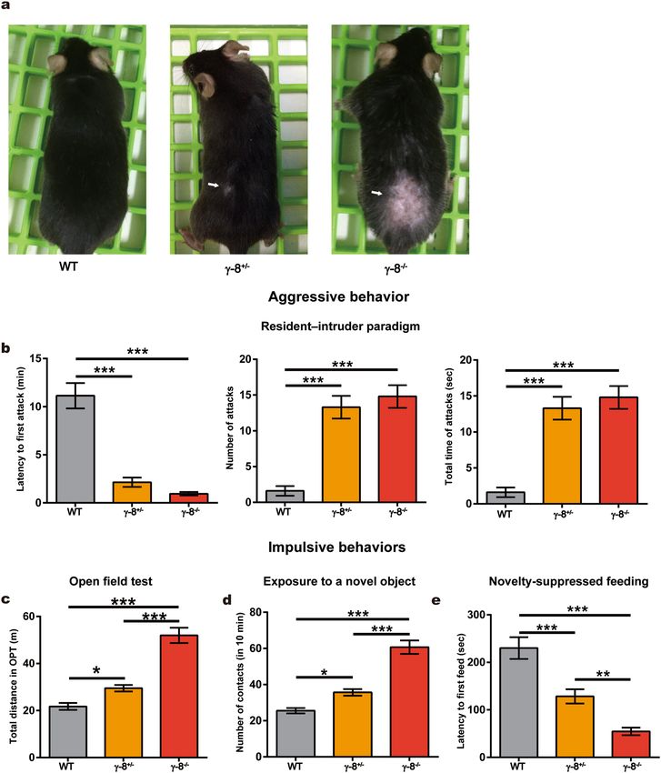
TARP γ‑8 knockout mice exhibit increased aggression and impulsivity. To understand the causal
relationship between the decrease of TARP γ-8 and ASPD, we designed systematic neurobehavioral tests and
analyzed the behavior of γ-8−/− and γ-8+/− mice27, in which either both or one of the γ-8 alleles were knocked
out, respectively. The γ-8−/− and γ-8+/− mice exhibited bare patches in their coats from severe bite wounds, sug-
gesting that these mice were more aggressive than wild-type (WT) animals, which did not have bite wounds
(Fig. 3a). For confirmation, we used the resident–intruder paradigm test to evaluate aggression34,35, which is a
standardized method to measure offensive aggression and defensive behavior in a semi natural setting35. The
experiments were performed and analyzed by persons blinded to the mouse genotypes. As expected, the γ-8−/−
and γ-8+/− mice exhibited a shorter latency to the first biting attack, a higher attack frequency and longer total
duration of attack episodes than WT mice (n = 10, Fig. 3b), demonstrating that reduced γ-8 expression led to
aggressive behaviors in mice.
Next, the general behavioral characteristics of the γ-8−/− and γ-8+/− mice were investigated using the open
field test. We found that the total distance traveled was greater for γ-8−/− and γ-8+/− mice than for WT mice, and
the total distance of γ-8−/− mice higher than γ-8+/− mice (n = 20, Fig. 3c), while the time in central vs. periphery
was not different among γ-8−/−, γ-8+/− and WT mice (n = 20, Fig. S1). Those results suggested that reduced γ-8
expression caused an increased locomotor response to environmental novelty, a phenotype classically associated
with impulsivity36. We then further examined impulsivity by testing the response to novelty. A novel object (a
blue plastic cap from a 15 mL Falcon tube) was introduced into the same corner of the home cage of each mouse.
The number of physical and nonphysical (sniffing) interactions (referred to as contacts) of the animal with the
object were recorded for 10 min. Compared with the WT mice, both γ-8−/− and γ-8+/− mice (n = 12) interacted
more with the novel object (n = 12, Fig. 3d), indicating again that the mutant mice were more impulsive than
the control mice. Impulsivity was further analyzed with a novelty-suppressed feeding test, which examines the
consequences of competing motivations, i.e., the drive to eat versus fear of venturing into the center of a brightly
lit arena where the food is l ocated36, known as hyponeophagia. Mice were fasted for 18 h and then placed in a new
cage with a brightly lit arena containing a pellet of food. The latency to start eating (defined as the mouse sitting
on its haunches and biting the pellet while holding it with the forepaws) was recorded within a 5 min period.
Scientific Reports | (2021) 11:11997 | https://doi.org/10.1038/s41598-021-91415-9 5
Vol.:(0123456789)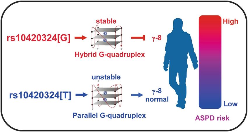
www.nature.com/scientificreports/

                                            Figure 3.  Aggression and impulsivity of WT and mutant mice. (a) Representative photos of WT and mutant
                                            mice at three-month-old. Four same genotype mice were housed in one cage. The arrows indicate areas where
                                            the animal was bitten. (b) Aggressive behavior test (resident-intruder paradigm). The latency to the first biting
                                            attack (left), the total number of attacks (middle) and the total duration of attack episodes (right) are presented.
                                            n = 10 cages (four mice in each cage). (c–e) Impulsive behavior tests. (c) Open field test. Total distance travelled
                                            in 5 min. n = 12. (d) The number of contacts with a novel object in 10 min. n = 12. (e) Novelty-suppressed
                                            feeding test. Degree of hyponeophagia in WT and mutant mice following an 18-h fast. n = 12. Data represent the
                                            mean ± s.e.m. one-way ANOVA, *p < 0.05, **p < 0.01, ***p < 0.001.

          Scientific Reports |   (2021) 11:11997 |               https://doi.org/10.1038/s41598-021-91415-9                                                    6

Vol:.(1234567890)
www.nature.com/scientificreports/
Compared to WT mice, γ-8−/− and γ-8+/− mice (n = 12) had shorter eating latencies and γ-8−/− mice shorter than
γ-8+/− mice (Fig. 3e), also indicating reduced γ-8 expression led to impulsive behaviors in mice.
The resident–intruder paradigm test, the open field, novel object exposure and novelty-suppressed feeding
tests suggested that γ-8 expression is negatively associated with aggression and impulsivity in mice.
TARP γ‑8‑null mice show increased antisocial and risk‑igonoring behaviors. To furtherly inves-
tigate the potential relationship between TARP γ-8 and mood disorders, we used a series of behavioral tests to
measure different endophenotypes of emotional disorders: social behavior, empathy-related behavior and risk-
taking behaviors37–39. We first tested their social behaviors by three-chamber test. The test mouse was free to
explore the apparatus, and the preference to contact the stranger mouse placed inside a wire cage vs. an empty
wire cage placed in the opposite chamber was assessed. Whereas the WT and γ-8+/− mice preferred to stay with
the stranger mice, γ-8−/− mice showed no preference (Fig. 4a,b). These results indicate that γ-8−/−mice have social
preference injury.
In addition, in contrast to the WT mice, the γ-8−/− and γ-8+/− mice showed little or no nesting behavior,
defined as crushing the paper and piling the pieces together to make a fluffy nest (Fig. 4c). Furthermore, the
γ-8−/− and γ-8+/− mice urinated throughout their cages, even on their nest (Fig. 4c, yellow spots marked with
arrows); thus, their cages were much dirtier than those of WT mice. We hypothesized that these reductions in
cage management behaviors in γ-8−/− and γ-8+/− mice indicated reduced social responsibility.
To further understand the social responsibility of these mouse genotypes, we designed an experiment called
the drowning rescue test (Fig. 4d) to examine their helping behavior. In this experiment, a mouse was placed in
a water-filled container measuring 15 cm in diameter in the center of a 60 × 60 c m2 field. A cage mate was then
introduced into the field outside of the container, and the level of interaction with the container containing the
struggling-against-drowning mouse were monitored. The WT mice spent more time near the container (defined
as keeping their own center of gravity less than 4 cm from the edge of the container) than did the γ-8−/− and
γ-8+/− mice, indicating empathetic behavior (Fig. 4e). Similarly, WT mice had significantly more contacting move-
ment traces than gene deficient mice (Fig. 4f). These data suggest that compared to the WT mice, the γ-8−/− and
γ-8+/− mice display callousness-like behaviors, which is classically associated with antisocial b
ehavior40,41.
42
Antisocial behavior often includes elevated risk-taking behaviors . To evaluate risk managing behavior, we
used the shuttle box apparatus, which divided into two halves, one with an electric shock at bottom43. During
the acquisition trial, immediately after the fasted mice entered the food-containing compartment, a five-second,
30-V electric shock was delivered to the mouse (Fig. 4g). In a recall trial 24 h later, compared to the WT mice,
the γ-8−/− and γ-8+/− mice exhibited a significantly reduced latency to enter the compartment (Fig. 4h) and a
significantly increased frequency of entries in 5 min (Fig. 4i), revealing that the γ-8−/− and γ-8+/− mice did not
learn to fear the shock lesson and thus exhibited risk-ignoring behaviors.
Three-chamber test, bedding management, the drowning rescue test and shuttle box test suggested that
γ-8 expression is negatively associated with social impairment, irresponsibility-like behaviors, callousness-like
behaviors and risk-ignoring behaviors.
TARP γ‑8 knockout impairs synaptic AMPAR function in medial prefrontal cortex. The medial
prefrontal cortex (mPFC) is a brain region that is liked to aggressive behaviors in humans and in r odents44; lesion
or inhibition leads to aggression. Since TARP γ-8 was known to regulate AMPAR functions, we thus chose to
analyze synaptic AMPAR function in the layer II/III pyramidal neurons of mPFC in γ-8−/− and γ-8+/− mice.
The amplitude of miniature excitatory postsynaptic currents (mEPSCs) was significantly reduced in γ-8−/− and
γ-8+/− neurons compared to WT neurons (Fig. 5a,b) while mEPSC frequency was not different among genotypes
(Fig. 5c,d). When EPSCs were evoked by stimulating layer IV with a bipolar tungsten electrode, the AMPA to
NMDA ratio was reduced in γ-8−/− and γ-8+/− mices compared to WT (Fig. 5e). Paired pulse ratio (PPR) of
AMPAR EPSCs evoked by a pair of stimuli with 50 ms intervals was not different among WT, γ-8−/− and γ-8+/−
(Fig. 5f), indicating neurotransmitter release probability was unaltered by γ-8 expression level.
Discussion
In this study, we aim to understand the role of the AMPA receptor regulatory protein TARP γ-8 in emotional
disorders. Our study shows that the expression level of γ-8 contributes to ASPD in humans. This conclusion is
supported by the evidence from ASPD patients and mouse models. In human, the SNP site rs10420324 in the 1st
intron of CACNG8 determines the transcription of TARP γ-8, likely by regulating the conformation and stability
of a local G-quadruplex. Compared to rs10420324T, rs10420324G suppresses transcription. The rs10420324G is
higher in patients diagnosed with ASPD than in controls, indicating that lower TARP γ-8 expression is associ-
ated with ASPD. In mice models, compared to the WT, the TARP γ-8 mutant mice exhibited greater impulsivity,
aggressiveness, risk-ignoring, irresponsibility-like and callousness-like behaviors, all of which are psychological
characteristics linked to ASPD in humans45. Reduction in TARP γ-8 impairs synaptic AMPAR function in mPFC,
a brain region linked to aggression44. Our study suggest a model shown in Fig. 6.
A previous study identified a two-SNP haplotype (rs10420331-rs11084307) in associated with s chizophrenia29.
These two SNPs, however, do not affect the expression of the reporter GFP, indicating they cannot regulate γ-8
expression. In contrast, SNP rs10420324, at a nearby locus, significantly affected the transcription of the GFP
reporter molecule. SNP rs10420324 is located in guanine-rich nucleic-acid sequences, which can form non-
canonical structure G-quadruplexes46. Substantial evidence now exists to support that formation of DNA or RNA
G-quadruplexes is coupled to altered gene expression by blocking the progression of RNA p olymerase46–48. The
variants of rs10420324 regulates the conformation and stability of the local G-quadruplex structure. The stable
hybrid G-quadruplex formed by rs10420324G can suppress reporter gene transcription in vitro and TARP γ-8
Scientific Reports | (2021) 11:11997 | https://doi.org/10.1038/s41598-021-91415-9 7
Vol.:(0123456789)www.nature.com/scientificreports/
Figure 4. Impaired prosocial behavior and lesson-learning ability in TARP γ-8 knockout mice. (a, b) Three-
chamber tests. (a) Contact times with the empty cage or stranger cage during the 10 min period and (b)
stranger/total contacts of the WT (n = 11), TARP γ-8+/− (n = 12) and TARP γ-8−/− (n = 11) mice. Data represent
the mean ± s.e.m. Independent-sample t-test used in (a), one-way ANOVA used in (b), *p < 0.05, **p < 0.01,
***p < 0.001. (c) Representative photos of mouse cages taken after four mice of same genotype housed in one
cage for 7 days without a change of bedding. The paper was the nest where the animals slept. The arrows
indicate yellow stains on the paper from the urine of TARP γ-8+/− and TARP γ-8−/− mice while no yellow stains
on the nest were observed in the cages of the WT mice. (d–f) Empathetic behavior tests. (d) Schematic of the
drowning rescue test. The mice in platform were the cage mate of mice in the water container. (e) Time spent
with the drowning cage mate during the 5 min period and (f) movement traces of the WT, TARP γ-8+/− and
TARP γ-8−/− mice (n = 12 for each) in the drowning rescue test. Red dots: experiment start position, Blue dots:
experiment end position, Yellow line: movement traces, the black circle: 4 cm from the edge of the container.
(g–i) Risk-taking behavior test. (g) Schematic of the shuttle box test. After receiving a plantar shock (defined
as acquisition), mice were fed freely for 6 h and then fasted for 18 h. The mice were tested for 180 s. Lightning
bolts: Bottom shock. (h) initial latency to enter and (i) total number of entries (n = 12 for each). Data represent
the mean ± s.e.m. one-way ANOVA, *p < 0.05, **p < 0.01, ***p < 0.001.
Scientific Reports | (2021) 11:11997 | https://doi.org/10.1038/s41598-021-91415-9 8
Vol:.(1234567890)www.nature.com/scientificreports/
Figure 5. Synaptic AMPAR functions are impaired in mPFC TARP γ-8 knockout mice. (a–d) mEPSCs from
γ-8+/− and γ-8−/− mPFC pyramidal cells had reduced amplitude but unchanged frequency. (a) Cumulative
frequency distribution of mEPSC amplitudes. Insert shows sample traces. Scale bar, 5 pA, 5 ms. (b) Amplitude
of mEPSC was reduced in mPFC pyramidal cells from γ-8+/− and γ-8−/− mice. (c) Cumulative frequency
distribution of mEPSC interevent interval. Sample traces are shown as insert. Scale bar, 10 pA, 500 ms. (d)
Frequency of mEPSC did not differ among WT, γ-8+/− and γ-8−/− neurons. (n = 12 for WT; n = 13 for γ-8+/−;
n = 13 for γ-8−/−). (e) AMPA/NMDA ratio was reduced in mPFC pyramidal neurons from γ-8+/− and γ-8−/− mice
(n = 14). Representative EPSCs recorded at holding of − 70 mV and + 40 mV are shown above the bar graph.
Arrows indicate where NMDAR-EPSCs were measured. Scale bar, 50 ms. (f) Paired-pulse ratio (PPR) does not
differ between WT, TARP γ-8+/− and TARP γ-8−/− neurons (n = 8 for each). The sample traces are shown above
the bar graph. Scale bar, 50 pA, 100 ms. Data represent the mean ± s.e.m. one-way ANOVA, *p < 0.05, **p < 0.01,
***p < 0.001.
transcription in vivo, supporting the notion that the G-quadruplex regulates transcription. Our observations
provide strong evidence in G-quadruplexes formation linking with biological processes of transcription (Fig. 6).
TARP γ-8 is highly expressed in the hippocampus, cortex and subcortical regions that are critical for emo-
tion generation in h umans19. Genetic deletion of TARP γ-8 led to reduced AMPARs on the neuronal surface26
and impaired synaptic AMPA-EPSCs and Long-term potentiation (LTP)27. LTP is a persistent strengthening
of synapses and widely considered one of the major cellular mechanisms that underlies learning and memory,
recognition and emotion49,50. Recent studies demonstrated that LTP induction required the PDZ binding motif
and CaMKII phosphorylation sites located at the C-terminal region of γ-851,52. It has been reported that γ-8
knockout mice showed hyperactivity28, a phenotype we also observed in this study. Interestingly, chemical
interference with TARP γ-8 function also led to hyperactivity in r ats53, indicating that TARP γ-8 is a valid drug
target. Most importantly, we observed that the γ-8−/− and γ-8+/− mice had multiple behavioral abnormalities that
likely mimicked antisocial behaviors associated with ASPD in humans.
ASPD is one of the leading risk factors associated with criminal behaviors, such as violent crime and murder,
in humans54. Impulsiveness, lack of foresight, and aggression are the most direct causes of crime, inflicting harm
on both the agent and s ociety55–57. Individuals with ASPD often engage in dangerous, risky, and potentially self-
damaging activities unnecessarily and without regard for consequences58. In our study, γ-8-null mice displayed
Scientific Reports | (2021) 11:11997 | https://doi.org/10.1038/s41598-021-91415-9 9
Vol.:(0123456789)www.nature.com/scientificreports/
Figure 6. A model illustrating a molecular mechanism that rs10420324 in CACNG8 regulates the susceptibility
to ASPD. The SNP site rs10420324 (T/G) in the 1st intron of CACNG8 determined TARP γ-8 expression by
regulating the conformation (parallel/hybrid) and stability of a local G-quadruplex. The rs10420324G suppresses
TARP γ-8 expression, thus increases ASPD risk.
many behaviors mimicking the above mentioned features of human ASPD. Firstly, γ-8−/− and γ-8+/− mice dis-
played elevated aggression with enhanced attacking behavior against intruders and engaged in intense fight-
ing and biting between cage mates. Secondly, these mice had greater impulsivity: they did not examine their
surroundings for safety when faced with novel objects or were presented with food in novel environments. In
addition, these animals showed a reduced lesson-learning ability and risk-ignoring behavior when facing danger,
similar to ASPD patients, who often disdain laws and regulations, have little regard for punishment and benefit
little from past punishment or other experiences59,60. Finally, individuals with ASPD tend to show diminished
responsibility and e mpathy45,58. ASPD can be identified according to the presence or absence of callous-une-
motional traits (deficits in empathy and guilt)61. TARP γ-8−/− and γ-8+/− mice failed to build nests or manage
their cages, indicating irresponsibility-like behaviors. These mice were also indifferent to their drowning cage
mates, indicating that a decrease in TARP γ-8 expression impairs empathetic behavior in mice. Interestingly, the
severity of all the ASPD-associated behaviors observed in this study was dependent on the γ-8 expression level,
with the γ-8−/− mice having a more severe phenotype than the γ-8+/− mice. Notably, to our knowledge, there is
no available animal model of ASPD. Therefore, γ-8 mutant mouse is a valuable animal model for studying ASPD.
Using mutant mice and human samples, we have demonstrated that the expression level of the AMPAR
regulatory subunit TARP γ-8 contributes to multiple behaviors linked to ASPD (Fig. 6). Our study lays a key
foundation for future study of ASPD at least in following two aspects. Firstly, the TARP γ-8 knockout mice is an
appropriate animal model for ASPD study. This animal model will setup a start point to dissect the neural circuits
related to ASPD. Then, TARP γ-8 and AMPARs are potential therapeutic targets for ASPD.
Materials and methods
All experiments, statistical analyzes, and information provided in this article were made in accordance with the
recommendations of the ARRIVE guideline.
Selection of SNP markers. Four tag SNPs (rs10420324, rs11084307, rs8111479, and rs34012356) and two
of their linked SNPs (rs4806677 and rs10420331) in CACNG8 were selected. Their variation and the linkage
disequilibrium (LD) in CHB populations was determined according to the SNPinfo web s erver30. population:
CHB; Minor allele frequency: > 0.05; LD threshold: > 0.8; Sort by LD similarity.
Cell culture and transfections. HEK 293 T cells were cultured in Dulbecco’s Modified Eagle Medium
(DMEM) supplemented with 10% FBS and penicillin/streptomycin (Thermo Fisher Scientific) in a humidi-
fied incubator at 37 °C with 5% CO2. The culture medium was replenished every other day. HEK293T Cell
were seeded in 24-well plates or 12-well plates. The HEK 293 T cells were transfected using Lipofectamine
2000 (11668030, Invitrogen) according to manufacturer’s instruction. Cells were collected for analysis 48 h after
transfection.
Participants and ethical approval. We recruited 2300 Chinese Han men: 1804 inmates from a prison33
and 496 healthy controls from the communities in Jiangsu Province, China. All procedures were approved by
the Institutional Review Board of Nanjing Brain Hospital and a consent form for this study was prepared. We
carried out the study in accordance with the principles of the Declaration of Helsinki. Individuals who had previ-
ously been diagnosed with chronic heart, liver and kidney diseases; had a history of nervous system diseases or
Scientific Reports | (2021) 11:11997 | https://doi.org/10.1038/s41598-021-91415-9 10
Vol:.(1234567890)www.nature.com/scientificreports/
mental disorders; or had a long history of medication use were excluded from the study. After informed consent
was obtained, psychiatrists clinically assessed and diagnosed participants with ASPD using the self-reported
Personality Diagnostic Questionnaire-4 (PDQ-4) and the Structured Clinical Interview Tool for DSM-IV Axis
II Disorders (SCID-II). Among all participants, based on the ASPD scores in the PDQ-4 (ASPD score ≥ 4) and
SCID-II (SCID-IIA score ≥ 3 or SCID-IIC score ≥ 2), 135 ASPD subjects and 487 healthy control subjects were
ultimately included in this study.
Human tissues. 101 human prefrontal cortex (PFC) brain tissues were provided by a Chinese brain bank
in Wuhan collecting the tissues of body donors. The tissues used in this study were all from male adults (25–
45 years old).
Genotyping. DNA was extracted from 5 ml of venous blood per person from 135 men with ASPD and
487 healthy men by the modified SDS method. PCR amplification (Taq DNA Polymerase, Vazyme) and Sanger
sequencing (Genscript, China) were used for genotyping of rs10420324. The primers for genotyping were as
follows: F: 5′-GCCTCTCCTGTGGAAGTTTGAG-3′; R: 5′-CCTCTTACTCCTACGTTCTCCG-3′. rs10420324
was successfully genotyped in all 135 ASPD subjects and all 487 healthy men.
Western blots. Proteins were extracted from HEK 293 T cells or human prefrontal cortex brain tissues
using tissue total protein lysis buffer (Beyotime, Shanghai, China) for each assay. Proteins were electrophoreti-
cally separated on SDS-PAGE gels and subsequently immunoblotted onto nitrocellulose membranes that were
blocked in skim milk and incubated at 4 °C for 16 h using antibodies against GFP (1:5000, ab290, Abcam),
β-tubulin (1:10,000, BS1482M, Bioworld), TARP γ-8 (1:500, abs132666a, Absin) and GAPDH (1:10,000, 60,004–
1-Ig, Proteintech). Detection was carried out using the horseradish peroxidase-conjugated secondary antibodies
goat anti-mouse IgG (1:10,000, BS12478, Bioworld) and goat anti-rabbit IgG (1:15,000, BS13278, Bioworld)
along with a High-sig ECL kit (Tanon, Shanghai, China), and signals were recorded using a Tanon 5200 multi
automatic chemiluminescence image analysis system (Tanon, Shanghai, China). For western blot quantitation,
images were analyzed using ImageJ and expressed as arbitrary units (AU). GFP protein expression was normal-
ized to β-tubulin protein expression, and TARP γ-8 expression was normalized to GAPDH expression.
Quantitative real‑time PCR analysis. Quantitative real-time PCR was performed to examine the rela-
tive expression levels of GFP in HEK 293 T cells and CACNG8 in the PFC of the human brain using an ABI
Prism 7300 sequence detector (ABI, Massachusetts, USA). The following primer sequences were used: primer
1(for qPCR detecting fusion GFP pre-mRNA) of fusion GFP forward(F1): 5′-TGCTGCCTGGAAGGGTTG
A-3′, reverse (R1): 5′-TGCCGTTCTTCTGCTTGTCG-3′; primer 2 (for qPCR detecting fusion GFP mRNA)
of fusion GFP forward (F2): 5′-CAGTGCTTCAGCCGCTACCC-3′, reverse (R2): 5′-AGTTCACCTTGATGC
CGTTCTT-3′; CACNG8 forward: 5′-GCGAGGGCTGAAGGGTAGTGA-3′, reverse: 5′-GCAATGGGTGAG
TGGCTGAAT-3′; β-actin forward: 5′-AGCGAGCATCCCCCAAAGTT-3′, reverse: 5′-GGGCACGAAGGC
TCATCATT-3′.Total RNA was extracted from HEK 293 T cells or the PFCs of human brains using TRIzol Rea-
gent (Invitrogen, California, USA) for each assay. RNA was converted to cDNA using a reverse transcription kit
HiScript Q RT SuperMix for qPCR (Vazyme, Biotech). qPCR reactions were performed with AceQ qPCR SYBR
Green Master Mix (Vazyme, Biotech) according to manufacturer’s instruction. The relative expression levels of
GFP or CACNG8 were normalized to the expression of β-actin.
CD spectroscopy. DNA oligonucleotides (Genscript, Jiangsu, China) were dissolved in 1X TE buffer
(10 mM Tris-HC, 1 mM EDTA, pH 7.4) with 100 mM KCl at a concentration of 5 μM. Before analysis, the oli-
gonucleotides were heated to 95 °C for 5 min, slowly cooled to room temperature, and incubated overnight. The
CD experiment was performed on a J-810 spectrophotometer (JASCO, Tokyo, JAPAN) using a quartz cell with
a 1 mm optical path length and an instrument scanning speed of 50 nm/min with a response time of 1 s and
over a wavelength range of 220–320 nm. Finally, three scans taken at 25 °C or 65 °C were averaged to obtain a
representative CD spectrum for each condition.
Mice. The TARP γ-8−/− mice27 were a gift from Roger A. Nicoll of UCSF and were backcrossed with C57BL/6 J
mice for at least 8 generations before these experiments. Four to six mice were housed in groups with a 12-h
dark/light cycle and free access to food and water in accordance with the Regulations on Mouse Welfare and
Ethics of Nanjing University, China. All procedures were conducted with the approval of the Model Animal
Research Center of Nanjing University, Nanjing, China. The same genotype mice were housed in one cage. Two-
to three-month-old male mice were used in all experiments. All behavioral tests and data analyses were carried
out by different investigators, and the one who analyzed the data was blind to the genotypes of the mice.
Behavioral tests. ZhengHua software (ZhengHua, Anhui, China) were used for recording the central locus
of mice and analysis of all behavioral data. A special room was used to conduct all behavioral experiments
separately. Mice were transferred to the room 2 h before the experiment. All experiments were performed in
triplicate.
Resident–intruder paradigm. An adult male WT mouse was used as an intruder. An intruder mouse was
transferred into the residents’ cage (four males per cage), and attack episodes by the resident mice were recorded
for 15 min. The intruder was typically attacked and defeated by the residents and showed submissive freezing
Scientific Reports | (2021) 11:11997 | https://doi.org/10.1038/s41598-021-91415-9 11
Vol.:(0123456789)www.nature.com/scientificreports/
behavior. Aggressive behaviors by the resident mice were scored; these behaviors were defined as biting, laterally
attacking and displaying piloerection. The latency to the first attack bite, the frequency of bites and the duration
of attack episodes were manually quantified. Data from four residents in the same cage were collectively analyzed
as one unit.
Open field test. The test arena consisted of a plastic-bottom plate (46.5 cm × 46.5 cm) and four surround-
ing plastic walls (35.0 cm high). A mouse was initially placed in a corner of the arena facing the center and then
allowed to freely explore the open field for 5 min. The arena was cleaned with 70% EtOH between trials. The
distance traveled (cm) by each mouse was quantified.
Novelty‑suppressed feeding. The testing apparatus consisted of a plastic box, 37 cm × 57 cm × 10 cm,
directly illuminated by white light. The floor was covered with 2 cm of corncob bedding. Six same genotype mice
were housed in one cage for 7 days. Eighteen hours before the test, food was removed from the cages. At the time
of testing, a single pellet of standard mouse chow was placed at the center of the box. The animal was placed in a
corner of the box, and a stopwatch was immediately started. The latency to start eating (defined as the mouse sit-
ting on its haunches and biting the pellet while holding it with the forepaws) was recorded within a 5 min period.
Exposure to a novel object. A novel object (a blue plastic cap from a 15 mL Falcon tube) was introduced
in the same corner of the home cage of each mouse. The object was placed in the cage after the animal was
momentarily removed into other cage. The mouse was then returned to the cage by gently setting it down in the
corner opposite the novel object. Data recording started immediately, and the physical and nonphysical (sniff-
ing) interactions of the animal with the object were recorded for 10 min. The latency to approach (measured as
first sniffing) and the number of contacts were analyzed by a researcher blinded to the genotypes.
Three‑chamber test. The social test apparatus was an opaque acrylic box with two pull-out doors and
three chambers. Each chamber was identical in size (40 × 20 cm), with the dimensions of the entire box being
63 cm × 42 cm × 21 cm. There was a 10-cm gap between adjacent chambers that could be opened or closed with
the removable doors. The transparent wire cage (12 cm in height and 9.5 cm wide) equipped with the novel,
stranger mouse was placed 2 cm away from the edge of the testing chamber to allow an interaction between
the mice. The whole experiment was performed under low illumination and quiet conditions. The unfamiliar,
stranger mouse was introduced into the wire cage in one side compartment, and an empty cage was placed in
the opposite side compartment. The test mouse was introduced into the middle chamber and habituated for at
least 5 min. The partitions were then removed, and the test mouse was permitted to explore all 3 compartments
for 10 min. The entire process was recorded by a camera hanging 1 m above the apparatus. The relative positions
of the empty cage and social cage were counterbalanced across test animals. The contact times with stranger or
empty cage was recorded using the automated ZhengHua software.
Risk‑taking behavior. The apparatus contained a chamber with two compartments (dark/light), which
were connected by a guillotine door43. The dark compartment contained a stainless steel grid floor that could
deliver electric shocks. The mice were first fasted for 18 h before the acquisition trial. Each mouse was habituated
to the light compartment for 180 s before the guillotine door was raised. Immediately after the mouse entered
the dark compartment containing food, the guillotine door was closed, and a five-second electric shock of 30 V
was delivered. The mouse was then returned to its home cage, fed freely for 6 h and then fasted for 18 h. The next
day, after the mice were habituated for 180 s in the light compartment, the guillotine door was raised, and the
mice were free to enter the electrically charged and food-containing dark compartment. The latency before the
first entry and the number of entries were recorded in a 5 min observation period.
Drowning rescue test. A mouse was placed into a 15 cm dimeter and 15 cm depth container filled with
25 °C water with a depth of 13 cm in the center of a 60 × 60 cm field. A cage mate was then introduced in the
field, and its attempts to contact the drowning mouse were monitored for 5 min. Mice were considered to display
empathetic behavior if they remained near the drowning cage mate (defined as keeping their central locus less
than 4 cm from the edge of the container).
Slice preparation and patch‑clamp recordings. Coronal brain slices (300 µm) were prepared from 4-
to 6-week-old C57BL6 male mice in ice-cold cutting solution containing (in mM): 210 Sucrose, 25 N aHCO3, 2.5
KCl, 7 MgSO4, 1.25 NaH2PO4, 1.3 Na-Ascorbic Acid, 10 glucose, and 0.5 CaCl2. After cutting, slices were allowed
to recover for 30 min at 32 °C and stored at room temperature in artificial cerebrospinal fluid (ACSF) contain-
ing (in mM): 119 NaCl, 26.2 N aHCO3, 2.5 KCl, 1 N
aH2PO4, 11 glucose, 1.3 MgSO4, and 2.5 C aCl2. All solutions
were constantly oxygenated with 95% O2/5% CO2. Slices containing the mPFC were transferred to a submersion
recording chamber, superfused with oxygenated ACSF at a speed of 1–2 ml/min. Whole-cell patchclamp record-
ings were performed using pipettes pulled from borosilicate glass capillaries (BF150-110-10, Sutter Instrument,
USA) with resistances of 4–6 MΩ. We used Cs-methanesulphonate based internal solution containing (mM):
135 Cs-methanesulphonate, 8 NaCl, 10 HEPES, 0.3 EGTA, 5 QX-314, 4 MgATP, 0.3 Na3GTP and 0.1 Spermine-
C14 (290–295 mOsm, pH 7.3–7.4). mEPSCs were recorded at − 70 mV in the presence of tetrodotoxin (0.5 M)
and bicuculline (100 M). For evoked EPSCs, Layer IV inputs were stimulated by bipolar tungsten electrodes
(Science Products). The stimulus was adjusted to evoke a measurable, monosynaptic eEPSC in both cells. We
identified the mPFC layer II/III PNs by somatic location and size. AMPAR eEPSCs were measured at a holding
Scientific Reports | (2021) 11:11997 | https://doi.org/10.1038/s41598-021-91415-9 12
Vol:.(1234567890)www.nature.com/scientificreports/
potential of − 70 mV, and NMDAR eEPSCs were measured at + 40 mV 150 ms after the stimulus, at which point
the AMPAR eEPSC has completely decayed. Paired-pulse ratios were measured by giving two pulses at a 50 ms
interval and taking the ratio of the peak of the second eEPSC over the first eEPSC from an average of 15–30
sweeps. Data were acquired with a Multiclamp 200B amplifier, Digidata1550, and Clampe10 software. Signals
were filtered at 2 kHz and digitized at 5 kHz.
Statistical analysis. Statistical analysis was performed using Statistical Product and Service Solutions
(SPSS) 22.0. The differences in demographic characteristics between the ASPD group and the control group
were examined by performing Hardy–Weinberg equilibrium and chi-square tests. Group comparisons of geno-
types and alleles were also achieved using chi-square tests. The independent-sample t-test was used for compar-
ing the two treatment groups, and one-way ANOVA was used for comparisons among more than two groups.
Data are presented as the means ± SEMs. P < 0.05 was set as the threshold for significance (*P < 0.05, **P < 0.01,
***P < 0.001).
Received: 18 February 2021; Accepted: 25 May 2021
References
1. LeDoux, J. E. Emotion circuits in the brain. Annu. Rev. Neurosci. 23, 155–184. https://doi.org/10.1146/annurev.neuro.23.1.155
(2000).
2. Phillips, M. L., Drevets, W. C., Rauch, S. L. & Lane, R. Neurobiology of emotion perception I: The neural basis of normal emotion
perception. Biol. Psychiatry 54, 504–514. https://doi.org/10.1016/s0006-3223(03)00168-9 (2003).
3. Kirkby, L. A. et al. An amygdala-hippocampus subnetwork that encodes variation in human mood. Cell 175, 1688–1700. https://
doi.org/10.1016/j.cell.2018.10.005 (2018).
4. Kavalali, E. T. & Monteggia, L. M. Targeting homeostatic synaptic plasticity for treatment of mood disorders. Neuron 106, 715–726.
https://doi.org/10.1016/j.neuron.2020.05.015 (2020).
5. Duman, R. S. Synaptic plasticity and mood disorders. Mol. Psychiatry 7(Suppl 1), S29-34. https://doi.org/10.1038/sj.mp.4001016
(2002).
6. Malinow, R. & Malenka, R. C. AMPA receptor trafficking and synaptic plasticity. Annu. Rev. Neurosci. 25, 103–126. https://doi.
org/10.1146/annurev.neuro.25.112701.142758 (2002).
7. Huganir, R. L. & Nicoll, R. A. AMPARs and synaptic plasticity: the last 25 years. Neuron 80, 704–717. https://doi.org/10.1016/j.
neuron.2013.10.025 (2013).
8. Zhuo, M. Cortical plasticity as synaptic mechanism for chronic pain. J. Neural Transm. https://d oi.o
rg/1 0.1 007/s 00702-0 19-0 2071-3
(2019).
9. Luscher, C. & Malenka, R. C. Drug-evoked synaptic plasticity in addiction: From molecular changes to circuit remodeling. Neuron
69, 650–663. https://doi.org/10.1016/j.neuron.2011.01.017 (2011).
10. Kessels, H. W. & Malinow, R. Synaptic AMPA receptor plasticity and behavior. Neuron 61, 340–350. https://doi.org/10.1016/j.
neuron.2009.01.015 (2009).
11. Volk, L., Chiu, S. L., Sharma, K. & Huganir, R. L. Glutamate synapses in human cognitive disorders. Annu. Rev. Neurosci. 38,
127–149. https://doi.org/10.1146/annurev-neuro-071714-033821 (2015).
12. Greger, I. H., Watson, J. F. & Cull-Candy, S. G. Structural and functional architecture of AMPA-type glutamate receptors and their
auxiliary proteins. Neuron 94, 713–730. https://doi.org/10.1016/j.neuron.2017.04.009 (2017).
13. Chen, S. et al. Activation and desensitization mechanism of AMPA receptor-TARP complex by Cryo-EM. Cell 170, 1234–1246.
https://doi.org/10.1016/j.cell.2017.07.045 (2017).
14. Twomey, E. C., Yelshanskaya, M. V., Grassucci, R. A., Frank, J. & Sobolevsky, A. I. Elucidation of AMPA receptor-stargazin com-
plexes by cryo-electron microscopy. Science 353, 83–86. https://doi.org/10.1126/science.aaf8411 (2016).
15. Yan, D. & Tomita, S. Defined criteria for auxiliary subunits of glutamate receptors. J. Physiol. 590, 21–31. https://doi.org/10.1113/
jphysiol.2011.213868 (2012).
16. Straub, C. & Tomita, S. The regulation of glutamate receptor trafficking and function by TARPs and other transmembrane auxiliary
subunits. Curr. Opin. Neurobiol. 22, 488–495. https://doi.org/10.1016/j.conb.2011.09.005 (2012).
17. Jackson, A. C. & Nicoll, R. A. The expanding social network of ionotropic glutamate receptors: TARPs and other transmembrane
auxiliary subunits. Neuron 70, 178–199. https://doi.org/10.1016/j.neuron.2011.04.007 (2011).
18. Milstein, A. D. & Nicoll, R. A. Regulation of AMPA receptor gating and pharmacology by TARP auxiliary subunits. Trends Phar-
macol. Sci. 29, 333–339. https://doi.org/10.1016/j.tips.2008.04.004 (2008).
19. Tomita, S. et al. Functional studies and distribution define a family of transmembrane AMPA receptor regulatory proteins. J. Cell
Biol. 161, 805–816. https://doi.org/10.1083/jcb.200212116 (2003).
20. Chen, L. et al. Stargazin regulates synaptic targeting of AMPA receptors by two distinct mechanisms. Nature 408, 936–943. https://
doi.org/10.1038/35050030 (2000).
21. Hashimoto, K. et al. Impairment of AMPA receptor function in cerebellar granule cells of ataxic mutant mouse stargazer. J. Neurosci.
19, 6027–6036 (1999).
22. Letts, V. A. et al. The mouse stargazer gene encodes a neuronal Ca2+-channel gamma subunit. Nat. Genet. 19, 340–347. https://
doi.org/10.1038/1228 (1998).
23. Noebels, J. L., Qiao, X., Bronson, R. T., Spencer, C. & Davisson, M. T. Stargazer: A new neurological mutant on chromosome 15
in the mouse with prolonged cortical seizures. Epilepsy Res. 7, 129–135. https://doi.org/10.1016/0920-1211(90)90098-g (1990).
24. Yamazaki, M. et al. Relative contribution of TARPs gamma-2 and gamma-7 to cerebellar excitatory synaptic transmission and
motor behavior. Proc. Natl. Acad. Sci. USA 112, E371-379. https://doi.org/10.1073/pnas.1423670112 (2015).
25. Burgess, D. L., Gefrides, L. A., Foreman, P. J. & Noebels, J. L. A cluster of three novel Ca2+ channel gamma subunit genes on
chromosome 19q13.4: Evolution and expression profile of the gamma subunit gene family. Genomics 71, 339–350. https://doi.org/
10.1006/geno.2000.6440 (2001).
26. Fukaya, M. et al. Abundant distribution of TARP gamma-8 in synaptic and extrasynaptic surface of hippocampal neurons and
its major role in AMPA receptor expression on spines and dendrites. Eur. J. Neurosci. 24, 2177–2190. https://doi.org/10.1111/j.
1460-9568.2006.05081.x (2006).
27. Rouach, N. et al. TARP gamma-8 controls hippocampal AMPA receptor number, distribution and synaptic plasticity. Nat. Neurosci.
8, 1525–1533. https://doi.org/10.1038/nn1551 (2005).
Scientific Reports | (2021) 11:11997 | https://doi.org/10.1038/s41598-021-91415-9 13
Vol.:(0123456789)You can also read