FAK displacement from focal adhesions: a promising strategy to target processes implicated in cancer progression and metastasis - Cell ...
←
→
Page content transcription
If your browser does not render page correctly, please read the page content below
Antoniades et al. Cell Communication and Signaling                (2021) 19:3
https://doi.org/10.1186/s12964-020-00671-1

 RESEARCH                                                                                                                                           Open Access

FAK displacement from focal adhesions: a
promising strategy to target processes
implicated in cancer progression and
metastasis
Ioanna Antoniades†, Maria Kyriakou†, Anna Charalambous, Katerina Kalalidou, Andri Christodoulou,
Maria Christoforou and Paris A. Skourides*

  Abstract
  Background: Focal adhesion kinase (FAK) is a non-receptor tyrosine kinase that is overexpressed or activated in
  several advanced-stage solid cancers. It is known to play both kinase-dependent and -independent roles in
  promoting tumor progression and metastasis. Numerous inhibitors, targeting either the enzymatic or scaffolding
  activities of FAK have been generated, with varying degree of success. Here, we describe a novel approach to site-
  specifically target both kinase-dependent and -independent FAK functions at focal adhesions (FAs), the primary
  sites at which the kinase exerts its activity.
  Methods: We took advantage of the well-characterized interactions between the paxillin LD motifs and the FAK
  FAT domain and generated a polypeptide (LD2-LD3-LD4) expected to compete with interactions with paxillin. Co-
  immunoprecipitation experiments were performed to examine the interaction between the LD2-LD3-LD4
  polypeptide and FAK. The effects of LD2-LD3-LD4 in the localization and functions of FAK, as well as FA
  composition, were evaluated using quantitative immunofluorescence, cell fractionation, FA isolation and Western
  Blot analysis. Live cell imaging, as well as 2-D migration and cell invasion assays were used to examine the effects
  on FA turnover and tumor cell migration and invasion.
  Results: Expression of the LD2-LD3-LD4 polypeptide prevents FAK localization at FAs, in a controlled and dose-
  dependent manner, by competing with endogenous paxillin for FAK binding. Importantly, the LD2-LD3-LD4
  peptide did not otherwise affect FA composition or integrin activation. LD2-LD3-LD4 inhibited FAK-dependent
  downstream integrin signaling and, unlike existing inhibitors, also blocked FAK’s scaffolding functions. We further
  show that LD2-LD3-LD4 expression markedly reduces FA turnover and inhibits tumor cell migration and invasion.
  Finally, we show that dimers of a single motif, linked through a flexible linker of the proper size, are sufficient for
  the displacement of FAK from FAs and for inhibition of tumor cell migration. This work raises the possibility of
  using a synthetic peptide as an antimetastatic agent, given that effective displacement of FAK from FAs only
  requires dimers of a single LD motif linked by a short flexible linker.
  (Continued on next page)

* Correspondence: skourip@ucy.ac.cy
†
 Ioanna Antoniades and Maria Kyriakou contributed equally to this work.
Department of Biological Sciences, University of Cyprus, P.O. Box 20537, 2109
Nicosia, Cyprus

                                        © The Author(s). 2021 Open Access This article is licensed under a Creative Commons Attribution 4.0 International License,
                                        which permits use, sharing, adaptation, distribution and reproduction in any medium or format, as long as you give
                                        appropriate credit to the original author(s) and the source, provide a link to the Creative Commons licence, and indicate if
                                        changes were made. The images or other third party material in this article are included in the article's Creative Commons
                                        licence, unless indicated otherwise in a credit line to the material. If material is not included in the article's Creative Commons
                                        licence and your intended use is not permitted by statutory regulation or exceeds the permitted use, you will need to obtain
                                        permission directly from the copyright holder. To view a copy of this licence, visit http://creativecommons.org/licenses/by/4.0/.
                                        The Creative Commons Public Domain Dedication waiver (http://creativecommons.org/publicdomain/zero/1.0/) applies to the
                                        data made available in this article, unless otherwise stated in a credit line to the data.
Antoniades et al. Cell Communication and Signaling   (2021) 19:3                                                    Page 2 of 22

 (Continued from previous page)
 Conclusion: In conclusion, these results suggest that FAK displacement from FAs is a promising new strategy to
 target critical processes implicated in cancer progression and metastasis.
 Keywords: Focal adhesion kinase (FAK), Paxillin, Focal adhesions, Cell migration, Cell invasion, Cancer

Background                                                         hydrophobic pockets (HPs), one located at the surface of
Cells and their associated microenvironment exhibit a              helices 1 and 4, and the second located at the surface of
complicated bidirectional communication that is critical           helices 2 and 3 [32]. These are absolutely essential for
for both normal tissue homeostasis and for tumor cell              FAK targeting to FAs [30, 33, 34]. The best characterized
growth, progression and metastasis [1]. The Focal                  interaction of the FAT HPs is that with the short (9 amino
Adhesion Kinase (FAK) has been identified as a critical            acids) Leucine-rich LD motifs of Paxillin [23, 24]. This
regulator and signal transducer for these Extracellular            interaction is important for FAK FA localization, suggest-
Matrix (ECM)-tumor cell interactions [2, 3] and has                ing that Paxillin is one of the major proteins responsible
been implicated in many aspects of the metastatic                  for the kinase’s recruitment to these complexes [33–35].
process including adhesion, migration, secretion of                Paxillin itself, is targeted to FAs via the C-terminal LIM
MMPs, spindle orientation and invasion [4–7]. FAK has              (Lin11, Isl-1 & Mec-3) domains, however the interacting
also been found to exhibit altered (usually elevated) ex-          proteins responsible for its localization have not been
pression and/or activation in most human epithelial                identified [24, 36]. Once FAK is localized at FAs, it phos-
cancers, resulting in enhanced invasive potential and              phorylates various proteins either directly or through the
poor overall patient survival [8]. FAK has therefore, be-          recruitment of Src kinases [37, 38], leading to efficient in-
come an attractive target for anti-cancer therapies.               tegrin signal transduction and cell migration.
   FAK has kinase-dependent and -independent functions,               The aim of the present study was to develop and test a
both of which are involved in cancer progression. However,         new strategy for the simultaneous inhibition of both en-
most of the recently developed inhibitors, have focused on         zymatic and scaffolding functions of FAK, specifically at
blocking the protein’s enzymatic activity. These include           FAs, by interfering with FAK targeting to these com-
antibodies [9, 10], dominant negative constructs [11–14]           plexes. To do so, we took advantage of the well charac-
and small molecule inhibitors, that primarily target the           terized binding of the LD motifs of Paxillin with the
ATP binding site, or allosterically inhibit the kinase domain      FAK FAT HPs, to prevent interactions with endogenous
[15], leading to decreased tumor cell viability, growth or         proteins, responsible for FA targeting. We show that a
apoptosis. However, targeting the kinase domain of FAK             polypeptide including the LD2 and LD4 motifs of Paxil-
has been complicated by the fact that the ATP-binding site         lin, specifically displaces endogenous FAK from FAs in a
shares consensus sequences and structural domains across           dose dependent manner without otherwise affecting FA
many different tyrosine kinases, making it less suitable for       composition or integrin activation. We go on to show
clinical testing, due to off-target effects [16].                  that these effects are the result of a competing inter-
   An alternative approach is to inhibit FAK’s kinase-             action of the polypeptide with endogenous paxillin, for
independent activities by blocking specific scaffolding            binding to the FAT domain of FAK. Furthermore, we
functions of the protein. This has been attempted using            show that effective displacement of FAK from FAs can
peptides, small molecule and antibody inhibitors that dis-         be accomplished using dimers of a single LD motif,
rupt interactions between FAK and various binding part-            linked by a short flexible linker. FAK displacement from
ners including VEGFR-3 [17], IFGR1 [18], c-Met [19, 20],           FAs leads to inhibition of downstream integrin signaling,
Mdm-2 [21] and p53 [22], with variable efficiency.                 reduced FA turnover, defects in cell spreading and in-
   One of the most highly studied multi-protein com-               hibition of cell migration and invasion. Our findings
plexes, that serve as sites of integration of growth factor        demonstrate that preventing FAK targeting and func-
signaling and integrin pathways, directing changes as di-          tions, specifically at FAs, represents a promising new
verse as gene expression and cytoskeletal reorganization,          strategy to prevent molecular and cellular processes im-
are Focal Adhesions (FAs). The focal adhesion targeting            plicated in tumor cell metastasis.
(FAT) domain of FAK is both necessary and sufficient for
localization at FAs and facilitates interactions with FA-          Materials and methods
associated proteins including Paxillin [23, 24], Talin [25,        Plasmids and DNA constructs
26], p130Cas [27], Grb2, ASAP1 [28] and p85α of PI3K               FLAG LD2-LD3-LD4
[29]. It is a highly conserved four helix bundle with a large      The DNA encoding amino acids 54–279 of human
hydrophobic core [30, 31]. It includes two surface exposed         Paxillin, was amplified via PCR using pCS108 GFP LD2-
Antoniades et al. Cell Communication and Signaling   (2021) 19:3                                                     Page 3 of 22

LD3-LD4 as template and primers F2 and R2                          GFP LD2-LD2
(Additional File 1: Table S1).                                     A two-step cloning strategy was followed using GFP
  The PCR program was as follows: 2 min at 95 °C for               LD2-LD4 6x short as template.
initial denaturation, followed by 35 cycles of 15 s at               An LD2-linker fragment was generated using primers
95 °C, 30 s at 67 °C, 1 min at 68 °C and final extension at        F4, R11 and R12 (Additional File 1: Table S1).
68 °C for 10 min.                                                    The PCR product was cloned in frame and down-
  The PCR product was cloned in frame and down-                    stream to GFP, in pCS108-GFP using the NotI and XbaI
stream to FLAG, in pFLAG CMV-2, using the NotI and                 restriction sites.
EcoRI restriction sites.                                             A linker-LD2 fragment was generated using primers
                                                                   F7, F8 and R13 (Additional File 1: Table S1):
GFP LD2-LD3-LD4 and mCherry LD2-LD3-LD4                              The PCR product was cloned in frame in the pCS108-
The DNA encoding amino acids 54–279 of human                       GFP LD2-linker plasmid, using the XbaI and XhoI
Paxillin, was amplified via PCR using pCS2++ GFP-                  restriction sites.
Paxillin as template and primers F1 and R1 (Additional
File 1: Table S1).                                                 GFP LD4-LD4
                                                                   A multi-step PCR was performed, using GFP LD2- LD4
GFP LD2-LD3-LD4 ΔLR                                                as template and primers F9, F10, R14, R15 and R12
The DNA encoding amino acids 139–279 of Paxillin,                  (Additional File 1: Table S1):
was amplified via a two-step PCR using GFP LD2-LD3-                  The PCR program was as follows: 2 min at 95 °C for initial
LD4 as template and primers F3, F4 and R2 (Additional              denaturation, followed by 35 cycles of 15 s at 95 °C, 30 s at
File 1: Table S1).                                                 67 °C, 1 min at 68 °C and final extension at 68 °C for 10 min.
                                                                     The PCR product was cloned in frame and down-
GFP LD2-LD4                                                        stream to GFP, in pCS108-GFP using the NotI and XhoI
For the generation of GFP LD2-LD4 constructs with                  restriction sites.
linkers of various sizes, multi-step PCRs using the fol-
lowing templates and primers were performed.                       pLV-tetO-LD2-LD4.
   GFP LD2-LD4 30aa (referred in the text as GFP LD2-              The DNA encoding motifs LD2 and LD4 connected with
LD4): GFP LD2-LD3-LD4 ΔLR was used as template and                 a 30 amino acid-long linker composed of 6 GGGS re-
primers F4, R4, R5, R6 and R3 (Additional File 1: Table S1).       peats was amplified via PCR using pCS108 GFP LD2-
   GFP LD2-LD4 15aa: GFP LD2-LD4 30aa was used as                  LD4 as template and primers F11 and R16 (Additional
template and primers F4, R7 and R3 (Additional File 1:             File 1: Table S1). The PCR program was as follows: 2
Table S1).                                                         min at 95 °C for initial denaturation, followed by 35 cy-
   GFP LD2-LD4 25aa: GFP LD2-LD4 15aa was used as                  cles of 15 s at 95 °C, 30 s at 67 °C, 1 min at 68 °C and
template and primers F4, R8 and R3 (Additional File 1:             final extension at 68 °C for 10 min. The PCR product
Table S1).                                                         was cloned in pLV-tetO-Oct4 vector, using the EcoRI
                                                                   restriction site (to replace Oct4) [39].
GFP LD2                                                              pCS2++ TagRFP FAK, pCS108 FusionRed Vinculin
The DNA encoding amino acids 420–449 of human Paxillin,            and pCS108 RFP Vinculin were generated by replacing
was amplified via PCR using pCS2++ GFP LD2-LD4 as tem-             the GFP sequence with that of TagRFP or FusionRed in
plate and primers F5 and R9 (Additional File 1: Table S1).         pCS2++ GFP FAK and pCS108 GFP Vinculin respect-
                                                                   ively [40]. pCS2++ mKate FAK was described elsewhere
GFP LD4                                                            [34]. pCS2-myc-GFP-dSH2 was obtained from Addgene.
The DNA encoding amino acids 783–845 of human
Paxillin, was amplified via PCR using pCS2++ GFP LD2-              Cells, cell culture and transfection
LD4 as template and primers F6 and R10 (Additional                 HeLa (CCL-2) and MDA MB-231 (HTB-26) cells were
File 1: Table S1).                                                 obtained from ATCC and were tested for mycoplasma
  The PCR program for the above was as follows: 2 min              contamination. HeLa and MDA MB-231 cells were
at 95 °C for initial denaturation, followed by 35 cycles of        maintained in DMEM (Biosera) supplemented with 10%
15 s at 95 °C, 30 s at 67 °C, 1 min at 68 °C and final ex-         FBS (Biosera) and 1X Antibiotic-Antimycotic (Gibco).
tension at 68 °C for 10 min.                                       H460 (HTB-177) cells were maintained in RPMI 1640
  The PCR products of the above were cloned into a                 medium (ThermoFisher Scientific) with 10% FBS, 1 mM
pCS108 vector (between NotI and XhoI restriction sites)            sodium pyruvate (Gibco) and 1X Antibiotic-Antimycotic.
already including either the EGFP or mCherry gene (be-             HCT 116 (CCL-247) cells were maintained in McCoy’s
tween EcoRI and NotI).                                             5A medium (ThermoFisher Scientific) supplemented with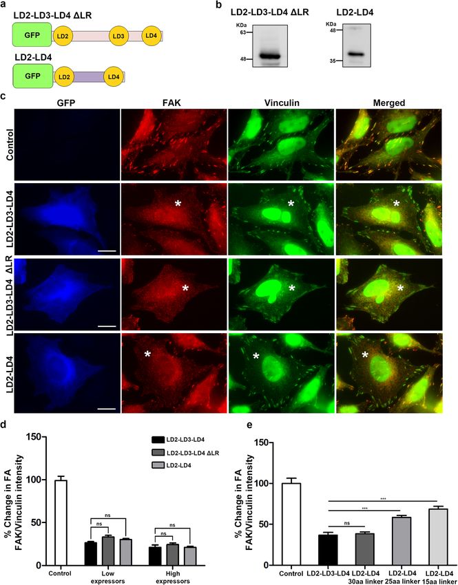
Antoniades et al. Cell Communication and Signaling   (2021) 19:3                                                    Page 4 of 22

10% FBS and 1X Antibiotic-Antimycotic. Transient trans-            Focal adhesion isolation
fections with Lipofectamine 2000 (Invitrogen) were per-            The isolation of focal adhesion complexes was based on
formed according to manufacturer’s instructions. Cells             the protocol of Jones et al. (2015) with some modifica-
were observed 24 h after transfection to verify expression         tions [42]. Briefly, cells were plated on 10-cm cell culture
and then used for subsequent experiments.                          dishes and grown for 48 h (10 plates per condition).
                                                                   Cells were fixed with 1% formaldehyde for 5 min at
                                                                   room temperature and formaldehyde was quenched with
GFP and GFP LD2-LD4 stable cell line generation
                                                                   0.125 M glycine for 10 min. Cells were washed with ice
For the production of infectious viral particles, HEK 293
                                                                   cold PBS and incubated with modified RIPA buffer for 5
T cells were transfected (calcium phosphate) with
                                                                   min on ice. Cell bodies were removed with high-
lentiviral plasmids encoding either a) reverse
                                                                   pressure water and the remaining focal adhesion were
tetracycline-controlled     trans-activator    (rtTA-N144)
                                                                   collected by scraping in Laemmli sample buffer.
(Addgene), or b) GFP LD2-LD4 (pLV-tetO-LD2-LD4) or
c) GFP (pLenti-CMV-GFP-Hygro) (Addgene), together
with the packaging (pCMV-dR8.91) and envelope                      Cell Lysis, Western blot and Immunoprecipitation
(pCMV-VGV-G) plasmids. Viral supernatants were col-                For western blot analysis cells were rinsed with ice-cold
lected after 48 and 72 h, filtered using a 0.45 mm syringe         PBS and lysed in RIPA buffer supplemented with
and stored at 4ο C.                                                protease inhibitors (2x Halt Protease inhibitor cocktail,
  GFP Hela cells were generated by transduction of                 Thermo Scientific) and sodium orthovanadate (5 mM,
pLenti-CMV-GFP-Hygro, while GFP LD2-LD4 Hela                       Sigma) for 10 min. Lysates were cleared by centrifuga-
cells were generated by co-transduction of pLV-tetO-               tion (15,000 g, 4 °C for 10 min) and 40 μg of extracted
LD2-LD4 and rtTA-N144 viral supernatants, in the pres-             protein were used for Western blot analysis as previously
ence of 10 μg/ml Polybrene (Merck Millipore). 10 μg/ml             described [34].
doxycycline was added for 48 h to induce GFP-LD2-LD4                 For GFP or FLAG pull-down, cells from two 10-cm
expression [41]. Cells were allowed to recover for 72 h            plates for each condition, were lysed in 1.5 ml lysis buf-
prior to selection with hygromycin B (Sigma-Aldrich)               fer (20 mM Tris pH 7.5, 150 mM NaCl, 1% v/v NP40)
(400 μg/ml for 10 days).                                           supplemented with protease inhibitors and sodium
                                                                   orthovanadate. Cleared protein extracts were gently
                                                                   mixed with 5 μl of GFP-Trap Agarose beads (Chromotek)
Antibodies
                                                                   or 30 μl anti-FLAG M2-Agarose affinity gel (Sigma Al-
Antibodies used for western blot analysis: mouse anti-
                                                                   drich), overnight at 4 °C. The beads or gel were washed 3
GFP (1:1000; Proteintech, #50430-2-AP), rabbit anti-
                                                                   times with lysis buffer and then resuspended in 50 μl 2x
pFAK Y576 (1:200; Santa Cruz, #sc-16563), rabbit anti-
                                                                   Laemli sample buffer (Bio-Rad) supplemented with 2-
pFAK Y397 (1:200; Novus, #NBPI-60837), rabbit anti-
                                                                   mercaptoethanol (Fluka). Samples were boiled for 10 min
pPaxillin Y31 (1:200; Santa Cruz, #sc-14035), mouse
                                                                   at 95 °C and the supernatant containing the immunocom-
anti-FAK (1:1000; Proteintech, #66258-I-1g), mouse
                                                                   plexes was collected by centrifugation (2500 g, 4 °C for 5
anti-Paxillin (1:5000; BD Biosciences, #610051), rabbit
                                                                   min) and analysed by western blot.
anti-FLAG (1:1000; Proteintech, #80010-1-RR).
                                                                     For FAK immunoprecipitation, five 10-cm plates of
  Antibodies used in immunofluorescence experiments:
                                                                   GFP, uninduced GFP LD2-LD4 or induced LD2-LD4
mouse anti-FAK 4.47 (1:1000; Millipore, #05-537),
                                                                   cells were lysed in 1.5 ml lysis buffer (as described
mouse anti-FAK (1:1000; Proteintech, #66258-I-1g),
                                                                   above). Protein extracts were incubated with 10 μg of
rabbit anti-FAK C-20 (1:200; Santa Cruz, #sc-558),
                                                                   anti-FAK antibody (Proteintech), overnight at 4 °C (ex-
rabbit anti-pFAK Y576 (1:200; Santa Cruz, #sc-16563),
                                                                   tracts of uninduced GFP LD2-LD4 cells incubated with
rabbit anti-pFAK Y397 (1:200; Novus, #NBPI-60837)
                                                                   no antibody were used as a negative control.). Extracts
goat anti-Talin C-20 (1:750; Santa Cruz, #sc-7534),
                                                                   were then incubated with 30 μl Protein-G Sepharose CL-
mouse anti-Paxillin (1:750; R&D Systems, #AF4259),
                                                                   4B beads (GE Healthcare) for 2 h at 4 °C. Beads were
mouse anti-Vinculin (1:1000; Sigma, #V9131), rabbit
                                                                   washed 5 times with lysis buffer and boiled in 40 μl 2x
anti-Vinculin (1:2000; Proteintech, #26520-I-AP), mouse
                                                                   Laemli sample buffer and analysed by western blot.
anti-av Integrin H-2 (1:100; Santa Cruz, #sc-376156),
rabbit anti-pPaxillin Y31 (1:200; Santa Cruz, #sc-14035),
mouse anti-p-Tyr (pY20) (1:200; Santa Cruz, #sc-508),              Inhibitor treatment
rat anti-active β1-Integrin 9EG7 (1:700; BD Bioscences,            Cells were incubated at 37 °C with either 50 μM
#550531), mouse anti-p130 Cas 35B.1A4 (1:500; Santa                Chloropyramine hydrochloride (C4) inhibitor (Santa
Cruz, #sc-20029), mouse anti-Tensin C-2 (1:1000; Santa             Cruz Biotechnology) for 24 h, or 10 μM PF228 inhibitor
Cruz, #sc-376367).                                                 (Santa Cruz Biotechnology) for 3 h.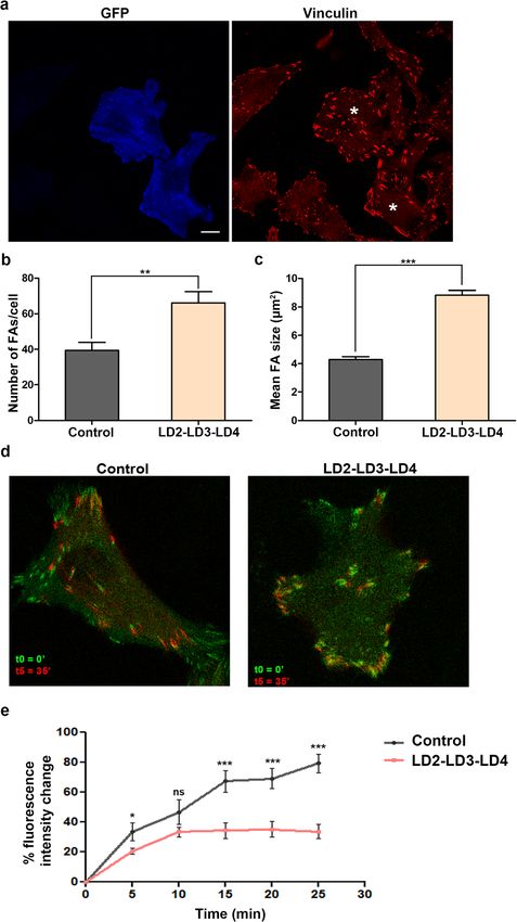
Antoniades et al. Cell Communication and Signaling   (2021) 19:3                                                   Page 5 of 22

Immunofluorescence                                                 Plan-Apochromat10x/0.4 Ph1 objective. Western blot
Cells (~ 7 × 10⁴) seeded on HCl-charged glass coverslips           imaging was performed using a UVP Biospectrum im-
(15 mm), were PFA or methanol/acetone fixed and im-                aging system.
munostained as described elsewhere [40]. Briefly, cells              Image analysis was performed using Zen 2010 and
were rinsed with cold PBS and fixed with 4% PFA for 10             Axiovision 4.8 software (Carl Zeiss AG, Germany) and
min, followed by quenching with 10 mM Glycine                      Adobe Photoshop (Adobe Inc., San Jose CA) to adjust
(Sigma) for 10 min and permeabilization with 0.2% Tri-             brightness and contrast. Figures were constructed with
ton X-100 (Bio-Rad) for 10 min. Alternatively cells were           Adobe Photoshop.
fixed with cold methanol/acetone (1:1) for 20 min at −
20 °C. Cells were then blocked with 10% donkey serum               Quantification
(Jackson Immumoresearch) in 1X PBS for 30 min and                  VisionWorks software (UVP LLC, CA) was used for the
incubated with primary antibodies for 1 h. This was                quantification of western blot results using raw data
followed by several washes in 1X PBS and incubation                from non-processed images for densitometry analysis.
with secondary antibodies for 1 h. Cells were thoroughly              Imaris image analysis software (Oxford Instruments,
washed with 1X PBS and mounted in ProLong Diamond                  UK) and Zen 2010 software (Carl Zeiss AG, Germany)
antifade mountant (Molecular Probes).                              were used for the quantification of fluorescence inten-
                                                                   sities. Classification was according to GFP expression
Invasion assay                                                     levels and was performed by determining the median
25–30,000 MDA MB-231 cells, transiently transfected                GFP intensity of expressing cells in each experiment and
with LD2-LD3-LD4, were resuspended in a solution of                comparing this to the GFP intensity of individual cells
30% Matrigel (Corning), 0.02% Hoechst (Invitrogen) and             from the same experiment. Non-expressing cells were
1% FBS in DMEM (final volume: 20 μl) and placed as a               used as negative controls and were intrinsic to each ex-
droplet on a rectangular chambered coverslip                       periment, since transient transfections never lead to
(24x60mm), previously treated with organosilane                    100% transfection efficiency.
(RainX). A round 10 mm coverslip, with 2 mm spacers                   To quantify FA-localization of specific proteins, cells
was placed on top, flattening the droplet into a round             were stained for the protein of interest as well as an add-
disc. The Matrigel was allowed to set in a humidifying             itional FA marker, shown to retain its localization upon
chamber (37 °C) for 30 min. A second layer of 30%                  LD2-LD3-LD4 expression (i.e. vinculin or talin). Individ-
Matrigel (with 5% FBS) containing 0,02% fluorescent                ual FAs were automatically selected, based on the stain-
beads (Molecular Probes) (final volume: 50 μl), was                ing of the unaffected FA marker, and the mean cytosolic
added under the round coverslip, to occupy the area sur-           intensity of each protein was subtracted from that on in-
rounding the disc and allowed to set as described above.           dividually selected FAs, thus enabling specific protein
The chambered rectangular coverslip was then filled                quantification on individual FAs. To quantify the
with DMEM (containing 10% FBS). The setup was used                 localization of the Src SH2 domain at FAs, cells were
for time-lapsed imaging of cell invasion over a period of          transfected with GFP Src dSH2 alone or together with
48–72 h. Alternatively, it was used to obtain static im-           mCherry LD2-LD3-LD4. Cells were stained for Vinculin
ages after 48 or 72 h.                                             and individual FAs were selected based on Vinculin
                                                                   staining. FA localization was then assessed as de-
Imaging                                                            scribed above. For the quantification of FA turnover
Confocal imaging was performed on a Zeiss LSM 710                  rates, time-lapse images of cells expressing RFP Vin-
laser scanning confocal microscope (Carl Zeiss AG,                 culin (used as an FA marker) were aligned in ImageJ
Germany), with a Plan-Apochromat 63x/1.40 oil DIC                  (NIH, USA) and analyzed. Individual FAs were manu-
immersion objective using lasers 488 nm, 543 nm and                ally selected (ROI) at time point t0 and the mean
633 nm. Super-resolution imaging was performed on a                fluorescence intensity of each ROI was documented
Zeiss LSM 900 laser scanning confocal microscope with              for each time point of the recording (t0 = 0 min, t1 =
Airyscan 2 (Carl Zeiss AG, Germany) with a Plan-                   5 min, t2 = 10 min, t3 = 15 min, t4 = 20 min, t5 = 25
Apochromat 63x/1.40 oil DIC immersion objective using              min). The mean cytosolic intensity for each time
lasers 475 nm, 555 nm, 630 nm. Widefield fluorescence              point was subtracted from that of each ROI.
imaging was performed on a Zeiss Axio Imager Z1                       Quantification of cell migration rates was performed
microscope (Carl Zeiss AG, Germany), with a Plan-                  using the Imaris image analysis software and performing
Apochromat 63x/1.40 oil Ph3 immersion objective. Live              automatic tracking of individual cells. The migration ef-
cell migration imaging was performed on a Zeiss Axio               ficiency was calculated as the percentage change of the
Imager Z1 microscope and an Axiovert 200 motorized                 migration rate of GFP expressing cells, compared to that
inverted microscope (Carl Zeiss AG, Germany), with a               of non-expressing control cells.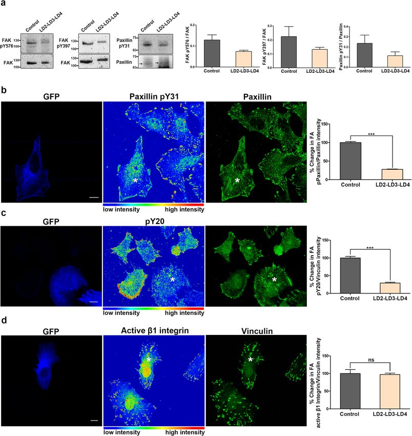
Antoniades et al. Cell Communication and Signaling   (2021) 19:3                                                   Page 6 of 22

  Quantification of cell invasion efficiency was per-              included in the construct [46]. We therefore generated a
formed with the Imaris image analysis software, using              construct containing LD2-LD3-LD4 and intermediate
static images of cells at the end of 72 h. Total cell num-         linking regions, fused to GFP, hereunto referred to as
bers were determined by automatic selection of individ-            LD2-LD3-LD4 (Fig. 1a). This construct led to expression
ual nuclei (stained with Hoechst). LD2-LD3-LD4                     of a stable protein, at the expected molecular weight,
expressing cell numbers were determined by automatic               which localized primarily in the cytosol (Fig. 1b and c).
selection of GFP positive cells. Control cell numbers                 We went on to examine if LD2-LD3-LD4 interacted
were determined by subtracting GFP positive cell num-              with FAK, in co-immunoprecipitation experiments,
bers from total cell numbers. Invading cell numbers                using extracts of HeLa cells transiently transfected with
were determined by automatic detection of cells within a           GFP LD2-LD3-LD4 or GFP (negative control). As shown
specific ROI. ROIs included areas defined by a fluores-            in Fig. 1d, a band corresponding to co-precipitated FAK
cently delineated boundary between a fluorescently-                (at 125kD), was detected only in the precipitates from
labeled low-serum concentration gel and a non-labeled              cells expressing GFP LD2-LD3-LD4, showing that LD2-
high-serum concentration gel. The invasion efficiency              LD3-LD4 specifically interacts with FAK, as expected.
was calculated as the percentage of cells that traversed           Overall, these experiments show that LD2-LD3-LD4 in-
the boundary.                                                      teracts with FAK directly and given its cytosolic
                                                                   localization it could potentially prevent FAK localization
Statistical analysis                                               at FAs.
Graph generation and statistical analysis were performed
using Prism software (GraphPad, San Diego, CA). All                Expression of LD2-LD3-LD4 leads to the specific and
graph data are shown as mean values while error bars               dose-dependent displacement of FAK from FAs
represent S.E.M. Statistical analysis was performed using          In order to examine if LD2-LD3-LD4 expression could
two-tailed unpaired t-tests with 95% confidence interval.          specifically disrupt FAT domain interactions and
For experiments examining FAs, all statistical analysis            displace FAK from FAs, HeLa cells were transiently
was performed on total FA numbers from all cells con-              transfected with LD2-LD3-LD4, seeded on FN coated
sidered. All experiments have been performed at least 3            coverlips for two hours, fixed and immuno-stained for
times.                                                             FAK and Talin. Talin was selected as a stable marker of
                                                                   mature FAs, given the fact that its recruitment to the
Results                                                            complex relies on direct binding to β integrin cytoplas-
Using the Paxillin LD motifs to interfere with interactions        mic tails. As shown, expression of LD2-LD3-LD4 led to
targeting FAK to FAs, as a strategy to inhibit FAK                 the clear displacement of FAK from FAs, while Talin
Our main aim was to develop and test a new strategy                localization was unaffected (Fig. 2a). This effect was con-
that would block both enzymatic and scaffolding func-              firmed using a second FAK antibody and, in addition,
tions of FAK, specifically at FAs, as a possible new ap-           exogenous mKate FAK (Additional File 2: Fig. S1 a-c).
proach for FAK inhibition. To achieve this, we set out to          We quantified this displacement, by calculating the ratio
interfere with interactions responsible for FA targeting.          of FAK in the cytosol to FAK at FAs, revealing that
The FAT domain is both necessary and sufficient to                 LD2-LD3-LD4 expression led to a 4-fold reduction of
drive FAK at FAs. Previous work from our group and                 FA-localized FAK (Fig. 2b). Interestingly, a similar quan-
others revealed that the two hydrophobic pockets (HPs)             tification for Talin showed that LD2-LD3-LD4 expres-
formed by the FAT domain are essential for FA targeting            sion leads to an increase in FA localized Talin, possibly
[33, 34, 43]. The best characterized interaction of the            due to enlargement of the FA complexes (Fig. 2c). In
HPs is with the LD2 and LD4 motifs of Paxillin [30, 35].           order to account for this, we calculated the FAK to Talin
We postulated that a peptide encoding these LD motifs,             ratio at FAs, which revealed a dramatic 5-fold reduction
but lacking FA targeting sequences (LIM domains),                  in LD2-LD3-LD4 expressing cells, suggesting that LD2-
would interfere with interactions responsible for FAK              LD3-LD4 is very effective in displacing FAK from FAs
FA targeting. We also took into account the fact that              (Fig. 2d), unlike expression of GFP, which was used as a
several cancer-linked Paxillin mutations have been                 negative control (Additional File 2: Fig. S1d). We then
mapped to the intrinsically disordered regions between             examined how the levels of LD2-LD3-LD4 affected dis-
LDs and not on the motifs themselves, such as P30S,                placement efficiency, and revealed a clear dose response
G105A and A127T that lie between LD1 and LD2 and                   relationship; in cells expressing relatively high levels of
P233L and T255I that lie between LD3 and LD4 [44,                  GFP, we observed complete loss of FAK from FAs while
45]. Given the significance of this intermediate linking           in cells expressing moderate or low levels of GFP, we
region, in LD interactions with binding partners and in            could still detect FAK at FAs, albeit at significantly
LD scaffolding functions, we decided that it should be             reduced levels (Fig. 2e).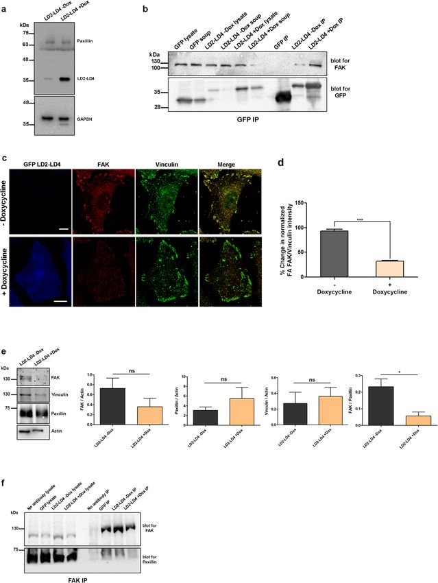
Antoniades et al. Cell Communication and Signaling        (2021) 19:3                                                                  Page 7 of 22

 Fig. 1 LD2-LD3-LD4 interacts with FAK directly a) Schematic representation of the LD2-LD3-LD4 polypeptide, composed of amino acids 54–279
 of Paxillin, including the LD2-LD3-LD4 motifs and fused to GFP. b) Representative Western Blot (using anti-GFP) showing expression of a stable
 protein encoding GFP-fused LD2-LD3-LD4, in HeLa cells (expected molecular weight ~ 51 kDa). c) Confocal images of PFA-fixed HeLa cells
 expressing GFP LD2-LD3-LD4. The protein is primarily localized in the cytosol (Scale bar: 10 μm). d) Western blots showing immunoprecipitated
 GFP (left) and GFP LD2-LD3-LD4 (right), blotted for GFP and FAK. Co-precipitation of FAK (125 kDa) is only observed in HeLa cells co-expressing
 GFP LD2-LD3-LD4

  Fluorescent proteins, despite mutations to reduce their                  LD4 is sufficiently stable and functional in the absence
ability to dimerize, still maintain some capacity to do so.                of a large globular protein.
Additionally, given their globular nature and relatively                     A previously generated inhibitor of the interaction of
large size (27kD) they tend to stabilize fused peptides.                   FAK with VEGFR3 (C4), was also reported to displace
Furthermore, GFP displays inherent accumulation to the                     FAK from FAs [47]. This interaction, as characterized by
nucleus and could thus be influencing peptide function,                    docking studies, takes place through binding of C4 to
by affecting cellular distribution. To ensure that the                     His 1025 on Helix 4 of the FAT domain of FAK, adja-
LD2-LD3-LD4 peptide is stable and can be used effect-                      cent to HP1, to which it may sterically hinder access
ively in the absence of GFP, we generated a FLAG-                          [17]. We thus decided to compare the efficiency of C4
tagged peptide which is much smaller in size (1kD). As                     with that of LD2-LD3-LD4, to displace FAK from FAs.
shown in Fig. S1e and S1f, the FLAG-tagged peptide can                     We examined the distribution of FAK and Talin in HeLa
efficiently interact with FAK and lead to effective dis-                   cells, following treatment with high concentrations of
placement from FAs., This confirms that the LD2-LD3-                       C4 (50 μM) for 48 h. Surprisingly, C4, failed to visibly
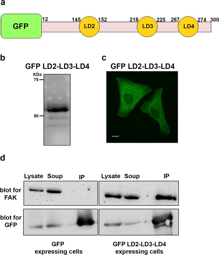
Antoniades et al. Cell Communication and Signaling   (2021) 19:3   Page 8 of 22

 Fig. 2 (See legend on next page.)
Antoniades et al. Cell Communication and Signaling           (2021) 19:3                                                                      Page 9 of 22

 (See figure on previous page.)
 Fig. 2 Expression of LD2-LD3-LD4 leads to the dose-dependent displacement of FAK from FAs without affecting overall FA composition a)
 Confocal images of methanol/acetone fixed HeLa cells, transiently transfected with GFP LD2-LD3-LD4 and immunostained for FAK and Talin. In
 control cells, FAK strongly localizes at FAs, labeled with Talin. In cells expressing LD2-LD3-LD4 (marked with asterisk) there is no detectable
 enrichment of FAK at FAs. In contrast, Talin localization at FAs is unaffected. b-c) Quantification of the % change in the mean FA/cytosolic FAK (b)
 and Talin (c) intensity ratios. FA/Cytosolic ratio of FAK in control cells is ~ 4.2 fold higher (100 ± 2.86, n = 469 FAs from 30 cells) compared to cells
 expressing GFP LD2-LD3-LD4 (23.78 ± 1.25, n = 409 FAs from 30 cells), indicating that LD2-LD3-LD4 leads to the displacement of FAK from FAs. In
 contrast, the FA/cytosolic ratio of Talin is higher in cells expressing GFP LD2-LD3-LD4, possibly due to increased FA size (109.7 ± 3.22 in
 expressing, compared to 100 ± 2.59 in controls). d) Quantification of the % change in the mean FAK/Talin intensity (based on b and c) reveals a
 ~ 5-fold drop of FA localized FAK in cells expressing GFP LD2-LD3-LD4 (19.01 ± 0.91 in expressing, compared to 100 ± 4.8 in control cells). e)
 Dose-dependent displacement of FAK from FAs as indicated by quantification of the % change in the mean FAK/Talin intensity in control
 compared to expressing cells (high and low) (15.55 ± 1.25, n = 188 FAs from 13 cells, expressing higher, and 28.52 ± 1.14, n = 218 FAs from 17
 cells, expressing lower amount of GFP LD2-LD3-LD4, compared to 100 ± 3.1, n = 229 FAs from 28 control cells). To discriminate between high and
 low expressing cells, we initially determined the mean GFP intensity of all expressing cells, and then compared this to the GFP intensity of
 individual cells, so as to classify them as high or low expressors. (f-i) Confocal images of PFA-fixed control and GFP LD2-LD3-LD4 (marked with
 asterisk) HeLa cells immunostained for Vinculin (f), Paxillin (g), av. Integrin (h) and Tensin (i) showing that localization of these proteins is not
 affected by the expression of GFP LD2-LD3-LD4. (j-m) Corresponding quantification of the % change in the mean FA/cytosolic intensity for each
 protein presented in f-i. FA/cytosolic ratio of Vinculin (j) and Paxillin (k) is higher in cells expressing GFP LD2-LD3-LD4, possibly due to increased
 FA size (111.8 ± 2.86, n = 460 FAs from 30 expressing, compared to 100 ± 2.57, n = 455 FAs from 30 control cells for Vinculin; 107.5 ± 2.29, n = 402
 FAs from 30 expressing, compared to 100 ± 2.64, n = 432 FAs from 30 control cells for Paxillin). There is no significant difference in the FA/
 cytosolic ratio of av. Integrin (l) and Tensin (m) (102.5 ± 1.94, n = 568 FAs from 30 expressing, compared to 100 ± 2.16, n = 485 FAs from 30 control
 cells for av. Integrin; 105 ± 2.24, n = 532 FAs from 30 expressing, compared to 100 ± 2.09, n = 568 FAs from 30 control cells for Tensin). Scale bars:
 10 μm. The error bars represent standard error of the mean (S.E.M). ***; p < 0.0001, **; p < 0.005, *; p < 0.05

displace FAK from FAs, compared to controls. Quantifi-                         from FAs, we went on to address its effects on FAK acti-
cation of FAK to Talin signal ratios on FAs of control                         vation. To do so, we examined the phosphorylation state
and treated cells, confirmed that C4 did not affect FAK                        of a) Tyr397, the major FAK auto-phosphorylation site
FA localization. However, after inhibitor treatment,                           required for activation, b) Tyr576, which resides in the
some cells appeared to have smaller FAs, with low FAK                          activation loop of the kinase domain and has been
and Talin signals, because they were detaching from the                        shown to lead to full activation upon phosphorylation
substrate. These data show that C4 does not specifically                       and c) paxillin Tyr31, one of the major FAK/Src down-
block FAK targeting to FAs and suggest that in order to                        stream targets [37, 48]. As shown, LD2-LD3-LD4
efficiently displace the protein, disruption of interactions                   expression led to a significant reduction of phosphoryl-
taking place at the HPs is necessary (Additional File 2:                       ation at these sites, suggesting that LD2-LD3-LD4
Fig. S1g).                                                                     expression blocks FAK activation and downstream sig-
   Given the potent displacement of FAK from FAs in-                           naling (Fig. 3a). Importantly, this reduction becomes
duced by LD2-LD3-LD4, we wanted to examine the spe-                            even more significant, since transient transfection effi-
cificity of this effect and possible consequences on FA                        ciency is never 100% and thus what we observe repre-
composition. We therefore examined the localization of                         sents an underestimation of the effect. In order to
additional core FA proteins including Integrins (av), Pax-                     examine the effects of LD2-LD3-LD4 on FAK phosphor-
illin, Tensin and Vinculin. As shown, similarly to Talin,                      ylation in individual cells, we carried out Immunofluor-
FA localization of these proteins was not reduced by                           escence (IF) using phospho-specific antibodies. As
LD2-LD3-LD4 expression, suggesting that the effect of                          shown, LD2-LD3-LD4 expression led to a dramatic drop
LD2-LD3-LD4 is specific to FAK and that the compos-                            in FAK phosphorylation (on Tyr397) at FAs, suggesting
ition of FA complexes is broadly unaltered in expressing                       that it effectively eliminates FAK activation at these
cells (Fig. 2f-m). Overall, these data provide evidence                        complexes (Additional File 2: Fig. S1h).
that LD2-LD3-LD4 could serve as an effective, site-                              One of the best characterized downstream targets of
specific inhibitor of interactions at the HP sites within                      FAK is Paxillin, which becomes phosphorylated on Ty-
the FAT domain of FAK and prevent FAK localization                             rosines 31 and 118, in response to integrin activation in
at FAs in a dose-dependent manner.                                             wild type but not in FAK null cells [49]. We thus went
                                                                               on using quantitative immunofluorescence and calcu-
LD2-LD3-LD4 inhibits both kinase-dependent and                                 lated the ratio of phosphorylated-Paxillin (pPax) to
scaffolding functions of FAK at FAs                                            Paxillin, in order to assess the effects of LD2-LD3-LD4
FAK is a major transducer of integrin signaling and be-                        on Paxillin phosphorylation, specifically at FAs. As
comes phosphorylated and activated in response to                              shown, expression of LD2-LD3-LD4 led to a significant
integrin-dependent adhesion. Given that LD2-LD3-LD4                            reduction of the levels of pPax, suggesting that it not
interacts with FAK directly, leading to its displacement                       only blocks FAK activation but also downstream
Antoniades et al. Cell Communication and Signaling (2021) 19:3 Page 10 of 22 Fig. 3 Expression of LD2-LD3-LD4 blocks kinase-dependent functions of FAK, downstream of integrin activation a) Representative Western Blots and quantification from control cells and cells expressing GFP LD2-LD3-LD4, indicating the phosphorylation status of FAK Tyr 397 and Tyr 576 and Paxillin Tyr 31. Quantification of the ratio of phosphorylated FAK over total FAK shows reduction of the phosphorylation at both Tyr 576 (0.13 ± 0.024 in control, 0.074 ± 0.005 in GFP LD2-LD3-LD4 expressing samples) and Tyr 397 (0.22 ± 0.0073 in control, 0.13 ± 0.015 in GFP LD2-LD3- LD4 expressing samples). Paxillin Tyr 31 phosphorylation is also reduced, as indicated by the ratio of phosphorylated over total paxillin (0.24 ± 0.08 in control, 0.11 ± 0.04 in GFP LD2-LD3-LD4 expressing samples). b) Confocal images of PFA-fixed HeLa cells transfected with GFP LD2-LD3-LD4 and immunostained for Paxillin and phosphorylated Paxillin (pY31). Phosphorylated Paxillin signal in control and GFP LD2-LD3-LD4 expressing cells (marked with asterisk) is presented in the middle panel as an intensity color-coded image. Quantification of the % change in the mean intensity of phosphorylated (pPaxillin) to total Paxillin reveals significant reduction of Paxillin phosphorylation at FAs in cells expressing GFP LD2- LD3-LD4 (27.93 ± 0.83, n = 331 FAs from 24 cells) compared to control cells (100 ± 2.92, n = 371 FAs from 25 cells). c) Confocal images of PFA-fixed HeLa cells transfected with GFP LD2-LD3-LD4, immunostained against phosphorylated tyrosine (pY20) and Vinculin. Intensity of tyrosine phosphorylation in control and GFP LD2-LD3-LD4 expressing cells (marked with asterisk) is presented in the middle panel in a color-coded image. Quantification of the % change in the mean pY20/Vinculinintensity reveals a 3.4-fold decrease in total phosphorylation at FAs in cells expressing GFP LD2-LD3-LD4 (29.29 ± 2.12, n = 414 FAs from 30 cells) compared to control cells (100 ± 4.44, n = 435 FAs from 30 cells). d) Confocal images of PFA-fixed cells transfected with GFP LD2-LD3-LD4 and immunostained against active β1 Integrin and Vinculin. Active β1 Integrin signal in control and GFP LD2-LD3-LD4 expressing cells (marked with asterisk) is presented in the middle panel as an intensity color-coded image. Quantification of the % change in the mean active β1 Integrin/Vinculin intensity shows that expression of GFP LD2-LD3-LD4 does not affect integrin activation at FAs (100 ± 11.18, n = 346 FAs from 30 control, compared to 97.10 ± 3.98, n = 457 FAs from 30 expressing cells). Scale bars: 10 μm. The error bars represent standard error of the mean (S.E.M). ***; p < 0.001 signaling from FAs (Fig. 3b). In agreement with this re- displacement (Fig. 3c). Given the dramatic reduction of sult, staining of LD2-LD3-LD4 expressing cells with a tyrosine phosphorylation at FAs, we examined whether well characterized pY antibody (pY20) revealed that LD2-LD3-LD4 somehow prevents integrin activation. overall tyrosine phosphorylation is dramatically reduced Quantification of the ratio of active Integrin β1 to Vin- at FAs, suggesting that signaling is impaired due to FAK culin at FAs showed that LD2-LD3-LD4 has no effect on
Antoniades et al. Cell Communication and Signaling   (2021) 19:3                                                  Page 11 of 22

integrin activation (Fig. 3d). Therefore, the above data           formed a characteristic pattern of FAs, mainly found at
clearly show that LD2-LD3-LD4 expression blocks FAK                the cell periphery. In contrast, LD2-LD3-LD4 expressing
kinase-dependent signal transduction events, down-                 cells displayed a significant increase in both the number
stream of integrin activation.                                     and size of FAs with prominent ventral FAs (Fig. 5b and
  Upon recruitment at FAs, FAK is auto-phosphorylated              c). This result, suggests a defect in FA turnover and is
on Tyr397, creating a high-affinity binding site for the           consistent with previous findings in FAK −/− cells [53].
SH2 domain of Src, which further phosphorylates FAK                We went on to directly examine the effects of LD2-LD3-
on Tyr576 and Tyr577 within the activation loop, lead-             LD4 expression on FA turnover. HeLa cells were trans-
ing to maximal enzymatic activity [50]. In order to                fected with RFP-Vinculin alone or co-transfected with
examine the effects of LD2-LD3-LD4 expression on                   RFP-Vinculin and LD2-LD3-LD4, seeded on fibronectin-
FAK-mediated Src recruitment to FAs we expressed the               coated chambered slides and time-lapse sequences were
SH2 domain of Src fused to GFP (GFP Src_dSH2), previ-              recorded, over a period of 35 min. Cells expressing the
ously shown to be necessary and sufficient for FA target-          construct displayed markedly slower FA turnover com-
ing of Src [51]. As expected, given the previous data              pared to control cells (Fig. 5d and e). Therefore, these
indicating FAK FA displacement and abolishment of                  data show that LD2-LD3-LD4 expression elicits defects
Tyr397 phosphorylation, expression of LD2-LD3-LD4                  in FA turnover, leading to the appearance of more and
led to a significant reduction in Src_dSH2 FA                      larger FAs, in a similar manner to defects reported in
localization, indicating an inability of Src to target FAs         FAK null fibroblasts [53].
(Fig. 4 a and b).                                                     Given the central role of FA turnover in cell migration,
  FAK also has well-established scaffolding functions, in-         we decided to examine how LD2-LD3-LD4 affected cell
cluding a kinase independent role in the recruitment of            spreading and migration. Control and LD2-LD3-LD4 ex-
the FAK-Src substrate, p130Cas to FAs [27]. This is                pressing HeLa cells were seeded on fibronectin-coated
achieved through an SH3-dependent interaction with                 coverslips and monitored using time-lapse video micros-
the C terminal proline-rich regions of FAK [27]. In order          copy over a period of 16 h. We used a motorized stage
to determine if, unlike kinase inhibitors, LD2-LD3-LD4             to image multiple areas simultaneously, so as to record
could also suppress kinase-independent, scaffolding                and track large numbers of cells. Analysis of the record-
functions, we examined p130Cas localization. LD2-LD3-              ings revealed that LD2-LD3-LD4 elicited dose-
LD4 expressing and control cells, as well as cells treated         dependent defects in both cell spreading and migration
with a previously characterized FAK kinase inhibitor               (Fig. 6a and b and Additional File 3: Movie S1). In
(PF228) [52], were immunostained for Talin and                     addition, analysis of the time-lapse images revealed that
p130Cas. There was a visible reduction of FA-localized             cells expressing high levels of LD2-LD3-LD4, displayed
p130Cas in LD2-LD3-LD4 expressing cells, unlike con-               slightly increased apoptosis (16,9% compared to 6,2% in
trol and PF228-treated cells in which no change was ob-            control cells). Similar effects were observed in other
served as confirmed by quantification of the ratio of              highly migratory and metastatic cell lines, namely MDA
p130Cas to Talin (Fig. 4c and d). As expected PF228                MB-231 (Additional File 2: Fig. S3a), H460 (Additional
treatment led to a clear reduction of tyrosine phosphory-          File 2: Fig. S3b) and HCT-116 (Additional File 2: Fig.
lated FAK at FAs but did not interfere with its                    S3c), in which FAK is effectively displaced from FAs,
localization; thus, as expected, p130Cas is maintained at          upon expression of LD2-LD3-LD4 (Additional File 2:
the complex (Additional File 2: Fig. S2a-c). The above             Fig. S3d-f). Overall, these data show that LD2-LD3-LD4,
results show that unlike inhibitors of FAK’s enzymatic             not only elicits defects in cell spreading and FA turnover,
activity, expression of LD2-LD3-LD4 blocks both kinase-            consistent with phenotypes observed in FAK null cells,
dependent and independent functions at FAs.                        but is also an effective inhibitor of two-dimensional (2-D)
                                                                   cell migration.
Expression of LD2-LD3-LD4 affects FA dynamics, and                    Although active cell migration is a prerequisite for me-
inhibits migration and invasion of tumor cells                     tastasis, there is strong evidence suggesting that 3-D cul-
It is well established that FAK is a critical regulator of         ture and gel invasion assays better mimic the tumor
FA assembly and disassembly, processes that are funda-             microenvironment and predict therapeutic responses,
mental for efficient, directional cell migration [53–55].          in vivo, more accurately [56, 57]. To examine the effects
Given that expression of LD2-LD3-LD4 displaces FAK                 of LD2-LD3-LD4 on tumor cell invasion, we developed
from FAs, we initially examined whether this would                 a modified Boyden-chamber gel invasion assay, which al-
elicit changes in FA dynamics. For this purpose, we eval-          lows live and end-point evaluation of cell invasion and
uated the number and size of FAs in LD2-LD3-LD4 ex-                permits imaging, tracking and quantification of both in-
pressing vs control HeLa cells that were seeded on glass           vading and non-invading cells. The highly invasive MDA
coveslips for 12 h. As shown in Fig. 5a, control cells             MB-231 cells were used for these experiments and both
Antoniades et al. Cell Communication and Signaling (2021) 19:3 Page 12 of 22 Fig. 4 Expression of LD2-LD3-LD4 blocks kinase-independent, scaffolding functions of FAK at FAs a-b) Super resolution images and quantification of the FA-localization of GFP Src_dSH2 in control (GFP Src_dSH2 only) and mCherry LD2-LD3-LD4 expressing cells (marked with asterisk). Cells were fixed with PFA and immunostained for Vinculin. Expression of mCherry LD2-LD3-LD4 leads to 84.3% decrease in the Src_dSH2/Vinculin ratio compared to control cells (100 ± 6.68, n = 419 FAs from 24 control, 15.67 ± 0.93, n = 373 FAs in 22 mCherry LD2-LD3-LD4 expressing cells). c-d) Confocal images (c) and quantification (d) of control (top panel), LD2-LD3-LD4 expressing (intermediate panel) and PF228 treated (bottom panel) cells, fixed with methanol/acetone and immunostained for p130Cas and Talin. Expression of LD2-LD3-LD4 leads to a 68% decrease in the p130Cas/Talin ratio compared to control cells, whereas treatment with PF228 does not elicit any significant change (100 ± 3.56, n = 505 FAs from 33 control, 31.89 ± 2.46, n = 564 FAs from 35 LD2-LD3-LD4 expressing and 100.2 ± 5.6, n = 515 FAs from 30 PF228-treated cells). F Scale bars: 10 μm. The error bars represent standard error of the mean (S.E.M). ***; p < 0.0001
Antoniades et al. Cell Communication and Signaling          (2021) 19:3                                                                   Page 13 of 22

 Fig. 5 Expression of LD2-LD3-LD4 leads to an increase in number and size of FAs and a reduction of FA turnover a) Confocal images of PFA-fixed
 HeLa cells transfected with GFP LD2-LD3-LD4 and immunostained for Vinculin. Cells expressing GFP LD2-LD3-LD4 (marked with asterisk) form
 more and larger FAs, that are localized more ventrally. (b-c) Quantification of the total number of FAs per cell (b) and mean FA size (c), confirm
 significant increase of FA number (39.41 ± 4.48 FAs, n = 27 control cells compared to 66.17 ± 6.35 FAs, n = 29 GFP LD2-LD3-LD4 expressing cells)
 and size (4.29 ± 0.21 μm2, n = 526 FAs from 27 control cells, compared to 8.83 ± 0.33 μm2, n = 915 FAs from 29 GFP LD2-LD3-LD4 expressing cells)
 and. d) Overlayed confocal images from time lapse recordings, showing FA turnover in control and GFP LD2-LD3-LD4 expressing cells. Cells were
 imaged over a period of 25 min and FAs were visualized using RFP Vinculin. e) Quantification of the FA turnover rate assessed as the percentage
 change in the average intensity of selected FAs over time. FAs in GFP LD2-LD3-LD4 expressing cells display significantly slower turnover rates
 (33.77% ± 4.86 change at 25 min, n = 52 FAs) compared to control cells (79.39% ± 6.22change at 25 min, n = 38 FAs). **; p < 0.005, ***; p < 0.0001, *;
 p = 0.0118, ***; p ≤ 0.0005
Antoniades et al. Cell Communication and Signaling (2021) 19:3 Page 14 of 22
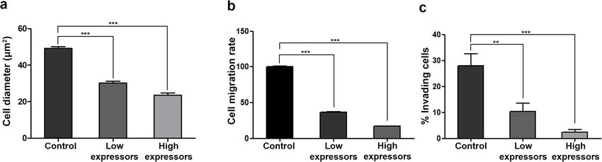
Fig. 6 LD2-LD3-LD4 expression inhibits tumor cell spreading, migration and invasion in a dose-dependent manner a) Expression of GFP LD2-LD3-
LD4 leads to dose-dependent defects in Hela cell spreading. Spread area is reduced by 38.4 and 52.2% in low-and high-expressing cells
respectively (49.16 ± 0.97 μm, n = 216 control cells, compared to 30.27 ± 0.92 μm, n = 102 low-expressing cells and 23.52 ± 1.28 μm, n = 67 high-
expressing cells). Cell spreading was calculated as a function of the diameter of the attached area on fibronectin coated coverslips, 1 h after
seeding. b) Expression of GFP LD2-LD3-LD4 leads to dose-dependent defects in migration of HeLa. The rate of migration is reduced by 63 and
83% in low and high-expressing cells respectively, following 16 h of recording (100 ± 1.32, n = 549 in control, compared to 36.62 ± 0.28, n = 263 in
low-expressing and 17.03 ± 0.2, n = 227 in high-expressing cells). c) Expression of GFP LD2-LD3-LD4 leads to a dose-dependent reduction in the
capacity of MDA-MB231 cells to invade Matrigel, as indicated by quantification, using Hoechst staining to detect all cells and GFP to determine
expressors. Invasion efficiency is reduced by 63 and 92% in low-and high-expressing cells respectively, following 72 h of incubation (27.95 ± 4.5%,
n = 22,718 control, compared to 10.39 ± 03.06%, n = 8950 low-expressing and 2.3 ± 1.02%, n = 5930 high-expressing cells). Invasion efficiency is
calculated as the percentage of cells traversing a fluorescently delineated boundary from a low- to high-serum concentration gel, in a
chemotactic gradient. To discriminate between high and low expressing cells, we initially determined the mean GFP intensity of all expressing
cells, and then compared this to GFP intensity of individual cells, so as to classify them as high or low expressors. ***; p < 0.0001, **; p < 0.005
end-point measurements, as well as time-lapse record- since it is well beyond the size limit for effective peptide
ings were generated with mixed populations of LD2- synthesis (100-120aa). Therefore, we decided to deter-
LD3-LD4 expressing and control cells, in the same mine the minimum paxillin sequences required for effi-
setup. As shown, LD2-LD3-LD4 expression, inhibited in- cient displacement of FAK from FAs. To this end we
vasion of MDA MB-231 cells in a dose-dependent man- implemented a subtractive approach, removing individ-
ner; in fact, high expression drastically reduced invasion ual linking regions in a stepwise fashion and assessing
of this highly metastatic cell line (Fig. 6c and Additional the activity of each construct.
File 4: Movie S2). These results show that displacement We initially deleted the region upstream of LD2 (LD1-
of FAK from FAs is an effective strategy to block both LD2 linking region-LR), previously reported to be
cell migration, as well as tumor cell invasion. It could necessary for optimal binding to FAK [23, 24, 30] and
therefore form the basis for the development of anti- examined how it affected the peptide’s capacity to dis-
metastatic drugs. place FAK from FAs (Fig. 7a). This construct led to ex-
pression of a stable polypeptide, at the expected
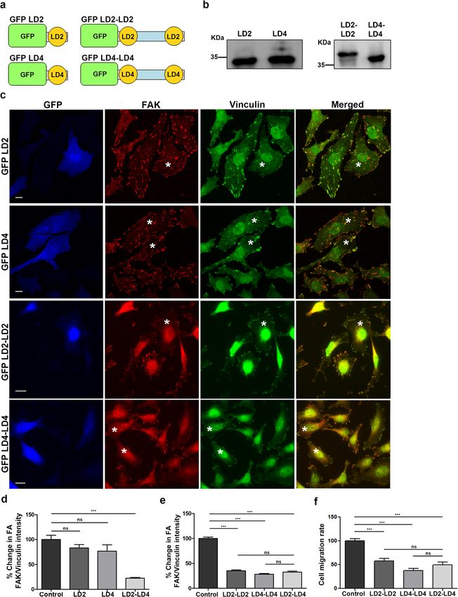
The LD2 and LD4 motifs are sufficient for effective FAK molecular weight, hereunto referred as LD2-LD3-LD4
displacement from FAs ΔLR (Fig. 7b). As shown, expression of LD2-LD3-LD4
Previous work revealed that LD2 and LD4 are respon- ΔLR led to clear displacement of FAK from FAs, while
sible for the interaction with the HPs of FAK [30, 35]. In Vinculin localization (used as an FA marker) was un-
the work described above, the construct used to displace affected as expected (Fig. 7c). Quantification of the FAK
FAK from FAs also contained LD3, as well as intermedi- to Vinculin ratio showed that LD2-LD3-LD4 ΔLR dis-
ate linking regions (Fig. 1a). This was initially deemed placed FAK with the same efficiency as the original pep-
necessary given the significant regulatory role assigned tide, suggesting that the linking segment upstream of LD2
to these unstructured linking segments for the FAK- does not play a pivotal role for efficient binding of the
paxillin interaction [44–46]. However, these regions also LD2 and LD4 motifs to the FAT HPs in the cell (Fig. 7d).
bare numerous phosphorylation sites and binding sites Next, we went on to examine whether the intermedi-
for proteins other than FAK, thus their presence would ate linking region between LD2 and LD4, containing the
be expected to be detrimental to the specificity of the LD3 motif, plays a role in the ability of the polypeptide
polypeptide and lead to off target effects. In addition, the to displace FAK. Using the DNA encoding for LD2-
large size of the polypeptide containing these regions LD3-LD4 ΔLR as template, we replaced the region be-
(24 kDa, 226aa) poses restrictions in its potential use as tween the LD2 and LD4 motifs with a flexible standard
a metastatic inhibitor, in the form of a synthetic peptide linker (GGGGS). Optimization of the length of theAntoniades et al. Cell Communication and Signaling (2021) 19:3 Page 15 of 22 Fig. 7 The LD2 and LD4 motifs of paxillin, are sufficient for displacement of FAK from FAs a) Schematic representation of LD2-LD3-LD4 ΔLR, composed of amino acids 139–279 of Paxillin fused to GFP; and LD2-LD4, composed of LD2 and LD4 motifs of paxillin, amino acids 139–162 and 261–279 respectively, joined together by a 30 amino acid-long flexible linker and fused to GFP. b) Representative Western Blot showing expression of stable proteins, encoding GFP fused LD2-LD3-LD4 ΔLR and LD2-LD4, in HeLa cells (expected molecular weight ~ 44 kDa and ~ 34 kDa respectively). c) Widefield images of HeLa cells, control or transiently transfected with GFP-fused LD2-LD3-LD4, LD2-LD3-LD4 ΔLR or LD2-LD4 and immunostained for FAK and Vinculin. Expressing cells are marked with an asterisk. Control cells display strong localization of FAK at FAs unlike cells expressing GFP-fused LD2-LD3-LD4, LD2-LD3-LD4 ΔLR or LD2-LD4. d) Quantification of the % change in the mean FAK/Vinculin intensity at FAs in control cells (100 ± 4.56, n = 335 FAs from control cells) and cells expressing GFP LD2-LD3-LD4 (26.33 ± 1.46, n = 163 FAs from 16 low-expressing and 21.2 ± 2.73, n = 147 FAs from 15 high-expressing cells), GFP LD2-LD3-LD4 ΔLR (33.04 ± 2, n = 150 FAs from 16 low- expressing and 24.32 ± 1.84, n = 133 FAs from 14 high-expressing cells) or GFP LD2-LD4 (30 ± 1.3, n = 140 FAs from 15 low-expressing and 21.22 ± 1.22, n = 225 FAs from 17 high-expressing cells). Both GFP LD2-LD3-LD4 ΔLR and GFP LD2-LD4 displace FAK from FAs in a dose dependent manner and as efficiently as GFP LD2-LD3-LD4 does. e) Quantification of the % change in the mean FAK/Vinculin intensity at FAs in control cells (100 ± 6.5, n = 119 FAs from 15 cells) and cells expressing GFP LD2-LD3-LD4 (36.65 ± 3.4, n = 140 FAs from 14 cells), or GFP LD2-LD4 with either a 30 amino acid linker (38.3 ± 2.35, n = 100 FAs from 14 cells), 25 amino acid linker (58.47 ± 2.44, n = 117 FAs from 15 cells) or a 15 amino acid linker (68.69 ± 3.31, n = 110 FAs from 15 cells) It is evident that the 30 amino acid long linker displays equivalent efficiency to displace FAK from FAs to LD2-LD3-LD4. Scale bars 10 μm. The error bars represent standard error of the mean (S.E.M). ***; p < 0.0001
You can also read