Dendritic cells in Sjögren's syndrome functional analyses
←
→
Page content transcription
If your browser does not render page correctly, please read the page content below
Dendritic cells in Sjögren’s syndrome ―
           functional analyses

                            Gry Sveri Lier

    A thesis submitted in partial fulfilment of the requirements for the
                      degree of Master of Science

               University of Bergen
            Department of Biomedicine
                       and
Broegelmann Research Laboratory, The Gade Institute

                              June 2009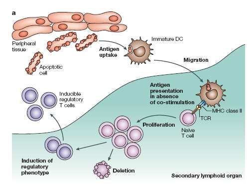
Dendritic cells in Sjögren’s syndrome

Acknowledgements

The work for this master thesis was carried out at Broegelmann Research Laboratory, The
Gade Institute, from August 2008 to June 2009 with support of Department of Biomedicine,
University of Bergen.

First I would like to begin by thanking my supervisor Silke Appel who has lent her support
and expertise to me over this last year and for help in writing this thesis.
I would also like to thank my co-supervisor Petra Vogelsang for invaluable technical
assistance and for always being patient and helpful.
I also need to thank Dagny Ann Sandnes for technical assistanse .
Thanks also to all the other members of Broegelmann Research Laboratory and especially
Professor Roland Jonsson
Of course I also need to thank all of the Sjögren’s syndrome patients for donating blood to
this study and to all the healthy controls who kindly volunteered to give their blood for my
experiments.

I would also like to express my gratitude to my fellow students, especially Marie Karlsen and
Rania Al-Mahdi who were always encouraging and supportive.

To Vemund who has put up with me through this year and given invaluable support, a special
thanks.
Last but not least a thanks to my family who has always been encouraging in everything I
have done.

                                                                                               2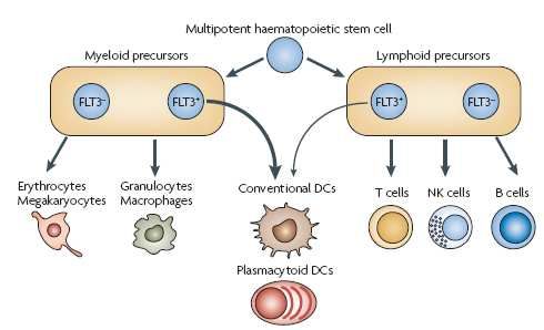
Dendritic cells in Sjögren’s syndrome

Contents

ACKNOWLEDGEMENTS.................................................................................................................................. 2

ABBREVIATIONS ............................................................................................................................................... 5

ABSTRACT........................................................................................................................................................... 7

1. INTRODUCTION............................................................................................................................................. 8

    1.1 SJÖGREN’S SYNDROME .................................................................................................................................. 8
        1.1.1 History .................................................................................................................................................. 8
        1.1.2 Classification criteria ........................................................................................................................... 8
        1.1.3 Clinical features ................................................................................................................................. 10
        1.1.4 Pathology............................................................................................................................................ 11
    1.2 THE IMMUNE SYSTEM .................................................................................................................................. 12
    1.3 DENDRITIC CELLS ........................................................................................................................................ 13
        1.3.1 DC subtypes........................................................................................................................................ 13
        1.3.2 Antigen recognition ............................................................................................................................ 15
        1.3.3 Antigen-processing and presentation ................................................................................................. 15
        1.3.4 DC are involved in inducing tolerance............................................................................................... 16
    1.4 ROLE OF RO52 AND IRF IN DC ................................................................................................................... 18
    1.5 DENDRITIC CELLS IN SS .............................................................................................................................. 20
    1.6 AIM OF THE STUDY. ..................................................................................................................................... 22

2. MATERIALS .................................................................................................................................................. 23

    2.1 STUDY SUBJECTS ......................................................................................................................................... 23
    2.2 EQUIPMENT ................................................................................................................................................. 23
    2.3 PLASTIC WARE ............................................................................................................................................ 24
    2.4 KITS ............................................................................................................................................................ 24
    2.5 REAGENTS ................................................................................................................................................... 24
    2.6 ANTIBODIES ................................................................................................................................................ 25
    2.7 BUFFERS AND MEDIA ................................................................................................................................... 26
    2.8 SOFTWARE .................................................................................................................................................. 28

3. METHODS ...................................................................................................................................................... 29

    3.1 GENERATION OF MONOCYTE-DERIVED DC (MODC).................................................................................... 29
        3.1.1 Isolation of PBMC from fresh blood................................................................................................... 29
        3.1.2 Isolation of monocytes by plastic adherence ...................................................................................... 29
        3.1.3 Generation of immature moDC .......................................................................................................... 30
        3.1.4 Maturation of moDC .......................................................................................................................... 30
    3.2 HARVESTING OF MODC............................................................................................................................... 30
    3.3 ENDOCYTOSIS ............................................................................................................................................. 30

                                                                                                                                                                         3
Dendritic cells in Sjögren’s syndrome
3.4 CHEMOTAXIS............................................................................................................................................... 31
3.5 PROTEIN LYSATE ......................................................................................................................................... 31
3.6 BCA ASSAY ................................................................................................................................................ 32
3.7 SDS-PAGE ................................................................................................................................................. 32
3.7.1 Gel casting.......................................................................................................................................... 32
3.7.2 Sample preparation ............................................................................................................................ 33
3.8 WESTERN BLOT........................................................................................................................................... 33
3.9 IL-12P70 ELISA ......................................................................................................................................... 34
3.10 STATISTICS ................................................................................................................................................ 35
4. RESULTS ........................................................................................................................................................ 36
4.1 DC FROM SS PATIENTS MAY HAVE HIGHER ENDOCYTIC CAPACITY THAN DC FROM HEALTHY CONTROLS .. 36
4.2 MATURE MODC FROM SS PATIENTS HAVE HIGHER MIGRATORY CAPACITY THAN MATURE MODC FROM
HEALTHY CONTROLS. ........................................................................................................................................ 40
4.3 EXPRESSION OF IRF-8 AND RO52................................................................................................................ 41
4.4 SLIGHT DECREASE IN IL-12P70 PRODUCTION IN MATURE MODC FROM SS PATIENTS. ................................ 46
5. DISCUSSION .................................................................................................................................................. 48
5.1 TECHNICAL ISSUES ...................................................................................................................................... 52
5.2 CONCLUSION ............................................................................................................................................... 54
5.3 FUTURE PERSPECTIVES ................................................................................................................................ 54
6. REFERENCES................................................................................................................................................ 56
4Dendritic cells in Sjögren’s syndrome
Abbreviations
AECC American-European Consensus group criteria
APC Antigen presenting cells
BAFF B cell activating factor
BCA Bicinchoninic acid
CCL Chemokine (C-C motif) ligand
CCR Chemokine (C-C motif) receptor
CD Cluster of differentiation
cDC Conventional dendritic cells
DMSO Dimethyl sulfoxide
DC Dendritic cells
EDTA Ethylenediaminetetraacetic acid
ELISA Enzyme-linked immunosorbent assay
FDC Follicular dendritic cells
FSC Forward scatter channel
GM-CSF Granulocyte macrophage-colony stimulating factor
HRP Horseradish Peroxidase
ICSBP Interferon consensus sequence-binding protein
IL Interleukin
IFN Interferon
Ig Immunoglobulin
IRF Interferon regulatory factor
LC Langerhans cells
PAMP Pathogen-associated molecular patterns
PBMC Peripheral blood mononuclear cells
PBS Phosphate buffered saline
pDC Plasmacytoid dendritic cells
pSS Primary Sjögren’s syndrome
MFI Median fluorescence intensity
MHC Major histocomatibility complex
moDC Monocyte derived dendritic cells
MR Mannose receptor
5Dendritic cells in Sjögren’s syndrome
NK cells Natural killer cells
n.s Non significant
RIPA Radioimmunoprecipitation assay
RT Room temperature
SDS-PAGE Sodium dodecyl sulfate-polyacrylamide gel
electrophoresis
SS Sjögren’s syndrome
SSC Side scatter channel
TBS Tris-buffered saline
TLR Toll like receptor
TMB Tetramethylbenzidine
TNF Tumor necrosis factor
TRIM Tripartite motif
Treg Regulatory T cells
WR Working reagent
6Dendritic cells in Sjögren’s syndrome
Abstract
Sjögren’s syndrome (SS) is an autoimmune disease of unknown etiology. It is characterized
by chronic inflammation of the exocrine glands which leads to dryness of the mouth and eyes.
Dendritic cells (DC) are the most potent antigen presenting cells of the immune system. They
take up antigens in the periphery and migrate to secondary lymphoid tissues where specific T
lymphocytes recognize the presented antigen and mount an immune response. So far not
much work has been done to examine the possible role of DC in SS. Aim of this study was to
functionally analyze monocyte-derived DC (moDC) from patients diagnosed with primary SS
compared to healthy controls. Peripheral blood mononuclear cells were isolated from freshly
heparinized blood samples from pSS patients fulfilling the American European Consesus
group criteria (AECC) and gender- and age-matched healthy controls. Monocytes were
isolated by plastic adherence, and immature DC were generated by culturing the monocytes
with IL-4 and GM-CSF. A fraction of the cells was stimulated with the TLR7/8 ligand CL097
for 48 hours.
In order to investigate functional aspects of moDC, a FITC-dextran assay to explore the
endocytic capacity of DC, and a chemotaxis assay to examine migratory capacity of the cells
towards CCL19 were performed. Moreover an IL-12p70 ELISA on cell culture supernatants
was performed as IL-12 is an inflammatory cytokine which is produced and secreted by
mature DC. Finally protein expression of Ro52 and IRF-8 was analyzed on whole cell lysates
from moDC by Western blot. Ro52 is a commonly detected autoantigen in patients with SS
and IRF-8 is a transcription-factor involved in DC function and differentiation.
The results from the endocytosis assay revealed that immature DC from SS patients have a
higher endocytic capacity than the cells from healthy controls. The chemotaxis assay showed
that mature moDC from patients have higher migratory capacity than the mature moDC from
controls. Furthermore the Western blot analysis indicated that Ro52 expression is
significantly lower in mature moDC from patients compared to controls. IRF-8 expression
was significantly lower in immature DC from SS patients compared to the control group. The
ELISA result proposed a slightly higher production of IL-12p70 in mature moDC from
patients than from mature moDC from healthy controls. From the experiments which have
been performed in this study we conclude that there indeed is a functional difference between
the moDC from SS patient and controls. This difference might possibly be caused by an
inappropriate stimulus of the immature moDC in SS patients leading to dysfunctional DC.
This study also emphasizes the need to continue examining the possible role of DC in SS.
7Dendritic cells in Sjögren’s syndrome
1. Introduction
1.1 Sjögren’s syndrome
Sjögren’s syndrome (SS) is a chronic rheumatic autoimmune disease characterized by
lymphocyte infiltration in the exocrine glands, primarily the lacrimal and salivary glands. The
mechanisms behind the disease are still not understood and there is at this time no cure.
Today’s treatment is mainly focused on alliviating the symptoms. SS has until recently been
an under-diagnosed and under-treated disease mainly because it is a fairly newly discovered
disease and has low mortality, but the implications of having this chronic disease is in fact
very serious for the patient and goes way beyond just having dry eyes and mouth (1-3).
1.1.1 History
Sjögren’s syndrome is a fairly recently discovered disease, although there were some isolated
cases reported in the late 1800. It was not described until 1933 when the Swedish
ophthalmologist Henrik Sjögren reported on clinical and histological findings in his doctoral
dissertation, “Zur Kenntnis der Keratoconjuvitis Sicca”(4). His work was not immediately
acknowledged and a mediocre grade on his thesis prevented an academic career. Not until the
fifties was his achievement recognized when he published a series of 80 patients of which
62% had arthritis. In 1970 the Swedish government awarded him the title of professor and he
was then able to work as a university professor (3).
In 1965, the distinction between primary and secondary SS was suggested and it was not until
this time that it became featured as a rheumatic disease. The Ro (SS-A) and La (SS-B)
autoantibodies and the labial gland infiltration were first described in the seventies and routine
diagnostic tests were available in the 1990s (1, 2).
1.1.2 Classification criteria
The first International Symposium on Sjögren’s Syndrome was held in Copenhagen in 1986,
where for the first time classification criteria were suggested. Later the Californian and the
European classification standard have been established. Today it is the American-European
8Dendritic cells in Sjögren’s syndrome
classification criteria which is used in diagnosis of SS (Table 1) (5). To meet the classification
criteria the patient must have:
• Presence of four out of the six items indicative of SS and each patient must have an
abnormal biopsy (IV) or the presence of anti-Ro or anti-La autoantibodies (VI).
• Presence of three out of four objective criteria (Items III, IV, V, VI).
Exclusion criteria are: past head and neck injuries, hepatitis C infection, AIDS, pre-existing
lymphoma, sarcoidosis, graft versus host disease and use of anticholinergic drugs (5).
Table 1. American-European Consensus Group Classification criteria for Sjögren’s
Syndrome according to Vitali et al (5)
I Ocular symptoms:
1. Have you had daily, persistent, troublesome dry eyes for more than 3 months?
2. Do you have a recurrent sensation of sand and gravel in the eyes?
3. Do you use a tear substitute more than 3 times a day?
II Oral symptoms:
1. Have you had a daily feeling of dry mouth for more than 3 months?
2. Have you had recurrent or persistent swollen salivary glands as an adult?
3. Do you frequently drink liquids to aid in swallowing dry foods?
III Ocular signs-positive result of one of the following tests:
1. Schirmer’s I test (≤ 5mm in 5 minutes)
2. Rose Bengal score ≥4 according to van Bijsterveld’s scoring system)
IV Histopathology: Abnormal biopsy of minor salivary gland
V Salivary gland involvement: positive result for one of following diagnostic test:
1. Unstimulated whole salivary flow (≤ 1,5 ml in 15 minutes)
2. Parotid sialography showing the presence of diffuse sialectasias, without
evidence of obstruction in the major ducts
3. Salivary scintigraphy showing delayed uptake, reduced concentration and/or
delayed excretion of tracer
VI Antibodies to Ro(SSA) or La(SSB) antigens, or both
To classify secondary SS in patients with a potential associated disease fewer criteria need to
be met. Only the presence of item I or II plus 2 items from III, IV, V, VI is required to be
indicative of secondary SS (5).
9Dendritic cells in Sjögren’s syndrome
1.1.3 Clinical features
SS can occur at any age and in people of both sexes, but it mainly appears in middelaged and
elderly women; the ratio between women and men is 9:1 (1, 2). The prevalence of the disease
varies in the literature and depending on which classification criteria is used, but recent data
suggest a prevalence of approximately 0,3% to 0,6% which is comparable to rheumatoid
arthritis (6). SS can occur alone in primary Sjögrens (pSS) or in association with other
rheumatic diseases as secondary SS.
SS is characterized by persistent lymphocyte infiltration in the exocrine glands primarily
salivary and lacrimal glands leading to dry mouth (xerostomia) as a result of deficient saliva
production and dry eyes (keraconjunctivitis sicca) due to lack of tear production.
The patient will generally present subjective symptoms of dry eyes. They can be described as
feeling sandy, gritty, itchy or as having a burning sensation. The physical implications of
deficient tear production can include destruction of corneal epithelium, conjunctivitis, dilation
of the bulbar conjunctival vessels, irregularity of the corneal image and lacrimal gland
enlargement. Patients can also have an inability to tolerate smoke and light (photosensitivity)
(1, 7, 8). Oral dryness, due to lack of saliva secretion can be quite debilitating in daily life,
interfering with the patients’ ability to eat, sleep and speak. They can have difficulties in
chewing and swallowing without liquid, develop fissures on the tongue, loose taste and smell
sensation, have increase in dental caries and develop candidasis (1-3, 8). Chronic persistence
can also result in more serious conditions such as chelitis and mouth ulcers (9).
Other common symptoms of pSS are dryness of the skin and vagina, joint pain and a feeling
of extreme fatigue (2).
In addition to exocrine dysfunction about 50% of the patients also have systemic
manifestations. Other extraglandular manifestations can include; respiratory tract,
gastrointestinal tract, genitourinary tract, skin, muscle and joints, neurological and
vasculopathies (1, 3, 7). One of the most serious complications of SS is an increased risk of
developing non-Hodgkins lymphoma due to chronic B cell stimulation (2).
10Dendritic cells in Sjögren’s syndrome
1.1.4 Pathology
A characteristic feature of SS is a chronic inflammation of the exocrine glands, mainly the
salivary and lacrimal glands. Histopathological analysis in salivary gland biopsies show
lymphocytic infiltrates consisting of a majority of T cells with fewer B cells, macrophages
and mast cells (2). Furthermore it could be shown that 17% of patients have germinal center
(GC)-like structures in their salivary glands. These structures consisted of T and B cell
aggregates within a follicular DC (FDC) and endothelial cell network (10).
B cells make up for about 20% of the lymhocytes in the lymphocyte infiltrates. There is
evidence of hyperactive B cells which have increased amounts of antibodies with
autoantibody activity including rheumatoid factors, Ro and La (11). Infiltrating B cells
predominantly express the IgG isotype in contrast to IgA which is expressed in normal
salivary glands (2). A member of the tumor necrosis factor (TNF) family, B cell activating
factor (BAFF), has been found to be upregulated in serum of patients with SS and also in
patients with other autoimmune diseases (12-14). BAFF is considered to be involved in the
proliferation and survival of B cells and might be involved in the pathogenesis of SS (14).
Infiltrating T cells are mainly activated CD4+ helper T cells which very likely contribute to
the hypereactivity of B cells. They produce high levels of IFN-γ and IL-10, while high levels
of IL-1β, IL-6 and TNF-α are produced by epithelial cells which might stimulate B cell
proliferation and differentiation (2).
It has for a long time been suspected that viruses could be involved in the pathogenesis of SS
and several different candidates have been listed, but there was not found a real connection (1,
2, 15, 16). In recent years several studies have revealed an activated type I interferon (IFN)
signature in salivary glands of SS patients (15, 17, 18). Plasmacytoid dendritic cells (pDC),
which are known as natural IFN producing cells, were found in salivary glands of patients
with pSS, but not in controls (15).
SS has a generally low mortality rate, in a study examining long term outcome of ~4500
patients with SS there were 39 deaths recorded of which 1 in 5 was from lymphoma. Since
this is mainly a disease of morbidity an important issue is the quality of life for the patients. It
has been shown that SS patients and particular those with pSS have very low scores on
general quality of life (19).
11Dendritic cells in Sjögren’s syndrome
1.2 The immune system
The body is constantly attacked by harmful microorganisms like bacteria, virus, fungus and
protozoa. To protect us from these pathogens our body has aquired the immune system. It is a
highly specialized system involving several different cell-types and molecules used for
communication between them. Immune cells have to distinguish self from non-self and
harmful from benign. The immune system is divided into two different systems, the innate
and the adaptive immune systems which both work separately but also communicate and
function collectively.
The innate immune response is the body’s first line of defence and has a very rapid response
time. Within the first 12 hours of an infection it is only the innate immune system which is
active. The components of this system are first physical and chemical barriers such as the skin
and the low pH of the stomach. If the microorganisms manage to pass these barriers, defence
mechanisms in the form of phagocytic cells like macrophages and neutrophils which ingest
and destroy foreign material are recruited. Other components of the innate immune response
are cytokines, which are secreted by cells in the innate and adaptive immune system, and
components of the complement system. They are involved in mediating an immune and
inflammatory response.
Many microorganisms manage to bypass all of these defence systems, and in that case the
adaptive immune response gets activated. This is a late response and is often called the
specific immune response because it targets very specific pathogens and it also has memory.
While the innate immune system responds to structures that are shared by many groups of
microbes, the adaptive immune system responds to antigens which are very specific for
different pathogens.
The major players of the adaptive response are B and T lymphocytes. T lymphocytes interact
with cells of the innate immune system and produce effector T cells that have many functions
such as secretion of cytokines and activation of other immune cells including B cells.
B lymphocytes are the only cells capable of producing antibodies which can neutralize or
initiate phagocytosis of the microbe.
The immune system is very intricate and both the innate and adaptive immune system work
together and communicate in several different ways, they are not isolated systems
As mentioned earlier the T lymphocytes are activated by cells of the innate immune system,
these are called antigen presenting cells (APC). The most potent APC are the dendritic cells
12Dendritic cells in Sjögren’s syndrome
which are central mediator between the innate and adaptive immune system. Their role is to
capture and process antigens and present them to T lymphocytes.
1.3 Dendritic cells
Dendritic cells (DC) got their name from their branchlike, dendritic shape. Their long
projections increase surface area and make it possible for them to effectively probe their
surrounding tissues for antigens (20). The first DC was visualized 140 years ago by Paul
Langerhans, who first characterized Langerhans cells (LC), but further description of the DC
did not start until the seventies by Steinman and Cohn (21).Since then DC have been
recognized as the most potent antigen presenting cells of the immune system and being
central in initiating an immune response. DC capture and process antigens from both
pathogens like viruses and bacteria and from apoptotic material. The processed antigens are
then presented on major histocompatability complex (MHC) class I and II molecules to naïve
T lymphocytes. Recognition of the antigens by T cells leads to either activation of the
adaptive immune response or induction of tolerance (22-24).
1.3.1 DC subtypes
Like all blood cells the dendritic cells are of hematopoietic lineage and originate from
pluripotent hematopoietic stem cells in the bonemarrow (figure 1.1). Downstream from the
multipotent hematopoietic stem cell precursors divide into two main lineages, myeloid and
lymphoid (25). DC are mainly generated from the myeloid lineage from a common myeloid
progenitor, but can also stem from common lymphoid precursors, both have been isolated
from the bone marrow. DC also have several different subsets which have different surface
markers and functions (20, 24). Different DC subtypes are present in most of the tissues in the
body, in the skin, mucus membranes, blood, lymph and visceral organs.
13Dendritic cells in Sjögren’s syndrome
Figure 1.1. Development of DC from a multipotent hematopoietic stem cell.
DC originate from a multipotent hematopoietic stem cell in the bone marrow. Downstream from this stem cell
precursors divide into two different lineages, myeloid and lymphoid. DC can originate from both lineages. DC
have several different specialised subsets with different functions and surface markers, but the two main subsets
are the conventional DC and the pDC. From Shortman .& Naik. Nat Rev Immunol 2007 (26).
The two main subsets of DC are plasmacytoid (pDC) and conventional DC (cDC).
Plasmacytoid DC are found as pre DC in steady state. They are circulating, long lived cells
which upon stimuli from virus acquire a dendritic form and start producing large amounts of
type I interferons (IFN) (26, 27). IFN are cytokines which mediate activation of several other
immune cells including T and B lymphocytes and cDC (27). When acquiring a dendritic
shape, pDC are also capable of antigen capture and presentation although not as efficient as
the cDC (26).
Conventional DC, also known as myeloid DC (mDC), do not develop from pre DC, but have
the shape and function of DC in steady state. This subset is known to contain the most potent
antigen presenting cells. They can be further divided into subgroups of migratory and
lymphoid-tissue-resident DC (26). Migratory DC are the textbook example of DC which catch
antigens in the periphery and migrate through lymphatics to secondary lymphoid tissue where
they present antigens to T cells. Included in this group are the Langerhans cells (LC) which
were the first DC to be described. They have been used as a model to study the function of the
DC (26, 28). LC reside in the epidermis and have upon activation been found to loose their
connection with neighboring cells and start to migrate to the dermis. From there they have
been traced through afferent lymphatic vessels and to T cell areas of cuteneous draining
lymphnodes (29).
14Dendritic cells in Sjögren’s syndrome
Another important DC subset are the monocyte-derived DC (moDC). They are inflammatory
DC not present in the steady state of the body(26). The ability of human peripheral monocytes
to differentiate into DC when cultured with IL-4 and GM-CSF was first published by Sallusto
and Lanzavecchia (30). This pathway of DC development has been utilized in vitro by
culturing blood monocytes with GM-CSF and IL 4. Today this is an established procedure for
generation of DC in the laboratory (30-32). The reason why this is such a conventional way of
generating DC instead of just isolating them from the blood is because pDC and cDC are very
rare and make up for a very small percentage of the blood cells, and they do not proliferate.
1.3.2 Antigen recognition
The antigen presentation ability of DC is an essential part of their function.
Immature DC recognize pathogens by different surface and intracellular receptors. One
important family of receptors are the Toll like receptors (TLR) which is a large family of
conserved proteins. These are transmembrane receptors recognizing pathogen-associated
molecular patterns (PAMP) on pathogens like viruses, fungi, gram positive, gram negative
bacteria and parasites (33, 34). TLR are expressed both on the plasma membrane and
intracellularly in a variety of cells of the innate immune system in addition to the DC,
including macrophages, mast cells and neutrophils. For instance, TLR3, TLR7, TLR8 and
TLR9, which are expressed intracellularly on endosomes, are important for viral recognition.
These receptors are activated by viral nucleic acids like RNA and DNA. TLR7 and TLR8 can
also be activated by synthetic imidazoquinoline compounds (34). Activation leads to the
increased expression of IFNα and inflammatory cytokines (34). TLR activation in immature
DC leads to upregulation of several costimulatory molecules like CD80 and CD86 and
induction of cytokine production, for example IL-6 and IL-12 (35-37). Secretion of IL-12 by
DC in response to TLR activation leads to IFNγ production of T and natural killer (NK) cells.
It is also important in promoting differentiation of CD4+ T cells into Th 1 cells, which are
essential in immunity towards intracellular pathogens.
1.3.3 Antigen-processing and presentation
After the immature DC recognize and are activated by foreign antigens many intracellular
processes start taking place. These include upregulation of co-stimulatory molecules and
15Dendritic cells in Sjögren’s syndrome
production of cytokines which start the maturation process in the DC. Lately it has been
proposed that the terms immature and mature DC are quite insufficient (24), but I will stick to
these terms throughout this thesis for the sake of simplicity.
Once antigens bind to the immature DC they are taken up in the cell by endocytosis.
Immature DC use several different pathways to internalize the antigens such as receptor
mediated endocytosis via C-type lectins and Fcγ receptors, phagocytosis and
macropinocytosis (22, 38). Once internalized the antigens are taken up into endosomes and
phagosomes where they are cleaved into peptides which can be presented to T lymphocytes
on major histocomatibility complex (MHC) class II molecules. In immature DC MHC class II
molecules are produced in the endoplasmic reticulum and transported in vesicles to
endosomes or phagosomes where they bind the processed peptides. The MHC class II/peptide
complex is then transported to the surface where it can be recognized by naïve T lymphocytes
(39). An intracellular infection, for instance by a virus, leads to presentation of viral peptides
on MHC class I molecules. MHC class I exist on all cells of the body and therefore make all
cells able to present peptides to CD8+-effector T cells. Usually MHC class I molecules
present peptides from degraded proteins in the cytosol or nucleus. Presentation of a foreign or
unknown peptide stimulates the CD8+ cell and leads to destruction of the cell (39).
Another important process is the relocation of DC from peripheral tissues to secondary
lymphoid organs through the lymphatic vessels (40). DC must undergo several functional and
phenotypical changes to be able to migrate to secondary lymphoid tissue. Some of the
important functional changes that take place during maturation is downregulation of receptors
responding to inflammatory cytokines and upregulating of several trafficking molecules
including chemokine (C-C motif) receptor 7 (CCR7) which bind to the chemokine (C-C
motif) ligands 19 (CCL19) and CCL21 (40, 41).
CCL19 and CCL21 are chemokines produced by stromal cells in the T lymphocyte area of the
lymph node and they are important in homing the DC to this area through chemotaxis (42).
1.3.4 DC are involved in inducing tolerance
DC are considered sentinels of the immune system due not only to their central position in
initiating a primary immune response but also because of their involvement in inducing
tolerance.
16Dendritic cells in Sjögren’s syndrome
The way T and B cells develop their antigen receptors by genomic recombination leads to the
possibility of billions of different combination. Many of these antigen receptors are
potentially autoantigenic and recognize self antigens. To avoid autoimmune reactions these T
lymphocytes must be deleted. Self–reacting thymocytes (T lymphocyte precursors) are mostly
deleted by negative selection in the thymus (39, 43). Both thymic DC and peripheral DC
migrated from peripheral tissues have been found to be efficient mediators of negative
selection in the thymus (44-46). Some auto-reactive T cells, mainly those with low affinity,
manage to escape from the clonal deletion and will have to be controlled by peripheral
tolerance to prevent tissue damage (47, 48).
Peripheral tolerance mechanisms include anergy, deletion and suppression to inhibit self-
reacting T lymphocytes. Anergy is thought to take place when APC present autoantigen to
naïve T cells in the absence of co-stimulatory molecules. This will lead to alterations
inhibiting the T cell to react to this antigen if restimulated (39). Activation of an autoreactive
T lymphocyte by repeated simulation or in the absence of inflammation can also lead to
activation of death receptors and apoptosis (39). Another mechanism to induce tolerance is
inhibition of the autoreactive T cell by regulatory T cells (Treg). It is thought that a failure of
these mechanisms is the cause of the development of autoimmunity.
Figure 1.2 shows a schematic representation of how immature DC might be involved in the
induction of tolerance. Immature DC constantly capture antigens in their environment from
apoptotic cells and then migrate to secondary lymphoid tissues. In the absence of
inflammation the DC do not mature and they will present the self-antigens to naïve T cells in
absence of co-stimulatory molecules. This will then lead to deletion of the autoreactive T cell
or induction of Treg (49).
17Dendritic cells in Sjögren’s syndrome
Figure 1.2. Role of immature DC in induction of tolerance. This figure illustrates one hypothesis on the role
of immature DC in development of tolerance. The immature DC constantly take up antigens from both the
environment and from apoptotic cells from the body’s own tissues. They then travel to secondary lymphoid
organs where they present the antigens to naïve T cells. In the absence of inflammation the DC stay immature
and present the antigens in absence of co-stimulatory molecules. This will lead to either deletion of the T cells or
induction of Treg (49). Modified from Banchereau & Palucka. Nat Rev Immunol, 2005.
Based on the role DC have both in central and peripheral tolerance there have been
speculations on whether or not they protect us from the development of autoimmune diseases.
To support this hypothesis it has been reported recently that constitutively DC depleted mice
develop spontaneous and severe autoimmune diseases (50).
1.4 Role of Ro52 and IRF in DC
Autoantibodies against the Ro52 protein are found in sera of many patients with SS and they
are also used as a diagnostic marker (5). The reason why Ro52 is a target for autoantibodies is
not known, and the function of the protein has been unknown for decades. In the recent years
it has been found that Ro52 is an E3 ubiqutin ligase with the ability to ubiquitinate itself and
other proteins (51). Ro52 is also known as TRIM21 because it contain a tripartite motif
(TRIM) (52, 53). Ubiquitin is a small regulatory protein with wide functional diversity. A key
task is targeting proteins in the cell for degradation, but it can also regulate other functions
18Dendritic cells in Sjögren’s syndrome
like translation, activation of transcription factors and DNA repair (54). Studies done on a
mouse B cell line showed that overexpression of Ro52 can have anti-proliferative and cell-
death-mediating properties (51). Its targets for ubiquitination are for the most part illusive, but
it has been found to regulate two members of the interferon regulatory factor (IRF) protein
family, IRF-3 and IRF-8. It has been suggested that Ro52 functions as a E3 ubiquitin ligase
for IRF-8 and that this leads to increased cytokine expression in macrophages (55). Ro52 has
also been suggested to regulate levels of IRF-3 by polyubiquitin–mediated degradation (56).
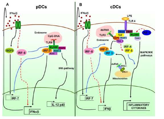
The IRF family of proteins are transcription factors which have been found to be key players
in the development maturation and function of DC (57, 58). Figure 3 illustrates the role of
different IRF proteins in regulating transcription of IFNα/β and inflammatory cytokines upon
activation.
IRF-8, also known as interferon consensus sequence-binding protein (ICSBP), is suggested to
have a pivotal role in DC maturation, trafficking, antigen uptake and presentation. It has for
instance been shown that IRF-8 knock out mice have defective endocytic capacity and antigen
presenting abilities (59). IRF-3 plays a critical role in the induction of type I interferons after a
viral infection (60).
Type I IFN are cytokines which mediate early response to pathogens and the most potent
stimuli for IFN production is binding of viral nucleic acids (39). IFNα/β produced at an early
period in an inflammatory response has the ability to activate other cells. As illustrated in
figure 1.3, different IRF proteins participate in these pathways. For instance IRF-8 is both
activated by an IFN stimulus and also contributes to an increased IFN production.
19Dendritic cells in Sjögren’s syndrome
Figure 1.3. Regulation of type I IFN and inflammatory cytokines by IRF proteins. IRF family of proteins
are involved in regulation of IFNα/β and inflammatory cytokines in DC through several pathways. For instance
is IRF-8 both activated by IFNα/β stimuli and a regulator of transcription of IFNα/β. From Gabriele & Ozato.
Cytokine Growth Factor Rev. 2007. (57)
1.5 Dendritic cells in SS
Until now not much work has been done on examining the role of DC in SS. Most of the
studies performed on SS have focused on B and T cell. Only recently first studies which
implicate a role for DC in the pathogenesis of SS have been conducted. The focus mainly has
been on the presence of follicular DC (FDC) in salivary gland infiltrates of patients with SS
(61). GC-like structures which have been found in minor salivary glands of some patients
with SS, consist of T and B cell aggregates within a FDC and endothelial cell network (10).
In SS there has also as mentioned earlier been discovered an activation of type I IFN signature
in salivary glands of patient with SS and the presence of type I IFN producing pDC in patients
with SS but not in healthy controls (15, 17, 62). Viral infections have been proposed to be the
initial cause of the IFN production. The secretion of IFN-α from pDC may lead to apoptosis
of cells and autoantibody production towards the apoptotic material (63). This may initiat a
vicious cycle where elevated levels of IFN attracts more pDC to the the affected area (64).
A hypothesis that has been proposed on how dysregulation of the DC might lead to
autoimmunity is shown in figure 1.4. An immature DC that has taken up an autoantigen is
20Dendritic cells in Sjögren’s syndrome
stimulated by an elevated level of IFNα/β caused by a viral infection. This leads to
inappropriate maturation of the DC and presentation of the autoantigen together with co-
stimulatory molecules. In a healthy individual this should lead to apoptosis induction in the
DC or anergy, where Treg inhibit presentation to autoreactive T cells. In the case of
autoimmunity the DC activate autoreactive T cells that then activate autoreactive B cell which
start producing autoantibodies towards the autoantigen (64).
Figure 1.4. The hypothesis on how the dysregulation of the immature DC might lead to autoimmunity.
An initial viral infection stimulates pDC to produce type I IFN. Due to continual stimulation the IFN levels
become elevated. This leads to inappropriate stimulation of an immature DC carrying an autoantigen. The
immature DC is inappropriately matured and presents both autoantigens and co-stimulatory molecules. In a
healthy individual this is controlled by induction of apoptosis or inhibition of autoantigen presentation by Treg.
In autoimmunity this leads to activation of autoreactive T cells which again activate autoreactive B cells that
start to produce autoantibodies towards the autoantigen presented by the DC. From Vogelsang et al. Scand J
Immunol, 2006 (64)
21Dendritic cells in Sjögren’s syndrome
1.6 Aim of the study
The aim of this master thesis was to perform functional analyses of moDC from patients with
SS and compare them to moDC from healthy controls.
Functional analyses of immature and mature moDC regarding endocytic and migratory
capacity were to be performed as both are crucial functions of DC. IL-12p70 production was
to be analyzed since this cytokine is one of the most important one for T cell stimulation.
Protein expression of Ro52 and IRF-8 was analyzed as Ro52 is a commonly detected
autoantigen in patients with SS and IRF-8 is involved in differentiation and function of DC
and is one of the targets of ubiquitination by Ro52.
22Dendritic cells in Sjögren’s syndrome
2. Materials
2.1 Study subjects
Heparinized blood from SS patients fulfilling the AECC criteria was collected at the
Department of Rheumatology at the Haukeland University Hospital. The control group
consisted of gender and age matched healthy persons that were recruited from the same
geographical area as the patients.
2.2 Equipment
Name Company
Cell counter: CASY Schärfe system GmbH, Germany
Centrifuge: Kubota 8700. Kubota. Tokyo Japan
Thermo Heraeus multifuge1S-R. Mandel Scientific company
Mini Spin Eppedorf, Germany
Heraeus Fresco 17 Thermo Scientific
Flow cytometer: FACS Canto BD Biosciences, USA
CO2 Incubator: Forma scientific, USA
Microplate reader: EMax Molecular Devices, USA
Microscope: Leica DMIL Leica, Germany
Mini Trans-blot electrophoretic cell BioRad, USA
MP3 system BioRad, USA
10-well, 1.0 mm thickness
Molecular imager ChemiDoc XRS system BioRad, USA
Safety cabinet: Nuair biological safety cabinet class II Nuaire INC, USA
23Dendritic cells in Sjögren’s syndrome
2.3 Plastic ware
Name Company
50ml tubes Sarstedt. Germany
15ml tubes Sarstedt. Germany
6well plates NUNC. Denmark
Cryotubes NUNC. Denmark
HTS Transwell- 96 system Corning. NY, USA
Insert with 8µm membrane pores
96 well U shaped plate NUNC,Denmark
2.4 Kits
Name Company
BCATM protein assay kit Pierce, USA
Immunstar WesternCTM kit Biorad, USA
TM
ELISA MAX SET Deluxe. Human IL12p70 BioLegend, USA
2.5 Reagents
Name Company
25x Proteinase inhibitor, complete EDTA-free Roche, Germany
BSA Sigma, USA
CCL19 Immunotools. Germany
CL097 Invivogen, USA
Casyton Innovatis, Germany
Dextran Fluorescein. 40 000MW Invitrogen. USA
DMSO Sigma, UK
24Dendritic cells in Sjögren’s syndrome
Dual color Protein Kaleidoscope Standards Biorad, USA
EDTA 0,5M Sigma, USA
GM-CSF ImmunoTools. Germany
IL-4 ImmunoTools. Germany
Isopropanol Arcus, Oslo, Norway
Lymphoprep Axis-Shield Poc AS, Norway
Milk powder Frema, Germany
Methanol Merck, Germany
NaF Merck, Germany
NaOrthovanadat Merck, Germany
Ponceu S solution Sigma Aldrich, USA
Penicillin/Streptomyocin Invitrogen, USA
PMSF Roche, Germany
RPMI 1640 Lonza, Belgium
Tween20 Merck, Germany
X-Vivo 20 medium Cambrex (Biowhittaker)
2.6 Antibodies
Name Company
Primary antibodies
Actin (I-19) Santa Cruz Biotechnology INC
Goat polyclonal
ICSBP (c 19). Santa Cruz Biotechnology INC
Goat polyclonal
52 kDa Ro/SSA (D12) Santa Cruz Biotechnology INC
Mouse monoclonal
25Dendritic cells in Sjögren’s syndrome
Secondary antibodies
Goat anti-mouse IgG (H+L)/HRP Bio-Rad. CA, USA
Rabbit anti-goat IgG/HRP DakoCytomation
2.7 Buffers and media
Name Reagents
Blocking buffer TBST 0.1%
5% skim milk powder
1x Blotting buffer pH 8.3 Running buffer without SDS
20% methanol
FACS buffer 0.5 % BSA in PBS
6x Lämmli buffer 375 mM Tris HCl pH 6.8
9% SDS
50% Glycerol
9% β-ME
0.03% Bromphenolblue
PBS 137mM NaCl
2.7mM KCl
8.1mM Na2HPO4
1.5mM KH2PO4
26Dendritic cells in Sjögren’s syndrome
RIPA 50mM Tris pH 7.4
1% NP40
0.25 % Nadeoxycholat
150mM NaCl
1mM EDTA
+
1x proteinase inhibitor (25xstock)
1mM PMSF
1mM Na-orthovanadat
1mM NaF
1x Running buffer 25mM Tris
192 mM Glycine
0.1% SDS
Do not adjust pH
10 x TBS 1M Tris pH 8.0
1.4 M NaCl
1 x TBST 0.1% 1 x TBS
0.1% Tween
RP10 medium RPMI 1640
2 mM glutamine
10 % FBS
50 units/ml Penicillin G sodium
50 µl streptomycin sulfate
Washing buffer (ELISA) 0,05% Tween in PBS
27Dendritic cells in Sjögren’s syndrome
2.8 Software
Name Company
FlowJo Tree Star, Inc.
SoftMaxPro Molecular Devices, USA
Quantity one BioRad, USA
All reagents not listed here were purchased from Sigma Aldrich, USA or BioRad, USA.
28Dendritic cells in Sjögren’s syndrome
3. Methods
3.1 Generation of monocyte-derived DC (moDC)
To generate DC is a multistep process where the first method is to isolate peripheral blood
mononuclear cells (PBMC) which consist of lymphocytes and monocytes. This is done by
gradient centrifugation. The second step is to isolate the monocytes in the PBMC from the
lymphocytes, which is accomplished by using the ability the monocytes have to adhere to
plastic. The third step is to stimulate the monocytes to turn into immature DC, which is done
by adding the appropriate cytokines.
3.1.1 Isolation of PBMC from fresh blood.
20-35ml freshly heparinized blood was carefully layered on top of 10ml lymphoprep and
centrifuged for 20-30 minutes at 800g, 20ºC. The brake on the centrifuge was turned off to
make sure that the layers did not mix.
The PBMC layer, located in-between the plasma and lymphoprep layer, was transferred to a
new tube and washed 3x with cold PBS by centrifugation; 4 min, 400g, 4°C.
To determine the number of PBMC in the sample 10µl were removed after the second wash,
diluted 1:1000 in casyton, and counted with a CASY® automated cell counter.
3.1.2 Isolation of monocytes by plastic adherence
After the last wash the PBMC were resuspended in X-vivo20 medium and plated out in a
concentration of 1 x 10⁷ PBMC in 3ml X-vivo medium per well in a 6 well plate.
They were then incubated 1 hour at 37ºC, 5% CO2.
After incubation the monocytes were washed 2- 3x with PBS (RT) to remove all non adherent
cells
29Dendritic cells in Sjögren’s syndrome
3.1.3 Generation of immature moDC
The monocytes were cultured at 37ºC, 5% CO2 for 5- 6 days in RP10 medium supplemented
with IL-4 (20 ng/ml), and GM-CSF (100 ng/ml) which were replenished every 2-3 days.
3.1.4 Maturation of moDC
A fraction of the immature DC were stimulated with imidazoquinoline derivative CL097
(1µg/ml) for 48 hours. This compound activates the TLR 7/8 receptors of the DC which
induces activation of the NF-кB pathway in the cells and stimulate maturation.
3.2 Harvesting of moDC
The cells in medium where first transferred from the wells to a new tube and centrifuged for 5
minutes at 400g, 4ºC. PBS with 2mM EDTA was added to the wells to help loosen any
remaining cells from the plastic.
After centrifugation the supernatant was collected and stored at -20ºC for later use.
The cell pellet was resuspended in the remaining cells in PBS with EDTA from the wells.
Amount of moDC in the suspensions was determined by diluting 10µl 1:1000 in casyton to be
counted by a CASY cell counter.
The cells were then centrifuged for 5 minutes at 400g, 4ºC. The supernatant was discarded
and the pellet resuspended in PBS or RP10 medium (1 x 10⁵ cells pr 100µl). Cells required for
different experiments were then distributed to separate tubes.
3.3 Endocytosis
To measure the antigen uptake capacity of the DC dextran conjugated with FITC was used for
this assay. Dextran is a water soluble molecule which is taken up via endocytosis by the cells.
FITC is a fluorescent dye which can be detected by flow cytometry.
2x10⁵ moDC in 800µl RP10 medium were divided between two 96 well plates, 5x10⁴ cells in
200µl RP10 medium were plated out in two wells on each plate. They were then pre-
incubated approximately 20 minutes, one at 37ºC and the other at 4ºC before FITC-Dextran
30Dendritic cells in Sjögren’s syndrome
(0,25mg/ml) was added to one well of each sample on both plates. They were then incubated
for 1 hour at the same temperature.
After incubation the cells were washed 4x with 175µl FACS buffer by centrifugating for 5
minutes at 400g, 4ºC. Thereafter the DC were resuspended in 150µl FACS buffer and
immediately analyzed by flow cytometry.
3.4 Chemotaxis
To measure the migratory capacity of the moDC, a chemotaxis assay towards the chemokine
CCL19 was performed. This molecule binds to the receptor CCR7, a receptor which is
expressed by mature DC, and is found in high concentrations in the lymph nodes and thymus.
235µl RP10 medium + CCL19 (100ng/ml) was added to the bottom chamber of a 96 well
transwell plate. To the top of the membrane 5 x 10⁴ cells in 80µl RP10 medium was added.
The plate was incubated for 16, 5 or 4 hours at 37ºC, 5% CO2. After incubation the medium
with migrated cells from the lower chamber was transferred to appropriate tubes compatible
with the CASY cell counter. The wells were washed with 235µl PBS, which was also
transferred to the correlating casy tubes and 530µl PBS were added to all the samples (1ml
total volume). All the samples were then diluted in 9 ml casyton (1:10 dilution) before the
moDC were counted with CASY cell counter.
3.5 Protein Lysate
The cells in PBS or RP10 medium were first centrifuged 5 minutes at 300 g (cells in RP10
medium had to be washed by centrifugation 1x with PBS). The supernatant was discarded,
and the pellet resuspended in radioimmunoprecipitation assay (RIPA) buffer (100µl/1 x 106
cells). The cells were then incubated on ice for 10 min. After incubation the lysed cells were
centrifuged for 5 minutes at 17000g, 4ºC. The supernatant was transferred to a new tube and
stored at -80ºC. From the supernatant a 5µl aliquot was taken out and stored at −20ºC to be
used in the BCA assay.
31Dendritic cells in Sjögren’s syndrome
3.6 BCA Assay
The bicinchoninic acid (BCA) assay is used for colometric detection and quantification of
proteins.
The 5µl aliquots from protein lysates was first diluted 1:5 in PBS.
A standard two-fold serial dilution series were then prepared from BSA, 1,5mg/ml. 10µl of
the sample dilution and the dilution series were added in duplicates onto a 96 well plate, and
200µl working reagent was added to all the wells The plate was covered and incubated for 30
minutes at 37ºC before it was analyzed by an ELISA plate reader, absorbance at 595nm.
3.7 SDS-PAGE
SDS-PAGE (Sodium dodecyl sulfate-polyacrylamide gel electrophoresis) is used to separate
proteins based on molecular weight. SDS denatures the proteins and covers them with a
negative charge allowing them to be separated by mass only.
Polyacrylamide gel has a meshwork of tunnels and fibers which allows the smallest proteins
to move faster and thereby move further down the gel. A stacking gel was used which helps
focus the proteins and give a clearer band.
3.7.1 Gel casting.
Resolving gel 12 % Stacking gel 5 %
dH2O 3,4 ml dH2O 2,85ml
30 % Acrylamide 4,0 ml 30 % Acrylamide 0,85 ml
1,5M Tris-HCl pH 8.8 2,5 ml 0,5M Tris-HCl pH 6.8 1,25 ml
10 % SDS 100 µl 10 % SDS 50 µl
10% APS 50 µl 10 % APS 25µl
TEMED 10 µl TEMED 5µl
For gel casting the BioRad MP3 system, 10 well with 1,00mm thickness was used.
The glass plates were cleaned with 70% ethanol before use and then assembled in the casting
frame. The gels were prepared according to the recipies above (APS and TEMED were added
immediately before gel casting). The 12% unpolymerized resolving gel was first poured in
between the glass plates and layered with isopropanol to give the gel an even surface. The gel
was then left to polymerize for 15 – 30 minutes. Thereafter the isopropanol was rinsed off
32Dendritic cells in Sjögren’s syndrome
using distilled water before the stacking gel was layered on top. The comb was then inserted
before the stacking gel was left to polymerize for 15 – 30 minutes.
The gels were inserted into the electrode assembly and lowered into the tank. Thereafter the
inner camber and about halfway up the outer chamber was filled with 1 x running buffer. The
comb was carefully extracted and a syringe used to clean unpolymerized acrylamide from the
wells.
3.7.2 Sample preparation
Samples were prepared, consisting of 10µg protein, RIPA buffer and 4µl 6 x Lämmli buffer
(volume of 24µl total). The samples were denatured for 5 minutes at 100ºC before being
loaded onto the gel. In the first well Dual color protein standard was loaded (6µl). This is a
marker with bands of a known size. One sample was used in all gels to make it possible to
compare different blots with each other. The electrophoresis was started at 150V and ran until
the blue running front left the gel (ca 1 hour).
3.8 Western Blot
Western blot is a method used to identify specific proteins in a sample.
The transfer was performed with BioRad Mini Trans-blot electrophoretic cell system.
A nitrocellulose membrane, filter papers and fiber pads were equilibrated by soaking them in
blotting buffer. The nitrocellulose-membrane was placed on top of the polyacrylamide gel in a
sandwich between the fiber pads and filter paper. The sandwich was placed in a cassette
which was inserted into the electrophoresis module. To maintain an even buffer temperature
and ion distribution a frozen ice cooling unit and a magnetic stir bar was added to the tank
before it was completely filled with blotting buffer. The transfer was performed for 1 hour at
250 mA.
After transfer the membrane was washed with 1x TBST 0,1% and incubated with Ponceau S
solution for about 5 minutes. It was then destained with 1x TBST 0,1% to be able to visualize
all proteins on the membrane, and confirm equal loading. The membrane was then incubated
1 hour with blocking solution at RT with agitation to inhibit unspecific binding of the
antibodies. Thereafter the membrane was sealed in plastic with primary antibodies and
incubated with agitation for one hour RT or overnight at 4ºC.
33You can also read