STRUCTURE, FUNCTION, AND ANTIGENICITY OF THE SARS- COV-2 SPIKE GLYCOPROTEIN
←
→
Page content transcription
If your browser does not render page correctly, please read the page content below
Article

Structure, Function, and Antigenicity of the SARS-
CoV-2 Spike Glycoprotein
Graphical Abstract                                             Authors
                                                               Alexandra C. Walls, Young-Jun Park,
                                                               M. Alejandra Tortorici, Abigail Wall,
                                                               Andrew T. McGuire, David Veesler

                                                               Correspondence
                                                               dveesler@uw.edu

                                                               In Brief
                                                               SARS-CoV-2, a newly emerged pathogen
                                                               spreading worldwide, binds with high
                                                               affinity to human ACE2 and uses it as an
                                                               entry receptor to invade target cells.
                                                               Cryo-EM structures of the SARS-CoV-2
                                                               spike glycoprotein in two distinct
                                                               conformations, along with inhibition of
                                                               spike-mediated entry by SARS-CoV
                                                               polyclonal antibodies, provide a blueprint
                                                               for the design of vaccines and
                                                               therapeutics.

Highlights
d   SARS-CoV-2 uses ACE2 to enter target cells

d   SARS-CoV-2 and SARS-CoV bind with similar affinities
    to ACE2

d   Structures of SARS-CoV-2 spike glycoprotein in two
    conformations

d   SARS-CoV polyclonal antibodies inhibit SARS-CoV-2 spike-
    mediated entry into cells

           Walls et al., 2020, Cell 180, 1–12
           March 19, 2020 ª 2020 Elsevier Inc.
           https://doi.org/10.1016/j.cell.2020.02.058
Please cite this article in press as: Walls et al., Structure, Function, and Antigenicity of the SARS-CoV-2 Spike Glycoprotein, Cell (2020),
 https://doi.org/10.1016/j.cell.2020.02.058

Article

Structure, Function, and Antigenicity
of the SARS-CoV-2 Spike Glycoprotein
Alexandra C. Walls,1,5 Young-Jun Park,1,5 M. Alejandra Tortorici,1,2 Abigail Wall,3 Andrew T. McGuire,3,4
and David Veesler1,6,*
1Department    of Biochemistry, University of Washington, Seattle, WA 98195, USA
2InstitutePasteur & CNRS UMR 3569, Unité de Virologie Structurale, Paris 75015, France
3Vaccines and Infectious Diseases Division, Fred Hutchinson Cancer Research Center, Seattle, WA 98195, USA
4Department of Global Health, University of Washington, Seattle, WA 98195, USA
5These authors contributed equally
6Lead Contact

*Correspondence: dveesler@uw.edu
 https://doi.org/10.1016/j.cell.2020.02.058

SUMMARY                                                                 Hubei province of China and was sequenced and isolated by
                                                                        January 2020 (Zhou et al., 2020; Zhu et al., 2020). SARS-
The emergence of SARS-CoV-2 has resulted in                             CoV-2 is associated with an ongoing outbreak of atypical
>90,000 infections and >3,000 deaths. Coronavirus                       pneumonia (Covid-2019) that has affected over 90,000 people
spike (S) glycoproteins promote entry into cells and                    and killed more than 3,000 of those affected in >60 countries
are the main target of antibodies. We show that                         as of March 3, 2020. On January 30, 2020, the World Health
SARS-CoV-2 S uses ACE2 to enter cells and that                          Organization declared the SARS-CoV-2 epidemic a public
                                                                        health emergency of international concern.
the receptor-binding domains of SARS-CoV-2 S
                                                                           MERS-CoV was suggested to originate from bats, but the
and SARS-CoV S bind with similar affinities to human
                                                                        reservoir host fueling spillover to humans is unequivocally
ACE2, correlating with the efficient spread of SARS-                    dromedary camels (Haagmans et al., 2014; Memish et al.,
CoV-2 among humans. We found that the SARS-                             2013). Both SARS-CoV and SARS-CoV-2 are closely related
CoV-2 S glycoprotein harbors a furin cleavage site                      and originated in bats, who most likely serve as reservoir host
at the boundary between the S1/S2 subunits, which                       for these two viruses (Ge et al., 2013; Hu et al., 2017; Li et al.,
is processed during biogenesis and sets this virus                      2005b; Yang et al., 2015a; Zhou et al., 2020). Whereas palm
apart from SARS-CoV and SARS-related CoVs.                              civets and racoon dogs have been recognized as intermediate
We determined cryo-EM structures of the SARS-                           hosts for zoonotic transmission of SARS-CoV between bats
CoV-2 S ectodomain trimer, providing a blueprint                        and humans (Guan et al., 2003; Kan et al., 2005; Wang et al.,
for the design of vaccines and inhibitors of viral                      2005), the SARS-CoV-2 intermediate host remains unknown.
                                                                        The recurrent spillovers of coronaviruses in humans along
entry. Finally, we demonstrate that SARS-CoV S mu-
                                                                        with detection of numerous coronaviruses in bats, including
rine polyclonal antibodies potently inhibited SARS-
                                                                        many SARS-related coronaviruses (SARSr-CoVs), suggest
CoV-2 S mediated entry into cells, indicating that                      that future zoonotic transmission events may continue (Anthony
cross-neutralizing antibodies targeting conserved S                     et al., 2017; Ge et al., 2013; Hu et al., 2017; Li et al., 2005b; Men-
epitopes can be elicited upon vaccination.                              achery et al., 2015; Menachery et al., 2016; Yang et al., 2015a;
                                                                        Zhou et al., 2020). In addition to the highly pathogenic zoonotic
INTRODUCTION                                                            pathogens SARS-CoV, MERS-CoV, and SARS-CoV-2, all
                                                                        belonging to the b-coronavirus genus, four low-pathogenicity
Three coronaviruses have crossed the species barrier to                 coronaviruses are endemic in humans: HCoV-OC43, HCoV-
cause deadly pneumonia in humans since the beginning of                 HKU1, HCoV-NL63, and HCoV-229E. To date, no therapeutics
the 21st century: severe acute respiratory syndrome coronavi-           or vaccines are approved against any human-infecting
rus (SARS-CoV) (Drosten et al., 2003; Ksiazek et al., 2003),            coronaviruses.
Middle-East respiratory syndrome coronavirus (Zaki et al.,                 Coronavirus entry into host cells is mediated by the trans-
2012) (MERS-CoV), and SARS-CoV-2 (Huang et al., 2020;                   membrane spike (S) glycoprotein that forms homotrimers
Zhu et al., 2020). SARS-CoV emerged in the Guangdong prov-              protruding from the viral surface (Tortorici and Veesler, 2019).
ince of China in 2002 and spread to five continents through air         S comprises two functional subunits responsible for binding
travel routes, infecting 8,098 people and causing 774 deaths.           to the host cell receptor (S1 subunit) and fusion of the viral
In 2012, MERS-CoV emerged in the Arabian Peninsula, where               and cellular membranes (S2 subunit). For many CoVs, S is
it remains a major public health concern, and was exported to           cleaved at the boundary between the S1 and S2 subunits, which
27 countries, infecting a total of 2,494 individuals and claim-        remain non-covalently bound in the prefusion conformation
ing 858 lives. A previously unknown coronavirus, named                  (Belouzard et al., 2009; Bosch et al., 2003; Burkard et al.,
SARS-CoV-2, was discovered in December 2019 in Wuhan,                   2014; Kirchdoerfer et al., 2016; Millet and Whittaker, 2014,

                                                                                     Cell 180, 1–12, March 19, 2020 ª 2020 Elsevier Inc. 1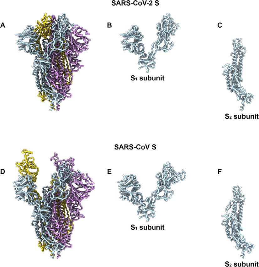
Please cite this article in press as: Walls et al., Structure, Function, and Antigenicity of the SARS-CoV-2 Spike Glycoprotein, Cell (2020),
 https://doi.org/10.1016/j.cell.2020.02.058

                                                                                                Figure 1. ACE2 Is a Functional Receptor for
                                                                                                SARS-CoV-2 S
                                                                                                (A) Entry of MLV pseudotyped with SARS-CoV-2 S,
                                                                                                SARS-CoV S and SARS-CoV-2 Sfur/mut in VeroE6
                                                                                                cells. Data are represented as mean ± standard
                                                                                                deviation of technical triplicates.
                                                                                                (B) Entry of MLV pseudotyped with SARS-CoV-2 S
                                                                                                or SARS-CoV-2 Sfur/mut in BHK cells transiently
                                                                                                transfected with hACE2. The experiments were
                                                                                                carried out with two independent pseudovirus
                                                                                                preparations and a representative experiment is
                                                                                                shown. Data are represented as mean ± standard
                                                                                                deviation of technical triplicates.
                                                                                                (C) Sequence alignment of SARS-CoV-2 S with
                                                                                                multiple related SARS-CoV and SARSr-CoV S gly-
                                                                                                coproteins reveals the introduction of an S1/S2 furin
                                                                                                cleavage site in this novel coronavirus. Identical and
                                                                                                similar positions are respectively shown with white
                                                                                                or red font. The four amino acid residue insertion at
                                                                                                SARS-CoV-2 S positions 681-684 is indicated with
                                                                                                periods. The entire sequence alignment is pre-
                                                                                                sented in Data S1.
                                                                                                (D) Western blot analysis of SARS-CoV S-MLV,
                                                                                                SARS-CoV-2 S-MLV, and SARS-CoV-2 Sfur/mut-
                                                                                                MLV pseudovirions. See also Data S1.

2015; Park et al., 2016; Walls et al., 2016a). The distal S1 subunit    antibodies (Abs) upon infection and the focus of therapeutic
comprises the receptor-binding domain(s) and contributes to             and vaccine design. S trimers are extensively decorated with
stabilization of the prefusion state of the membrane-anchored           N-linked glycans that are important for proper folding (Rossen
S2 subunit that contains the fusion machinery (Gui et al., 2017;        et al., 1998) and for modulating accessibility to host proteases
Kirchdoerfer et al., 2016; Pallesen et al., 2017; Song et al.,          and neutralizing Abs (Walls et al., 2016b; Walls et al., 2019; Xiong
2018; Walls et al., 2016a; Walls et al., 2017b; Yuan et al.,            et al., 2018; Yang et al., 2015b). We previously characterized
2017). For all CoVs, S is further cleaved by host proteases at          potent human-neutralizing Abs from rare memory B cells of
the so-called S20 site located immediately upstream of the fusion       individuals infected with SARS-CoV (Traggiai et al., 2004) or
peptide (Madu et al., 2009; Millet and Whittaker, 2015). This           MERS-CoV (Corti et al., 2015) in complex with SARS-CoV
cleavage has been proposed to activate the protein for mem-             S and MERS-CoV S to provide molecular-level information of
brane fusion via extensive irreversible conformational changes          the mechanism of competitive inhibition of SB attachment to
(Belouzard et al., 2009; Heald-Sargent and Gallagher, 2012; Mil-        the host receptor (Walls et al., 2019). The S230 anti-SARS-CoV
let and Whittaker, 2014, 2015; Park et al., 2016; Walls et al.,         Ab also acted by functionally mimicking receptor attachment
2017b). As a result, coronavirus entry into susceptible cells is a      and promoting S fusogenic conformational rearrangements
complex process that requires the concerted action of recep-            through a ratcheting mechanism that elucidated the unique
tor-binding and proteolytic processing of the S protein to pro-         nature of the coronavirus membrane fusion activation (Walls
mote virus-cell fusion.                                                 et al., 2019).
   Different coronaviruses use distinct domains within the S1              We report here that ACE2 could mediate SARS-CoV-2
subunit to recognize a variety of attachment and entry receptors,       S-mediated entry into cells, establishing it as a functional
depending on the viral species. Endemic human coronaviruses             receptor for this newly emerged coronavirus. The SARS-CoV-
OC43 and HKU1 attach via their S domain A (SA) to 5-N-                  2 SB engages human ACE2 (hACE2) with comparable affinity
acetyl-9-O-acetyl-sialosides found on glycoproteins and glyco-          to SARS-CoV SB from viral isolates associated with the 2002–
lipids at the host cell surface to enable entry into susceptible        2003 epidemic (i.e., binding with high affinity to hACE2). Tight
cells (Hulswit et al., 2019; Tortorici et al., 2019; Vlasak et al.,     binding to hACE2 could partially explain the efficient transmis-
1988). MERS-CoV S, however, uses domain A to recognize                  sion of SARS-CoV-2 in humans, as was the case for SARS-
non-acetylated sialoside attachment receptors (Li et al., 2017;         CoV. We identified the presence of an unexpected furin cleav-
Park et al., 2019), which likely promote subsequent binding             age site at the S1/S2 boundary of SARS-CoV-2 S, which is
of domain B (SB) to the entry receptor, dipeptidyl-peptidase 4          cleaved during biosynthesis—a novel feature setting this virus
(Lu et al., 2013; Raj et al., 2013). SARS-CoV and several               apart from SARS-CoV and SARSr-CoVs. Abrogation of this
SARS-related coronaviruses (SARSr-CoV) interact directly with           cleavage motif moderately affected SARS-CoV-2 S-mediated
angiotensin-converting enzyme 2 (ACE2) via SB to enter target           entry into VeroE6 or BHK cells but may contribute to expand
cells (Ge et al., 2013; Kirchdoerfer et al., 2018; Li et al., 2005a;    the tropism of this virus, as reported for several highly patho-
Li et al., 2003; Song et al., 2018; Yang et al., 2015a).                genic avian influenza viruses and pathogenic Newcastle dis-
   As the coronavirus S glycoprotein is surface-exposed and             ease virus (Klenk and Garten, 1994; Steinhauer, 1999). We
mediates entry into host cells, it is the main target of neutralizing   determined cryoelectron microscopy (cryo-EM) structures of

2 Cell 180, 1–12, March 19, 2020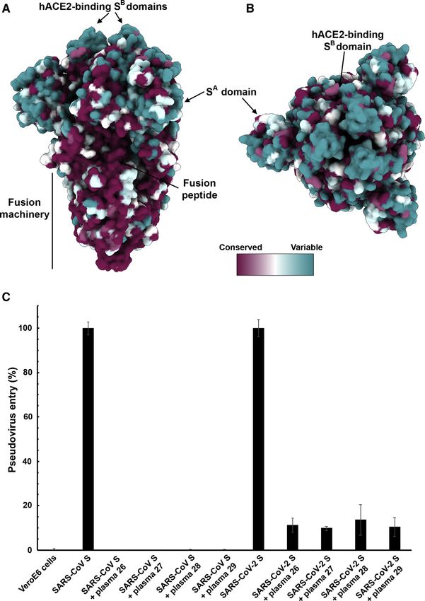
Please cite this article in press as: Walls et al., Structure, Function, and Antigenicity of the SARS-CoV-2 Spike Glycoprotein, Cell (2020),
 https://doi.org/10.1016/j.cell.2020.02.058

                                                                                                Figure 2. SARS-CoV-2 S Recognizes hACE2
                                                                                                with Comparable Affinity to SARS-CoV S
                                                                                                (A and B) Biolayer interferometry binding analysis of
                                                                                                the hACE2 ectodomain to immobilized SARS-CoV-
                                                                                                2 SB (A) or SARS-CoV SB (B). The experiments were
                                                                                                repeated with different protein preparations and one
                                                                                                representative set of curves is shown. Dotted lines
                                                                                                correspond to a global fit of the data using a 1:1
                                                                                                binding model.
                                                                                                (C) Sequence alignment of SARS-CoV-2 SB and
                                                                                                SARS-CoV SB Urbani (late phase of the 2002–2003
                                                                                                SARS-CoV epidemic). Identical and similar posi-
                                                                                                tions are respectively shown with white or red font.
                                                                                                The single amino acid insertion at position 483 of the
                                                                                                SARS-CoV-2 SB is indicated with a period at the
                                                                                                corresponding SARS-CoV SB position. The 14 res-
                                                                                                idues that are key for binding of SARS-CoV SB to
                                                                                                hACE2 are labeled with a star. See also Data S1.

                                                                                               SARSr-CoV WIV-1 and WIV-16 were iso-
                                                                                               lated (Ge et al., 2013; Yang et al.,
                                                                                               2015a). Furthermore, Zhou et al. (2020)
                                                                                               recently reported that SARS-CoV-2 is
                                                                                               most closely related to the bat SARSr-
                                                                                               CoV RaTG13, with which it forms a
                                                                                               distinct lineage from other SARSr-CoVs,
                                                                                               and that their S glycoproteins share 97%
                                                                                               amino acid sequence identity. SARS-
                                                                                               CoV recognizes its entry receptor hACE2
                                                                                               at the surface of type II pneumocytes us-
                                                                                               ing SB, which shares 75% overall amino
                                                                                               acid sequence identity with SARS-CoV-2
                                                                                               SB and 50% identity within their recep-
                                                                                               tor-binding motifs (RBMs) (Li et al.,
                                                                                               2005a; Li et al., 2003; Li et al., 2005c;
                                                                                               Wan et al., 2020). Previous studies also
                                                                                               showed that the host proteases cathepsin
                                                                                               L and TMPRSS2 prime SARS-CoV S for
                                                                                               membrane fusion through cleavage at
                                                                                               the S1/S2 and at the S20 sites (Belouzard
                                                                                               et al., 2009; Bosch et al., 2008; Glowacka
the SARS-CoV-2 S ectodomain trimer and reveal that it adopts            et al., 2011; Matsuyama et al., 2010; Millet and Whittaker, 2015;
multiple SB conformations that are reminiscent of previous re-          Shulla et al., 2011).
ports on both SARS-CoV S and MERS-CoV S. Finally, we                       We set out to investigate the functional determinants of
show that SARS-CoV S mouse polyclonal sera potently in-                 S-mediated entry into target cells using a murine leukemia virus
hibited entry into target cells of SARS-CoV-2 S pseudotyped vi-         (MLV) pseudotyping system (Millet and Whittaker, 2016). To
ruses. Collectively, these results pave the way for designing           assess the ability of SARS-CoV-2 S to promote entry into target
vaccines eliciting broad protection against SARS-CoV-2,                 cells, we first compared transduction of SARS-CoV-2 S-MLV
SARS-CoV, and SARSr-CoV.                                                and SARS-CoV S-MLV into VeroE6 cells, that are known to ex-
                                                                        press ACE2 and support SARS-CoV replication (Drosten et al.,
RESULTS                                                                 2003; Ksiazek et al., 2003). Both pseudoviruses entered cells
                                                                        equally well (Figure 1A), suggesting that SARS-CoV-2 S-MLV
ACE2 Is an Entry Receptor for SARS-CoV-2                                could use African green monkey ACE2 as entry receptor. To
The SARS-CoV-2 S glycoprotein shares 76% amino acid                     confirm these results, we evaluated entry into BHK cells and
sequence identity with the SARS-CoV S Urbani and 80% iden-              observed that transient transfection with hACE2 rendered
tity with bat SARSr-CoV ZXC21 S and ZC45 S glycoprotein. The            them susceptible to transduction with SARS-CoV-2 S-MLV
latter two SARSr-CoV sequences were identified from Rinolo-             (Figure 1B). These results demonstrate hACE2 is a functional re-
phus sinicus (Chinese horseshoe bats), the species from which           ceptor for SARS-CoV-2, in agreement with recently reported

                                                                                                            Cell 180, 1–12, March 19, 2020 3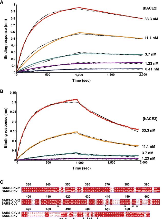
Please cite this article in press as: Walls et al., Structure, Function, and Antigenicity of the SARS-CoV-2 Spike Glycoprotein, Cell (2020),
 https://doi.org/10.1016/j.cell.2020.02.058

Table 1. Kinetic Analysis of hACE2 Binding to SARS-CoV-2 SB and SARS-CoV SB by Biolayer Interferometry
                                                 SARS-CoV-2 SB                                                     SARS-CoV SB
KD (nM)                                          1.2 ± 0.1                                                         5.0 ± 0.1
      1    1
kon (M .s )                                      1.4 3 10 (2.3 ± 1.4 3 10 )
                                                            5                 5
                                                                                                                   1.4 3 105 (1.7 ± 0.7 3 105)
     1                                                     4                    4
koff (s )                                        1.6 3 10        (1.7 ± 0.8 3 10 )                                 7.1x 10⁴ (8.7 ± 5.1 3 104)
Values reported represent the global fit to the data shown in Figures 2A and 2B and the averages obtained from five (SARS-CoV-2) or four (SARS-CoV)
replicates carried out with different protein preparations are shown in parentheses.

findings (Hoffmann et al., 2020; Letko et al., 2020; Zhou                     transmissibility and disease severity (Guan et al., 2003; Li
et al., 2020).                                                                et al., 2004; Li et al., 2005c; Wan et al., 2020). Indeed, specific
   Sequence analysis of SARS-CoV-2 S reveals the presence of                  SB mutations enabled efficient binding to hACE2 of SARS-CoV
a four amino acid residue insertion at the boundary between                   isolates from the three phases of the 2002–2003 epidemic, which
the S1 and S2 subunits compared with SARS-CoV S and                           were associated with marked disease severity (Consortium,
SARSr-CoV S (Figure 1C). This results in the introduction of a                2004; Kan et al., 2005; Li et al., 2005c; Sui et al., 2004). In
furin cleavage site, a feature conserved among the 144 SARS-                  contrast, SARS-CoV isolates detected during the brief 2003–
CoV-2 isolates sequenced to date but not in the closely related               2004 re-emergence interacted more weakly with hACE2 and
RaTG13 S (Zhou et al., 2020). Using western blot analysis, we                 had low pathogenicity and transmissibility (Consortium, 2004;
observed that SARS-CoV-2 S was virtually entirely processed                   Kan et al., 2005; Li et al., 2005c).
at the S1/S2 site during biosynthesis in HEK293T cells, presum-                  To understand the contribution of receptor interaction to the
ably by furin in the Golgi compartment (Figure 1D). This observa-             infectivity of SARS-CoV-2, we characterized engagement of
tion contrasts with SARS-CoV S, which was incorporated into                   hACE2 by SARS-CoV-2 SB and SARS-CoV SB side-by-side.
pseudovirions largely uncleaved (Figure 1D). To study the influ-              We used biolayer interferometry to study binding kinetics
ence on pseudovirus entry of the SARS-CoV-2 S1/S2 furin                       and affinity of the purified hACE2 ectodomain to SARS-CoV-
cleavage site, we designed an S mutant lacking the four                       2 SB and SARS-CoV SB immobilized at the surface of biosen-
amino acid residue insertion and the furin cleavage site by                   sors. We found that hACE2 bound to SARS-CoV-2 SB and
mutating Q677TNSPRRARYSV687 (wild-type SARS-CoV-2 S) to                       SARS-CoV SB with respective equilibrium dissociation con-
Q677TILRYSV683 (SARS-CoV-2 Sfur/mut). SARS-CoV-2 Sfur/mut                     stants of 1.2 nM (Figure 2A) and 5.0 nM (Figure 2B), and com-
preserves only the conserved arginine residue at position 685                 parable kinetic rate constants, although the off-rate, were
of wild-type SARS-CoV-2 S, thereby mimicking the S1/S2                        slightly higher for SARS-CoV SB (Table 1). The affinity deter-
cleavage site of the related SARSr-CoV S CZX21 (Figure 1D).                   mined here for SARS-CoV SB binding to hACE2 is in line
SARS-CoV-2 Sfur/mut is therefore expected to undergo process-                 with prior studies and the tighter apparent interactions formed
ing at the S1/S2 site upon encountering a target cell, similar to             by SARS-CoV SB compared with the SARS-CoV S1 subunit or
SARS-CoV S and SARSr-CoV S, i.e., via TMPRSS2 and/or                          the SARS-CoV S trimer (Kirchdoerfer et al., 2018; Li et al.,
cathepsin L (Belouzard et al., 2009; Bosch et al., 2008; Glowacka             2005c; Sui et al., 2004; Walls et al., 2019; Wong et al.,
et al., 2011; Matsuyama et al., 2010; Millet and Whittaker, 2015;             2004). Previous structural work identified 14 positions that
Shulla et al., 2011). As expected, SARS-CoV-2 Sfur/mut-MLV                    are key for binding of SARS-CoV SB to hACE2: T402, R426,
harbored uncleaved S upon budding (Figure 1D). The observed                   Y436, Y440, Y442, L472, N473, Y475, N479, Y484, T486,
transduction efficiency of VeroE6 cells was higher for SARS-                  T487, G488, and Y491 (Li et al., 2005a). Analysis of the 144
CoV-2 Sfur/mut-MLV than for SARS-CoV-2 S-MLV (Figure 1A),                     SARS-CoV-2 genome sequences available from the Global
whereas the opposite trend was observed for transduction                      Initiative on Sharing All Influenza Data (GISAID) (Elbe and
of hACE2-expressing BHK cells (Figure 1B). These results                      Buckland-Merrett, 2017) shows that 8 out of these 14
suggest that S1/S2 cleavage during S biosynthesis was not                     positions are strictly conserved in SARS-CoV-2 SB, whereas
necessary for S-mediated entry in the conditions of our experi-               the other 6 positions are (semi)conservatively substituted:
ments (Figures 1C and 1D). We speculate that the detection                    R426SARS-CoVN439SARS-CoV-2,          Y442SARS-CoVL455SARS-CoV-2,
of a polybasic cleavage site in the fusion glycoprotein of                    L472SARS-CoVF486SARS-CoV-2,          N479SARS-CoVQ493SARS-CoV-2,
SARS-CoV-2 could putatively expand its tropism and/or                         Y484SARS-CoVQ498SARS-CoV-2, and T487SARS-CoVN501SARS-CoV-2
enhance its transmissibility, compared with SARS-CoV and                      (Figure 2C). The conservation of many key contact residues
SARSr-CoV isolates, due to the near-ubiquitous distribution                   could explain the similar binding affinities of SARS-CoV-2 SB
of furin-like proteases and their reported effects on other                   and SARS-CoV SB for hACE2. No mutations of residues pre-
viruses (Klenk and Garten, 1994; Millet and Whittaker, 2015;                  dicted to contact hACE2 have been observed among SARS-
Steinhauer, 1999).                                                            CoV-2 S sequences available to date. However, we note that
                                                                              SARSr-CoV ZXC21 and ZC45, which harbor the most closely
SARS-CoV-2 Recognizes hACE2 with Comparable                                   related S sequences after SARSr-CoV RaTG13, have a deletion
Affinity to SARS-CoV                                                          in the RBD that could affect binding to hACE2 (between SARS-
The binding affinity of SARS-CoV for hACE2 correlates with the                CoV-2 S residues 473–486 ) (Data S1). Collectively, these results
overall rate of viral replication in distinct species as well as with         suggest that SARS-CoV-2 is at least as well adapted to the

4 Cell 180, 1–12, March 19, 2020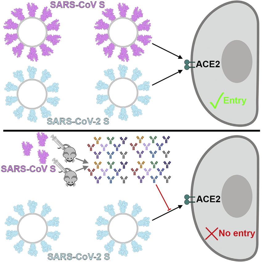
Please cite this article in press as: Walls et al., Structure, Function, and Antigenicity of the SARS-CoV-2 Spike Glycoprotein, Cell (2020),
 https://doi.org/10.1016/j.cell.2020.02.058

                                                                                                Figure 3. Cryo-EM Structures of the SARS-
                                                                                                CoV-2 S Glycoprotein
                                                                                                (A) Closed SARS-CoV-2 S trimer unsharpened cryo-
                                                                                                EM map.
                                                                                                (B and C) Two orthogonal views from the side (B)
                                                                                                and top (C) of the atomic model of the closed SARS-
                                                                                                CoV-2 S trimer.
                                                                                                (D) Partially open SARS-CoV-2 S trimer un-
                                                                                                sharpened cryo-EM map (one SB domain is open).
                                                                                                (E-F) Two orthogonal views from the side (E) and top
                                                                                                (F) of the atomic model of the closed SARS-CoV-2 S
                                                                                                trimer. The glycans were omitted for clarity. See also
                                                                                                Figures S1 and S2.

                                                                                                   We determined a reconstruction of the
                                                                                                 closed SARS-CoV-2 S ectodomain trimer
                                                                                                 at 2.8 Å resolution (applying 3-fold sym-
                                                                                                 metry) and an asymmetric reconstruction
                                                                                                 of the trimer with a single SB domain
                                                                                                 opened at 3.2 Å resolution (Figures 3A–
                                                                                                 3H and S1; Table S1). The S2 fusion ma-
                                                                                                 chinery is the best resolved part of the
                                                                                                 map, whereas the SA and SB domains
                                                                                                 are less well resolved, presumably
                                                                                                 because of conformational heterogeneity.
                                                                                                 The atomic model comprises residues 27-
                                                                                                 1147, with internal breaks corresponding
                                                                                                 to flexible regions (including part of the
                                                                                                 RBM), and lacks the C-terminal most
                                                                                                 segment (including the heptad repeat 2)
hACE2 ortholog as the 2002–2003 epidemic strains of SARS-                that is not visible in the map, as is the case for all S structures
CoV, which could explain the efficient transduction efficiency           determined to date. Overall, the SARS-CoV-2 S ectodomain is
mediated by their respective S glycoproteins (Figures 1A and             a 160-Å-long trimer with a triangular cross-section, resembling
1B) and the current rapid SARS-CoV-2 transmission in humans.             the closely related SARS-CoV S structure (Figures S2A–S2F)
                                                                         (Gui et al., 2017; Kirchdoerfer et al., 2018; Song et al., 2018;
Architecture of the SARS-CoV-2 S Glycoprotein Trimer                     Walls et al., 2019; Yuan et al., 2017).
To enable single-particle cryo-EM study of the SARS-CoV-2 S                 As is the case for other b-coronavirus S glycoproteins,
glycoprotein, we designed a prefusion stabilized ectodomain              including SARS-CoV S (Gui et al., 2017; Kirchdoerfer et al.,
trimer construct with an abrogated furin S1/S2 cleavage site (Tor-       2018; Pallesen et al., 2017; Park et al., 2019; Song et al., 2018;
torici et al., 2019; Walls et al., 2017a; Walls et al., 2016a; Walls     Tortorici et al., 2019; Walls et al., 2016a; Walls et al., 2016b; Walls
et al., 2019), two consecutive proline stabilizing mutations (Kirch-     et al., 2019; Yuan et al., 2017), the S1 subunit has a V-shaped
doerfer et al., 2018; Pallesen et al., 2017), and a C-terminal foldon    architecture (Figures S2B–S2E). In the closed S trimer, the three
trimerization domain (Miroshnikov et al., 1998). Three-dimensional       hACE2-recognition motifs, whose location was inferred based
(3D) classification of the cryo-EM data revealed the presence of         on the crystal structure of SARS-CoV SB in complex with
multiple conformational states of SARS-CoV-2 S corresponding             hACE2 (Li et al., 2005a), are buried at the interface between
to distinct organization of the SB domains within the S1 apex.           protomers. As a result, SARS-CoV-2 SB opening is expected
Approximately half of the particle images selected correspond to         to be necessary for interacting with ACE2 at the host cell surface
trimers harboring a single SB domain opened whereas the remain-          and initiating the conformational changes leading to cleavage of
ing half was accounted for by closed trimers with the three SB do-       the S20 site, membrane fusion and viral entry (Gui et al., 2017;
mains closed. A recently determined SARS-CoV-2 S structure               Kirchdoerfer et al., 2018; Millet and Whittaker, 2014; Pallesen
also detected trimers with a single SB domain opened but none            et al., 2017; Park et al., 2016; Song et al., 2018; Walls et al.,
entirely closed (Wrapp et al., 2020). The observed conformational        2019; Yuan et al., 2017).
variability of SB domains is reminiscent of observations made with          As the SARS-CoV-2 and SARS-CoV S2 subunits share 88%
SARS-CoV S and MERS-CoV S trimers, although we did not                   sequence identity, they are structurally conserved and can be
detect trimers with two SB domains open and the distribution of          superimposed with 1.2 Å root-mean-square deviation (rmsd)
particles across the S conformational landscape varies among             over 417 aligned Ca positions (Figures S2E and S2F). Only the
studies (Gui et al., 2017; Kirchdoerfer et al., 2018; Pallesen et al.,   most N-terminal part of the fusion peptide is resolved in the
2017; Song et al., 2018; Walls et al., 2019; Yuan et al., 2017).         map, as was the case for previously determined SARS-CoV S

                                                                                                            Cell 180, 1–12, March 19, 2020 5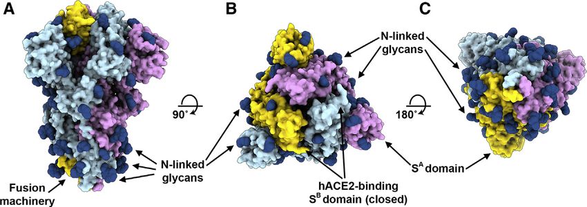
Please cite this article in press as: Walls et al., Structure, Function, and Antigenicity of the SARS-CoV-2 Spike Glycoprotein, Cell (2020),
 https://doi.org/10.1016/j.cell.2020.02.058

                                                                                                Figure 4. Organization of the SARS-CoV-2 S
                                                                                                N-Linked Glycans
                                                                                                (A–C) Ribbon diagrams of the SARS-CoV-2 S
                                                                                                closed structure rendered as a surface with glycans
                                                                                                resolved in the cryo-EM map rendered as dark blue
                                                                                                spheres. See also Table 2 and Data S1.

                                                                                              SARS-CoV S-mediated entry into target
                                                                                              cells. All sera tested completely inhibited
                                                                                              transduction of SARS-CoV S-MLV and
                                                                                              reduced SARS-CoV-2 S-MLV transduc-
                                                                                              tion to 10% of control in VeroE6 cells
structures. The sequence and conformational conservation of             (Figure 5C). The elicitation of a heterotypic response blocking
the fusion peptide region observed across SARS-CoV-2 S and              SARS-CoV-2 S-mediated entry into host cells concurs with the
SARS-CoV S suggests that Abs targeting this functionally impor-         sequence and structural conservation of SARS-CoV-2 S and
tant motif might cross-react and neutralize the two viruses as          SARS-CoV S along with their comparable glycans shields and
well as related coronaviruses.                                          suggests that immunity against one virus of the sarbecovirus
   We previously showed that coronavirus S glycoproteins                subgenus can potentially provide protection against related
are densely decorated by heterogeneous N-linked glycans                 viruses.
protruding from the trimer surface (Walls et al., 2016b; Walls
et al., 2019; Xiong et al., 2018). These oligosaccharides               DISCUSSION
participate in S folding (Rossen et al., 1998), affect priming
by host proteases (Yang et al., 2015b), and might modulate              Receptor recognition is the first step of viral infection and is a
antibody recognition (Pallesen et al., 2017; Walls et al.,              key determinant of host cell and tissue tropism. Enhanced
2019). SARS-CoV-2 S comprise 22 N-linked glycosylation                  binding affinity between SARS-CoV S and hACE2 was pro-
sequons per protomer and oligosaccharides are resolved in               posed to correlate with increased virus transmissibility and
the cryo-EM map for 16 of these sites (Figure 4). By compar-            disease severity in humans (Li et al., 2005c). Indeed, SARS-
ison, SARS-CoV S possesses 23 N-linked glycosylation se-                CoV isolates from the three phases of the 2002–2003
quons per protomer, and we previously experimentally                    epidemic were more efficiently transmitted among humans
confirmed that at least 19 of them are glycosylated (Walls              and more pathogenic than the isolates associated with the
et al., 2019). 20 out of 22 SARS-CoV-2 S N-linked glycosyl-             2003–2004 re-emergence that caused only a few cases, in
ation sequons are conserved in SARS-CoV S (Table 2).                    line with their binding affinities for hACE2 (Consortium,
Specifically, 9 out of 13 glycans in the S1 subunit and all 9           2004; Kan et al., 2005; Li et al., 2005c). Moreover, the ability
glycans in the S2 subunit are conserved among SARS-                     to engage ACE2 from different animal species appears to
CoV-2 S and SARS-CoV S. Furthermore, S2 N-linked glyco-                 reflect host susceptibility to SARS-CoV infection and facili-
sylation sequons are mostly conserved across SARSr-CoV S                tated the jump of the virus from animals to humans (Li,
glycoproteins, suggesting that accessibility of the fusion              2008; Li et al., 2004). We report here that SARS-CoV-2 uses
machinery to Abs will be comparable among these viruses                 hACE2 as an entry receptor and recognizes it with a similar
(Table 2 and Data S1).                                                  affinity to the 2002–2003 SARS-CoV isolates, which suggests
                                                                        it can spread efficiently in humans, in agreement with the
SARS-CoV S Elicits Neutralizing Abs Against                             numerous SARS-CoV-2 human-to-human transmission
SARS-CoV-2 S                                                            events reported to date.
Mapping of the sequence conservation across multiple S se-                 Besides binding to host cell receptors, priming of the S
quences from the sarbecovirus subgenus underscores that the             glycoprotein by host proteases through cleavage at the S1/S2
S2 fusion machinery is more conserved than the S1 subunit               and the S20 sites is another crucial factor modulating tropism
with the highest divergence found within SA and SB (Figures 5A          and pathogenicity (Millet and Whittaker, 2015). For instance,
and 5B). These observations are in line with (1) the fact that          entry of the MERS-CoV-related bat coronavirus HKU4 into
some, but not all, of these viruses use ACE2 as entry receptor          human cells required addition of exogenous trypsin, indicating
(Ge et al., 2013; Ren et al., 2008; Yang et al., 2015a); and (2)        that S proteolytic activation of this bat virus did not occur in
that the S1 subunit is more exposed at the viral surface than           human cells (despite its ability to recognize human DPP4)
the fusion machinery and is likely to be subject to a more              (Wang et al., 2014; Yang et al., 2014). Subsequent work
stringent selection pressure from the immune system.                    suggested that a glycan present near the S1/S2 boundary
   Based on these observations, we hypothesized that exposure           accounted for the lack of proteolytic priming of HKU4 S and
to one of the two viruses could elicit cross-reactive and               that its removal enhanced pseudovirus entry in human cells
potentially neutralizing Abs against the other virus. We therefore      (Yang et al., 2015b). The presence of a polybasic cleavage
investigated the ability of plasma from four mice immunized             site, that can be processed by furin-like proteases, is a signature
with a stabilized SARS-CoV S to inhibit SARS-CoV-2 S- and               of several highly pathogenic avian influenza viruses and

6 Cell 180, 1–12, March 19, 2020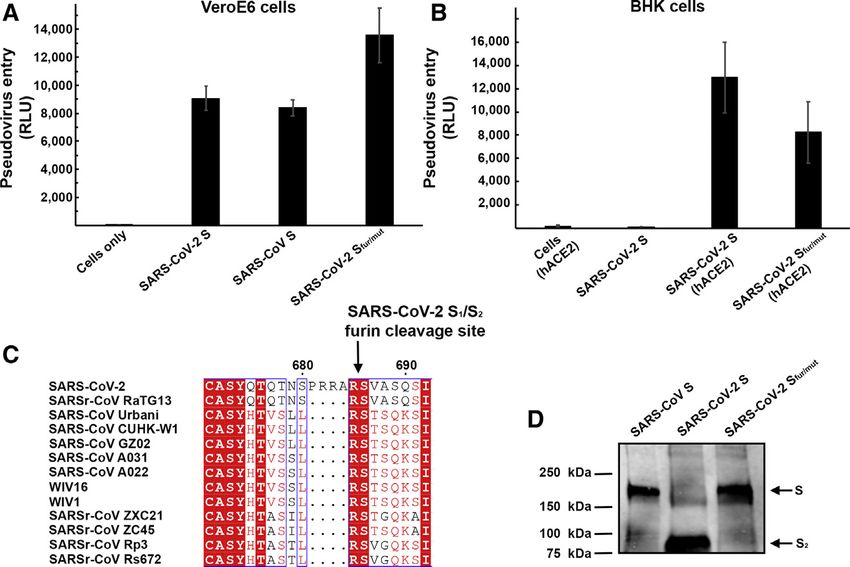
Please cite this article in press as: Walls et al., Structure, Function, and Antigenicity of the SARS-CoV-2 Spike Glycoprotein, Cell (2020),
 https://doi.org/10.1016/j.cell.2020.02.058

Table 2. Conservation of N-Linked Glycosylation Sequons in                  distinct conformational states resulting from SB opening at
SARS-CoV-2 S and SARS-CoV S                                                 the trimer apex. These structural changes are necessary for
SARS-CoV-2 S                                            SARS-CoV S          receptor engagement of these three viruses and lead to initiation
                                                                            of fusogenic conformational changes (Song et al., 2018; Walls
N17LT                                                   T21FD
                                                                            et al., 2017b; Walls et al., 2019). In contrast, only closed S
P25PA                                                   N29YT
                                                                            trimers have been detected for the four other human-infecting
N61VT                                                   N65VT               coronaviruses: HCoV-NL63 (Walls et al., 2016b), HCoV-OC43
H69VS                                                   N73HT               (Tortorici et al., 2019), HCoV-HKU1 (Kirchdoerfer et al., 2016),
N74GT                                                   ...                 and HCoV-229E (Li et al., 2019). As HCoV-NL63 and HCoV-
S112KT                                                  N109KS              229E are known to engage protein receptors through SB
N121NA                                                  N118NS              (Wong et al., 2017; Wu et al., 2009), trimer opening is also
N122AT                                                  N119ST
                                                                            expected to occur to expose their RBMs that are otherwise
                                                                            buried at the interface between protomers in the closed S
N149KS                                                  T146QT
                                                                            trimers (Li et al., 2019; Walls et al., 2016b). Regardless of the
N165CT                                                  N158CT
                                                                            nature of the receptor and the location of the receptor-binding
N234IT                                                  N227IT              domains, removal of the trimeric S1 crown is expected to be
N282GT                                                  N269GT              necessary for all coronaviruses to allow the large-scale S2
N331IT                                                  N318IT              conformational changes leading to fusion of the viral and
N343AT                                                  N330AT              host membranes (Walls et al., 2017b). Collectively, these data
N370SA                                                  N357ST              underscore that S glycoprotein trimers found in highly
N603TS                                                  N589AS
                                                                            pathogenic human coronaviruses appear to exist in partially
                                                                            opened states, while they remain largely closed in human
N616CT                                                  N602CT
                                                                            coronaviruses associated with common colds. Based on the
N657NS                                                  D643TS
                                                                            aforementioned data correlating the binding affinity of SARS-
N709NS                                                  N691NT              CoV for hACE2 with the rate of transmissibility, viral replication
N717FT                                                  N699FS              in distinct species, and disease severity (Guan et al., 2003; Li
N801FS                                                  N783FS              et al., 2004; Li et al., 2005c; Wan et al., 2020), we hypothesize
N1074FT                                                 N1056FT             that the most pathogenic coronaviruses will exhibit S glyco-
N1098GT                                                 N1080GT             protein trimers spontaneously sampling closed and open
N1134NT                                                 N1116NT
                                                                            conformations, as is the case for SARS-CoV-2, SARS-CoV and
                                                                            MERS-CoV.
N1158HT                                                 N1140HT
                                                                               The striking structural similarity and sequence conservation
N1173AS                                                 N1155AS
                                                                            among the SARS-CoV-2 S and SARS-CoV S glycoproteins
N1194ES                                                 N1176ES             emphasize the close relationship between these two viruses
Italic font indicates the absence of a glycosylation sequon and deletions   that recognize hACE2 to enter target cells. This resemblance
are indicated with periods. Glycans observed in the SARS-CoV-2 S cryo-      is further strengthened by our finding that SARS-CoV S
EM map are underlined. See also Data S1.
                                                                            elicited polyclonal Ab responses, potently neutralizing
                                                                            SARS-CoV-2 S-mediated entry into cells. We surmise most
pathogenic Newcastle disease virus (Klenk and Garten, 1994;                 of these Abs target the highly conserved S2 subunit (including
Steinhauer, 1999). Strikingly, SARS-CoV-2 S harbors a furin                 the fusion peptide region) based on its structural similarity
cleavage site at the S1/S2 boundary, which is processed during              across SARS-CoV-2 and SARS-CoV, the lack of cross-reac-
biosynthesis. The presence of a furin cleavage site sets                    tivity of several SB-directed Abs (Tian et al., 2020; Wrapp
SARS-CoV-2 S apart from SARS-CoV S (and SARSr-CoV S)                        et al., 2020), and previous reports showing that sera from
that possesses a monobasic S1/S2 cleavage site processed                    SARS-CoV-infected individuals target this region (Zhang
upon entry of target cells (Belouzard et al., 2009; Bosch et al.,           et al., 2004). We note that most SARS-CoV neutralizing Abs
2008; Glowacka et al., 2011; Matsuyama et al., 2010; Millet                 isolated to date target the SB domain and that several of
and Whittaker, 2015; Shulla et al., 2011). We speculate that the            them recognize the RBM and prevent receptor engagement
almost ubiquitous expression of furin-like proteases could                  (Hwang et al., 2006; Rockx et al., 2008; Rockx et al., 2010;
participate in expanding SARS-CoV-2 cell and tissue tropism,                Traggiai et al., 2004; Walls et al., 2019). As the SARS-CoV-2
relative to SARS-CoV, as well as increasing its transmissibility            and SARS-CoV SB domains share 75% amino acid sequence
and/or altering its pathogenicity.                                          identity, future work will be necessary to evaluate whether
   We previously suggested that coronaviruses use conforma-                 any of these Abs neutralize the newly emerged coronavirus.
tional masking and glycan shielding to limit recognition by the             These findings also indicate that it might be difficult to
immune response of infected hosts (Walls et al., 2016b; Walls               distinguish exposure to SARS-CoV-2 from other SARSr-
et al., 2019; Xiong et al., 2018). Similarly to SARS-CoV S and              CoVs in serological studies using S ectodomain trimers and
MERS-CoV S (Gui et al., 2017; Kirchdoerfer et al., 2018; Pallesen           that specific assays will need to be designed. Our results
et al., 2017; Song et al., 2018; Walls et al., 2019; Yuan et al.,           provide a structural framework to identify conserved and
2017), we found that the SARS-CoV-2 S trimer exists in multiple,            accessible epitopes across S glycoproteins that will support

                                                                                                            Cell 180, 1–12, March 19, 2020 7
Please cite this article in press as: Walls et al., Structure, Function, and Antigenicity of the SARS-CoV-2 Spike Glycoprotein, Cell (2020),
https://doi.org/10.1016/j.cell.2020.02.058
Figure 5. SARS-CoV S Elicits Antibodies
Neutralizing SARS-CoV-2 S-Mediated Entry
into Host Cells
(A and B) Sequence conservation of sarbecovirus S
glycoproteins plotted on the SARS-CoV-2 S struc-
ture viewed from the side (A) and top (B). The
sequence alignment was generated using 48 SARS-
CoV-2 S sequences obtained from GISAID in addi-
tion to the sequences listed in Data S1.
(C) Entry of SARS-CoV-2 S-MLV and SARS-CoV
S-MLV is potently inhibited by four SARS-CoV S
mouse polyclonal immune plasma.
ongoing vaccine design efforts. Finally, elicitation of diverse, d METHOD DETAILS
polyclonal Ab responses might prove key in light of the B Transient expression of SARS-CoV-2 and SARS-
diversity of viruses circulating in animal reservoirs and in CoV SB
preventing the possible emergence of viral neutralization B Transient expression of hACE2
escape mutants. B Pseudovirus production
B Western Blotting
B Pseudovirus entry assays
STAR+METHODS
B Biolayer interferometry
B Protein expression and purification
Detailed methods are provided in the online version of this paper
B CryoEM sample preparation and data collection
and include the following:
B Cryo-EM data processing
d KEY RESOURCES TABLE B CryoEM model building and analysis
d LEAD CONTACT AND MATERIALS AVAILABILITY B Immunizations with SARS-CoV S
d EXPERIMENTAL MODEL AND SUBJECT DETAILS d QUANTIFICATION AND STATISTICAL ANALYSIS
B Cell lines d DATA AND CODE AVAILABILITY
8 Cell 180, 1–12, March 19, 2020Please cite this article in press as: Walls et al., Structure, Function, and Antigenicity of the SARS-CoV-2 Spike Glycoprotein, Cell (2020),
https://doi.org/10.1016/j.cell.2020.02.058
SUPPLEMENTAL INFORMATION cryo-microscopy reconstructions. Acta Crystallogr. D Biol. Crystallogr. 71,
136–153.
Supplemental Information can be found online at https://doi.org/10.1016/j. Burkard, C., Verheije, M.H., Wicht, O., van Kasteren, S.I., van Kuppeveld, F.J.,
cell.2020.02.058. Haagmans, B.L., Pelkmans, L., Rottier, P.J., Bosch, B.J., and de Haan, C.A.
(2014). Coronavirus cell entry occurs through the endo-/lysosomal pathway
ACKNOWLEDGMENTS in a proteolysis-dependent manner. PLoS Pathog. 10, e1004502.
Chen, V.B., Arendall, W.B., 3rd, Headd, J.J., Keedy, D.A., Immormino, R.M.,
This study was supported by the National Institute of General Medical Sci-
Kapral, G.J., Murray, L.W., Richardson, J.S., and Richardson, D.C. (2010).
ences (R01GM120553 to D.V.), the National Institute of Allergy and Infectious
MolProbity: all-atom structure validation for macromolecular crystallography.
Diseases (HHSN272201700059C to D.V.), a Pew Biomedical Scholars Award
Acta Crystallogr. D Biol. Crystallogr. 66, 12–21.
(D.V.), an Investigators in the Pathogenesis of Infectious Disease Award from
the Burroughs Wellcome Fund (D.V.), the University of Washington Arnold Consortium, C.S.M.E.; Chinese SARS Molecular Epidemiology Consortium
and Mabel Beckman cryoEM center, the Washington Research Foundation, (2004). Molecular evolution of the SARS coronavirus during the course of the
and the Pasteur Institute (M.A.T.). We are grateful to Lynda Stuart for sharing SARS epidemic in China. Science 303, 1666–1669.
the full-length hACE2 plasmid, to Gary Whittaker for the MLV pseudotyping Corti, D., Zhao, J., Pedotti, M., Simonelli, L., Agnihothram, S., Fett, C., Fer-
system, and to Ning Zheng for providing access to the Octet device. nandez-Rodriguez, B., Foglierini, M., Agatic, G., Vanzetta, F., et al. (2015).
Prophylactic and postexposure efficacy of a potent human monoclonal anti-
AUTHOR CONTRIBUTIONS body against MERS coronavirus. Proc. Natl. Acad. Sci. USA 112,
10473–10478.
A.C.W., Y.-J.P., A.T.M., and D.V. designed the experiments. A.C.W. and Drosten, C., Günther, S., Preiser, W., van der Werf, S., Brodt, H.R., Becker, S.,
M.A.T. expressed and purified the proteins. A.C.W. carried out binding assays Rabenau, H., Panning, M., Kolesnikova, L., Fouchier, R.A., et al. (2003). Iden-
and pseudovirus entry assays. Y.-J.P. prepared samples for cryo-EM and tification of a novel coronavirus in patients with severe acute respiratory syn-
collected the data. Y.-J.P. and D.V. processed the data and built and refined drome. N. Engl. J. Med. 348, 1967–1976.
the atomic models. A.W. and A.T.M. immunized mice. A.C.W., Y.-J.P., and
Elbe, S., and Buckland-Merrett, G. (2017). Data, disease and diplomacy: GI-
D.V. analyzed the data and prepared the manuscript with input from all
SAID’s innovative contribution to global health. Glob Chall. 1, 33–46.
authors.
Emsley, P., Lohkamp, B., Scott, W.G., and Cowtan, K. (2010). Features
DECLARATION OF INTERESTS and development of Coot. Acta Crystallogr. D Biol. Crystallogr. 66,
486–501.
The authors declare no competing financial interests. Frenz, B., Rämisch, S., Borst, A.J., Walls, A.C., Adolf-Bryfogle, J., Schief,
W.R., Veesler, D., and DiMaio, F. (2019). Automatically fixing errors in glyco-
Received: February 19, 2020 protein structures with Rosetta. Structure 27, 134–139.e3.
Revised: February 24, 2020 Ge, X.Y., Li, J.L., Yang, X.L., Chmura, A.A., Zhu, G., Epstein, J.H., Mazet,
Accepted: February 26, 2020 J.K., Hu, B., Zhang, W., Peng, C., et al. (2013). Isolation and characterization
Published: March 9, 2020 of a bat SARS-like coronavirus that uses the ACE2 receptor. Nature 503,
535–538.
REFERENCES
Glowacka, I., Bertram, S., Müller, M.A., Allen, P., Soilleux, E., Pfefferle, S., Stef-
fen, I., Tsegaye, T.S., He, Y., Gnirss, K., et al. (2011). Evidence that TMPRSS2
Agirre, J., Iglesias-Fernández, J., Rovira, C., Davies, G.J., Wilson, K.S., and
activates the severe acute respiratory syndrome coronavirus spike protein for
Cowtan, K.D. (2015). Privateer: software for the conformational validation of
membrane fusion and reduces viral control by the humoral immune response.
carbohydrate structures. Nat. Struct. Mol. Biol. 22, 833–834.
J. Virol. 85, 4122–4134.
Anthony, S.J., Gilardi, K., Menachery, V.D., Goldstein, T., Ssebide, B., Mba-
Goddard, T.D., Huang, C.C., and Ferrin, T.E. (2007). Visualizing density maps
bazi, R., Navarrete-Macias, I., Liang, E., Wells, H., Hicks, A., et al. (2017).
with UCSF Chimera. J. Struct. Biol. 157, 281–287.
Further Evidence for Bats as the Evolutionary Source of Middle East Respira-
tory Syndrome Coronavirus. MBio 8, e00373-17. Goddard, T.D., Huang, C.C., Meng, E.C., Pettersen, E.F., Couch, G.S., Morris,
Ashkenazy, H., Abadi, S., Martz, E., Chay, O., Mayrose, I., Pupko, T., and Ben- J.H., and Ferrin, T.E. (2018). UCSF ChimeraX: Meeting modern challenges in
Tal, N. (2016). ConSurf 2016: an improved methodology to estimate and visu- visualization and analysis. Protein Sci. 27, 14–25.
alize evolutionary conservation in macromolecules. Nucleic Acids Res. 44, Guan, Y., Zheng, B.J., He, Y.Q., Liu, X.L., Zhuang, Z.X., Cheung, C.L., Luo,
W344-50. S.W., Li, P.H., Zhang, L.J., Guan, Y.J., et al. (2003). Isolation and characteriza-
Barad, B.A., Echols, N., Wang, R.Y., Cheng, Y., DiMaio, F., Adams, P.D., and tion of viruses related to the SARS coronavirus from animals in southern China.
Fraser, J.S. (2015). EMRinger: side chain-directed model and map validation Science 302, 276–278.
for 3D cryo-electron microscopy. Nat. Methods 12, 943–946. Gui, M., Song, W., Zhou, H., Xu, J., Chen, S., Xiang, Y., and Wang, X. (2017).
Belouzard, S., Chu, V.C., and Whittaker, G.R. (2009). Activation of the SARS Cryo-electron microscopy structures of the SARS-CoV spike glycoprotein
coronavirus spike protein via sequential proteolytic cleavage at two distinct reveal a prerequisite conformational state for receptor binding. Cell Res. 27,
sites. Proc. Natl. Acad. Sci. USA 106, 5871–5876. 119–129.
Bosch, B.J., van der Zee, R., de Haan, C.A., and Rottier, P.J. (2003). The Haagmans, B.L., Al Dhahiry, S.H., Reusken, C.B., Raj, V.S., Galiano, M.,
coronavirus spike protein is a class I virus fusion protein: structural and Myers, R., Godeke, G.J., Jonges, M., Farag, E., Diab, A., et al. (2014). Middle
functional characterization of the fusion core complex. J. Virol. 77, East respiratory syndrome coronavirus in dromedary camels: an outbreak
8801–8811. investigation. Lancet Infect. Dis. 14, 140–145.
Bosch, B.J., Bartelink, W., and Rottier, P.J. (2008). Cathepsin L functionally Heald-Sargent, T., and Gallagher, T. (2012). Ready, set, fuse! The corona-
cleaves the severe acute respiratory syndrome coronavirus class I fusion pro- virus spike protein and acquisition of fusion competence. Viruses 4,
tein upstream of rather than adjacent to the fusion peptide. J. Virol. 82, 557–580.
8887–8890. Hoffmann, M., Kleine-Weber, H., Krüger, N., Müller, M., Drosten, C., and
Brown, A., Long, F., Nicholls, R.A., Toots, J., Emsley, P., and Murshudov, G. Pöhlmann, S. (2020). The novel coronavirus 2019 (2019-nCoV) uses
(2015). Tools for macromolecular model building and refinement into electron the SARS-coronavirus receptor ACE2 and the cellular protease TMPRSS2
Cell 180, 1–12, March 19, 2020 9Please cite this article in press as: Walls et al., Structure, Function, and Antigenicity of the SARS-CoV-2 Spike Glycoprotein, Cell (2020), https://doi.org/10.1016/j.cell.2020.02.058 for entry into target cells. bioRxiv. https://doi.org/10.1101/2020.01.31. Li, W., Hulswit, R.J.G., Widjaja, I., Raj, V.S., McBride, R., Peng, W., Wi- 929042. dagdo, W., Tortorici, M.A., van Dieren, B., Lang, Y., et al. (2017). Identifi- Hu, B., Zeng, L.P., Yang, X.L., Ge, X.Y., Zhang, W., Li, B., Xie, J.Z., Shen, X.R., cation of sialic acid-binding function for the Middle East respiratory syn- Zhang, Y.Z., Wang, N., et al. (2017). Discovery of a rich gene pool of bat SARS- drome coronavirus spike glycoprotein. Proc. Natl. Acad. Sci. USA 114, related coronaviruses provides new insights into the origin of SARS coronavi- E8508–E8517. rus. PLoS Pathog. 13, e1006698. Li, Z., Tomlinson, A.C., Wong, A.H., Zhou, D., Desforges, M., Talbot, P.J., Huang, C., Wang, Y., Li, X., Ren, L., Zhao, J., Hu, Y., Zhang, L., Fan, G., Xu, J., Benlekbir, S., Rubinstein, J.L., and Rini, J.M. (2019). The human corona- Gu, X., et al. (2020). Clinical features of patients infected with 2019 novel coro- virus HCoV-229E S-protein structure and receptor binding. eLife 8, navirus in Wuhan (China: Lancet). e51230. Hulswit, R.J.G., Lang, Y., Bakkers, M.J.G., Li, W., Li, Z., Schouten, A., Ophorst, Liebschner, D., Afonine, P.V., Baker, M.L., Bunkóczi, G., Chen, V.B., Croll, B., van Kuppeveld, F.J.M., Boons, G.J., Bosch, B.J., et al. (2019). Human co- T.I., Hintze, B., Hung, L.W., Jain, S., McCoy, A.J., et al. (2019). Macromo- ronaviruses OC43 and HKU1 bind to 9-O-acetylated sialic acids via a lecular structure determination using X-rays, neutrons and electrons: conserved receptor-binding site in spike protein domain A. Proc. Natl. Acad. recent developments in Phenix. Acta Crystallogr. D Struct. Biol. 75, Sci. USA 116, 2681–2690. 861–877. Hwang, W.C., Lin, Y., Santelli, E., Sui, J., Jaroszewski, L., Stec, B., Farzan, M., Lu, G., Hu, Y., Wang, Q., Qi, J., Gao, F., Li, Y., Zhang, Y., Zhang, W., Yuan, Y., Marasco, W.A., and Liddington, R.C. (2006). Structural basis of neutralization Bao, J., et al. (2013). Molecular basis of binding between novel human corona- by a human anti-severe acute respiratory syndrome spike protein antibody, virus MERS-CoV and its receptor CD26. Nature 500, 227–231. 80R. J. Biol. Chem. 281, 34610–34616. Madu, I.G., Roth, S.L., Belouzard, S., and Whittaker, G.R. (2009). Character- Kan, B., Wang, M., Jing, H., Xu, H., Jiang, X., Yan, M., Liang, W., Zheng, ization of a highly conserved domain within the severe acute respiratory syn- H., Wan, K., Liu, Q., et al. (2005). Molecular evolution analysis and drome coronavirus spike protein S2 domain with characteristics of a viral geographic investigation of severe acute respiratory syndrome coronavi- fusion peptide. J. Virol. 83, 7411–7421. rus-like virus in palm civets at an animal market and on farms. J. Virol. Matsuyama, S., Nagata, N., Shirato, K., Kawase, M., Takeda, M., and Taguchi, 79, 11892–11900. F. (2010). Efficient activation of the severe acute respiratory syndrome corona- Kirchdoerfer, R.N., Cottrell, C.A., Wang, N., Pallesen, J., Yassine, H.M., virus spike protein by the transmembrane protease TMPRSS2. J. Virol. 84, Turner, H.L., Corbett, K.S., Graham, B.S., McLellan, J.S., and Ward, A.B. 12658–12664. (2016). Pre-fusion structure of a human coronavirus spike protein. Nature Memish, Z.A., Mishra, N., Olival, K.J., Fagbo, S.F., Kapoor, V., Epstein, J.H., 531, 118–121. Alhakeem, R., Durosinloun, A., Al Asmari, M., Islam, A., et al. (2013). Middle Kirchdoerfer, R.N., Wang, N., Pallesen, J., Wrapp, D., Turner, H.L., Cottrell, East respiratory syndrome coronavirus in bats, Saudi Arabia. Emerg. Infect. C.A., Corbett, K.S., Graham, B.S., McLellan, J.S., and Ward, A.B. (2018). Sta- Dis. 19, 1819–1823. bilized coronavirus spikes are resistant to conformational changes induced by Menachery, V.D., Yount, B.L., Jr., Debbink, K., Agnihothram, S., Gralinski, receptor recognition or proteolysis. Sci. Rep. 8, 15701. L.E., Plante, J.A., Graham, R.L., Scobey, T., Ge, X.Y., Donaldson, E.F., et al. Klenk, H.D., and Garten, W. (1994). Host cell proteases controlling virus path- (2015). A SARS-like cluster of circulating bat coronaviruses shows potential ogenicity. Trends Microbiol. 2, 39–43. for human emergence. Nat. Med. 21, 1508–1513. Ksiazek, T.G., Erdman, D., Goldsmith, C.S., Zaki, S.R., Peret, T., Emery, S., Menachery, V.D., Yount, B.L., Jr., Sims, A.C., Debbink, K., Agnihothram, S.S., Tong, S., Urbani, C., Comer, J.A., Lim, W., et al.; SARS Working Group Gralinski, L.E., Graham, R.L., Scobey, T., Plante, J.A., Royal, S.R., et al. (2016). (2003). A novel coronavirus associated with severe acute respiratory syn- SARS-like WIV1-CoV poised for human emergence. Proc. Natl. Acad. Sci. drome. N. Engl. J. Med. 348, 1953–1966. USA 113, 3048–3053. Letko, M., Marzi, A., and Munster, V. (2020). Functional assessment of cell en- Millet, J.K., and Whittaker, G.R. (2014). Host cell entry of Middle East respira- try and receptor usage for SARS-CoV-2 and other lineage B betacoronavi- tory syndrome coronavirus after two-step, furin-mediated activation of the ruses. Nat. Microbiol. Published online February 24, 2020. https://doi.org/10. spike protein. Proc. Natl. Acad. Sci. USA 111, 15214–15219. 1038/s41564-020-0688-y. Millet, J.K., and Whittaker, G.R. (2015). Host cell proteases: Critical deter- Li, F. (2008). Structural analysis of major species barriers between humans and minants of coronavirus tropism and pathogenesis. Virus Res. 202, palm civets for severe acute respiratory syndrome coronavirus infections. 120–134. J. Virol. 82, 6984–6991. Millet, J.K., and Whittaker, G.R. (2016). Murine Leukemia Virus (MLV)-based Li, W., Moore, M.J., Vasilieva, N., Sui, J., Wong, S.K., Berne, M.A., Somasun- Coronavirus Spike-pseudotyped Particle Production and Infection. Bio Pro- daran, M., Sullivan, J.L., Luzuriaga, K., Greenough, T.C., et al. (2003). Angio- toc. 6, e2035. tensin-converting enzyme 2 is a functional receptor for the SARS coronavirus. Miroshnikov, K.A., Marusich, E.I., Cerritelli, M.E., Cheng, N., Hyde, C.C., Nature 426, 450–454. Steven, A.C., and Mesyanzhinov, V.V. (1998). Engineering trimeric Li, W., Greenough, T.C., Moore, M.J., Vasilieva, N., Somasundaran, M., Sulli- fibrous proteins based on bacteriophage T4 adhesins. Protein Eng. 11, van, J.L., Farzan, M., and Choe, H. (2004). Efficient replication of severe acute 329–332. respiratory syndrome coronavirus in mouse cells is limited by murine angio- Pallesen, J., Wang, N., Corbett, K.S., Wrapp, D., Kirchdoerfer, R.N., Turner, tensin-converting enzyme 2. J. Virol. 78, 11429–11433. H.L., Cottrell, C.A., Becker, M.M., Wang, L., Shi, W., et al. (2017). Immunoge- Li, F., Li, W., Farzan, M., and Harrison, S.C. (2005a). Structure of SARS coro- nicity and structures of a rationally designed prefusion MERS-CoV spike anti- navirus spike receptor-binding domain complexed with receptor. Science 309, gen. Proc. Natl. Acad. Sci. USA 114, E7348–E7357. 1864–1868. Park, J.E., Li, K., Barlan, A., Fehr, A.R., Perlman, S., McCray, P.B., Jr., and Gal- Li, W., Shi, Z., Yu, M., Ren, W., Smith, C., Epstein, J.H., Wang, H., Crameri, G., lagher, T. (2016). Proteolytic processing of Middle East respiratory syndrome Hu, Z., Zhang, H., et al. (2005b). Bats are natural reservoirs of SARS-like coro- coronavirus spikes expands virus tropism. Proc. Natl. Acad. Sci. USA 113, naviruses. Science 310, 676–679. 12262–12267. Li, W., Zhang, C., Sui, J., Kuhn, J.H., Moore, M.J., Luo, S., Wong, S.K., Park, Y.J., Walls, A.C., Wang, Z., Sauer, M.M., Li, W., Tortorici, M.A., Bosch, Huang, I.C., Xu, K., Vasilieva, N., et al. (2005c). Receptor and viral determi- B.J., DiMaio, F., and Veesler, D. (2019). Structures of MERS-CoV spike glyco- nants of SARS-coronavirus adaptation to human ACE2. EMBO J. 24, protein in complex with sialoside attachment receptors. Nat. Struct. Mol. Biol. 1634–1643. 26, 1151–1157. 10 Cell 180, 1–12, March 19, 2020
Please cite this article in press as: Walls et al., Structure, Function, and Antigenicity of the SARS-CoV-2 Spike Glycoprotein, Cell (2020),
  https://doi.org/10.1016/j.cell.2020.02.058

Punjani, A., Rubinstein, J.L., Fleet, D.J., and Brubaker, M.A. (2017). cryo-              Walls, A.C., Tortorici, M.A., Bosch, B.J., Frenz, B., Rottier, P.J.M., DiMaio, F.,
SPARC: algorithms for rapid unsupervised cryo-EM structure determination.                 Rey, F.A., and Veesler, D. (2016a). Cryo-electron microscopy structure of a co-
Nat. Methods 14, 290–296.                                                                 ronavirus spike glycoprotein trimer. Nature 531, 114–117.
Raj, V.S., Mou, H., Smits, S.L., Dekkers, D.H., Müller, M.A., Dijkman, R., Muth,         Walls, A.C., Tortorici, M.A., Frenz, B., Snijder, J., Li, W., Rey, F.A., DiMaio, F.,
D., Demmers, J.A., Zaki, A., Fouchier, R.A., et al. (2013). Dipeptidyl peptidase 4        Bosch, B.J., and Veesler, D. (2016b). Glycan shield and epitope masking of a
is a functional receptor for the emerging human coronavirus-EMC. Nature 495,              coronavirus spike protein observed by cryo-electron microscopy. Nat. Struct.
251–254.                                                                                  Mol. Biol. 23, 899–905.
Ren, W., Qu, X., Li, W., Han, Z., Yu, M., Zhou, P., Zhang, S.Y., Wang, L.F.,              Walls, A., Tortorici, M.A., Bosch, B.J., Frenz, B., Rottier, P.J., DiMaio, F., Rey,
Deng, H., and Shi, Z. (2008). Difference in receptor usage between severe                 F.A., and Veesler, D. (2017a). Crucial steps in the structure determination of a
acute respiratory syndrome (SARS) coronavirus and SARS-like coronavirus                   coronavirus spike glycoprotein using cryo-electron microscopy. Protein Sci.
of bat origin. J. Virol. 82, 1899–1907.                                                   26, 113–121.
Rockx, B., Corti, D., Donaldson, E., Sheahan, T., Stadler, K., Lanzavecchia,              Walls, A.C., Tortorici, M.A., Snijder, J., Xiong, X., Bosch, B.-J., Rey, F.A., and
A., and Baric, R. (2008). Structural basis for potent cross-neutralizing hu-              Veesler, D. (2017b). Tectonic conformational changes of a coronavirus spike
man monoclonal antibody protection against lethal human and zoonotic se-                  glycoprotein promote membrane fusion. Proc. Natl. Acad. Sci. USA 114,
vere acute respiratory syndrome coronavirus challenge. J. Virol. 82,                      11157–11162.
3220–3235.                                                                                Walls, A.C., Xiong, X., Park, Y.J., Tortorici, M.A., Snijder, J., Quispe, J., Camer-
Rockx, B., Donaldson, E., Frieman, M., Sheahan, T., Corti, D., Lanzavecchia,              oni, E., Gopal, R., Dai, M., Lanzavecchia, A., et al. (2019). Unexpected Recep-
A., and Baric, R.S. (2010). Escape from human monoclonal antibody neutrali-               tor Functional Mimicry Elucidates Activation of Coronavirus Fusion. Cell 176,
zation affects in vitro and in vivo fitness of severe acute respiratory syndrome          1026–1039.
coronavirus. J. Infect. Dis. 201, 946–955.                                                Wan, Y., Shang, J., Graham, R., Baric, R.S., and Li, F. (2020). Receptor recog-
Rossen, J.W., de Beer, R., Godeke, G.J., Raamsman, M.J., Horzinek, M.C.,                  nition by novel coronavirus from Wuhan: An analysis based on decade-long
Vennema, H., and Rottier, P.J. (1998). The viral spike protein is not involved            structural studies of SARS. J. Virol. Published online January 29, 2020.
in the polarized sorting of coronaviruses in epithelial cells. J. Virol. 72,              https://doi.org/10.1128/JVI.00127-20.
497–503.                                                                                  Wang, M., Yan, M., Xu, H., Liang, W., Kan, B., Zheng, B., Chen, H., Zheng, H.,
Scheres, S.H., and Chen, S. (2012). Prevention of overfitting in cryo-EM struc-           Xu, Y., Zhang, E., et al. (2005). SARS-CoV infection in a restaurant from palm
ture determination. Nat. Methods 9, 853–854.                                              civet. Emerg. Infect. Dis. 11, 1860–1865.
Shulla, A., Heald-Sargent, T., Subramanya, G., Zhao, J., Perlman, S., and Gal-            Wang, Q., Qi, J., Yuan, Y., Xuan, Y., Han, P., Wan, Y., Ji, W., Li, Y., Wu, Y.,
lagher, T. (2011). A transmembrane serine protease is linked to the severe                Wang, J., et al. (2014). Bat origins of MERS-CoV supported by bat corona-
acute respiratory syndrome coronavirus receptor and activates virus entry.                virus HKU4 usage of human receptor CD26. Cell Host Microbe 16,
J. Virol. 85, 873–882.                                                                    328–337.
Song, W., Gui, M., Wang, X., and Xiang, Y. (2018). Cryo-EM structure of the               Wang, R.Y., Song, Y., Barad, B.A., Cheng, Y., Fraser, J.S., and DiMaio, F.
SARS coronavirus spike glycoprotein in complex with its host cell receptor                (2016). Automated structure refinement of macromolecular assemblies from
ACE2. PLoS Pathog. 14, e1007236.                                                          cryo-EM maps using Rosetta. eLife 5, e17219.
Steinhauer, D.A. (1999). Role of hemagglutinin cleavage for the pathogenicity             Wong, S.K., Li, W., Moore, M.J., Choe, H., and Farzan, M. (2004). A 193-amino
of influenza virus. Virology 258, 1–20.                                                   acid fragment of the SARS coronavirus S protein efficiently binds angiotensin-
Sui, J., Li, W., Murakami, A., Tamin, A., Matthews, L.J., Wong, S.K., Moore,              converting enzyme 2. J. Biol. Chem. 279, 3197–3201.
M.J., Tallarico, A.S., Olurinde, M., Choe, H., et al. (2004). Potent neutralization       Wong, A.H.M., Tomlinson, A.C.A., Zhou, D., Satkunarajah, M., Chen, K.,
of severe acute respiratory syndrome (SARS) coronavirus by a human mAb to                 Sharon, C., Desforges, M., Talbot, P.J., and Rini, J.M. (2017). Receptor-bind-
S1 protein that blocks receptor association. Proc. Natl. Acad. Sci. USA 101,              ing loops in alphacoronavirus adaptation and evolution. Nat. Commun.
2536–2541.                                                                                8, 1735.
Suloway, C., Pulokas, J., Fellmann, D., Cheng, A., Guerra, F., Quispe, J.,                Wrapp, D., Wang, N., Corbett, K.S., Goldsmith, J.A., Hsieh, C.L., Abiona, O.,
Stagg, S., Potter, C.S., and Carragher, B. (2005). Automated molecular micro-             Graham, B.S., and McLellan, J.S. (2020). Cryo-EM structure of the 2019-
scopy: the new Leginon system. J. Struct. Biol. 151, 41–60.                               nCoV spike in the prefusion conformation. Science. Published online February
Tegunov, D., and Cramer, P. (2019). Real-time cryo-electron microscopy data               19, 2020. https://doi.org/10.1126/science.abb2507.
preprocessing with Warp. Nat. Methods 16, 1146–1152.                                      Wu, K., Li, W., Peng, G., and Li, F. (2009). Crystal structure of NL63 respiratory
Tian, X., Li, C., Huang, A., Xia, S., Lu, S., Shi, Z., Lu, L., Jiang, S., Yang, Z., Wu,   coronavirus receptor-binding domain complexed with its human receptor.
Y., and Ying, T. (2020). Potent binding of 2019 novel coronavirus spike protein           Proc. Natl. Acad. Sci. USA 106, 19970–19974.
by a SARS coronavirus-specific human monoclonal antibody. Emerg. Mi-                      Xiong, X., Tortorici, M.A., Snijder, J., Yoshioka, C., Walls, A.C., Li, W., McGuire,
crobes Infect. 9, 382–385.                                                                A.T., Rey, F.A., Bosch, B.J., and Veesler, D. (2018). Glycan shield and fusion
Tortorici, M.A., and Veesler, D. (2019). Structural insights into coronavirus en-         activation of a deltacoronavirus spike glycoprotein fine-tuned for enteric infec-
try. Adv. Virus Res. 105, 93–116.                                                         tions. J. Virol. 92, e01628-17.
Tortorici, M.A., Walls, A.C., Lang, Y., Wang, C., Li, Z., Koerhuis, D., Boons,            Yang, Y., Du, L., Liu, C., Wang, L., Ma, C., Tang, J., Baric, R.S., Jiang, S., and
G.J., Bosch, B.J., Rey, F.A., de Groot, R.J., and Veesler, D. (2019). Structural          Li, F. (2014). Receptor usage and cell entry of bat coronavirus HKU4 provide
basis for human coronavirus attachment to sialic acid receptors. Nat. Struct.             insight into bat-to-human transmission of MERS coronavirus. Proc. Natl.
Mol. Biol. 26, 481–489.                                                                   Acad. Sci. USA 111, 12516–12521.
Traggiai, E., Becker, S., Subbarao, K., Kolesnikova, L., Uematsu, Y., Gis-                Yang, X.L., Hu, B., Wang, B., Wang, M.N., Zhang, Q., Zhang, W., Wu, L.J.,
mondo, M.R., Murphy, B.R., Rappuoli, R., and Lanzavecchia, A. (2004).                     Ge, X.Y., Zhang, Y.Z., Daszak, P., et al. (2015a). Isolation and Character-
An efficient method to make human monoclonal antibodies from memory                       ization of a Novel Bat Coronavirus Closely Related to the Direct Progenitor
B cells: potent neutralization of SARS coronavirus. Nat. Med. 10,                         of Severe Acute Respiratory Syndrome Coronavirus. J. Virol. 90,
871–875.                                                                                  3253–3256.
Vlasak, R., Luytjes, W., Spaan, W., and Palese, P. (1988). Human and bovine               Yang, Y., Liu, C., Du, L., Jiang, S., Shi, Z., Baric, R.S., and Li, F. (2015b). Two
coronaviruses recognize sialic acid-containing receptors similar to those of              Mutations Were Critical for Bat-to-Human Transmission of Middle East Respi-
influenza C viruses. Proc. Natl. Acad. Sci. USA 85, 4526–4529.                            ratory Syndrome Coronavirus. J. Virol. 89, 9119–9123.

                                                                                                                                 Cell 180, 1–12, March 19, 2020 11
You can also read