Antagonising Chromatin Remodelling Activities in the Regulation of Mammalian Ribosomal Transcription - MDPI
←
→
Page content transcription
If your browser does not render page correctly, please read the page content below
G C A T
T A C G
G C A T
               genes
Review
Antagonising Chromatin Remodelling Activities in the
Regulation of Mammalian Ribosomal Transcription
Kanwal Tariq              and Ann-Kristin Östlund Farrants *

                                          Department of Molecular Biosciences, The Wenner-Gren Institute, The Arrhenius Labs F4, Svante Arrhenius väg
                                          20C, Stockholm University, SE-106 91 Stockholm, Sweden; kanwal.tariq@su.se
                                          * Correspondence: anki.ostlund@su.se; Tel.: +46-8164-097

                                          Abstract: Ribosomal transcription constitutes the major energy consuming process in cells and is
                                          regulated in response to proliferation, differentiation and metabolic conditions by several signalling
                                          pathways. These act on the transcription machinery but also on chromatin factors and ncRNA. The
                                          many ribosomal gene repeats are organised in a number of different chromatin states; active, poised,
                                          pseudosilent and repressed gene repeats. Some of these chromatin states are unique to the 47rRNA
                                          gene repeat and do not occur at other locations in the genome, such as the active state organised
                                          with the HMG protein UBF whereas other chromatin state are nucleosomal, harbouring both active
                                          and inactive histone marks. The number of repeats in a certain state varies on developmental stage
                                          and cell type; embryonic cells have more rRNA gene repeats organised in an open chromatin state,
                                          which is replaced by heterochromatin during differentiation, establishing different states depending
                                          on cell type. The 47S rRNA gene transcription is regulated in different ways depending on stimulus
                                          and chromatin state of individual gene repeats. This review will discuss the present knowledge
                                          about factors involved, such as chromatin remodelling factors NuRD, NoRC, CSB, B-WICH, histone
                                modifying enzymes and histone chaperones, in altering gene expression and switching chromatin

                                          states in proliferation, differentiation, metabolic changes and stress responses.
Citation: Tariq, K.; Östlund Farrants,
A.-K. Antagonising Chromatin
                                          Keywords: rRNA gene repeats; chromatin states; NuRD; B-WICH; NoRC; histone modifications;
Remodelling Activities in the
                                          non-coding RNA
Regulation of Mammalian Ribosomal
Transcription. Genes 2021, 12, 961.
https://doi.org/10.3390/genes12070961

Academic Editor: Konstantin Panov         1. Introduction
                                                Ribosomal genes, both the nucleolar 47S genes and the 5S rRNA genes, are highly
Received: 24 May 2021                     transcribed and contribute to the major part of all transcripts. The expression is tightly
Accepted: 19 June 2021                    coupled to proliferation and cell growth with a high demand for protein synthesis [1–3]
Published: 24 June 2021                   in mammalian cells. Therefore, ribosomal gene transcription is often dysregulated in
                                          highly proliferating cancer cells and is a target for novel cancer drugs [2–6]. Ribosomal
Publisher’s Note: MDPI stays neutral      transcription is regulated by environmental cues, such as changes in nutrient and energy
with regard to jurisdictional claims in   supply, and thereby adjusts transcription and energy consumption [1,7,8]. The two ribo-
published maps and institutional affil-   somal genes are transcribed by their own transcription machineries: 47S rRNA genes by
iations.
                                          RNA polymerase I (RNA pol I) and 5S rRNAs by RNA pol III. In addition, ribosomal
                                          transcription encompasses the transcription of ribosomal protein-coding genes by RNA pol
                                          II which is coordinated with the transcription by RNA pol I and RNA pol III [9]. Although
                                          the ribosomal genes are regarded as house-keeping genes recent studies have identified
Copyright: © 2021 by the authors.         cell-type-specific expression, giving rise to tissue specific ribosomes with varying activity
Licensee MDPI, Basel, Switzerland.        and specify [10,11]. Variations in ribosomal biogenesis, in particular in rRNA modifications,
This article is an open access article    also contribute to the heterogeneity of ribosomes [12,13].
distributed under the terms and                 Ribosomal biogenesis takes place in the nucleolus, a membrane-less nuclear body
conditions of the Creative Commons
                                          that forms around active Nuclear Organising Regions (NORs), after each mitosis [14].
Attribution (CC BY) license (https://
                                          The NORs comprise gene clusters of the rRNA gene organised head to tail separated by
creativecommons.org/licenses/by/
                                          an intergenic spacer region (IGS) [1,7,15]. Transcription of the genes, the processing and
4.0/).

Genes 2021, 12, 961. https://doi.org/10.3390/genes12070961                                                   https://www.mdpi.com/journal/genes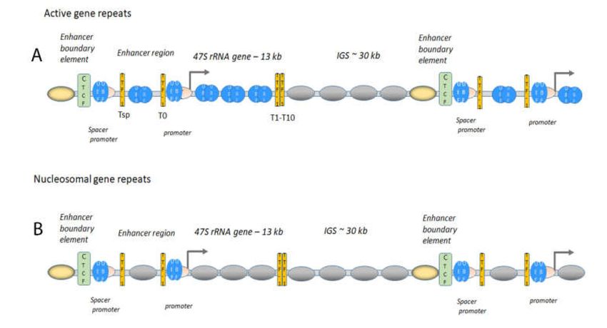
Genes 2021, 12, 961 2 of 21
cleavage of the 47S rRNA transcript and the subsequent assembly of ribosomal RNAs and
protein subunits occur in specific internal nucleolar structures dedicated to the different
processes. The fibrillar centre (FC) assembles around active rRNA gene repeats associated
with the RNA pol I factor upstream binding factor (UBF), the gene transcription occurs
in the interface between the FC and the dense fibrillary centre (DFC), where processing
and cleavage of the 47S rRNA take place, followed by the assembly of the rRNAs with
ribosomal proteins to ribosomal subunits in the granular centre (GC) [16–19]. The formation
of the nucleolar structures depends on transcriptional activity and when inhibited or very
low, nucleolar structures are disrupted and nucleolar proteins are delocalised; fibrillarin,
which is responsible for the 20 -O-methylation of rRNA early in the FC [6,20], and UBF
translocate to the periphery, forming nucleolar caps (Figure 1A). Other abundant nucleolar
proteins, such as the multifunctional proteins nucleolin, involved in activating rRNA gene
transcription [21,22], and nucleophosmin (NPM), mainly involved in ribosomal assembly
and export [22,23], are also leaving the nucleolus upon transcriptional inhibition and
stress. The formation of the different nucleolar centres and the integrity of the nucleolus
are based on phase separation and each centre contains many RNAs and several RNA
binding proteins with intrinsically disordered domains [24]. Fibrillarin associates co-
transcriptionally with the 5’-end of the growing rRNA chain in the FC and forms phase
separated condensates representing DFCs [25]. Nucleolin and NPM are also involved in
phase separation; nucleolin retains histone H2B in the nucleolus in a pre-rRNA dependent
manner [26] and NPM is involved in protein quality control by forming phase separation
condensates with misfolded proteins in the GC [27].
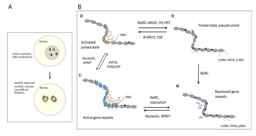
Figure 1. (A) The nucleolar integrity in active and dispersed nucleoli. Nucleolus with several FCs reflecting active
transcription, with UBF (green) and fibrillarin (red) assemblies surrounded by GC. (top panel), and nucleus exposed to
stress such as DNA damage or starvation, reducing transcription, leading dispersed nuclei with a different organisation,
here shown with nucleolar caps, UBF (green) and fibrillarin (red) at the periphery and GC-component in the interior (bottom
panel). (B) Different chromatin states of the mammalian rDNA and the chromatin remodelling complex identified. (1)
The active chromatin state is organised with UBF (bleu ovals), which imposes an open, decondensed chromatin state
that is transcriptionally active if induced by transcription factors (RNA pol I is marked with a yellow triangle), with
nucleosomal chromatin (grey ovals) in the IGS; (2) the activated poised state, organised with nucleosomes (grey ovals)
and is transcriptionally active (RNA pol I is depicted with a yellow triangle, (3) the permissive poised state or the pseudo-
silent state, organised with nucleosomes carrying histone PMTs and heterochromatin factors (marked with purple circles),
and transcriptionally inactive, (4) the constitutively repressed chromatin state, organised in heterochromatin with DNA-
methylations and is transcriptionally silent. The chromatin remodelling complexes and histone chaperones involved in the
switches between states are depicted around the arrows. RNAs are shown, the sense 47S rRNA transcript in (1) and (2) and
ncRNA species from the IGS in (3) and (4).Genes 2021, 12, 961 3 of 21
The nucleolus is also the target of several stress responses, such as heat shock, hypoxia,
and DNA damage, all of which affect the expression of the 47S rRNA transcription [1,7,8].
In addition, nucleolar structure and function are disturbed in many viral infections to
channel the resources to promote viral replication [27]. Many viruses accumulate in the
nucleolus and bind to nucleolar proteins such as nucleolin, NPM and fibrillarin, to interfere
with their function and 47S rRNA gene expression [28]. The many functions that congregate
in the nucleolus and impact the 47S rRNA gene expression link nucleolar function closely
to the status of the cell. The nucleolus constitutes a hub for responses, hence the 47S rRNA
transcription is regulated by environmental states and stress on several levels; modification
of the RNA pol I machinery and the chromatin landscape as well as altering nucleolar
morphology and structure to alter the expression level. This review will focus on the
regulation of 47S rRNA gene repeats in mammalian cells.
1.1. The Active 47S rRNA Gene Transcription Is Organised in an Open Chromatin State
The human haploid genome harbours approximately 200 gene repeats found in
clusters located on five acrocentric chromosomes, chromosome 13, 14, 15, 21 and 22 [7,8].
Not all genes are active in differentiated cells and some gene repeats are gradually silenced
by the establishment of heterochromatin during differentiation [7,29–31]. The number
of active gene repeats correlates to the requirement for protein synthesis in each cell but
since the 47S rRNA transcription also needs to be adjusted according to cellular state and
environmental conditions, several regulatory mechanisms exist also in differentiated cells.
During differentiation, the gene repeats acquire different chromatin states with different
compactions to meet changes in the cell. At least four different states, from open active
gene repeats and to closed repressed gene repeats, have been identified (Figure 1B) [32–37].
The active 47S rRNA gene repeats are organised with the high motility group (HMG)
protein UBF, which binds at the at the enhancer element, UCE, in the promoter, but
also in the gene body and the upstream enhancer region [31,33,35,37]. UBF binds to
UCE as a dimer covering approximately 140 bp of DNA in a single turn [35,38], and
interacts with the TBP-containing TAFI-SL1 complex which binds at the core promoter at
the transcription start site [39,40]. The UBF and SL1 recruit the RNA pol I bound to the
axillary factor TIF-1A/RRN3 to form the pre-initiation complex (PIC) (Figure 2) [39–41].
UBF establishes an open chromatin state specific to the active 47S gene repeats [42] and
is associated with GC-rich DNA at approximately every 170 bp in the gene body [37]. A
further promoter [43], the spacer promoter binding UBF and SL1, is located approximately
two kb upstream of the transcription start [35,37]. RNA pol I and TIF-1A/RRN3 are
also found at the spacer promoter and a non-coding RNA (ncRNA), the spacer RNA, is
produced in mouse [44–46]. Upstream of the spacer promoter is the boundary element,
binding CTCF and cohesin, separating the nucleosomal IGS from the active UBF associated
gene repeat [35,37,47–49]. The enhancer boundary element is in turn flanked by three
to four positioned nucleosomes containing H3K4me2/3 and H2A-Z, indicating that the
boundary constitutes an insulator (Figure 2). Similar enrichment of RNA pol I factors has
been found in human cells approximately 800 bp upstream of transcription start site, with
an enrichment of CTCF [37,48]. In the human IGS, a third promoter two kb upstream of
the transcription start site in human cells was identified, from which an antisense RNA of
unknown function is produced [50]. The open structure organised with UBF is particularly
abundant in ES cells (embryonic stem cells) and mouse embryonic fibroblasts (MEFs),
where few histone marks are found in the 47S rRNA gene repeat (35, 37), whereas that
of differentiated cells may contain more histones (49). Interestingly, malignant human
hepatocytes, HepG2, exhibit an increase of histone H3K4me3 approximately at the spacer
promoter compared to control cells, and this is reduced when UBF is silenced.Genes 2021, 12, 961 4 of 21
Figure 2. Chromatin architecture at the rDNA at the active (A) and the poised state (B). (A) The
genes are organised head-to-tail; UBF (bleu ovals) and SL1 (pink crescent) are bound to the promoter
and the spacer promoter (depicted in the figure). TTF-1 (yellow bars) is bound to its binding sites,
depicted Tsp downstream of the spacer promoter, T0 at the promoter, and T1-T10, at the end of the
47S rRNA gene. UBF is associated with the active 47S rRNA gene body and with the upstream
enhancer region and the IGS downstream of the termination of the gene (T1-T10) is nucleosomal
(grey ovals). CTCF (green rectangle) is flanking the spacer promoter with positioned nucleosomes
with active histone marks and H2AZ (yellow oval nucleosome). These actively marked nucleosome
are also present in the poised state in the bottom panel). (B) The poised state have nucleosomes
(grey ovals) in the gene repeat, UBF (bleu ovals and SL1 (pink crescent) at the promoter and spacer
promoter. TTF-1 (yellow bars) is binding to its sites, Tsp, T0, T1-T10 in the gene repeat. CTCF (green
rectangle) is also here flanking the spacer promoter with positioned nucleosomes with active histone
marks and H2AZ (yellow oval nucleosome).
1.2. The Boundary Element, a Form of Insulator, Is Important in the Maintenance of Chromatin
States and Factor Loading
The boundary element functions as an insulator between chromatin configurations and
may also represent a nucleation site for assemble factor that are responsible for switching
chromatin structures (Figure 2). Several line of evidence support such an organisation:
interfering with the composition of the element by siRNA silencing of CTCF impairs the
loading of UBF on the 47S gene repeats [47] allowing for nucleosomes entering the gene
repeats [32,34–36,42,49]. The boundary element and the adjacent active histones are present
also in repressed gene repeats, suggesting that this site is a “marked” site and involved in
establishing the active chromatin state [31,35,37]. Furthermore, the boundary element is
enriched in chromatin remodelling ATPase SNF2h and the histone acetyl-transferase (HAT)
p300, both of which are involved in altering chromatin configurations. The organisation of
gene repeats with UBF is not sufficient for transcription, some of the active repeats are not
engaged in transcription [35,37], and other factors, such as transcription factors, may be
necessary for proper PIC formation to induce transcription.
1.3. Gene Expression of Active Gene Repeats Requires Transcription Factors
Initiation and elongation of the 47S rRNA transcription are rate limiting steps in re-
sponses to environmental cues without changing the chromatin state [42,51–53]. Enhancing
elongation is the mauclein regulatory step in response to acute growth factor stimulation
when the PIC is still in place, possibly by phosphorylation of UBF in the gene body [52,54].
Growth factor stimulation for cells in G1/G0, for instance after long-term, chronic star-
vation, requires PIC formation, RNA pol I loading as well as elongation regulation [52].
None of these regulatory steps require a change in chromatin states. This is further sup-Genes 2021, 12, 961 5 of 21
ported by the activation of 47S rRNA gene transcription of the proliferative transcription
factor c-MYC in pre-malignant murine B-cells that occurs without a change in open UBF
gene repeats organised with UBF [53]. Taken together, this suggest that the decondenced
chromatin state organised with UBF forms the basis for transcription, as the competent
state [53,55] (Figure 1B, panel 1), which is activated by specific transcription factors and by
post-translational modification (PMT) of general factors.
1.4. Active Gene Repeats Form Loops
Directly upstream of the UCE in the promoter, a binding site, T0, for transcription
termination factor 1 (TTF-1) is located [56], and a similar site, Tsp, is also found at the
spacer promoter, located 40 to 50 bp downstream of the UCE in mouse and human cells
(Figure 2) [25,37]. Several TTF-1 sites, the Sal-boxes or T1-T10, are located at the end of the
rRNA gene repeat to terminate the 47S rRNA transcription and it is also a replication fork
barrier [57,58]. The function of the gene upstream sites is possibly to terminate ncRNAs
produced upstream of the promoter, but the spatial organisation promotes interactions to
form loops [59–61]. The spatial organisation of the active gene repeats in the nucleolus
studied by super-resolution imaging techniques shows that RNA pol I forms ring structures
with a size suggesting that active genes are organised in loops of one or a few genes being
loaded with UBF or nucleosomes [25,62]. These loops may represent the loops between
TTF-1 identified with 3C-techniques [59] or loops with CTCF [60,63]. It is not clear if the
loops are formed between the promoter and the terminator of the same gene or proximal
gene repeats or in what chromatin state these gene repeats are present, but the looping
makes transcription more efficient.
1.5. Acetylation of UBF Enhances 47S rRNA Gene Transcription
Similar to histones, UBF is acetylated by histone acetyl-transferases (HATs), particular
p300/CBP, and deacetylated by histone deacetylases (HDACs) to regulate the 47S rRNA
expression [64,65]. Ribosomal transcription is altered through the cell cycle and UBF-Ac is
higher in G2 when the transcription also is highest [66]. CBP acetylates UBF in S-phase to
increase the interaction with PAF53, a RNA pol I associated factor, resulting in an increased
47S rRNA expression. Lack of acetylation of UBF also contributes to the reduced 47S rRNA
transcription observed in a human Huntington disease model, suggested to be caused
by the mutated Huntingtin protein sequestering the CBP protein [67]. Other HATs have
been identified among processing factors and hALP and 16A/DRIM, t-UTP U3 processing
factors, acetylate UBF which increases its interaction with RNA pol I [68,69].
2. The Inactive 47S rRNA Gene Repeats Are DNA Methylated and Organised in
Constitutive Heterochromatin
The 47S rRNA gene repeats are maintained in and open chromatin state early in
development, and then subsequent silencing of gene repeats is an important process
during differentiation [70]. In the early embryo, most rRNA gene repeats are loaded with
UBF and actively transcribed to meet the requirement of protein synthesis in the rapidly
dividing cells [34,36,70,71] (Figure 1B, panel 1). The chromatin remodelling complex NoRC,
composed of TIP5 (TIF interacting protein 5), the ATPase SNF2h, HDAC1, the histone
methyl-transferase (HMT) SUV39H1, the DNA methylase DNMT3b is instrumental in
establishing heterochromatin at the rDNA to reduce the 47S rRNA gene transcription [72].
It is also involved in establishing heterochromatin outside of the nucleolus, on centric
and peri-centric repeats as well as major and minor repeats and at LINE elements [73].
The NoRC complex contributes to the restructuring of the nuclear architecture during
embryogenesis by silencing active chromatin regions at repetitive regions.
The repressed state established by NoRC is characterised by repressive chromatin
marks, heterochromatin factors, and methylated DNA [72,74–76] (Figure 1B, panel 4).
The complex is recruited to the promoter by TIP5 binding to TTF-1 [56,77], histone
H4Ac [76,78,79] and linker DNA [80]. At the promoter, NoRC moves a promoter-bound
nucleosome to a “closed” position incompatible with the assembly of the pre-initiationGenes 2021, 12, 961 6 of 21
complex [76,80,81]. The closed promoter-bound nucleosome position (−132 to +22 bp)
covers the core promoter and transcription start site, preventing SL1 from binding and
initiating transcription. Subsequently the p-RNA, a fragment from the spacer-RNA, forms
a triple helix with the T0 site, replaces TTF-1 and binds to TIP5 [45,46,75]. The p-RNA
inhibits the ATPase activity of NoRC, which instead recruits the silencing factors HDACs,
SUV39H1, and DNMT3 [81]. The conversion to heterochromatin involves the deacetylation
of histones at the promoter, and the introduction of histone methylation, H3K9me2/3, and
DNA methylation, which together reduces the binding of UBF [74]. The DNA-methylated
promoter-bound nucleosome in quiescent cells also carries the histone modification histone
H4K20me3 [82]. This histone modification is established by SUV420, and the protein is re-
cruited to the rRNA promoter by the anti-sense transcript PAPAS (promoter and pre-rRNA
antisense) [82]. This transcript is produced by RNA pol II in the antisense direction to the
47S rRNA, originates from the gene body but also includes reads through to the promoter,
and is increased upon reduced levels of sense-transcription [82].
Histone variants of the canonical histones have been associated with rDNA. The
DNA-methylated state binds macroH2A, which is associated with silent chromatin [83]. It
accumulates on the rRNA gene promoter and maintains a silent chromatin state. Histone
H1 is also associated with the silent gene repeats. Different H1 variants interact with
silencing factors in the nucleolus, such as macroH2A, heterochromatin proteins 1α (HP1α),
HP1β, and HP1γ and DNMT3a/b, but also with active factors, such as UBF, nucleolin
and topoisomerase I [84–86]. Histone H1.2 interacts with UBF in mitotic NORs [87].
The binding of H1 variants also affect the rRNA gene transcription, where deletion of
UBF [32] and Actinomycin D [88] increase H1 and HP1 association with the rRNA gene
repeat. However, phosphorylated H1.4 co-localises with fibrillarin and actively transcribed
rRNA [88]). Whether histone H1 and HP1 is functioning with NoRC is not clear but it does
not exclusively associate with DNA methylated promoters, which suggests that it also is
present in other chromatin states.
3. The Switch between Active and DNA-Methylated Inactive 47S rRNA Gene Repeats
The switch between active and DNA methylated inactive states involves the NoRC
complex, which replaces UBF for nucleosomes in a process that occurs mainly during
differentiation, embryonic development and malignant transformation. The switch em-
ploys specific transcription factors, chromatin remodelling and histone PTMs (Figure
1B, panel 1 and 4). The transcription factors recruit HDACs and SIRTs (NAD-dependent
histone deacetylases) as well as histone HMTs, such as SUV39H1, either directly or as
part of the NoRC complex to silence the genes [7,72]. The general transcription repressor
BEND3 associates with the 47S rRNA and in its SUMOlated form interacts with NoRC and
stabilises TIP5 by inhibiting its degradation through ubiquitination [89]. Furthermore, a
number of bridging factors have been identified that respond to signalling pathways and
affect the chromatin state. The activation of the 47S rRNA gene transcription by mTOR
in human cell is counteracted by the tumour suppressor inhibitor of growth (ING1) [90].
ING1 is recruited to active rDNA by histone H3K4me3, and it recruits in turn the NoRC
complex, which results in the establishment of heterochromatin and reduced 47S rRNA
gene transcription. ING1 also inhibits the localisation of mTOR to the nucleolus. RUNX2,
important in osteoblast differentiation, is not silencing rRNA gene repeats by NoRC, in-
stead it binds the 47S rRNA gene promoters, recruit HDAC1 to deacetylase UBF, which
leads to a decrease in RNA pol I transcription [91].
During development of certain lineages, transcription must be reactivated to allow for
cellular transitions. This occurs in the epithelial-to-mesenchymal transition (EMT) in cells
that need to develop a migratory cellular program, such as neural crest progenitor cells
in early development. A temporary increased ribosomal transcription in G1/S-phase is
required for EMT and is executed by the EMT-specific transcription factor Snail and the
mTORC2 component Rictor [92]. The increase in transcription is associated with reduced
DNA-methylation and replacement of TIP5 by UBF. A similar process is also found inGenes 2021, 12, 961 7 of 21
cancer cells that rely on EMT for metastasis. It is not known if a chromatin-remodelling
factor is involved in the antagonistic effect to NoRC in this process.
Little is known about the switch between rRNA repeats silenced with the histones
variants macroH2A and H1 and active genes. If these gene repeats also exhibit further
attributes of NoRC silenced chromatin than DNA-methylation is not known. Nevertheless,
nucleolin counteracts the silent state imposed by macroH2A by increasing UBF loading
along the rRNA gene body and increasing the presence of active histone marks at the
promoter [83,93]. Nucleolin may act as a FACT-like H2A-H2B histone chaperon [94,95],
removing macroH2A which leads to UBF loading, preventing TTF-1 binding to the T0 and
the subsequent recruitment of TIP5 and NoRC [93]. In addition, inhibition of rRNA gene
transcription results in higher levels of macroH2A at the 47S rRNA gene repeat, suggesting
that both changes in nucleolin levels in the nucleolus and transcription leads to switches
in chromatin state. Depletion of nucleolin affects the RNA pol I levels at the 5´end of the
rRNA gene body, suggesting that not only transcriptional initiation but also elongation
is affected. A similar effect is shown by the H2A-H2B histone chaperone FACT, which
facilitate elongation through the gene body [96,97]. NPM/B23 also function as a histone
chaperone at the promoter, requiring its RNA-binding domain and UBF to be recruited to
the gene [98]. NPM inhibits histones and possible histone H1 [99] to associate with the 47S
rRNA gene repeat and maintain and open chromatin structure [98]. Altering the binding
of core histones and linker histones to the promoter or the gene body is one way to change
the chromatin states of rRNA gene repeats and regulate gene expression. Nucleolar histone
H1 interacts with both activating and repressing factors and is suggested to be involved in
regulating different states. Several of the H1 isoforms have been localised to the nucleolus;
H1.0, H1.2, H1.3 [86] and phosphorylated forms of H1.2 and H1.4 [88]. Phosphorylated
histone H1.2 and in particular histone H1.4 associate with active 47S rRNA genes repeats
and rRNA inhibition reduces the phosphorylation level. Histone H1 interacts with the
histone chaperones nucleolin, NPM, subunits of FACT as well as the heterochromatic
proteins macroH2A, and HP1 and the active proteins UBF, histone H2AZ, histone H3.3
and DNA topoisomerase II [84,86,87]. The interaction of histone H1 with several histone
chaperones indicates that one function of the chaperones is to regulate histone H1 and UBF
and their PMT at the promoter. Interestingly, H1 knock out gives more DNMT3a/b as
well as proteins involved in chromatin remodelling, such as WSTF and BRG1, enriched
in the nucleolus, indicating that chromatin changes and DNA methylation are involved
in the regulation [85,100]. The changes caused by the histone variants and nucleolar
chaperone proteins do not change the DNA-methylation fraction of gene repeats [49,83]
which suggests that more chromatin states exist, constituted of nucleosomal chromatin on
non-DNA-methylated 47S rRNA repeats.
3.1. The Permissive Poise Chromatin State Is Organised in Bivalent Chromatin on
Unmethylated DNA
The rRNA gene promoters adopt further chromatin states than the active and re-
pressed gene states referred to as the poised and pseudo-silent states, which in contrast to
the repressed promoters silenced by NoRC, assemble on unmethylated DNA. The exact
compositions of these states are largely unknown and they may constitute a group of
differently organised nucleosomal gene repeats [31,37,49,101] with cell type specificity. The
promoters carry UBF and SL1 as well as nucleosomes with bivalent chromatin marks [101]
or histone H1 and HP1 [32,34]. The ChIP seq studies [33,35,37,49] of mouse and human
rDNA suggest that the poised or pseudo-silent state is essentially nucleosomal and the
active gene repeats with an open chromatin configuration are organised with UBF. In
mouse, the poised state is associated with the two ATP-dependent chromatin remodelling
complexes Cockayens syndrome protein B (CSB) and CHD4/ nucleosome remodelling
deacetylase (NuRD), which act on the promoter nucleosomes (Figure 1B, panel 2 and
3) [101]. CSB and CHD4, the ATPase in the NuRD complex, are recruited to the promoter
by TTF-1 during growth arrest and differentiation [101,102]. NuRD is responsible for
the positioning of the promoter-bound nucleosome in the closed state covering the coreGenes 2021, 12, 961 8 of 21
promoter and transcription start site [101]. Although the position of the “closed” poised
nucleosome is the same as that of the “closed” nucleosome formed by NoRC [81], the
associating factors are different; the poised nucleosome carries both activate and silence
histone marks, the active H3K4me3 and the two silencing H3K27me3 and H3K9me2/3,
but no H4K20me3. It also differs from the NoRC “closed” nucleosome in that it is devoid
of methylated DNA and associates with both UBF and SL1. Despite that UBF and SL1 are
present at the promoter, TIF-1/RRN3 and RNA pol I are excluded [101], which shows that
the poised closed nucleosome is not compatible with the assembly of the PIC. NuRD is
suggested to prevent DNA methylation of the promoter, but still keep a poised pool of 47S
rRNA gene repeats available for activation [101].In support of this, NuRD also inhibits TIP5
expression, indirectly contributing to inhibit the DNA methylation of rRNA repeats [103].
3.2. The Activation of the Permissive Poised State Involves Establishing an Active
Nucleosome State
The poised “closed” marked nucleosome at the promoter is moved by the ATPase CSB
to an active position (−157 to −2), compatible with TIF-1A/RRN3 and RNA pol I binding
(Figure 1B, panel 2 and 3) [101]. CSB then recruits PCAF to the promoter to acetylate
histone H4 and histone H3K9, creating an active configuration with histone H4Ac and
histone H3K9Ac [104]. CSB associates also with the gene body, where it recruits the histone
methyl transferase G9a to activate transcription by increasing the level of histone H3K9me2
and HP1γ in the region [102]. Recently, CSB together with CSA was shown to regulate
transcription elongation mainly by recruiting nucleolin in human cells [105].
In addition to CSB, the poised state induced by the NuRD complex is counteracted by
the ATP-dependent chromatin remodelling complex B-WICH (Figure 1B, panel 2 and 3). B-
WICH, comprising WSTF, the ATPase SNF2h and nuclear myosin [106,107], activates rRNA
gene transcription by establishing a more open chromatin configuration at the promoter to
allow SL1, TIF-1A/RRN3 and RNA pol I to bind [108,109]. This state is characterised by
the binding of UBF and a high histone H3K9Ac level at the promoter in human cells [108].
An impaired B-WICH complex, obtained by RNAi silencing of WSTF, results in CHD4 not
being released from the promoter after glucose stimulation, suggesting that B-WICH is
required to replace NuRD to open up the chromatin to allow the pre-initiation complex to
form [109]. B-WICH also allows other factors, such as c-MYC, to bind to the promoter and
enhance expression of the genes [109].
3.3. Transcriptions Factors, c-MYC, Induce Chromatin Changes and Activate Gene Expression
c-MYC is a transcription factor that regulate ribosomal and metabolic genes in re-
sponse to proliferative stimulus and in complex with MAX binds to several position in
rDNA, including a site in the 47S gene promoter [110,111]. NPM facilitates the nucle-
olar entry of c-MYC and its transcription activation [112]. c-MYC recruits the TRRAP
co-regulatory complex that contain the HATs GCN5, TIP60 and PCAF, which acetylate
histones in the promoters [113]. c-MYC is also involved in the induction 47S rRNA gene
transcription in hypertrophic mouse muscle, where it associates with the promoter together
with WSTF [114]. B-WICH opens up chromatin for c-MYC-MAX binding in the intergenic
spacer between the 5S rRNA genes, which is a pre-requisite for histone modifications and
transcription [115]. Along with regulating the ribosomal gene transcription directly, c-MYC
regulates the expression of RNA pol I factors [116]. The regulation of c-MYC influences
ribosomal transcription and some pathways are targeting both c-MYC and ribosomal
factors; c-MYC and UBF are degraded following SUMOlation by PIAS E3 ligases [117] and
c-MYC mRNA stability is coordinated with 47S rRNA gene transcription through the RNA
guanine-7 methyltransferase in the mRNA cap [118].
c-MYC is dysregulated in cancer [119] usually with a concomitant increase in 47S
rRNA gene transcription. This increase may be linked to a change in chromatin state at 47S
rRNA gene repeats but may also result in other changes. In contrast to c-Myc expression
in murine pre-malignant B-cells which have an enhanced rRNA transcription without
chromatin changes, the malignant transformed cells have an increased number of activeGenes 2021, 12, 961 9 of 21
gene repeats, but no change in gene expression occurred [53]. Instead, a higher extent
of interactions between UBF organised rRNA gene repeats and RNA pol II genes in the
peri-nucleolar region appeared [53]. These UBF organised gene repeats were shown to
display an enhanced interaction with RNA pol II gene enhancers in the peri-nucleolar
region. The UBF has been shown to associate with a subset of highly expressed RNA pol
II genes [120]. The change in chromatin state in malignant transformed cells, referred to
as the rDNA class switching, does not involve changes in DNA methylation, most likely
switching an unmethylated repressed pool of 47S rRNA gene repeats [53], such as the
poised or pseudo-silent state.
In addition to c-MYC, other factors, such as ING4, are involved in activating transcrip-
tion by altering the chromatin configuration. ING4 activates transcription by inducing
histone H3K9Ac and histone H4Ac and increasing the level of UBF at promoters [121]. It is
not known whether these transcription factors activate 47S rRNA gene transcription by
altering both active gene repeats and permissive poised repeats at the same time and by
the same mechanism, recruiting chromatin remodellers or histone modifying complexes.
3.4. HATs and HDACs Are Involved in Chromatin Changes
HATs, such as MOF, p300/CBP, PCAF, GCN5, TIP60, are associated with the switch to
an active state introducing histone H3Ac and histone H4Ac [49,72,78,101,108], functioning
with transcription factors, such a c-MYC. In tumour cells, overexpression of LYAR, a
transcription factor in inflammatory pathways, also MYST/KAT7 bound to BRD2 and
BRD4 associates to UBF to acetylate histone H4 and H3 [122]. Not only acetylation of
histones and UBF is associated with activation; subunits in SL1 acetylated by PCAF [123],
which are deacetylated by the NAD-dependent SIRT1 to inhibit transcription during
mitosis [124]. SIRT7 is a further NAD-dependent deacetylase associated with ribosomal
transcription, but unlike SIRT1, it is an activator [125]. SIRT7 localises to the nucleolus,
it interacts with UBF and is required for the release from mitosis [126,127]. SIRT7 has
many targets in the nucleolus affecting both transcription and processing that have been
acetylated by CBS; PAF53, an RNA pol I associated factor, to activate transcription [128],
the helicase DDX21 to resolve R-loop [129] and U3-155, NOP56 and fibrillarin, to promote
processing and cleavage of the 47S rRNA [130,131]. SIRT7 also deacetylate fibrillarin at
the exit of mitosis promoting methylation of histone H2A at glutamine 104 (H2AQ104),
which decompacts the promoter to resume transcription [129,132,133]. Other active histone
marks in RNA pol II transcription, such as H3K4me3 and H3K36me3, are also present 47S
rRNA gene repeats, most likely at the permissive poised state [101]. These modifications
are altered by the histone demethylases KDM2A/B, in response to glucose starvation
and metabolites through AMPK pathway and HP1γ recruitments [134–138]. Two histone
demethylases, PHD8 and KDM4B, are involved in switching to an active chromatin state
by removing methyl groups from histone H3K9me3/2n [139], PHD8 in response to PIP2-
binding [140]. The fact that many histone modifying enzymes associate with the promoter
suggests that they modify nucleosomes in the permissive poised or pseudo-silent state and
facilitate the switch to an active nucleosomal state.
3.5. eNOSC Induces Silencing in Response to Low NADH-Levels
A low energy level is sensed as an increased AMP/ATP ratio by the AMPK pathway,
which downregulates several processes to save energy, among those the 47S rRNA gene
transcription [141]. The AMPK inhibits mTOR which reduces the rRNA gene transcrip-
tion by inhibitory phosphorylation of TIF-1A/RRN3 and UBF [142–145] to prevent the
preinitiation complex from forming [146]. In HeLa cells, which lack the AMPK-pathway,
a chromatin-remodelling complex, eNoSC, which senses a reduced energy level in the
form of an increased NAD+ level has been identified [147]. The eNoSC, comprising nu-
cleomethylin (NML), SIRT1 and SUV39H1, is recruited to the promoter by nucleosomes
harbouring H3K9me2 and in turn establishes heterochromatin by deacetylation and methy-
lation of histone H3K9 [147]. NML also binds to the 47S rRNA and associates with theGenes 2021, 12, 961 10 of 21
promoter first when released after reduction in transcription [148,149]. The NML protein
is also involved in rRNA methylation and function through p53 in senescence [150,151].
Since eNoSC only associates with the promoter after transcription is reduced it is most
likely that the complex functions in the maintenance of a silent chromatin in the energy
saving pathway rather than in establishing a silent chromatin state. In addition to eNOSC,
NuRD establishes a repressed chromatin state in glucose starved cells, and we propose that
B-WICH counteracts this chromatin structure upon glucose stimulation [109].
4. Acute Stress Responses and the Regulation of rRNA Transcription
The nucleolus acts as a hub for different stress responses, such as viral infections,
hypoxia and heat shock, and these responses reduce the 47S rRNA gene transcription.
Many stress responses are sensed by signals and targets the RNA pol I factors directly,
which inhibit the assembly of the PIC but others are altering the chromatin states at the
genes. Hypotonic stress [152] and heat shock [153,154] require the NuRD complex to
establish a silent state at the promoter. SUV420, which is recruited by PAPAS during
growth arrest to establish a compaction of the chromatin, is degraded by the E3-ligase
NEDD4 during stress [152]. This leaves the PAPAS, which forms a triple helix with the
enhancer element at the promoter, to bind to and recruit the dephosphorylated CHD4 in
the NuRD complex to the promoter [154]. The mouse cells, the PAPAS is induced by heat
shock and hypotonic stress by the simultaneous dephosphorylation of TIF-1A/RRN3 as
CHD4 [153,155], and this results in reduced rRNA gene transcription and an increased
PAPAS production.
4.1. lncRNA Originating from the Human IGS Are INVOLVED in Stress Responses with
Reduced 47S rRNA Gene Transcription
pRNA and PAPAS are not found in human cells, but a number of ncRNAs that origi-
nate from the IGS have been identified. These are associated with different stress responses;
heat shock induces the expression of transcripts, IGS16, and IGS22, originating from loci 16
kb and 22 kb from the transcription start site and acidosis induces a transcript, IGS28, from a
locus 28 kb, all of which in the sense direction [156,157]. These ncRNAs bind stress proteins,
such as the chaperon HSP70 and the acidosis responsive von Hippel– Lindau (VHL) protein
and sequester them from their site of action. DNMT1, POLD1, and RNA biogenesis factors,
such as the RNA pol I and III factors RPA16 and RPA40, are other proteins that bind to these
ncRNAs, further emphasising the role of these transcripts in sequestering and immobilising
factors important in different pathways during stress [156,157]. Another long ncRNA, the
PNCTR (pyrimidine-rich noncoding transcript), originating from 28 kb, is proposed to
sequester the RNA binding protein PTBP1 to the peri-nucleolar centre, to interfere with
splicing and prevent apoptosis [158]. This stress related RNA is overexpressed in cancers
and suggested to form a scaffold for the peri-nucleolar centre, a structure which is larger
in cancer cells. The shorter ncRNAs from the IGS also form scaffolds in phase separated
condensates, liquid-like detention centres, with sequestered mobile proteins. These conden-
sates disrupt the nucleolar structure and reduces 47S rRNA gene transcription [159–161].
In particular, IGS16, IGS22, and IGS23 contain low-complexity RNA structures formed by
repetitive cytosine/uracil (CU) or adenosine/guanine (AG) sequences, which are proposed
to immobilise proteins with cationic domain and fibrillation-propensity domains [160]. The
transcription of these short ncRNA initiate the formation of phase-separated aggregates,
which resembles solid-like amyloid bodies, in response to thermal stress and extracellular
acidosis [160]. It has been suggested that these aggregates are under the surveillance of
chaperones in the protein quality control to protect against nucleolar disruption [160,161].
SincRNA transcripts produced by RNA pol I have been identified from the same
region in the human IGS as the stress-induced ncRNA [162]. Similar to the stress-induced
ncRNAs, the sincRNAs disrupt nucleolar structure and reduce 47S gene transcription. The
expression of sincRNAs is reduced by R-loops formed by RNA pol II antisense transcription
at the same locus. The R-loops produced prevent RNA pol I from loading and transcribe
and is regulated by the helicase Sentaxin, which facilitates RNA pol II loading at the IGS.Genes 2021, 12, 961 11 of 21
R-loops are found at many location, which suggests that the whole region is transcribed
and possibly is under control to reduce detrimental ncRNA transcription that can feed
phase separation condensates [163,164].
4.2. Nuclear Integrity Is Important for 47S rRNA Gene Transcription
Nucleolar integrity is tightly coupled to 47S rRNA gene transcription, with many
studies even showing that the assembly of nucleoli at NORs depends on active transcrip-
tion [6,16–20,165–167], and a reduced transcription results in in reorganised or dispersed nu-
cleoli [18,19,24,165,166,168–170]. DNA damage and inhibited 47S gene transcription lead to
a reorganisation of the nucleolar structure, with the typical nucleolar caps [166,168,171,172].
A region immediately outside of the rDNA locus on all five acrocentric chromosomes
harbours conserved sequences, the Distal Junction (DJ), of about 400 kb. This region is
located at the peri-nucleolar heterochromatin and anchors the rDNA [172]. It is actively
transcribed by RNA pol II from at least four promoters, but the function of these transcripts
is unknown. The DJ regions, perhaps their transcripts, enhance 47S rRNA gene transcrip-
tion by promoting active NORs from different chromosomes to coalescence possible by
promoting phase separation [167,173].
Intron-derived Alu-repeat sequences in human cells, or B1 elements in mouse cells,
originating from RNA pol II genes in the nucleoplasm are also important for proper
nucleolar assembly, possibly by promoting coalescence of individual small nucleoli driven
by phase separation. Alu-repeat sequences bind to nucleolin, NPM and fibrillarin and are
required for nucleolin and NPM pre-nucleolar bodies to form larger nucleolar structures
after exit from mitosis [174]. Inhibition of RNA pol II results in dispersed nucleoli and the
depletion of Alu-sequences reduces 47S rRNA gene transcription as a consequence of low
levels of transcription factors in these smaller nucleoli [168,174].
The stress-related ncRNAs are specific for human cells, suggesting that the trigger
for regulation of nucleolar integrity of nucleoli differs between species but the outcome
is the same. In addition to these RNAs, further species-specific RNAs that regulate 47S
rRNA gene transcription have been identified. The SLERT sno-containing RNA produced
by alternative splicing from the TBRG4 locus affects RNA pol I transcription in human
cells [175]. It is present in ES cells and cancer cells and increases 47S rRNA gene transcrip-
tion by inhibiting the helicase DDX21. This RNA is not found in mouse cells, but regulation
of the 47S rRNA gene transcription by ncRNAs occurs. Nucleolar lncRNAs have been
identified from mouse brain, and the LoNa (nucleolar-specific lncRNA) binds nucleolin
and fibrillarin and reduces 47S rRNA gene transcription [176].
5. The Trigger to Switch between Different States
Regulation of 47S rRNA gene transcription employ a network of factors and pathways,
with several layers of cross-talk between the processes. Cellular states, such as proliferation,
differentiation, metabolic changes and cellular stress conditions, regulate rRNA gene
transcription by phosphorylation of the RNA pol I factors to change their interaction in
the PIC formation [1–3,7,8]). Proliferation activates kinases in the RAS-MAPK pathway
and PI3/AKT-kinase pathway as well as cyclin-CDKs (cyclin dependent kinases) during
the G1 and S-phases, which phosphorylate UBF, SL1 and TIF-1A/RRN3 [177–180]. mTOR
is induced in response to nutrient availability [142,143] and AMPK in response to energy
starvation [146], and both pathways activate kinases that phosphorylate RNA pol I factors.
In addition, cellular stress induces JNK2 [155] and PTEN [181,182] to inhibit the RNA
pol machinery. These signalling pathways are most probable functioning on genes in an
active chromatin state, and only stimuli requiring long-term changes in expression cause
alterations of chromatin states. How long term external or internal signals are conveyed
and the mechanism behind switching between chromatin states is less clear.Genes 2021, 12, 961 12 of 21
TTF-1 Appears to Be a Determining Factor in the Switch
Many factors have been associated with the switch between the permissive poised
state and the active state. In mouse cells, the NoRC, NuRD and CSB complexes bind to
TTF-1 and are recruited to the promoter region where they remodel the chromatin [80,101].
TTF-1 is in itself an important factor in the activation of 47S rRNA gene transcription
by forming loops between of active genes [59–61]. The level of TTF-1 in the nucleolus is
changed upon DNA damage and oncogenic stress, when the tumour suppressor p19/14
ARF is induced and inhibits the nucleolar localisation of TTF-1 [183]. In addition, nucleolin
reduces the TTF-1 bound to the promoter [93]. The lower level of TTF-1 at the rRNA gene
results in a reduced 47S rRNA gene transcription [93,183], possibly by reducing the ability
to form TTF-1 loops and induce heterochromatin formation. TTF-1-mediated increase of
47S rRNA gene transcription occurs in insulin-stimulated adipocytes by activation of the
caveolar protein Cav-1/PTRF [184,185]. Cav-1/PTRF is phosphorylated upon stimulation,
localises to the nucleus, binds the TTF-1 at the 47 rRNA gene repeat, and enhances the loop
formation between the promoter and the termination of the rRNA gene [184]. In addition
to TTF-1, other proteins in the rRNA gene repeat also are involved in loop formation, such
as c-MYC, which is responsible for loops formed in response to transcription [186] and
possibly CTCF although two inverted binding sites have not been identified [35,37,47–49].
These looping factors may compensate for each other, and siRNA silencing of TTF-1 in
HeLa cells does not reduce c-MYC recruitment [109].
How TTF-1 regulate its interactions with chromatin remodellers and affects gene
expression is not fully understood. Nucleolin reduces the binding of TTF-1 to T0, which de-
creases the association of TIP5 with the promoter [93] but whether this also affects CHD4 is
not known. The interaction between TTF-1 and TIP5 in NoRC is further regulated by PTM;
TIP5 is acetylated by MOF and this inhibits the interaction with TTF-1 and pRNA [187] and
is deacetylated by SIRT1 upon glucose deprivation allowing for its interaction with TTF-1
to establish heterochromatin [75,81,187–189]. TTF-1 binding to CHD4 in mouse cells re-
quires the nucleosomes at the promoter to be modified by the MLL/SET to contain histone
H3K4me3 [101]. NuRD then establishes a bivalent mark consisting of histone H3K4me3
and histone H3K27me3/H3K9me3 on nucleosomes by promoting histone H3K4me3 for-
mation, which affects the binding of other regulating proteins. The higher level of histone
H3K4me3 counteracts the binding of SHPRH, a human protein that enhances 47S rRNA
gene transcription by recruiting RNA pol I to the promoter [190,191]. The SHPRH is
released from the promoter upon starvation or mTOR inhibition and binds instead to
gene body, which correlates with a NuRD-dependent histone H3K4me3 increase at the
promoter, and a simultaneous increase of histone H3K4me2 in the gene body. However,
siRNA silencing of TTF-1 in HeLa cells suggests that TTF-1 is not necessary for targeting
NuRD to the promoter; instead, NuRD enhances TTF-1 binding at the promoter [109].
B-WICH subunits, which counteract NuRD silencing, do not interact with TTF-1 directly,
but the complex restrains the level associated with the promoter. A reduced level of TTF-1
results in an enhanced level of UBF at the promoter, suggesting that TTF-1 is involved in
establishing or stabilising chromatin states at the promoter which in turn is regulated by the
NuRD and B-WICH. This raises the question how the chromatin remodelling complexes
are recruited to the promoter. B-WICH association with the promoter is independent of
transcription, suggesting that the subunits interact with the chromatin at the promoter
directly. In addition, WSTF is hyper-phosphorylated and stays at the rDNA throughout
mitosis [192]. The WSTF also interacts with nucleolar proteins at the promoter, such CSB,
DDX21 and the Myb-binding protein [107] and SIRT7 [193,194].
6. Conclusions
The 47S rRNA gene repeats are organised in several in different chromatin states and
differently regulated to meet the need from the environment. In mammalian cells, the
active genes are organised with UBF in an open chromatin state, constitutively repressed
genes are heterochromatinised on methylated-DNA, and further gene repeats are in theGenes 2021, 12, 961 13 of 21
less defined permissive poised state or pseudo silent state (Figure 1B). It is tempting to
speculate that the UBF-organised gene repeats are regulated by PTMs of the different RNA
pol transcription factors and by transcription factors, such as c-MYC; either to enhance or
inhibit the formation of the PIC or change elongation rate in response to environmental
conditions. During differentiation, active UBF-organised gene repeats are silenced by
NoRC, which establishes a constitutively repressed nucleosomal state with the silent
histone modifications H3K9me3, H4K20me3 at a DNA methylated promoter. In cancer,
this repressed state is derepressed into an active state loaded with UBF, but how this
switch is achieved is not known. It appears to require dysregulated oncogenes, such as
c-MYC or Snail, binding at the promoter [52,53,92], but if this switch also occurs in non-
malign cells is not clear. In addition to the active and the constitutively repressed state, a
permissive poised state and a pseudo-silent state have been identified, possibly providing
the cell with a pool of genes that can be switched on and off to preserve homeostasis
in response to metabolic states. These chromatin states appear to be heterogeneous but
represents a nucleosomal state at low-methylated DNA promoters that carries bivalent
histone modifications and chromatin proteins. Different chromatin remodellers and factors,
such as NuRD or histone H1, are associated with the permissive silent poised state and
are counteracted by activating complexes, CSB [101] and B-WICH [108,109]. How these
factors are recruited and regulated are poorly understood, but TTF-1 and ncRNAs appear
to be involved, in addition to possible PMTs of the chromatin remodelling factors. Histone
chaperones, nucleolin, NPM, and FACT, also function to regulate the chromatin state on the
47S rRNA gene repeat, but it is unclear if they work to clear histones from UBF-associated
active repeats, which has been suggested for nucleolin [83], or if they are involved in a
switch between chromatin states on nucleosomal repeats. Which chromatin states present
in each cell may be cell type dependent and rely on which factors that are expressed, WSTF
in B-WICH is highly expressed in neural crest cells [195].
Several ncRNA from both the rDNA and outside of the nucleolus are involved in the
regulation of 47S rRNA gene transcription. These work in two ways, either by influencing
the chromatin structure at the promoter or by binding proteins influencing the structure
of the nucleolus (Figure 1A,B). These mechanisms are general and found in most species.
Inhibited transcription results in a reorganisation of the nucleolar structure and the fibril-
larin and UBF form nucleolar caps both in mouse and human cells [167,196]. However,
the ncRNAs that changes the chromatin structure, pRNA or the PAPAS, have still to be
identified in human cells. In human cells, ncRNAs are formed in response to different stress
conditions to form phase separated condensates and disperse the nucleoli [156,166,170],
which leads to a reduce transcription. The differences between mouse and man may reflect
differences in the architecture of the IGSs and the induction of ncRNAs but the ability
and mechanisms to respond to environmental cues and stress is most likely the same.
Nevertheless, we have identified ncRNAs that affect the accessibility of the promoter and
the spacer promoter and regulate the 47S rRNA gene transcription originating from the
region 30kb to 40 kb from the transcription start site of the human IGS (Tariq and Östlund
Farrants, unpublished results), indicating that also human ribosomal gene expression relies
directly on chromatin changes imposed by RNA. Still many questions remain about the
composition of different chromatin states and the mechanisms behind chromatin switches.
Investigations has been hampered because of technical difficulties in studying repetitive
gene and to distinguish between chromatin states of individual 47S rRNA gene repeats.
With new imaging techniques and transcriptomics and proteomics techniques we will
obtain new insights into the nature of ribosomal gene regulation.
Author Contributions: Conceptualization, A.-K.Ö.F. and K.T..; resources, A.-K.Ö.F.; writing—original
draft preparation, A.-K.Ö.F.; writing—review and editing, A.-K.Ö.F. and K.T.; visualization, A.-K.Ö.F.
and K.T.; supervision, A.-K.Ö.F.; project administration, A.-K.Ö.F.; funding acquisition, A.-K.Ö.F. All
authors have read and agreed to the published version of the manuscript.Genes 2021, 12, 961 14 of 21
Funding: This work was supported by Stockholm University and The Swedish Cancer Society (grant
number 19 0453 Pj).
Institutional Review Board Statement: Not applicable.
Informed Consent Statement: Not applicable.
Data Availability Statement: Not applicable.
Conflicts of Interest: The authors declare no conflict of interest.
References
1. Kusnadi, E.P.; Hannan, K.M.; Hicks, R.J.; Hannan, R.D.; Pearson, R.B.; Kang, J. Regulation of rDNA transcription in response to
growth factors, nutrient and energy. Gene 2015, 556, 27–34. [CrossRef] [PubMed]
2. Ferreira, R.; Schneekloth, J.S., Jr.; Panov, K.I.; Hannan, K.M.; Hannan, R.D. Targeting the RNA Polymerase I Transcription for
Cancer Therapy Comes of Age. Cells 2020, 9, 266. [CrossRef]
3. Gaviraghi, M.; Vivori, C.; Tonon, G. How Cancer Exploits Ribosomal RNA Biogenesis: A Journey beyond the Boundaries of
rRNA Transcription. Cells 2019, 17, 1098. [CrossRef] [PubMed]
4. Sanij, E.; Hannan, K.M.; Xuan, J.; Yan, S.; Ahern, J.E.; Trigos, A.S.; Brajanovski, N.; Son, J.; Chan, K.T.; Kondrashova, O.; et al.
CX-5461 activates the DNA damage response and demonstrates therapeutic efficacy in high-grade serous ovarian cancer. Nat.
Commun. 2020, 11, 2641. [CrossRef]
5. Low, J.Y.; Sirajuddin, P.; Moubarek, M.; Agarwal, S.; Rege, A.; Guner, G.; Liu, H.; Yang, Z.; De Marzo, A.M.; Bieberich, C.; et al.
Effective targeting of RNA polymerase I in treatment-resistant prostate cancer. Prostate 2019, 79, 1837–1851. [CrossRef]
6. Catez, F.; Dalla Venezia, N.; Marcel, V.; Zorbas, C.; Lafontaine, D.L.J.; Diaz, J.J. Ribosome biogenesis: An emerging druggable
pathway for cancer therapeutics. Biochem. Pharmacol. 2019, 159, 74–81. [CrossRef]
7. Sharifi, S.; Bierhoff, H. Regulation of RNA Polymerase I Transcription in Development, Disease, and Aging. Annu. Rev. Biochem.
2018, 87, 51–73. [CrossRef]
8. Piazzi, M.; Bavelloni, A.; Gallo, A.; Faenza, I.; Blalock, W.L. Signal Transduction in Ribosome hamdaneBiogenesis: A Recipe to
Avoid Disaster. Int. J. Mol. Sci. 2019, 20, 2718. [CrossRef] [PubMed]
9. Hu, H.; Li, X. Transcriptional regulation in eukaryotic ribosomal protein genes. Genomics 2007, 90, 421–423. [CrossRef] [PubMed]
10. Guimaraes, J.C.; Zavolan, M. Patterns of ribosomal protein expression specify normal and malignant human cells. Genome Biol.
2016, 17, 236. [CrossRef] [PubMed]
11. Li, X.; Zheng, Y.; Hu, H.; Li, X. Integrative analyses shed new light on human ribosomal protein gene regulation. Sci. Rep. 2016, 6,
28619. [CrossRef]
12. Simsek, D.; Tiu, G.C.; Flynn, R.A.; Byeon, G.W.; Leppek, K.; Xu, A.F.; Chang, H.Y.; Barna, M. The Mammalian Ribo-interactome
Reveals Ribosome Functional Diversity and Heterogeneity. Cell 2017, 169, 1051–1065. [CrossRef]
13. Genuth, N.R.; Barna, M. The Discovery of Ribosome Heterogeneity and Its Implications for Gene Regulation and Organismal
Life. Mol. Cell 2018, 71, 364–374. [CrossRef] [PubMed]
14. Hernandez-Verdun, D. Assembly and disassembly of the nucleolus during the cell cycle. Nucleus 2011, 2, 189–194. [CrossRef]
[PubMed]
15. Moss, T.; Langlois, F.; Gagnon-Kugler, T.; Stefanovsky, V. A housekeeper with power of attorney: The rRNA genes in ribosome
biogenesis. Cell. Mol. Life Sci. 2007, 64, 29–49. [CrossRef] [PubMed]
16. Grob, A.; Colleran, C.; McStay, B. Construction of synthetic nucleoli in human cells reveals how a major functional nuclear
domain is formed and propagated through cell division. Genes Dev. 2014, 28, 220–230. [CrossRef] [PubMed]
17. Farley, K.I.; Surovtseva, Y.; Merkel, J.; Baserga, S.J. Determinants of mammalian nucleolar architecture. Chromosoma 2015, 124,
323–331. [CrossRef] [PubMed]
18. McStay, B. Nucleolar organizer regions: Genomic ‘dark matter’ requiring illumination. Genes Dev. 2016, 30, 1598–1610. [CrossRef]
19. Cerqueira, A.V.; Lemos, B. Ribosomal DNA and the Nucleolus as Keystones of Nuclear Architecture, Organization, and Function.
Trends Genet. 2019, 35, 710–723. [CrossRef]
20. Rodriguez-Corona, U.; Sobol, M.; Rodriguez-Zapata, L.C.; Hozak, P.; Castano, E. Fibrillarin from Archaea to human. Biol Cell.
2015, 107, 159–174. [CrossRef]
21. Durut, N.; Sáez-Vásquez, J. Nucleolin: Dual roles in rDNA chromatin transcription. Gene 2015, 556, 7–12. [CrossRef]
22. Scott, D.D.; Oeffinger, M. Nucleolin and nucleophosmin: Nucleolar proteins with multiple functions in DNA repair. Biochem. Cell
Biol. 2016, 94, 419–432. [CrossRef]
23. López, D.J.; Rodríguez, J.A.; Bañuelos, S. Nucleophosmin, a multifunctional nucleolar organizer with a role in DNA repair.
Biochim. Biophys Acta Proteins Proteom. 2020, 1868, 140532. [CrossRef]
24. Mangan, H.; Gailín, M.Ó.; McStay, B. Integrating the genomic architecture of human nucleolar organizer regions with the
biophysical properties of nucleoli. FEBS J. 2017, 284, 3977–3985. [CrossRef] [PubMed]
25. Yao, R.W.; Xu, G.; Wang, Y.; Shan, L.; Luan, P.F.; Wang, Y.; Wu, M.; Yang, L.Z.; Xing, Y.H.; Yang, L.; et al. Nascent Pre-rRNA
Sorting via Phase Separation Drives the Assembly of Dense Fibrillar Components in the Human Nucleolus. Mol. Cell 2019, 76,
767–783. [CrossRef]You can also read