The role of ADAM17 during liver damage - De Gruyter
←
→
Page content transcription
If your browser does not render page correctly, please read the page content below
Biol. Chem. 2021; 402(9): 1115–1128

Review

Mazin Al-Salihi, Anna Bornikoel, Yuan Zhuang, Pawel Stachura, Jürgen Scheller, Karl S. Lang
and Philipp A. Lang*

The role of ADAM17 during liver damage
https://doi.org/10.1515/hsz-2021-0149                                     has a central role in liver injury and recovery from it.
Received February 15, 2021; accepted June 2, 2021;                        Furthermore, inactive rhomboid proteins (iRhom) are
published online June 30, 2021                                            involved in the trafficking and maturation of ADAM17 and
                                                                          have been linked to liver damage. Taken together, ADAM17
Abstract: A disintegrin and metalloprotease (ADAM) 17 is a
                                                                          can contribute in a complex way to liver damage and injury.
membrane bound protease, involved in the cleavage and
thus regulation of various membrane proteins, which are                   Keywords: ADAM17; IL-6; iRhom; liver damage; regenera-
critical during liver injury. Among ADAM17 substrates are                 tion; TNF.
tumor necrosis factor α (TNFα), tumor necrosis factor re-
ceptor 1 and 2 (TNFR1, TNFR2), the epidermal growth factor
receptor (EGFR) ligands amphiregulin (AR) and heparin-
                                                                          Introduction
binding-EGF-like growth factor (HB-EGF), the interleukin-6
receptor (IL-6R) and the receptor for a hepatocyte growth
                                                                           Chronic liver disease and liver damage are a major public
factor (HGF), c-Met. TNFα and its binding receptors can
                                                                          health challenge, accounting for more than 1 million deaths
promote liver injury by inducing apoptosis and necroptosis
                                                                          worldwide annually (Byass 2014; Koyama and Brenner
in liver cells. Consistently, hepatocyte specific deletion of
                                                                          2017). Caused by viral infections, alcoholic and non-
ADAM17 resulted in increased liver cell damage following
                                                                          alcoholic liver disease, as well as auto-immune diseases
CD95 stimulation. IL-6 trans-signaling is critical for liver
                                                                          affecting the liver or bile duct, hepatic inflammation and
regeneration and can alleviate liver damage. EGFR ligands
                                                                          injury can result in liver fibrosis, cirrhosis, and even hepa-
can prevent liver damage and deletion of amphiregulin and
                                                                          tocellular carcinoma (Byass 2014; Iwaisako et al. 2014;
HB-EGF can result in increased hepatocyte death and
                                                                          Koyama and Brenner 2017). Tissue damage can cause
reduced proliferation. All of which indicates that ADAM17
                                                                          further immune activation, which can substantially promote
                                                                          liver inflammation, liver damage, and the development of
                                                                          pathologic changes such as liver fibrosis (Heymann and
                                                                          Tacke 2016; Robinson et al. 2016). Several cell death re-
Mazin Al-Salihi and Anna Bornikoel contributed equally to this article.
                                                                          ceptors have been associated with liver damage during liver
*Corresponding author: Philipp A. Lang, Department of Molecular           disease (Luedde et al. 2014).
Medicine II, Medical Faculty, Heinrich Heine University,                        A disintegrin and metalloprotease 17 is a membrane
Universitätsstr. 1, D-40225 Düsseldorf, Germany, E-mail: langp@uni-       bound protease, which cleaves membrane proteins to
duesseldorf.de. https://orcid.org/0000-0001-5341-0407                     shed them from the plasma membrane (Lambrecht et al.
Mazin Al-Salihi, Department of Molecular Medicine II, Medical Faculty,
                                                                          2018; Zunke and Rose-John 2017). Originally, ADAM17 was
Heinrich Heine University, Universitätsstr. 1, D-40225 Düsseldorf,
Germany; and School of Medicine, University of Central Lancashire,        identified through its role in the proteolytic cleavage of
Preston, PR1 2HE, UK, E-mail: alsalihi@hhu.de                             membrane bound TNFα (Kriegler et al. 1988; Mohler et al.
Anna Bornikoel, Yuan Zhuang and Pawel Stachura, Department of             1994), and named TNFα converting enzyme (TACE) (Black
Molecular Medicine II, Medical Faculty, Heinrich Heine University,        et al. 1997; Moss et al. 1997). Proteolytic cleavage by
Universitätsstr. 1, D-40225 Düsseldorf, Germany,
                                                                          ADAM17 is involved in modulating several important
E-mail: anna_bornikoel@t-online.de (A. Bornikoel),
yzhuang@bidmc.harvard.edu (Y. Zhuang),                                    signaling pathways, which also have been shown to play
pawell.stachura@gmail.com (P. Stachura)                                   an important role in liver regeneration acting as mitogens
Jürgen Scheller, Department of Biochemistry and Molecular Biology II,     for hepatocytes (Berasain et al. 2005a; Kiso et al. 2003;
Medical Faculty, Universitätsstr. 1, D-40225 Düsseldorf, Germany,         Michalopoulos 2007; Mitchell et al. 2005; Zarnegar et al.
E-mail: jscheller@uni-duesseldorf.de
                                                                          1991). This review will focus on the role of ADAM17, its
Karl S. Lang, Institute of Immunology, Medical Faculty, University of
Duisburg-Essen, Hufelandstr. 55, D-45147 Essen, Germany,                  regulation, and ADAM17 dependent pathways during
E-mail: karlslang@gmail.com                                               liver damage.
  Open Access. © 2021 Mazin Al-Salihi et al., published by De Gruyter.       This work is licensed under the Creative Commons Attribution 4.0
International License.
1116 M. Al-Salihi et al.: ADAM17 during liver damage
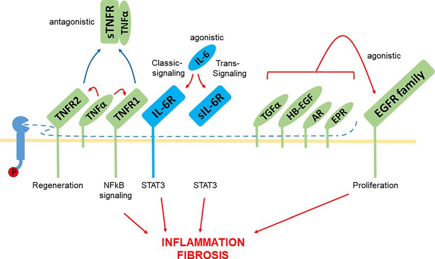
ADAM17 protease and Rose-John 2017). Briefly, after ADAM17 is synthesized in
the endoplasmic reticulum (ER), it requires various modifi-
ADAM17 is comprised of an N-terminal signal peptide fol- cations before it becomes catalytically active. Activation of
lowed by a pro-domain, the metalloproteinase domain and a ADAM17 is mediated by cleavage of the pro-domain by furin-
disintegrin-like domain, a cysteine-rich membrane proximal like proprotein convertases in the Golgi (Endres et al. 2003;
domain (MPD), and a unique Conserved Adam 17 Dynamic Schlondorff et al. 2000; Wong et al. 2015) and phosphory-
Interaction Sequence (CANDIS) region, succeeded by the lation of its cytosolic tail, relieving dimerization inhibition of
stalk region, transmembrane helix and intracellular cyto- ADAM17, via the ERK1/2 and p38 MAPK pathways (Figure 1)
solic tail (Düsterhöft et al. 2019). ADAM17 and ADAM10, (Fan and Derynck 1999; Fan et al. 2003; Gechtman et al. 1999;
which is closest to ADAM17 in structure and function, are Killock and Ivetic 2010; Lemjabbar-Alaoui et al. 2011; Saad
atypical members of the ADAM family, as they contain the et al. 2019; Schwarz et al. 2014; Xu and Derynck 2010). In the
MPD region, instead of the cysteine-rich and EGF-like ER, the pro-protein is suggested to act as a chaperone for
domain found in other ADAM family proteins (Takeda ADAM17, ensuring correct folding and processing, while
2009). The MPD region and the CANDIS, which is not found protecting ADAM17 from degradation (Milla et al. 1999). In
in ADAM10, have been reported to be involved in the addition to this, the pro-protein has been reported to
dimerization of ADAM17, while the CANDIS region has contribute to active site inhibition of ADAM17. Thus, cleav-
further been suggested to facilitate the interaction of age of the pro-protein is important to release the inhibition of
ADAM17 with specific substrates, such as IL-6R (Düsterhöft the catalytic site (Milla et al. 1999). Hence, the processing of
et al. 2014; Riethmueller et al. 2016). Notably, in contrast to ADAM17 has been suggested to contribute to its activity
ADAM10, ADAM17 is not constitutively active, and multiple following phorbol 12-myristate 13-acetate (PMA) stimulation
additional mechanisms for how ADAM17 activity is regu- (Nagano et al. 2004; Soond et al. 2005). However, the
lated have been brought forward (Grötzinger et al. 2017). absence of the cytoplasmic tail is dispensable for PMA
While ADAM17 transcription can be induced by inflam- mediated ADAM17 activation (Horiuchi et al. 2007b; Reddy
mation and in neoplastic tissue, systemic over-expression et al. 2000). Consistently, rapid ADAM17 response following
was not found to result in upregulated levels of substrate physiological stimuli requires its transmembrane domain,
shedding in vivo (Düsterhöft et al. 2019; Yoda et al. 2013). This but not its cytoplasmic tail (Le Gall et al. 2010).
indicated that ADAM17’s proteolytic activity is regulated Additionally, the disintegrin domain has been sug-
at the post-transcriptional level (Armstrong et al. 2006; gested to play a role in the conformation of the ADAM17
Kornfeld et al. 2011; Ringel et al. 2006; Rzymski et al. 2012; protein, acting as a scaffold ensuring the C-shaped confor-
Satoh et al. 2004, 2008). A detailed overview of ADAM17 mation and bridging between the catalytic and the MDP
activity, regulation and substrates is given elsewhere (Zunke domain (Takeda et al. 2006). This may play a role in substrate
Figure 1: Schematic representation of the ADAM17 trafficking.
A simplified schematic illustrating that iRhom2 creates a stable complex with ADAM17 in the Endoplasmic Reticulum, which leads to
translocation of the complex to the Golgi apparatus and maturation of ADAM17 by removing the pro-domain by furin-like proprotein
convertases. The mature form consists of a metalloprotease domain and cytosolic tail, which is next phosphorylated by several kinases, like
ERK1/2, p38 MAPK and PLK2. The complex translocates to the cell membrane, where iRhom2 can dissociate from ADAM17.M. Al-Salihi et al.: ADAM17 during liver damage 1117
binding. Protein disulphide isomerase (PDI) has been et al. 2005). In addition to their role in trafficking ADAM17
demonstrated to inactivate ADAM17, by changing the from the ER to the Golgi, iRhoms have been demonstrated to
conformation of the MDP region of ADAM17 from an open to play a role in the regulation of ADAM17 activation at the cell
a closed conformation (Düsterhöft et al. 2013, 2014; Lorenzen membrane, with ERK1/2-dependent phosphorylation of the
et al. 2012). In addition, this has also been suggested to affect cytoplasmic iRhom2 resulting in the release of mature
the shedding activity of ADAM17, as only the open form of ADAM17 and increased ADAM17 mediated shedding of TNFα
MDP binds to phospholipid phosphatidylserines, which are (Adrain et al. 2012; Grieve et al. 2017; Maney et al. 2015).
translocated to the outer leaflet of the cell membrane by Notably, ADAM17 deficient mice are perinatally lethal,
scramblases in response to stimulation with PMA, for exhibiting a specific developmental phenotype relating to
example. This interaction between phospholipid phospha- epidermal growth factor receptor (EGFR) signaling
tidylserines and open form MDP has been reported to result (Peschon et al. 1998). These and other signaling pathways
in a conformational change, which may contribute to the are modulated by ADAM17’s proteolytic activity during
initialization of the shedding activity (Sommer et al. liver damage (Figure 2).
2016a,b). Likewise, the CANDIS region has also been sug-
gested to contribute to the conformational change in
ADAM17 activating its shedding activity. The CANDIS region ADAM17 during liver damage
has been reported to contain an amphipathic helix, whose
interaction with the cell membrane is enhanced in the ADAM17 deficient mice are perinatally lethal exhibiting
presence of phosphatidylserine. High cholesterol in the cell development defects in the eye, hair and skin, which are
membrane, on the other hand, has been found to counteract associated with defects in transforming growth factor alpha
this interaction, thus indicating that cell membrane (TGFα) shedding (Peschon et al. 1998). However, the
composition may be involved in the regulation of ADAM17 development of a hypomorph mouse model system, in
activity (Düsterhöft et al. 2015; Schütze et al. 2003; Tellier which Adam17 mRNA and protein levels are highly reduced,
et al. 2006). showed a critical role of ADAM17 in intestinal barrier func-
Dimerization of ADAM17 with α5β1 integrin via its dis- tion while mice were viable (Chalaris et al. 2010). Moreover,
integrin domain has been suggested to promote the binding the generation of conditional ADAM17 knockout mice
of active site tissue inhibitor of metalloproteinase 3 (TIMP3) showed that myeloid specific deletion resulted in the
(Wisniewska et al. 2008), while decreasing the accessibility protection of mice against LPS induced septic shock, with
of the active site by steric hindrance (Bax et al. 2004; Huang reduced serum TNFα levels following the challenge
Figure 2: Schematic representation of the ADAM17 enzymatic activities.
A simplified schematic illustrates that ADAM17 can shed substrates from the cell surface, thereby controlling pro-inflammatory and
proliferation pathways. Specifically, TNFα binds to TNFR1, which leads to activation of NF-κB and Jak2/STAT3 pathways respectively, whereas
cleaving the receptors from the cell membrane can result in limited signaling. Other substrates include growth factors like TGFα, heparin-
binding EGF, amphiregulin, and epiregulin, which can bind to EGFR and induce proliferation. Shedding of IL-6R can result in IL-6 trans-
signaling via gp130.1118 M. Al-Salihi et al.: ADAM17 during liver damage
(Horiuchi et al. 2007a). Both, specific deletion of ADAM17 in (Peschon et al. 1998). In addition to ADAM17 activation,
either myeloid cells or hepatocytes resulted in reduced iRhom proteins may play a role in substrate specificity of
serum TNFα levels following stimulation with LPS and ADAM17 (Maretzky et al. 2013). In mouse embryonic fibro-
partial hepatectomy although liver regeneration appeared blasts (MEF) iRhom2 triggers, among others, shedding of the
normal in these settings (McMahan et al. 2013). In turn, EGFR ligands HB-RGF, Amphiregulin, Epiregulin, but not
hepatocyte specific deletion of ADAM17 resulted in TGFα (Maretzky et al. 2013). IRhoms can also regulate ADAM17
increased liver cell death following injection with anti-Fas activation via their cytoplasmic tail. Deletion of the cyto-
antibodies, attributed to reduced TNFR1 and EGFR ligand plasmic tail results in increased ADAM17 activity and TNFR
shedding (Murthy et al. 2010). Consistently, the absence of shedding, which triggers resistance to TNF mediated cell
TNFR1 reduced CD95 induced apoptosis in this setting death (Maney et al. 2015). The mouse curly bare (cub) muta-
(Murthy et al. 2010). Furthermore, adenoviral mediated tion, which deletes most of the cytoplasmic domain of iRhom2
expression of ADAM17 prevented liver cell damage during showed alterations in EGFR signaling (Hosur et al. 2014; Siggs
acetaminophen induced toxicity (Murthy et al. 2010). In et al. 2014). Consistently, iRhom2 gain of function mutations in
turn, treatment of mice with α-1 antitrypsin (AAT) showed its N-terminal cytoplasmic tail, identified in the inherited
increased survival following CD95 stimulation, which Tylosis with oesophageal cancer (TOC) syndrome, result in
coincided with reduced activity of ADAM17 and decreased increased EGFR ligand and TNFR1 shedding (Blaydon et al.
serum levels of TNFα. Consistently, AAT treatment resulted 2012; Brooke et al. 2014; Maney et al. 2015; Saarinen et al.
in protection from liver failure following acetaminophen 2012). Dissociation of iRhom2 from ADAM17 takes place after
induced liver toxicity (Jedicke et al. 2014). These data sug- phosphorylation of the cytoplasmic tail of iRhom2, which can
gest that there are overlapping functions of ADAM17. occur at three different sites. Point mutations in each site
ADAM17 maturation is triggered by co-factors such as downregulated the activity of ADAM17, while additive muta-
iRhoms, which are inactive members of the rhomboid prote- tions resulted in complete loss of ADAM17 mediated shedding
ase family (Al-Salihi and Lang 2020). The founding member (Cavadas et al. 2017; Grieve et al. 2017). Moreover, co-
rhomboid-1 was identified in Drosophila as a Golgi protein, immunoprecipitation of the extracellular iRhom2 domain in
which cleaves the EGF like ligand spitz, promoting its secre- MEFs upon stimulation with PMA demonstrated reduced
tion (Lee et al. 2001; Urban et al. 2001). However, rhomboid interaction with ADAM17 compared to unstimulated cells.
proteins exist, which lack catalytic residues and are descrip- Grieve et al. concluded that phosphorylation of iRhom2 on the
tively named inactive rhomboids (iRhom). There are two cell membrane results in the dissociation from ADAM17 fol-
iRhom proteins in humans and mice, named iRhom1 and lowed by increased catalytic activity (Grieve et al. 2017).
iRhom2, whereas in Drosophila there is only one iRhom The regulation of ADAM17 via the interaction with
member (Lemberg and Freeman 2007). IRhoms are unable to iRhom2 has been suggested to play a significant role in liver
cleave traditional rhomboid substrates and iRhom deficient disease. Absence of iRhom2 resulted in impaired shedding
flies exhibit an extended sleep cycle, which has been associ- of TNFα and consequently reduced susceptibility towards
ated with enhanced EGF receptor signaling (Zettl et al. 2011). LPS mediated liver damage and septic shock (McIlwain et al.
While the single iRhom found in Drosophila induces ER 2012). Moreover, iRhom2 was significantly upregulated
associated ligand degradation (ERAD) and subsequent inhi- following exposure to a mixture of air pollutants (PM2.5),
bition of EGFR signaling, both iRhom1 and iRhom2 have been which was accompanied by augmented levels of TNFR2,
shown to interact with ADAM17 (Li et al. 2015; Zettl et al. 2011). ADAM17 and TNFα, hepatic steatosis, and dyslipidemia in
Deletion of both, iRhom1 and iRhom2 resembles the pheno- the livers of WT mice. Markedly, however, PM2.5-induced
type observed in ADAM17 deficient mice (Li et al. 2015). liver damage and dyslipidemia were attenuated in the
Moreover, examination of newborn iRhom 1 and 2 double absence of iRhom2, while TNFα expression, as well as the
knockout out mice, demonstrated a significant lack of mature expression of associated pro-inflammatory cytokines, were
ADAM17 in multiple tissues examined, including the liver. reduced in vitro (Ge et al. 2017). Moreover, mature ADAM17
Notably, mature ADAM17 expression was also found to be expression and TNFR shedding is increased during bile duct
significantly reduced in livers of iRhom2 knockout mice (Li ligation (BDL), a murine liver fibrosis model system (Sun-
et al. 2015). In turn, iRhom1 and iRhom2 single knockout mice daram et al. 2019). The absence of iRhom2 resulted in
are viable and fertile (Adrain et al. 2012; Li et al. 2015; McIlwain decreased ADAM17 activation and reduced shedding of
et al. 2012; Siggs et al. 2012). These data suggest that iRhom1 TNFRs following BDL. Consequently, hepatic stellate cell
and iRhom2 promote ADAM17 maturation in different com- proliferation and liver fibrosis was increased in iRhom2−/−
partments, which allows single knockout mice to overcome mice following BDL (Sundaram et al. 2019). Notably,
the severe phenotypes observed in ADAM17 deficient mice myeloid and hepatocyte specific deletions of ADAM17 haveM. Al-Salihi et al.: ADAM17 during liver damage 1119
also been associated with decreased TNFα serum levels and signaling and JNK/IRS1 signaling pathway activation, while
reduced TNFR1 shedding (McMahan et al. 2013; Murthy et al. impairing AKT/GSK3β-associated insulin signaling (Xu et al.
2010). Moreover, ursodeoxycholic acid (UDCA), used as a 2020). Similarly, hepatocyte specific deletion of ADAM17 has
therapeutic agent for primary biliary cirrhosis and intra- been shown to improve liver steatosis in a mouse model of
hepatic cholestasis, has not only been associated with NAFLD/NASH (Casagrande et al. 2017). Taken together,
regulating the expression of inflammatory cytokines, ADAM17 dependent signaling is critical during liver damage
including TNFα, but has also been demonstrated to improve and in liver disease (Table 1).
BDL-induced cholestasis in mice by inhibiting ADAM17 ac-
tivity and reducing sMet, with a similar trend for TNFα
(Buryova et al. 2013; Ishizaki et al. 2008; Neuman et al.
2002). In addition, iRhom2 is a key regulator of inflamma-
ADAM17-dependent pathways and
tion driving non-alcoholic fatty liver disease (Xu et al. 2020). their role during liver damage
The absence of iRhom2 significantly alleviated insulin
resistance, hepatic steatosis, and activation of inflammatory TNFα signaling
macrophages in iRhom2 knockout mice in response to high
fat diet. This was associated with the interaction of hepatic TNFα is a central cytokine during liver damage (Koyama and
iRhom2 with mitogen-activated protein kinase 7 (MAP3K7) Brenner 2017; Luedde et al. 2014). Named for its anti-tumor
and subsequent MAP3K7 phosphorylation, leading to NF-κB activity, it was originally identified in the late ’70s and is the
Table : Overview of the ADAM dependent pathways on liver disease.
Liver diseases Examples for relevant ADAM-regulated processes Reference studies
Liver fibrosis and cirrhosis TNFα was associated with mediating NF-κB induced HSC (Osawa et al. ; Pradere et al.
activation, as well as promoting HSC survival, thereby ; Tarrats et al. )
contributing to liver fibrosis indirectly.
Cleavage of MerTK, contributed to the activation of HSCs (Cai et al. )
and promoted liver fibrosis in NASH.
Amphiregulin induced fibrogenic activity of hepatic stel- (Berasain et al. b; McKee et al.
late cells and promoted liver fibrosis. ; Santamaria et al. )
Hepatocellular carcinoma TNF signaling via NF-κB was reported to have a protective, (Schwabe and Brenner )
anti-apoptotic effect, which contributes to hepatocyte
carcinogenesis.
EGFR activation was shown to have a tumor-promoting role. (Fuchs et al. ; Lanaya et al. )
Hepatic ADAM regulation by TIMP slowed (Casagrande et al. )
tumorgenesis.
Alcoholic and non-alcoholic fatty liver dis- Augmented levels of EGFR ligand amphiregulin was (McKee et al. )
ease (NAFLD) as well as non-alcoholic induced by fibrogenic activity of hepatic stellate cells,
steatohepatitis (NASH) driving liver fibrosis.
ADAM-mediated cleavage of MerTK was demonstrated (Cai et al. )
to contribute to the activation of HSCs, driving liver fibrosis
in NASH.
iRhom overexpressing mice showed inflammatory (Casagrande et al. ; Xu et al.
aggravation mediated by NF-κB activation, whereas in the )
ADAM KO model, mice had alleviated hepatic steatosis.
Viral hepatitis TNFα was shown to be crucial to mounting an appropriate (Beyer et al. ; Kasahara et al.
T-cell response to infection with hepatitis B virus. )
TNFα was associated with increased cell death via sup- (Park et al. ; Xu et al. )
pression of NF-κB during viral infection.
Liver regeneration HGF and EGFR ligands, HB-EGF and amphiregulin acted as (Berasain et al. a,b; Kiso et al.
mitogens for hepatocytes during liver regeneration. ; Mitchell et al. ; Takemura
et al. )
IL- was shown to have a transignaling role in liver (Fazel Modares et al. ; Jin et al.
regeneration. ; Riethmueller et al. )
IL- was shown to have a role in maintaining the biliary tree (Demetris et al. ; Nozaki et al.
in liver. )1120 M. Al-Salihi et al.: ADAM17 during liver damage
founding member of a superfamily of cytokine-like mole- pathway can induce several different cellular responses
cules (Carswell et al. 1975; Green S Fau-Chiasson et al.). including inflammation, apoptosis and necrosis by acti-
Along with their cognate receptors, the TNFR superfamily vating gene transcription in downstream signaling path-
control signaling pathways in immunity and disease (Dos- ways such as NF-κB, JNK, and p38.
tert et al. 2019; Kondylis and Pasparakis 2019). TNFα can Complex I activates downstream NF-κB signaling
induce signaling via two cognate receptors TNFR1 and resulting in the expression of pro-inflammatory and pro-
TNFR2. TNFα has been shown to promote liver damage survival genes (Dostert et al. 2019; Hsu et al. 1995, 1996;
during toxic liver damage (Yin et al. 1999). Injection of TNFα Kondylis and Pasparakis 2019; Ting et al. 1996). In turn, NF-
can cause severe liver damage and septic shock when κB inhibition and complex II formation results in cell death
applied in combination with D-Galactosamine (D-Gal), and induces tumor regression (Dostert et al. 2019; Fulda
which is dependent on TNFR1 (Luedde et al. 2014). Consis- 2015; Kondylis and Pasparakis 2019; Xie et al. 2016). The
tently, TNFR1 deficient mice are protected from liver injury mode of cell death induced depends on which type of com-
and septic shock following lipopolysaccharide (LPS) chal- plex II is formed and the presence or absence of active cas-
lenge (Pfeffer et al. 1993; Rothe et al. 1993). TNFR1 is ubiq- pase 8. In complex II FLIPL mirrors caspase 8 and is
uitously expressed and can be activated by both shed and catalytically inactive, but forms catalytically active hetero-
membrane bound TNFα. TNFR2 on the other hand is dimers, which have a reduced activity and altered targets
expressed in a more limited fashion on immune, neuronal, compared to caspase 8 homodimers (Dostert et al. 2019; Pop
cardiac, endothelial and stem cells and binds membrane et al. 2011). While the short isoforms (S and R) bind to cas-
bound TNFα with higher affinity (Dostert et al. 2019). pase 8 and inhibit its activation (Golks et al. 2005; Irmler
Notably, ADAM17 can cleave membrane bound TNFα into its et al. 1997). Complexes IIa and IIb induce apoptosis, are
soluble form as well as the binding receptors TNFR1 and caspase 8 dependent and contain either TRADD or RIPK1
TNFR2 (Peschon et al. 1998). Accordingly, ADAM17 con- respectively. Absence of active caspase 8 results in nec-
tributes to soluble TNFα’s systemic effects. Conversely, roptosis (Holler et al. 2000; Vercammen et al. 1998; Zhang
shedding of the TNFRs reduces TNFR1 signaling, and et al. 2009). Caspase 8 is an initiator caspase; when active, it
resulting soluble TNFRs can bind to TNFα thereby reducing triggers a downstream cascade of caspase cleavage, leading
its biological activity. Therefore, depending on the balance to activation of effector caspases such as caspase 3, ulti-
of TNFα versus TNFR cleavage and the predominant cell on mately leading to apoptosis (Dostert et al. 2019; Stennicke
which this happens, ADAM17 can have opposite effects on et al. 1998). Consistently, TNFα expression levels correlate
liver damage and fibrosis. Furthermore, the TNF pathway with active caspase 3 in patient cohorts suffering from hep-
once activated also has dual outcomes when it comes to atitis C virus infection, which can increase the sensitivity
hepatocyte death and proliferation. towards TNFα mediated cell death (Park et al. 2012; Walsh
The binding of TNFα to TNFR1 results in a conforma- et al. 2004). TNFα promotes chronic viral infection and
tional change exposing the death domain of TNFR1. This hepatitis in murine viral infection model systems (Beyer et al.
leads to the recruitment of the adaptor protein TNFR1- 2016; Suresh et al. 2005; Xu et al. 2014; Zhuang et al. 2020),
associated death domain (TRADD) via the interaction of the and Hepatitis B virus infection sensitizes hepatocytes to-
death domains. The interaction of TNFR1 and TRADD wards TNFα mediated cell death (Jia et al. 2015). In contrast
provides an assembly scaffold for the recruitment of further to Complexes IIa and IIb, Complex IIc, also known as the
adaptor molecules, resulting in the formation of complexes necrosome, depends on RIPK1, 3 and mixed lineage kinase
I and II and subsequent induction of TNF signaling. Com- domain-like (MLKL) to induce necroptosis via the insertion
plex I consists primarily of the receptor itself, the adaptor of MLKL into the plasma membrane creating an ion channel
protein TRADD, TNFR-associated factor 2 (TRAF-2), cellular (Cai et al. 2014; Dostert et al. 2019; Galluzzi et al. 2014;
inhibitors of apoptosis 1 and 2 (cIAP1/2), linear ubiquitin Kondylis and Pasparakis 2019; Wang et al. 2008, 2014;
chain assembly complex (LUBAC) and receptor-interacting Weber et al. 2018). Notably, complex II formation can occur
serine/threonine-protein kinase 1 (RIPK1). Complex II is independently of its role in cell death and inflammation. This
comprised of three distinct, Fas-associated death domain last has been linked to chromosomal stability, suggesting a
(FADD) and long isoform of FLICE-like inhibitory protein possible mechanism relating loss of caspase 8 to hepato-
(FLIPL) containing sub complexes, IIa-c. Phosphorylation, cellular carcinoma (HCC) among other cancers (Liccardi
ubiquitination and de-ubiquitination of RIPK1 controls et al. 2019; Soung et al. 2005).
whether TNFα binding to TNFR1 results in signaling via During liver fibrosis, TNFα produced by Kupffer cells
complex I or complex II (Feoktistova et al. 2011; Wang et al. and neutrophils induces Hepatic stellate cell (HSC) acti-
2008; Yu and Cleveland 2018). The TNFα-TNFR1 signaling vation and proliferation. Specifically, TNFα and LPSM. Al-Salihi et al.: ADAM17 during liver damage 1121
promote proliferation and survival of HSCs by activating STAT3 signaling in the liver promoted establishment of
NF-κB and down-regulating pro-apoptotic genes (Gandhi steatohepatitis (Kroy et al. 2010).
2017; Osawa et al. 2013; Seki et al. 2007). Accordingly, While classical signaling via the membrane-bound IL-6
liver damage and fibrosis were decreased in TNFR1 KO receptors is associated with the inflammatory role of IL-6,
mice and TNFR1&2 double knockout mice compared to IL-6 trans-signaling has been suggested to play a major role
wild-type or single TNFR2 KO mice in a murine model in liver regeneration. Hyper-IL-6 is a fusion protein between
system of fibrosis, suggesting TNF-TNFR1 but not TNFR2 IL-6 and soluble IL-6R, which stimulates trans-signaling in
is critical for HSC activation and liver fibrosis (Tarrats gp130 expressing cells (Schmidt-Arras and Rose-John 2016).
et al. 2011). Mice expressing Hyper-IL-6 exhibited hepatocellular hy-
pertrophy and accelerated liver regeneration following
partial hepatectomy (Peters et al. 2000). Consistently, in a
IL-6 receptor signaling mouse model exhibiting only IL-6 trans-signaling, liver
regeneration following partial hepatectomy was compara-
IL-6 signaling is induced by IL-6, secreted during the in- ble to control animals (Fazel Modares et al. 2019). Further-
flammatory response, binding to its non-signal transducing more, administration of Hyper-IL-6 during toxic liver injury
α-receptor IL-6R and the signaling receptor gp130 (Tanaka could alleviate liver damage and promote hepatocyte pro-
et al. 2014). IL-6 signaling can also be induced in cells liferation (Galun et al. 2000). Taken together, these findings
lacking membrane bound IL-6R, albeit IL-6 cannot bind to suggest a prominent role of IL-6 trans-signaling, triggered
gp130 in the absence of IL-6R. However, IL-6 cannot bind to by ADAM17 IL-6R cleavage, during liver regeneration.
gp130 on its own, but rather gp130 binds the IL-6/IL-6R Reduced IL6-trans-signaling might result in increased liver
complex (Schmidt-Arras and Rose-John 2016). This signaling damage and prolonged injury.
requires the shedding of membrane bound IL-6R. Active
ADAM17 and ADAM10 can cleave IL-6R, resulting in soluble
IL-6R (sIL-6R) (Müllberg et al. 1993; Riethmueller et al. 2017).
EGFR signaling
Accordingly, sIL-6R can still bind IL-6 and gp130 on cells,
which lack IL-6R, and induce gp130 mediated signaling, so-
ADAM17 can cleave a variety of EGFR ligands including
called IL-6 trans-signaling (Mackiewicz et al. 1992). IL-6
amphiregulin, heparin-binding-EGF-like growth factor, epi-
expression during the inflammatory response to injury, re-
regulin, TGFα and epigene (Sahin and Blobel 2007; Sahin
sults in the induction of acute phase proteins such as
et al. 2004; Sigismund et al. 2018). Conditional deletion of
C-reactive protein and fibrinogen. In the liver, IL-6,
EGFR on hepatocytes was associated with increased liver
expressed by neutrophils, monocytes and macrophages, is a
transaminases and reduced survival following partial hep-
major force driving the expression of acute phase proteins
atectomy (Natarajan et al. 2007). Consistently, mice with
(Schmidt-Arras and Rose-John 2016). Aside from its role
hepatic deletion of EGFR showed increased liver damage and
prominent as a potent pro-inflammatory cytokine, IL-6 has
cancer formation following DEN treatment (Lanaya et al.
been reported to play a critical role in liver regeneration.
2014). In turn, macrophage specific deletion of EGFR caused
Hepatectomy in rats results in increased TNFα levels, suc-
ceeded by significantly increased IL-6 expression (Trautwein reduced cancer growth (Lanaya et al. 2014). Treatment with
et al. 1996). Consistently, B cells and macrophages triggered the EGFR inhibitor erlotimib alleviated the establishment of
IL-6 expression in mice following partial hepatectomy liver fibrosis and the development of hepatocellular carci-
(Behnke et al. 2018). In the absence of IL-6 signaling tissue noma (Fuchs et al. 2014). In turn, ectodomain shedding of
regeneration after partial hepatectomy was impaired EGFR ligands and TNFR1 can critically regulate acute liver
(Cressman et al. 1996; Fazel Modares et al. 2019). Likewise, damage (Murthy et al. 2010). Conditional deletion of HB-EGF
inhibition of IL-6 signaling resulted in increased liver dam- results in increased liver injury following acute toxic hepa-
age in response to carbon tetrachloride (CCL4) induced liver titis (Takemura et al. 2013). Furthermore, overexpression of
damage (Gewiese-Rabsch et al. 2010). Moreover, liver injury HB-EGF results in aggravated liver fibrosis following chronic
was exacerbated by inhibition of IL-6 signaling in a non- liver injury (Guo et al. 2017). Deletion of amphiregulin pre-
alcoholic steatohepatitis (NASH) model (Yamaguchi et al. vented hepatocyte proliferation following partial hepatec-
2011). The absence of gp130 – abolishing all IL-6-type cyto- tomy. Furthermore, pretreatment with amphiregulin could
kine signaling – resulted in increased liver damage following prevent liver damage following CD95 stimulation (Berasain
BDL, which was associated with increased bacterial burden et al. 2005b). Consistently, amphiregulin deficient mice
(Wuestefeld et al. 2005). Moreover, lack of IL-6, gp130, and showed increased liver injury during bile duct ligation1122 M. Al-Salihi et al.: ADAM17 during liver damage
(Santamaria et al. 2019). Collectively, these reports hint to a Nevertheless, several promising approaches have been
rather protective role of EGFR ligands and thus a potentially explored recently and are reviewed in more detail by Calligaris
prominent role for ADAM17 shedding during liver injury but et al. (2021). These include the use of more specific small
cell, substrate, and context specific effects might apply. molecules, manipulating the selectivity of endogenous in-
hibitors of ADAM17, as well as targeting the ancillary domains
of ADAM17 or regulators, that are specific and essential for
Other substrates ADAM17 activation, such as iRhoms (Calligaris et al. 2021). For
example, Wong et al. developed a stable form of the auto-
Likewise, the receptor for hepatocyte growth factor, c-Met inhibitory pro-domain of ADAM17 (or TACE pro-domain), TPD,
has been reported to play a significant role in liver devel- which demonstrated promising attenuation of ADAM17-
opment and regeneration (Chalupský et al. 2013). Chalupský mediated disease in models of sepsis, rheumatoid arthritis
et al. demonstrate that ADAM17 along with ADAM10 are (RA) and inflammatory bowel disease as well as in RA patients
involved in releasing the soluble form of the c-Met receptor, (Wong et al. 2016).
sMet from the cell surface in human hepatocellular HepG2 Notably, targeting iRhom2 therapeutically is a particu-
and hepatic stellate cell LX2 lines, while postulating a larly promising route: Not only is iRhom2 specific for ADAM17,
dominant role for ADAM17. Moreover, serum levels of sMET but also it carries the advantage that, iRhom1 can substitute
in a hepatobiliary obstruction mouse model correlated with for the loss of iRhom2-mediated regulation of ADAM17. At the
the level of hepatic injury, expression of established markers same time, using iRhom2 as a therapeutic target could regu-
of liver damage including alanine aminotransferase (ALT), late several pro-inflammatory ADAM17-mediated factors,
aspartate transaminase (AST), alkaline phosphatase (ALP), including IL-6 signaling (Calligaris et al. 2021).
and total bilirubin, as well as with subsequent regeneration
(Chalupský et al. 2013). Hepatocyte specific, post partum
deletion of c-Met resulted in increased hepatocyte apoptosis Concluding remarks
and reduced proliferation following transfer experiments
(Kaldenbach et al. 2012). Consistently, hepatocyte specific ADAM17 substrates play a critical role during liver damage
deletion of c-Met resulted in fulminant liver damage and in various model systems. Furthermore, data from condi-
lower hepatocyte proliferation following bile duct ligation tional ADAM17 deficient mice corroborate the significance
(BDL) (Giebeler et al. 2009). of ADAM17 during liver damage. Myeloid deletion of
ADAM17 resulted in reduced soluble TNFα production. In
turn, hepatocyte specific deletion resulted in increased
ADAM17 as a therapeutic target liver damage resulting from reduced shedding of TNFRs.
Furthermore, IL-6 trans-signaling is important for liver
Since its discovery as a protease involved in the activation of regeneration and hepatocyte proliferation. Accordingly,
the TNF pathway, ADAM17 has been a focal point of research cell type specific regulation of ADAM17 is critical to
aiming to find new therapeutics for inflammatory diseases orchestrating liver regeneration and liver damage. In
(Calligaris et al. 2021). However, due to its involvement in the addition, adaptor molecules such as iRhoms might cause
regulation of multiple signaling pathways, with over 80 cell specific or substrate specific ADAM17 activity and thus
different substrates aside from TNF reported, and the simi- could critically regulate liver damage during disease model
larity of its catalytic domain to ADAM10, which plays an systems. However, further characterization of ADAM17 in
important role in vascularisation, cell proliferation and dif- different cell subsets is required to fully understand the
ferentiation, and several other metalloproteinases, targeting complete picture of ADAM17 during liver damage and
ADAM17 has proven difficult (Calligaris et al. 2021; Maskos disease. This is particularly important to further explore
et al. 1998; Riethmueller et al. 2016; Wetzel et al. 2017; Zunke ADAM17 or its regulators as a therapeutic target.
and Rose-John 2017). Initial drugs designed to target ADAM17
activity demonstrated severe side effects, including serious Author contributions: A.B., M.A., Y.Z., P.S., J.S., K.S.L., and
hepatotoxicity, outweighing their therapeutic benefits (Calli- P.A.L wrote the paper.
garis et al. 2021; Moss et al. 2008; Rossello et al. 2016). Thus, Research funding: This study was supported by the
the risk of deregulating numerous physiologically relevant German Research Council (SFB974, RTG1949) and the
processes and the broad expression of ADAM17 throughout Jürgen Manchot Graduate School MOI.
the body, pose a caveat for targeting ADAM17 therapeutically Conflict of interest statement: The authors declare no
(Calligaris et al. 2021; Zunke and Rose-John 2017). conflicts of interest regarding this article.M. Al-Salihi et al.: ADAM17 during liver damage 1123
References trimerized MLKL protein is required for TNF-induced necroptosis.
Nat. Cell Biol. 16: 55–65.
Calligaris, M., Cuffaro, D., Bonelli, S., Spanò, D.P., Rossello, A.,
Adrain, C., Zettl, M., Christova, Y., Taylor, N., and Freeman, M. (2012).
Nuti, E., and Scilabra, S.D. (2021). Strategies to target ADAM17
Tumor necrosis factor signaling requires iRhom2 to promote
in disease: from its discovery to the iRhom revolution.
trafficking and activation of TACE. Science 335: 225–228.
Molecules 26: 944.
Al-Salihi, M.A. and Lang, P.A. (2020). iRhom2: an emerging adaptor
Carswell, E.A., Old, L.J., Kassel, R.L., Green, S., Fiore, N., and
regulating immunity and disease. Int. J. Mol. Sci. 21: 6570.
Williamson, B. (1975). An endotoxin-induced serum factor that
Armstrong, L., Godinho, S.I., Uppington, K.M., Whittington, H.A., and
causes necrosis of tumors. Proc. Natl. Acad. Sci. U.S.A. 72: 3666.
Millar, A.B. (2006). Contribution of TNF-alpha converting Casagrande, V., Mauriello, A., Bischetti, S., Mavilio, M., Federici, M.,
enzyme and proteinase-3 to TNF-alpha processing in human and Menghini, R. (2017). Hepatocyte specific TIMP3 expression
alveolar macrophages. Am. J. Respir. Cell Mol. Biol. 34: prevents diet dependent fatty liver disease and hepatocellular
219–225. carcinoma. Sci. Rep. 7:6747.
Bax, D.V., Messent, A.J., Tart, J., van Hoang, M., Kott, J., Maciewicz, R.A., Cavadas, M., Oikonomidi, I., Gaspar, C.J., Burbridge, E., Badenes, M.,
and Humphries, M.J. (2004). Integrin α5β1 and ADAM-17 interact in Felix, I., Bolado, A., Hu, T., Bileck, A., Gerner, C., et al. (2017).
vitro and co-localize in migrating HeLa cells*. J. Biol. Chem. 279: Phosphorylation of iRhom2 controls stimulated proteolytic
22377–22386. shedding by the metalloprotease ADAM17/TACE. Cell Rep. 21:
Behnke, K., Zhuang, Y., Xu, H.C., Sundaram, B., Reich, M., Shinde, P.V., 745–757.
Huang, J., Modares, N.F., Tumanov, A.V., Polz, R., et al. (2018). B Chalaris, A., Adam, N., Sina, C., Rosenstiel, P., Lehmann-Koch, J.,
cell-mediated maintenance of cluster of differentiation 169-positive Schirmacher, P., Hartmann, D., Cichy, J., Gavrilova, O., Schreiber, S.,
cells is critical for liver regeneration. Hepatology 68: 2348–2361. et al. (2010). Critical role of the disintegrin metalloprotease ADAM17
Berasain, C., García-Trevijano, E.R., Castillo, J., Erroba, E., Lee, D.C., for intestinal inflammation and regeneration in mice. J. Exp. Med.
Prieto, J., and Avila, M.A. (2005a). Amphiregulin: an early trigger 207: 1617–1624.
of liver regeneration in mice. Gastroenterology 128: 424–432. Chalupský, K., Kanchev, I., Žbodáková, O., Buryová, H., Jiroušková, M.,
Berasain, C., Garcia-Trevijano, E.R., Castillo, J., Erroba, E., Kořínek, V., Gregor, M., and Sedláček, R. (2013). ADAM10/
Santamaria, M., Lee, D.C., Prieto, J., and Avila, M.A. (2005b). 17-dependent release of soluble c-Met correlates with
Novel role for amphiregulin in protection from liver injury. J. Biol. hepatocellular damage. Folia Biol. 59: 76–86.
Chem. 280: 19012–19020. Cressman, D.E., Greenbaum, L.E., DeAngelis, R.A., Ciliberto, G., Furth,
Beyer, M., Abdullah, Z., Chemnitz, J.M., Maisel, D., Sander, J., E.E., Poli, V., and Taub, R. (1996). Liver failure and defective
Lehmann, C., Thabet, Y., Shinde, P.V., Schmidleithner, L., hepatocyte regeneration in interleukin-6-deficient mice. Science
Kohne, M., et al. (2016). Tumor-necrosis factor impairs CD4 T 274: 1379.
cell-mediated immunological control in chronic viral infection. Demetris, A.J., Lunz, J.G., 3rd, Specht, S., and Nozaki, I. (2006). Biliary
Nat. Immunol. 17: 593–603. wound healing, ductular reactions, and IL-6/gp130 signaling in
Black, R.A., Rauch, C.T., Kozlosky, C.J., Peschon, J.J., Slack, J.L., the development of liver disease. World J. Gastroenterol. 12:
Wolfson, M.F., Castner, B.J., Stocking, K.L., Reddy, P., 3512–3522.
Srinivasan, S., et al. (1997). A metalloproteinase disintegrin that Dostert, C., Grusdat, M., Letellier, E., and Brenner, D. (2019). The TNF
releases tumour-necrosis factor-α from cells. Nature 385: family of ligands and receptors: communication modules in the
729–733. immune system and beyond. Physiol. Rev. 99: 115–160.
Blaydon, D.C., Etheridge, S.L., Risk, J.M., Hennies, H.C., Gay, L.J., Düsterhöft, S., Jung, S., Hung, C.-W., Tholey, A., Sönnichsen, F.D.,
Carroll, R., Plagnol, V., McRonald, F.E., Stevens, H.P., Spurr, N.K., Grötzinger, J., and Lorenzen, I. (2013). Membrane-proximal
et al. (2012). RHBDF2 mutations are associated with tylosis, a domain of a disintegrin and metalloprotease-17 represents the
familial esophageal cancer syndrome. Am. J. Hum. Genet. 90: putative molecular switch of its shedding activity operated by
340–346. protein-disulfide isomerase. J. Am. Chem. Soc. 135: 5776–5781.
Brooke, M.A., Etheridge, S.L., Kaplan, N., Simpson, C., O’Toole, E.A., Düsterhöft, S., Höbel, K., Oldefest, M., Lokau, J., Waetzig, G.H.,
Ishida-Yamamoto, A., Marches, O., Getsios, S., and Kelsell, D.P. Chalaris, A., Garbers, C., Scheller, J., Rose-John, S., Lorenzen, I.,
(2014). iRHOM2-dependent regulation of ADAM17 in cutaneous et al. (2014). A disintegrin and metalloprotease 17 dynamic
disease and epidermal barrier function. Hum. Mol. Genet. 23: interaction sequence, the sweet tooth for the human interleukin
4064–4076. 6 receptor. J. Biol. Chem. 289: 16336–16348.
Buryova, H., Chalupsky, K., Zbodakova, O., Kanchev, I., Jirouskova, M., Düsterhöft, S., Michalek, M., Kordowski, F., Oldefest, M., Sommer, A.,
Gregor, M., and Sedlacek, R. (2013). Liver protective effect of Röseler, J., Reiss, K., Grötzinger, J., and Lorenzen, I. (2015).
ursodeoxycholic acid includes regulation of ADAM17 activity. Extracellular juxtamembrane segment of ADAM17 interacts with
BMC Gastroenterol. 13: 155. membranes and is essential for its shedding activity.
Byass, P. (2014). The global burden of liver disease: a challenge for Biochemistry 54: 5791–5801.
methods and for public health. BMC Med. 12: 159. Düsterhöft, S., Babendreyer, A., Giese, A.A., Flasshove, C., and
Cai, B., Dongiovanni, P., Corey, K.E., Wang, X., Shmarakov, I.O., Ludwig, A. (2019). Status update on iRhom and ADAM17: it’s still
Zheng, Z., Kasikara, C., Davra, V., Meroni, M., Chung, R.T., et al. complicated. Biochim. Biophys. Acta Mol. Cell Res. 1866:
(2020). Macrophage MerTK promotes liver fibrosis in 1567–1583.
nonalcoholic steatohepatitis. Cell Metabol. 31: 406–421.e7. Endres, K., Anders, A., Kojro, E., Gilbert, S., Fahrenholz, F., and
Cai, Z., Jitkaew, S., Zhao, J., Chiang, H.C., Choksi, S., Liu, J., Ward, Y., Postina, R. (2003). Tumor necrosis factor-alpha converting
Wu, L.G., and Liu, Z.G. (2014). Plasma membrane translocation of enzyme is processed by proprotein-convertases to its mature1124 M. Al-Salihi et al.: ADAM17 during liver damage
form which is degraded upon phorbol ester stimulation. Eur. Grieve, A.G., Xu, H., Künzel, U., Bambrough, P., Sieber, B., and
J. Biochem. 270: 2386–2393. Freeman, M. (2017). Phosphorylation of iRhom2 at the plasma
Fan, H. and Derynck, R. (1999). Ectodomain shedding of TGF-alpha and membrane controls mammalian TACE-dependent inflammatory
other transmembrane proteins is induced by receptor tyrosine and growth factor signalling. eLife 6: e23968.
kinase activation and MAP kinase signaling cascades. EMBO Grötzinger, J., Lorenzen, I., and Düsterhöft, S. (2017). Molecular
J. 18: 6962–6972. insights into the multilayered regulation of ADAM17: the role of
Fan, H., Turck, C.W., and Derynck, R. (2003). Characterization of the extracellular region. Biochim. Biophys. Acta Mol. Cell Res.
growth factor-induced serine phosphorylation of tumor 1864: 2088–2095.
necrosis factor-alpha converting enzyme and of an Guo, Y., Ding, Q., Chen, L., Ji, C., Hao, H., Wang, J., Qi, W., Xie, X., Ma, J.,
alternatively translated polypeptide. J. Biol. Chem. 278: Li, A., et al. (2017). Overexpression of heparin-binding epidermal
18617–18627. growth factor-like growth factor mediates liver fibrosis in
Fazel Modares, N., Polz, R., Haghighi, F., Lamertz, L., Behnke, K., transgenic mice. Am. J. Med. Sci. 354: 199–210.
Zhuang, Y., Kordes, C., Haussinger, D., Sorg, U.R., Pfeffer, K., Heymann, F. and Tacke, F. (2016). Immunology in the liver–from
et al. (2019). IL-6 trans-signaling controls liver regeneration after homeostasis to disease. Nat. Rev. Gastroenterol. Hepatol. 13:
partial hepatectomy. Hepatology 70: 2075–2091. 88–110.
Feoktistova, M., Geserick, P., Kellert, B., Dimitrova, D.P., Langlais, C., Holler, N., Zaru, R., Micheau, O., Thome, M., Attinger, A., Valitutti, S.,
Hupe, M., Cain, K., MacFarlane, M., Hacker, G., and Leverkus, M. Bodmer, J.-L., Schneider, P., Seed, B., and Tschopp, J. (2000). Fas
(2011). cIAPs block Ripoptosome formation, a RIP1/caspase-8 triggers an alternative, caspase-8–independent cell death
containing intracellular cell death complex differentially pathway using the kinase RIP as effector molecule. Nat. Immunol.
regulated by cFLIP isoforms. Mol. Cell 43: 449–463. 1: 489–495.
Fuchs, B.C., Hoshida, Y., Fujii, T., Wei, L., Yamada, S., Lauwers, G.Y., Horiuchi, K., Kimura, T., Miyamoto, T., Takaishi, H., Okada, Y., Toyama, Y.,
McGinn, C.M., DePeralta, D.K., Chen, X., Kuroda, T., et al. (2014). and Blobel, C.P. (2007a). Cutting edge: TNF-alpha-converting
Epidermal growth factor receptor inhibition attenuates liver enzyme (TACE/ADAM17) inactivation in mouse myeloid cells
fibrosis and development of hepatocellular carcinoma. prevents lethality from endotoxin shock. J. Immunol. 179:
Hepatology 59: 1577–1590. 2686–2689.
Fulda, S. (2015). Targeting extrinsic apoptosis in cancer: challenges Horiuchi, K., Le Gall, S., Schulte, M., Yamaguchi, T., Reiss, K., Murphy, G.,
and opportunities. Semin. Cell Dev. Biol. 39: 20–25. Toyama, Y., Hartmann, D., Saftig, P., and Blobel, C.P. (2007b).
Galluzzi, L., Kepp, O., and Kroemer, G. (2014). MLKL regulates necrotic Substrate selectivity of epidermal growth factor-receptor ligand
plasma membrane permeabilization. Cell Res. 24: 139–140. sheddases and their regulation by phorbol esters and calcium
Galun, E., Zeira, E., Pappo, O., Peters, M., and Rose-John, S. (2000). influx. Mol. Biol. Cell 18: 176–188.
Liver regeneration induced by a designer human IL-6/sIL-6R Hosur, V., Johnson, K.R., Burzenski, L.M., Stearns, T.M., Maser, R.S.,
fusion protein reverses severe hepatocellular injury. Faseb. J. 14: and Shultz, L.D. (2014). Rhbdf2 mutations increase its protein
1979–1987. stability and drive EGFR hyperactivation through enhanced
Gandhi, C.R. (2017). Hepatic stellate cell activation and pro-fibrogenic secretion of amphiregulin. Proc. Natl. Acad. Sci. U.S.A. 111:
signals. J. Hepatol. 67: 1104–1105. E2200–E2209.
Ge, C.X., Qin, Y.T., Lou, D.S., Li, Q., Li, Y.Y., Wang, Z.M., Yang, W.W., Hsu, H., Huang, J., Shu, H.-B., Baichwal, V., and Goeddel, D.V. (1996).
Wang, M., Liu, N., Wang, Z., et al. (2017). iRhom2 deficiency TNF-dependent recruitment of the protein kinase RIP to the TNF
relieves TNF-α associated hepatic dyslipidemia in long-term receptor-1 signaling complex. Immunity 4: 387–396.
PM2.5-exposed mice. Biochem. Biophys. Res. Commun. 493: Hsu, H., Xiong, J., and Goeddel, D.V. (1995). The TNF receptor
1402–1409. 1-associated protein TRADD signals cell death and NF-κB
Gechtman, Z., Alonso, J.L., Raab, G., Ingber, D.E., and Klagsbrun, M. activation. Cell 81: 495–504.
(1999). The shedding of membrane-anchored heparin-binding Huang, J., Bridges, L.C., and White, J.M. (2005). Selective modulation
epidermal-like growth factor is regulated by the Raf/mitogen- of integrin-mediated cell migration by distinct ADAM family
activated protein kinase cascade and by cell adhesion and members. Mol. Biol. Cell 16: 4982–4991.
spreading. J. Biol. Chem. 274: 28828–28835. Irmler, M., Thome, M., Hahne, M., Schneider, P., Hofmann, K., Steiner, V.,
Gewiese-Rabsch, J., Drucker, C., Malchow, S., Scheller, J., and Rose- Bodmer, J.-L., Schröter, M., Burns, K., Mattmann, C., et al. (1997).
John, S. (2010). Role of IL-6 trans-signaling in CCl4 induced liver Inhibition of death receptor signals by cellular FLIP. Nature 388:
damage. Biochim. Biophys. Acta (BBA) – Mol. Basis Dis. 1802: 190–195.
1054–1061. Ishizaki, K., Iwaki, T., Kinoshita, S., Koyama, M., Fukunari, A., Tanaka, H.,
Giebeler, A., Boekschoten, M.V., Klein, C., Borowiak, M., Birchmeier, C., Tsurufuji, M., Sakata, K., Maeda, Y., Imada, T., et al. (2008).
Gassler, N., Wasmuth, H.E., Muller, M., Trautwein, C., and Ursodeoxycholic acid protects concanavalin A-induced mouse liver
Streetz, K.L. (2009). c-Met confers protection against chronic liver injury through inhibition of intrahepatic tumor necrosis factor-alpha
tissue damage and fibrosis progression after bile duct ligation in and macrophage inflammatory protein-2 production. Eur.
mice. Gastroenterology 137: 297–308, 308 e1-4. J. Pharmacol. 578: 57–64.
Golks, A., Brenner, D., Fritsch, C., Krammer, P.H., and Lavrik, I.N. Iwaisako, K., Jiang, C., Zhang, M., Cong, M., Moore-Morris, T.J., Park, T.J.,
(2005). c-FLIPR, a new regulator of death receptor-induced Liu, X., Xu, J., Wang, P., Paik, Y.H., et al. (2014). Origin of
apoptosis. J. Biol. Chem. 280: 14507–14513. myofibroblasts in the fibrotic liver in mice. Proc. Natl. Acad. Sci.
Green S Fau-Chiasson, M.A., Chiasson Ma Fau-Shah, R.G., and U.S.A. 111: E3297–E3305.
Shah, R.G. Evidence for the presence of an antitumor factor in Jedicke, N., Struever, N., Aggrawal, N., Welte, T., Manns, M.P., Malek, N.P.,
serum of normal animals (0304-3835 (Print)). Zender, L., Janciauskiene, S., and Wuestefeld, T. (2014). α-M. Al-Salihi et al.: ADAM17 during liver damage 1125
1-antitrypsin inhibits acute liver failure in mice. Hepatology 59: Lee, J.R., Urban, S., Garvey, C.F., and Freeman, M. (2001). Regulated
2299–2308. intracellular ligand transport and proteolysis control EGF signal
Jia, B., Guo, M., Li, G., Yu, D., Zhang, X., Lan, K., and Deng, Q. (2015). activation in Drosophila. Cell 107: 161–171.
Hepatitis B virus core protein sensitizes hepatocytes to tumor Lemberg, M.K. and Freeman, M. (2007). Functional and evolutionary
necrosis factor-induced apoptosis by suppression of the implications of enhanced genomic analysis of rhomboid
phosphorylation of mitogen-activated protein kinase kinase 7. intramembrane proteases. Genome Res. 17: 1634–1646.
J. Virol. 89: 2041–2051. Lemjabbar-Alaoui, H., Sidhu, S.S., Mengistab, A., Gallup, M., and
Jin, X., Zimmers, T.A., Perez, E.A., Pierce, R.H., Zhang, Z., and Koniaris, L.G. Basbaum, C. (2011). TACE/ADAM-17 phosphorylation by
(2006). Paradoxical effects of short- and long-term interleukin-6 PKC-epsilon mediates premalignant changes in tobacco smoke-
exposure on liver injury and repair. Hepatology 43: 474–484. exposed lung cells. PloS One 6: e17489.
Kaldenbach, M., Giebeler, A., Tschaharganeh, D.F., Erschfeld, S., Li, X., Maretzky, T., Weskamp, G., Monette, S., Qing, X., Issuree, P.D.,
Wasmuth, H.E., Dolle, L., Floege, J., Trautwein, C., and Streetz, K.L. Crawford, H.C., McIlwain, D.R., Mak, T.W., Salmon, J.E., et al.
(2012). Hepatocyte growth factor/c-Met signalling is important (2015). iRhoms 1 and 2 are essential upstream regulators of
for the selection of transplanted hepatocytes. Gut 61: ADAM17-dependent EGFR signaling. Proc. Natl. Acad. Sci. U.S.A.
1209–1218. 112: 6080–6085.
Kasahara, S., Ando, K., Saito, K., Sekikawa, K., Ito, H., Ishikawa, T., Liccardi, G., Ramos Garcia, L., Tenev, T., Annibaldi, A., Legrand, A.J.,
Ohnishi, H., Seishima, M., Kakumu, S., and Moriwaki, H. (2003). Robertson, D., Feltham, R., Anderton, H., Darding, M., Peltzer, N.,
Lack of tumor necrosis factor α induces impaired proliferation of et al. (2019). RIPK1 and caspase-8 ensure chromosome stability
hepatitis B virus-specific cytotoxic T lymphocytes. J. Virol. 77: independently of their role in cell death and inflammation. Mol.
2469–2476. Cell 73: 413–428 e7.
Killock, D.J. and Ivetic, A. (2010). The cytoplasmic domains of Lorenzen, I., Lokau, J., Düsterhöft, S., Trad, A., Garbers, C., Scheller,
TNFalpha-converting enzyme (TACE/ADAM17) and L-selectin are J., Rose-John, S., and Grötzinger, J. (2012). The membrane-
regulated differently by p38 MAPK and PKC to promote proximal domain of A disintegrin and metalloprotease 17
ectodomain shedding. Biochem. J. 428: 293–304. (ADAM17) is responsible for recognition of the interleukin-6
Kiso, S., Kawata, S., Tamura, S., Inui, Y., Yoshida, Y., Sawai, Y., receptor and interleukin-1 receptor II. FEBS Lett. 586:
Umeki, S., Ito, N., Yamada, A., Miyagawa, J., et al. (2003). Liver 1093–1100.
regeneration in heparin-binding EGF-like growth factor Luedde, T., Kaplowitz, N., and Schwabe, R.F. (2014). Cell death and
transgenic mice after partial hepatectomy. Gastroenterology cell death responses in liver disease: mechanisms and clinical
124: 701–707. relevance. Gastroenterology 147: 765–U110.
Kondylis, V. and Pasparakis, M. (2019). RIP kinases in liver cell death, Mackiewicz, A., Schooltink, H., Heinrich, P.C., and Rose-John, S.
inflammation and cancer. Trends Mol. Med. 25: 47–63. (1992). Complex of soluble human IL-6-receptor/IL-6 up-
Kornfeld, J.W., Meder, S., Wohlberg, M., Friedrich, R.E., Rau, T., regulates expression of acute-phase proteins. J. Immunol. 149:
Riethdorf, L., Loning, T., Pantel, K., and Riethdorf, S. (2011). 2021–2027.
Overexpression of TACE and TIMP3 mRNA in head and neck Maney, S.K., McIlwain, D.R., Polz, R., Pandyra, A.A., Sundaram, B.,
cancer: association with tumour development and progression. Wolff, D., Ohishi, K., Maretzky, T., Brooke, M.A., Evers, A., et al.
Br. J. Canc. 104: 138–145. (2015). Deletions in the cytoplasmic domain of iRhom1 and
Koyama, Y. and Brenner, D.A. (2017). Liver inflammation and fibrosis. iRhom2 promote shedding of the TNF receptor by the protease
J. Clin. Invest. 127: 55–64. ADAM17. Sci. Signal. 8: ra109.
Kriegler, M., Perez, C., DeFay, K., Albert, I., and Lu, S.D. (1988). A novel Maretzky, T., McIlwain, D.R., Issuree, P.D., Li, X., Malapeira, J., Amin,
form of TNF/cachectin is a cell surface cytotoxic transmembrane S., Lang, P.A., Mak, T.W., and Blobel, C.P. (2013). iRhom2
protein: ramifications for the complex physiology of TNF. Cell 53: controls the substrate selectivity of stimulated
45–53. ADAM17-dependent ectodomain shedding. Proc. Natl. Acad. Sci.
Kroy, D.C., Beraza, N., Tschaharganeh, D.F., Sander, L.E., Erschfeld, S., U.S.A. 110: 11433–11438.
Giebeler, A., Liedtke, C., Wasmuth, H.E., Trautwein, C., and Maskos, K., Fernandez-Catalan, C., Huber, R., Bourenkov, G.P.,
Streetz, K.L. (2010). Lack of interleukin-6/glycoprotein 130/signal Bartunik, H., Ellestad, G.A., Reddy, P., Wolfson, M.F., Rauch, C.T.,
transducers and activators of transcription-3 signaling in Castner, B.J., et al. (1998). Crystal structure of the catalytic
hepatocytes predisposes to liver steatosis and injury in mice. domain of human tumor necrosis factor-α-converting enzyme.
Hepatology 51: 463–473. Proc. Natl. Acad. Sci. U.S.A. 95: 3408–3412.
Lambrecht, B.N., Vanderkerken, M., and Hammad, H. (2018). The McIlwain, D.R., Lang, P.A., Maretzky, T., Hamada, K., Ohishi, K.,
emerging role of ADAM metalloproteinases in immunity. Nat. Maney, S.K., Berger, T., Murthy, A., Duncan, G., Xu, H.C., et al.
Rev. Immunol. 18: 745–758. (2012). iRhom2 regulation of TACE controls TNF-mediated
Lanaya, H., Natarajan, A., Komposch, K., Li, L., Amberg, N., Chen, L., protection against Listeria and responses to LPS. Science 335:
Wculek, S.K., Hammer, M., Zenz, R., Peck-Radosavljevic, M., 229–232.
et al. (2014). EGFR has a tumour-promoting role in liver McKee, C., Sigala, B., Soeda, J., Mouralidarane, A., Morgan, M.,
macrophages during hepatocellular carcinoma formation. Nat. Mazzoccoli, G., Rappa, F., Cappello, F., Cabibi, D., Pazienza, V.,
Cell Biol. 16: 1–7. et al. (2015). Amphiregulin activates human hepatic stellate cells
Le Gall, S.M., Maretzky, T., Issuree, P.D., Niu, X.D., Reiss, K., Saftig, P., and is upregulated in non alcoholic steatohepatitis. Sci. Rep. 5:
Khokha, R., Lundell, D., and Blobel, C.P. (2010). ADAM17 is 8812.
regulated by a rapid and reversible mechanism that controls McMahan, R.S., Riehle, K.J., Fausto, N., and Campbell, J.S. (2013). A
access to its catalytic site. J. Cell Sci. 123: 3913–3922. disintegrin and metalloproteinase 17 regulates TNF and TNFR1You can also read