The Role of the JC Virus in Central Nervous System Tumorigenesis - MDPI
←
→
Page content transcription
If your browser does not render page correctly, please read the page content below
International Journal of
            Molecular Sciences

Review
The Role of the JC Virus in Central Nervous
System Tumorigenesis
Nicholas Ahye, Anna Bellizzi , Dana May and Hassen S. Wollebo *
 Center for Neurovirology, Department of Neuroscience, Lewis Katz School of Medicine at Temple University,
 3500 N. Broad Street, Philadelphia, PA 19140, USA; nicholas.ahye@tuhs.temple.edu (N.A.);
 tuf81370@temple.edu (A.B.); tul10237@temple.edu (D.M.)
 * Correspondence: siraj123@temple.edu; Tel.: +1-215-707-7137; Fax: +1-215-707-4888

 Received: 19 June 2020; Accepted: 24 August 2020; Published: 28 August 2020

 Abstract: Cancer is the second leading cause of mortality worldwide. The study of DNA
 tumor-inducing viruses and their oncoproteins as a causative agent in cancer initiation and
 tumor progression has greatly enhanced our understanding of cancer cell biology. The initiation of
 oncogenesis is a complex process. Specific gene mutations cause functional changes in the cell that
 ultimately result in the inability to regulate cell differentiation and proliferation effectively. The human
 neurotropic Polyomavirus JC (JCV) belongs to the family Polyomaviridae and it is the causative agent
 of progressive multifocal leukoencephalopathy (PML), which is a fatal neurodegenerative disease in
 an immunosuppressed state. Sero-epidemiological studies have indicated JCV infection is prevalent
 in the population (85%) and that initial infection usually occurs during childhood. The JC virus
 has small circular, double-stranded DNA that includes coding sequences for viral early and late
 proteins. Persistence of the virus in the brain and other tissues, as well as its potential to transform
 cells, has made it a subject of study for its role in brain tumor development. Earlier observation of
 malignant astrocytes and oligodendrocytes in PML, as well as glioblastoma formation in non-human
 primates inoculated with JCV, led to the hypothesis that JCV plays a role in central nervous system
 (CNS) tumorigenesis. Some studies have reported the presence of both JC viral DNA and its proteins
 in several primary brain tumor specimens. The discovery of new Polyomaviruses such as the Merkel
 cell Polyomavirus, which is associated with Merkel cell carcinomas in humans, ignited our interest
 in the role of the JC virus in CNS tumors. The current evidence known about JCV and its effects,
 which are sufficient to produce tumors in animal models, suggest it can be a causative factor in
 central nervous system tumorigenesis. However, there is no clear association between JCV presence
 in CNS and its ability to initiate CNS cancer and tumor formation in humans. In this review, we will
 discuss the correlation between JCV and tumorigenesis of CNS in animal models, and we will give an
 overview of the current evidence for the JC virus’s role in brain tumor formation.

 Keywords: Polyomavirus JC; tumors of central nervous system; CY and Mad-4 NCCR-transgenic
 mice; p53 and pRB oncosuppressor; DNA damage response (DDR); Wnt pathway; insulin receptor
 substrate-1 IRS-1 signaling

1. Introduction
     The transformative properties of some viruses and their role in human cancers have been known
for many decades. An estimated 10–15% of human cancers worldwide are associated with one of seven
known viruses [1,2]. The Polyomaviridae family is one salient example of viruses whose known role in
the development of human diseases has been evolving [3–5]. The JC virus, BK virus, Simian virus 40
(SV40), and more recently discovered Merkel cell Polyomavirus (MCPyV) are some of the members
in this group that infect humans and are known to either cause or have an association with human

Int. J. Mol. Sci. 2020, 21, 6236; doi:10.3390/ijms21176236                         www.mdpi.com/journal/ijms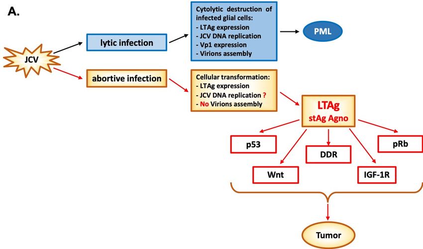
Int. J. Mol. Sci. 2020, 21, 6236                                                                     2 of 22

diseases and cancers. These viruses are widespread, but most often only responsible for a clinically
silent infection [6–8]. Detection of viral nucleic acid or proteins in tumor samples and the study of
effects on cellular regulatory pathways have illustrated the mechanisms by which they can transform
cells. Persistence of the JC virus in the brain and other human tissues, as well as its oncogenic potential,
has made it a subject of study for a role in brain tumor formation [9,10].
      Many primary central nervous system tumors remain incurable, even after multimodality
treatment with surgical resection, chemotherapy, and radiation therapy. Identification of some key
genetic mutations in these tumors that influence growth, affect treatment response, or predict outcomes
has increased understanding and influenced new treatment strategies. Yet there remains much
to be learned about risk factors and mechanisms of tumorigenesis. Many potential genetic and
environmental risk factors for brain tumor formation have been heavily studied, but few have been
identified [11]. Discovering the underlying mechanisms driving oncogenesis in the central nervous
system can reveal how these malignancies form, grow, and resist treatment, thereby steering the
development of more effective therapies. A variety of brain tumors, including glioblastomas [12–16],
ependymomas [17,18] medulloblastomas [19–23], astrocytomas and oligoastrocytomas [15,24],
and oligodendrogliomas [15,17,25], have been studied for an association with the JC virus.
The understanding of the complex role these viruses play in brain tumor formation requires a
wide variety of methods. It remains a very active area of research due to the potential benefits of being
able to treat better or prevent these cancers if a causative agent is identified. In this review, we present
an overview of the current evidence for the JC virus’s role in brain tumor formation.

2. History and Epidemiology of JC Virus
      The JC virus (JCV) is a human neurotropic Polyomavirus causing progressive multifocal
leukoencephalopathy (PML). The JC virus was discovered in 1971 from the brain of a patient
with PML [26]. PML lesions have multiple foci of myelin loss, which cause debilitating neurological
symptoms. PML lesions are characterized by oligodendrocytes with viral nuclear inclusion bodies
and JCV-infected bizarre astrocytes with no signs of apoptosis. The common underlying feature
of PML is a severe weakening of the immune system, especially from human immunodeficiency
virus (HIV-1)/ Acquired Immunodeficiency Syndrome (AIDS) [27–30]. PML also occurs in other
immunosuppressive conditions such as organ transplantation [31], CD40 ligand deficiency [32],
hyper-immunoglobulinemia [33], multiple sclerosis (MS) [34,35], and Crohn’s disease [36].
      Due to the asymptomatic nature of JCV infection, it is difficult to pinpoint the period of primary
infection. JCV infection occurs in childhood and, several seroprevalence studies have shown 8–10% of
children below six years have antibodies against the JC virus [37,38]. Sero-epidemiological data have
shown that JCV seropositivity increases during adult life, with rates up to 70% [39–43] or higher of the
world’s population having positive titers for JC virus [41–44]. Based on the structure of the non-coding
control region (NCCR), there are two types of JCV variants: Archetype and Mad-1. The Archetype is
the most abundant strain of the JC virus in the environment, and it is the transmittable form between
individuals [45]. Genotyping analysis based on DNA sequence polymorphism in the C-terminal
domain of VP1, intergenic region, and T-antigen gene indicates that JCV transmission occurs within the
same family [46,47]. Many different possible routes of infection have been proposed for these viruses,
but the definitive answer remains unclear. JCV is most often detected from urine samples of normal
and immunosuppressed individuals, indicating urine as a major source of viral transmission [48].
The tonsils and gastrointestinal tract can contain viral DNA and have been suggested as possible
initial infection sites [44,49–52]. After primary infection, JCV is believed to be disseminated by a
hematogenous route [53–56] and maintains in a persistent or latent state in kidneys, bone marrow,
and spleen tissue [57–59]. From the site of latency, the virus reactivates under immunosuppressed
conditions and can reach the brain using B-lymphocytes [60,61]. Although JCV DNA can be detected
in different blood cell types, a clear indication of whether the virus undergoes productive infection is
missing [62]. It has been reported that the JCV genomic DNA sequence can be found in the brain from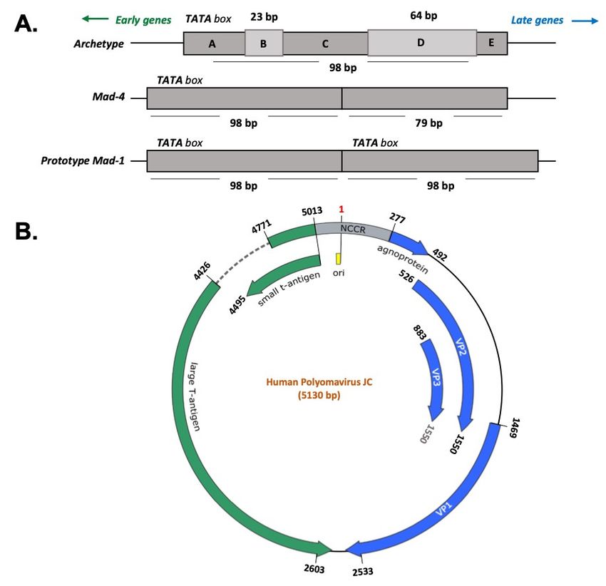
Int. J. Mol. Sci. 2020, 21, 6236                                                                     3 of 22

immunocompetent patients, implicating the brain as a possible site of latency [63–65]. There are many
critical areas where our understanding of JCV biology is incomplete. Specifically, the exact JCV latency
site, the cell type in which the Archetype form converts to neurotropic strains such as Mad-1, and the
mechanism of virus reactivation to cause disease. The finding of both Archetype and neurotropic
strains in a different subpopulation of PBMC suggests that the blood compartment might be the site of
JCV conversion [66].

3. JC Virus Characteristics
      The JC virus is a double-stranded DNA virus, with a circular genome contained in a small,
non-enveloped icosahedral capsid. The genome consists of early and late coding sequences that are
separated by a regulatory region [26,67,68]. The early coding region encodes large T antigen (LTAg)
and small t antigen (stAg). In contrast, the viral structural proteins (VP1, VP2, and VP3) and small
accessory protein, called Agno, are encoded by the late coding region. The viral structural proteins are
essential for early events of the viral life cycle, such as attachment to a cellular receptor, adsorption,
and penetration. Between early and late coding regions lies the non-coding control region (NCCR)
region, which contains the promoter/enhancer elements for expression of the early and late genes
and the origin of viral DNA replication [68,69]. The NCCR region also contains binding sites for a
number of transcriptional factors including a unique NF-kB site, C/EBPβ, NFAT4, Rad51, NF-1, SP1,
and others [70–73]. Sequence variation in the NCCR determines JCV tropism and its pathogenic
effect [74–77]. Figure 1 shows a representation of JCV genome with the genes involved in the production
of the viral proteins and the NCCR.
      The early coding region products are not only involved in DNA replication and transcription,
but they are also known to have a role in cellular transformation. The late coding region contains genes
that encode for viral capsid proteins, which after assembly, form the infectious virion. The Agno protein
is involved in different aspects of the JCV life cycle such as viral replication and transcription [78,79],
cell cycle arrest and deregulation [80], viroporin [81,82], and transport of the new virion from the
nucleus to the cytoplasm [83,84] without being packaged into the virion structure [85].
      The JCV life cycle begins with the interaction of VP1 with α-2,3-linked sialic acid. This binding
allows the virus to navigate in the cytoplasm through different cellular compartments to reach the
nucleus [86]. Inside the nucleus, viral transcription precedes viral DNA replication since the viral
early gene products LTAg, stAg, and splicing variants of LTAg are essential for the initiation and
progress of the lytic cycle [87,88]. LTAg of JCV is a multifunctional protein with many domains and is
essential for viral DNA replication, where it binds directly to the viral origin of replication. LTAg is
responsible for the transcriptional switch from early to late by directly activating late gene expression
and downregulating its early promoter [89–91]. It has been reported that the splicing variant of LTAg
(T prime or T0 ) cooperates to facilitate LTAg-mediated DNA replication [92]. LTAg modulates many
cellular functions through its many domains by interacting with cellular regulators such as pRB and
p53 to promote cell cycle progression.
(NCCR) region, which contains the promoter/enhancer elements for expression of the early and late
genes and the origin of viral DNA replication [68,69]. The NCCR region also contains binding sites
for a number of transcriptional factors including a unique NF-kB site, C/EBPβ, NFAT4, Rad51, NF-1,
SP1, and others [70–73]. Sequence variation in the NCCR determines JCV tropism and its pathogenic
effect [74–77]. Figure 1 shows a representation of JCV genome with the genes involved in the
Int. J. Mol. Sci. 2020, 21, 6236 4 of 22
production of the viral proteins and the NCCR.
Figure 1. Polyomavirus JC genome. (A) The Non-Coding Control Region (NCCR) is the most variable
Figure 1. Polyomavirus JC genome. (A) The Non-Coding Control Region (NCCR) is the most variable
region of the JCV genome and determines the viral strains. The Prototype Mad-1 strain is characterized
region of the JCV genome and determines the viral strains. The Prototype Mad-1 strain is
by a sequence of 98 bp repeated in tandem. Mad-4 differentiates from Mad-1 in the deletion of 19 base
characterized by a sequence of 98 bp repeated in tandem. Mad-4 differentiates from Mad-1 in the
pairs in the second 98 bp repeat. The Archetype strain is composed of a single sequence of 98 bp with
two insertions of 24 bp and 64 bp. (B) The NCCR is located between the two coding regions of the
JCV genome: the early and the late regions. The early region encodes for the large T antigen (LTAg)
and the small t antigen (stAg), whereas the late region contains the genes for the Agnoprotein and
the capsid proteins VP-1, VP-2, and VP-3. The numbering of the nucleotide positions refers to the
prototype Mad-1 strain (NCBI Reference Sequence: NC_001699.1).
4. JCV Early Gene Products and Agno Protein and Their Oncogenic Potential
Several lines of experimental evidence suggest that JC virus infection of primary cells in vitro
leads to transformation, and those cells with a transformed phenotype showed the expression of viral
early gene products. Although many studies confirm the presence of JC virus in human malignancies,
whether there is a direct association of JCV with human cancer remains a topic of debate. The expression
of JCV early gene products is strongly associated with the oncogenic potential of JCV, specifically viral
LTAg. The LTAg is a nuclear protein with multiple functional domains involved in different viral and
cellular functions [93,94].
LTAg is a nuclear phosphoprotein required for the JC virus to replicate its DNA. This protein is
ubiquitous amongst members of the Polyomavirus family. After viral infection and the beginning of
viral genome replication, LTAg protein interacts with the transcription origin. It also can promote
progression of the cell life cycle into S-phase by a variety of regulatory protein interactions. This step
is necessary for the completion of the viral life cycle. These significant protein interactions includeInt. J. Mol. Sci. 2020, 21, 6236 5 of 22
retinoblastoma protein (pRb) [95,96] and the tumor suppressor p53. The interaction of LTAg with pRb
leads to the activation of cellular elongation factors to promote cell cycle progression [97]. Normally,
pRb sequesters the E2F transcription factor and prevents cell cycle progression from G1 to S phase.
Inactivation of pRb by LTAg binding releases E2F and promotes cellular proliferation [95,98,99].
Besides, LTAg interacts with p53 [100,101] to inhibit DNA repair and apoptosis. Briefly, the release of
E2F from pRb by LTAg activates p14ARF expression, which leads to the stabilization of p53. However,
LTAg binds and inactivates p53, preventing the p53 action in response to the DNA damage or p14ARF
production [99]. In mice transgenic for JCV early region, LTAg may also inhibit the tumor suppressor
activity of p53 through the interaction with merlin, a product of the gene neurofibromatosis type 2
(NF-2), that neutralizes the inhibitory effect of Mdm2 on p53 [102]. The disease NF-2 is characterized
by an NF2 mutation resulting in the development of tumors which histologically resemble malignant
peripheral nerve sheath tumors [103,104] (Figure 2 and Table 1).
LTAg is also known to interact with components of different signaling pathways which are
associated with cellular transformation such as β-catenin [3,105], insulin receptor substrate -1
(IRS-1) [106], and survivin [107].
β-catenin is a crucial protein of the Wnt signaling pathway, usually located and degraded
in the cytoplasm. The central domain of LTAg can bind the C-terminus of this protein [105],
resulting in its nuclear translocation, and subsequent activation of c-Myc and cyclin D1 TCF promoter,
leading to cellular proliferation [108]. These signaling events occur in human cancers associated with
JCV, including medulloblastoma, colon cancer, and esophageal cancer [3,109–111]. LTAg can also
stabilize β-catenin through a non-canonical Wnt signal pathway, recruiting the GTPase protein Rac1.
Rac1 stabilizes β-catenin and prevents its degradation by ubiquitin-mediated proteasome, allowing its
nuclear translocation [112] (Figure 2 and Table 1).
IRS-1 is the downstream docking molecule of the insulin growth factor 1 receptor (IGF-1R)
pathway. As for β-catenin, LTAg is stabilized by binding IRS-1 in the cytoplasm with the result of its
nuclear translocation [106]. The unusual presence of IRS-1 in the nucleus enhances its binding and
inactivation of enzyme Rad51, which is involved in repairing DNA double-stranded breaks (DSBs)
by homologous recombination (HR). HR is a high-fidelity DNA repairing process characterized by
a significant amount of energy and an active cell division stage during which a homologous DNA
template is available. However, if the HR result is compromised, damaged cells are forced into
non-homologous end-joining (NHEJ) recombination, a primitive process in which the loose ends of
the DNA breakage are simply rejoined without the involvement of a homologous template, resulting
in accumulation of mutations. The inactivation of Rad51 by nuclear translocation of LTAg-stabilized
IRS-1 prevents HR, forcing the cell to repair its DSBs via NHEJ [113] (Figure 2 and Table 1).
LTAg cooperation with IGF-1R is linked to an increased level of survivin, a potent anti-apoptotic
protein normally expressed during development, but completely silenced in a differentiated tissue.
The in vitro induction of LTAg expression in wild type IGF-1R neural progenitors increased the
survivin expression threefold and accelerated cell proliferation. In contrast, in IGF-1R-knockout neural
progenitors, LTAg expression failed to increase survivin expression, resulting in massive apoptosis
induction [114]. It has also been shown that LTAg may activate the survivin promoter, and infection of
glial cell cultures with JCV resulted in a significant expression of survivin, which protected infected
cells from apoptosis [107] (Figure 2 and Table 1).
Infection of glial cells by JCV also inflicts severe DNA damage on host cell DNA, which is
manifested by an increase in ploidy, micronuclei formation, and expression of γH2AX, all of which
are indicative of DNA damage and involve the viral early transforming protein LTAg [115]. The first
indication of an association between Polyomavirus infection and chromosomal damage was reported by
Lazutka JR et al. [116]. They showed the correlation between high titers of JCV and BKV antibodies with
increased frequency of chromosomal damage in human lymphocytes. The ability of LTAg to influence
cell cycle regulation and DNA transcription and repair is part of the repertoire of transformative effects
on an infected cell. It has been recently reported that there is a direct association of PolyomavirusInt. J. Mol. Sci. 2020, 21, 6236 6 of 22
BK in some urothelial neoplasms arising from kidney transplantation. In this case, reports suggest
that BK virus integrated into human chromosomes in tumor cells with no productive infection but
with robust expression of LTAg. This dysregulation of LTAg expression in non-lytic cells might
drive cell growth, DNA damage, and tumorigenesis [117]. In the case of JCV, we and others have
reported the activation of DNA damage response during JCV infection or transient expression of
LTAg which is associated with activation of ataxia-telangiectasia mutated (ATM) and ATM- and
Rad3-Related (ATR) kinases [118,119]. This molecular interaction of JCV with the components of the
DDR facilitate conditions that promote viral replication at the cost of host genomic instability that
may lead to tumorigenicity [120]. The induction of the DDR by infection may be a general feature of
Polyomaviruses [121]. The two human Polyomaviruses BKV and MCPyV also induce the DDR through
activation of ATM and ATR kinases. However, these viruses utilize DDR not only to promote their
viral replication but also to cause carcinogenesis at the expense of host DNA damage [117,122–125]
(Figure 2 and Table 1).
Int. J. Mol. Sci. 2020, 21, x FOR PEER REVIEW 5 of 23
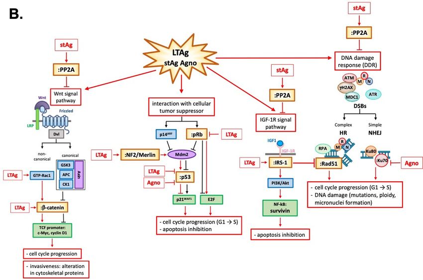
Figure 2. Figure 2. Polyomavirus JC: one virus, two stories. (A) In cells permissive to JCV infection, such as
Polyomavirus JC: one virus, two stories. (A) In cells permissive to JCV infection, such as
oligodendrocytes, viral transcription proceeds the viral DNA replication since the product of the viral
oligodendrocytes, viral transcription proceeds the viral DNA replication since the product of the viral early
early genes LTAg and stAg is essential for initiation and progression of the lytic cycle in
oligodendrocytes and progression of PML. JCV LTAg is a multifunctional protein with many domains
and it is important for viral DNA replication and for the transcriptional switch from early to late genes
which culminates in the production of capsid proteins VP1, VP2, and VP3 and final virions assembly.
In cells non permissive to the viral infection, LTAg protein starts modulating many cellular functions
through its many domains by interacting with cellular regulators, such as pRb, p53, Wnt, and IGF-1R
signaling pathways and DNA damage response factors, promoting cell cycle progression, apoptosis
inhibition and DNA damage which culminate in tumor onset. (B) In detail, the inactivation of pRb byInt. J. Mol. Sci. 2020, 21, 6236 7 of 22
genes LTAg and stAg is essential for initiation and progression of the lytic cycle in oligodendrocytes
and progression of PML. JCV LTAg is a multifunctional protein with many domains and it is
important for viral DNA replication and for the transcriptional switch from early to late genes
which culminates in the production of capsid proteins VP1, VP2, and VP3 and final virions assembly.
In cells non permissive to the viral infection, LTAg protein starts modulating many cellular functions
through its many domains by interacting with cellular regulators, such as pRb, p53, Wnt, and IGF-1R
signaling pathways and DNA damage response factors, promoting cell cycle progression, apoptosis
inhibition and DNA damage which culminate in tumor onset. (B) In detail, the inactivation of
pRb by LTAg promotes cell cycle progression through the release of E2F and activation of p14ARF
expression which leads to the stabilization of p53. However, LTAg binds and inactivates p53
preventing the p53 action in response to the DNA damage or p14ARF production. In mice transgenic
for the JCV early region, LTAg may also inhibit the tumor suppressor activity of p53 through
the interaction with neurofibromatosis type 2 (NF-2), a protein that neutralizes the inhibitory
effect of Mdm2 on p53, with the development of tumors. LTAg is also known to interact with
components of different signaling pathways which are associated with cellular transformation such
as β-catenin, insulin receptor substrate -1 (IRS-1), and survivin. β-catenin is a crucial protein of
the Wnt signaling pathway normally located and degraded in the cytoplasm. LTAg can bind the
C-terminus of this protein, resulting in its nuclear translocation and subsequent activation of c-Myc
and cyclin D1 TCF promoter, leading to cellular proliferation. LTAg can also stabilize β-catenin
through a non-canonical Wnt signal pathway, recruiting the GTPase protein Rac1 that stabilizes
β-catenin by inhibiting its ubiquitin-dependent proteasomal degradation. IRS-1 is the downstream
docking molecule of the insulin growth factor 1 receptor (IGF-1R) pathway. As for β-catenin,
LTAg binds and stabilizes g IRS-1 in the cytoplasm with the result of its nuclear translocation.
The unusual presence of IRS-1 in the nucleus enhances its binding and inactivation of enzyme
Rad51 which is involved in repairing of DNA double-stranded breaks (DSBs) by homologous
recombination (HR). The inactivation of Rad51 prevents the HR forcing the cell to repair its DSBs
via non-homologous end-joining (NHEJ). LTAg cooperates also with IGF-1R increasing the level of
survivin, which protects infected cells from apoptosis by dysregulation of cellular homeostasis and
oncogenesis. Infection of glial cells by JCV inflicts also severe cellular DNA damage throughout
LTAg which inactivates the ataxia-telangiectasia-mutated (ATM) and ATM- and Rad3-Related
(ATR) kinases. Small t antigen (stAg) is another significant viral protein that has roles in viral
production and influencing host cell growth. Its interactions with retinoblastoma proteins (pRbs)
and protein phosphatase 2A (PP2A) result in alterations of DNA damage response, inhibition of the
Wnt signaling pathway, and alteration in cytoskeletal proteins. Finally, Agno protein also affects the
DDR: Agno protein can bind to the Ku70 DNA repair protein, sequestering it in the perinuclear
space and impairing the NHEJ. In addition, the cooperation between Agno protein and p53 seems
to induce the activation of p21/WAF-1 gene expression.
Small t antigen (stAg) is another significant protein that has roles in viral production and influencing
host cell growth. Research on stAg has revealed its oncogenic effects [126]. Its interactions with specific
protein phosphatases, which generally suppress cellular growth pathways, permits increased activation
of those pathways. These include retinoblastoma proteins and protein phosphatase 2A (PP2A), both of
which have host cell functions that can lead to transformation when inhibited [78,126–129]. PP2A is
a serine/threonine phosphatase that regulates phosphorylation signals activated by kinases, such as
mitogen-activated protein kinase (MAPK), that promote cell proliferation. stAg can interact with PP2A
to regulate DNA repair mechanisms [130], inhibition of the Wnt signaling pathway, and alteration
in cytoskeletal proteins and tight junctions which increases invasiveness [131]. Recently it has also
been shown that MCPyV small t antigen is an oncoprotein that can transform rodent fibroblast in vitro
independent of LTAg [132,133]. Small t is an essential enhancer of cell proliferation in MCC [134] and
in vivo [135] (Figure 2 and Table 1).
Finally, it seems that the expression of the late JCV protein Agno also affects the DDR. The cells
expressing Agno were more sensitive to the cytotoxic effects of cisplatin with consequent increased
chromosome fragmentation, micronuclei formation, and cell cycle impairment [136]. NHEJ was
impaired in nuclear extracts from cells that expressed Agno. It has been hypothesized that defectsInt. J. Mol. Sci. 2020, 21, 6236 8 of 22
in NHEJ were caused by binding of Agno protein to the Ku70 DNA repair protein and subsequent
sequestering in the perinuclear space [115]. Moreover, in mouse fibroblasts constitutively expressing
Agnoprotein, an increased level of p21/WAF-1 protein was observed with dysregulation of cell cycle
progression, and an accumulation of cells in the G2/M phase. Besides, activation of p21/WAF-1 gene
expression in these cells is partly mediated through the cooperation of Agno protein with p53 [80]
(Figure 2 and Table 1).
5. JCV Oncogenicity in Animal Models
While the tumorigenic potential of polyomaviruses such as JCV and BKV in humans is still
a matter of debate (see Table 1), the JCV tumorigenic role in animal models is well documented.
The first evidence of the association of JC virus with cancer was reported with the development of
different brain tumors within 3–12 months when newborn golden Syrian hamsters were inoculated
with JCV. The majority of the tumors were glial origins, such as medulloblastomas that originate in the
cerebellum [137,138] and gliomas that originate in the brain [139]. Neuroblastoma, a solid embryonal
tumor of the sympathetic nervous system arising from the neural crest, was also reported [140].
The kind of tumors that developed in hamsters was strictly related to the site of injection and the
JCV strain. JCV Mad-1 strain isolated from a PML patient was able to induce malignant gliomas
within six months in 83% of newborn hamsters inoculated intracerebrally and subcutaneously [139].
Moreover, JCV from hamster tumor cells was capable of replicating after the fusion of these cells with
one permissive to the viral infection, confirming the persistence of the JCV genome in cultured tumor
cells [139]. Another study came to the same conclusion as reported by Walker and coworkers [138].
Additional studies have further confirmed the neuro-oncogenicity of other strains of JCV in newborn
hamsters. The 95% of hamsters inoculated with JCV Mad-2 developed cerebellar medulloblastomas
similar to those inoculated with the Mad-1 strain. In contrast, inoculation of the Mad-4 strain resulted
in 45% of animals with pineal gland tumors and 45% of animals with tumors in the cerebellum [137]
Although different strains of JCV were associated with the induction of various brain tumors, the route
of JCV inoculation has also been shown to correlate with the kind of tumor observed in newborn
hamsters. As reported by Varakis and colleagues, hamsters administered with JCV Mad-1 through the
eye developed neuroblastomas after 6–11 months, and primary tumors also arose in the abdominal
cavity with metastasis in several organs [140] (Table 2). Finally, there was no direct evidence of
viral replication or detection of viral structural proteins in all studies conducted to date in tumor
tissues [138,139,141–145].
The pioneering studies in hamsters prompted the researchers to focus on the association between
JCV and brain tumors in non-human primates. The first studies have reported that intracranial injection
of JC virus into owl monkeys and squirrel monkeys led to the development of astrocytoma within
14–36 months [142,146]. Analysis of the specimens from monkey tumors showed the presence of
LTAg in tumor tissue without active viral replication, as evident from the lack of detection of the
viral structural proteins [142] (Table 2). Interestingly, a suspension of tumor tissue isolated from owl
monkeys with JCV-induced astrocytoma [142] was successfully maintained in culture and analyzed for
phenotypic changes generally associated with expression of the LTAg [147]. JCV LTAg appeared in the
nucleus of these cultured cells, not in complex with the onco-suppressor p53 but in the presence of
actin dealignment features. These observations led the authors to conclude that JCV LTAg expression
can persist in brain tumor cells and correlate with cell phenotypes typical of Polyomaviruses-related
transformation, with tumor development in monkeys visible only several years after the first viral
inoculation [147].
Further support of the role of JC virus in tumor development was reported by Ohsumi’s laboratory
that showed the induction of neuroectodermal tumor after Tokio-1 JCV strain injection into the
brains of newborn Sprague–Dawley rats [148–150] (Table 2). However, compelling evidence linking
JCV with oncogenicity comes from a transgenic mice study. These transgenic mice were generated
by inserting the entire gene for JCV LTAg under the control of its promoter expressing only LTAg.Int. J. Mol. Sci. 2020, 21, 6236 9 of 22
In particular, Krynska and colleagues generated transgenic mice containing the early region of the
archetype strain CY. These mice exhibited paralysis of rear limbs, poor grooming, and hunched
posture within 9 and 13 months of age. The autopsy revealed the presence of primitive tumors from
the cerebellum, resembling the human medulloblastoma/primitive neuroectodermal tumor (PNETs).
Moreover, LTAg was expressed in the nuclei of all tumor tissue analyzed by immunohistochemistry,
strongly suggesting the potential of this animal model for the study of human CNS tumors in association
with the Polyomavirus JCV [151] (Table 2). A subsequent study in transgenic mice expressing LTAg
under the promoter region of JCV Mad-4 strain developed large pituitary neoplasia within one year
in 50% of animals. The same study also demonstrated the interaction of LTAg with p53 with the
overexpression of the p53-downstream target protein p21/WAF1. These results have contributed to
make this animal model a useful tool to evaluate further mechanisms of tumorigenesis mediated by
JCV LTAg [152] (Table 2). As observed in hamsters, it is essential to underlay that the type of malignant
transformations developed in murine models also seems to be dependent on the strain of JCV [151–154].
All in all, these findings suggested the tumorigenic role of JCV LTAg.
Table 1. JCV-related Oncogenic Mechanism.
Signaling Pathway Cellular Factor JCV Factor Oncogenic Effect References
p53, p21WAF1 Agno cell cycle arrest in G2/M in vitro Darbinyan A et al. 2002 [80]
pituitary neoplasia in LTAG
p53, p21WAF1 LTAg Gordon J et al. 2000 [152]
transgenic mice
pRb LTAg cell cycle progression in vitro Dyson N et al. 1990 [95]
pRb2/p130, E2F4/5 LTAg cell cycle progression in vitro Caracciolo V et al. 2007 [97]
Tumor suppressors cell cycle dysregulation in tumor
p53, pRb LTAg formation in LTAg Krynska B et al. 1997 [99]
transgenic mice
transgenic mouse model of
NF2 LTAg malignant peripheral nerve Shollar D et al. 2004 [103]
sheath tumors
Bollag B et al. 2010 [127];
cell cycle dysregulation and
pRb, PP2A stAg Sariyer IK et al. 2008 [128];
viral DNA replication
Pallas DC et al. 1990 [129]
-catenin, c-Myc, Enam S et al. 2002 [3];
LTAg oncogenesis of colon cancer
Cyclin D1 Ripple MJ et al. 2014 [108]
mouse medulloblastoma cell
line (BSB8), JCV-induced
Gan DD and Khalili K
-catenin LTAg hamster astrocytoma cell line
2004 [105]
(HJC2) and human astrocytoma
U-87MG cell line
Wnt -catenin,
murine medulloblastoma cell
LEF-1/TCF LTAg Gan DD et al. 2001 [111]
line (BsB8)
promoter
-catenin stabilization and cell Bhattacheryya R et al.
Rac1 GTPase LTAg
cycle progression in vitro 2007 [112]
Inhibition of Wnt signaling,
alteration in cytoskeleton Nunbhakdi-Craig V et al.
PP2A stAg
proteins and increase 2003 [131]
of invasiveness
translocation to the nucleus and
IRS-1 LTAg Lassal A et al. 2002 [106]
cell cycle progression
Piña-Oviedo S et al.
survivin LTAg apoptosis inhibition
IGF-1R 2007 [107]
apoptosis inhibition and
survivin LTAg proliferation of Gualco E et al. 2010 [114]
neural progenitors
HR dysregulation and
IGF-1R and DDR IRS-1, Rad51 LTAg Trojanek J et al. 2006 [113]
DNA damageInt. J. Mol. Sci. 2020, 21, 6236 10 of 22
Table 1. Cont.
Signaling Pathway Cellular Factor JCV Factor Oncogenic Effect References
HR dysregulation and
NHEJ Ku70 Agno Darbinyan A et al. 2004 [136]
DNA damage
HR dysregulation and DNA
HR Rad51, NHEJ
LTAg, Agno damage (mutation, ploidy, Darbinyan A et al. 2007 [115]
DDR Ku70, H2AX
and micronuclei formation)
White MK et al. 2014 [73];
HR Rad51, ATM LTAg DNA damage
White MK et al. 2017 [118]
PP2A stAg DNA damage Huang JL et al. 2015 [130]
HR: Homologous recombination; NHEJ: Non homologous end join.
Table 2. JCV oncogenesis in animal model.
Animal Model JCV Delivery Tumors Assay References
transplantation of tumors
subcutaneously and
isolation of JCV from 5/7
newborns inoculated tumors tested. Cells from
malignant gliomas:
intracerebrally and four of these tumors were
most of the tumors Walker DL et al.
subcutaneously with cultivated in vitro:
were glioblastomas 1973 [139]
JCV isolated from a intranuclear LTAg
and medulloblastomas
patient with PML antigenically related to
SV40 LTAg; JCV virions
after fusion of this culture
with permissive cells
cerebellar
three groups of medulloblastomas
newborns inoculated with Mad-2
intracerebrally with inoculation; pineal histologic characterization Padgett BL et al.
three different JCV gland tumors and of tumors. 1977b [137]
strains (Mad-2, Mad-3, tumors in the
Golden Syrian
and Mad-4) cerebellum with
Hamsters
Mad-4 inoculation.
(Mesocricetus
auratus) two neuroblastomas were
one group of newborns
neuroblastomas and transplanted serially, and a
inoculated
primary tumors in the tissue culture cell line was
intraocularly. Another
abdominal cavity with established from one of Varakis J et al.
group was inoculated
metastasis in liver, them. T-antigen was 1978 [140]
subcutaneously and
bone marrow, detected in 3/5 primary
intraperitoneally. Both
and lymph nodes. tumors tested and in the
with JCV Mad-1 strain
transplanted tumors.
medulloblastoma
newborns inoculated involved the internal
intracerebrally and granular layer of the
ZuRhein GM et al.
subcutaneously with cerebellum: lesion LTAg IF and histology
1979 [138]
JCV isolated from a comparable to
patient with PML childhood human
medulloblastoma
newborns inoculated
intracerebrally with
Tokio-1 JCV strain
cerebellar LTAg IF and histology Nagashima K et al.
(isolated form a patient
medulloblastoma (Homer-Wright rosettes) 1984 [148]
with PML,
serologically identical
to Mad-1 strain).
astrocytoma
two animals inoculated
(resembling human
intracerebrally,
glioblastoma
Owl Monkeys subcutaneously, London WT et al.
multiforme) and a TAg IF and histology
(Aotus trivirgatus) and intravenously with 1978 [142]
malignant tumor
JCV isolated from a
containing both glial
patient with PML
and neuronal cellsInt. J. Mol. Sci. 2020, 21, 6236 11 of 22
Table 2. Cont.
Animal Model JCV Delivery Tumors Assay References
six animals inoculated
intracerebrally,
Squirrel Monkeys subcutaneously, astrocytomas in 4/6 histologic characterization London WT et al.
(Saimiri sciureus) and intravenously with animals. of tumors 1983 [146]
JCV isolated from a
patient with PML
LTAg IHC and histology.
brain tumors in the Neuronal differentiation
cerebrum: was not proved. Glial
newborns inoculated Oshumi et al.
undifferentiated differentiation was
Sprague-DawleyRats intracranially with 1985 [149]; Oshumi
neuroectodermal confirmed by
Tokyo-1 JCV strain. et al. 1986 [150].
nature and subcutaneous
pseudo-rosettes. transplantation of cultured
tumor cells
primitive tumors
originating from the RT-PCR for LTAg mRNA,
transgenic mice for the cerebellum: close IHC for LTAg and p53, IP
Krynska B et al.
early region of JCV resemblance of human for LTAg and p53 and
1999b [151]
Transgenic Mice Archetype strain medulloblastoma/primitive Archetype NCCR
neuroectodermal sequencing
tumors (PNETs)
transgenic mice for the
IHC for LTAg and p53, IP Gordon J et al.
early region of JCV pituitary neoplasia
for LTAg, p53 and p21WAF1 2000 [152]
Mad-4 strain
pituitary neoplasia and IHC for LTAg, NF-1,
transgenic mice for the
signs resembling NF2,p53, and p21WAF1 and Shollar D et al.
early region of JCV
malignant peripheral IP for LTAg, NF-1, NF2 2004 [103]
Mad-4 strain
nerve sheath tumors. and p53,
6. Evidence of JCV Infectivity in Human Tumor Tissues
The direct link of JCV with human cancer is still a matter of debate. JCV causes tumors in rodents
and non-human primates, which are not its natural host. The JCV ubiquitous nature in the population
makes it hard to establish its role in human cancer. However, there have been reports that indicate
the expression of the viral LTAg in association with the transformation of neuronal cells in vitro and
the induction of tumors in monkeys, which lead to speculation of the possible association of JCV
with human CNS tumors. The first evidence linking the JCV association with a human brain tumor
(oligodendroglioma) was reported in a patient with chronic lymphocytic leukemia with PML [155].
JCV particles were detected in multiple gliomas and multiple and malignant astrocytomas in patient
with characteristic PML lesions [156,157]. However, the first direct evidence implicating JCV and its
viral protein in CNS neoplasms was described in a 21-year-old patient with immunodeficiency and
PML. Postmortem examination of brain tissue by immunohistochemistry and in situ hybridization
showed the expression of viral LTAg and mRNA, respectively [158]. The association of JCV with
CNS tumors in animal models prompted the undertaking of a large-scale analysis of human brain
tumor tissue samples for the presence of viral DNA or proteins [159]. The first systematic analysis of
brain tumor tissues for the detection of JCV DNA and proteins was conducted in 1996 by Rencic and
colleagues [24] in a patient with an oligoastrocytoma in the absence of PML. In this study, the presence
of JCV DNA was confirmed by sequencing of the PCR products. JCV RNA and T-Ag protein were
detected in the tumor tissue by primer extension analysis and Western blotting, respectively. Del Valle
and colleagues [15] examined 85 samples of glial tumors for the presence of JCV DNA sequences and
T-Ag expression, showing that 57% to 83% of tumors were positive for JCV.
Further studies by the same Authors linked JCV to other brain tumors, such as glioblastoma
multiforme, oligoastrocytoma, oligodendroglioma, and medulloblastoma (Table 3). In particular,
medulloblastoma is among the most frequent grade IV brain tumor with the highest number of cases in
children of age between six and eight years. Histologically and morphologically, medulloblastomas are
embryonal tumors derived from neuronal stem cells of the cerebellum [160]. Krynska and colleaguesInt. J. Mol. Sci. 2020, 21, 6236 12 of 22
investigated the association between JCV and pediatric tumors in humans. They showed the presence of
JCV DNA encoding N-terminal and C-terminal of LTAg in 11 out of 23 pediatric medulloblastoma tissue
samples [20]. They hypothesized the “hit-and-run” mechanism where LTAg expression may trigger a
cascade of tumorigenic events that do not require the presence of the viral protein in the advanced
stage of tumoral progression. In support of these outcomes, earlier studies by the same research group
in a transgenic murine model of medulloblastomas induced by JCV early gene expression showed
that not all the tumoral cells produced LTAg [151]. Moreover, the N-terminus region of JCV LTAg
that interacts with the onco-suppressor pRb was detected in 87% of the pediatric medulloblastomas.
In contrast, the C-terminal region was detected in only 56.5% of the samples. The finding of a particular
critical domain of LTAg, which is known to interact with onco-suppressor proteins, might implicate
Tag to play a role in this specific tumor [20].
Table 3. Association between JCV and human brain tumors in the absence of PML.
Brain Tumor JCV Factor Cellular Factor Assay References
Boldorini R et al.
PCR and sequencing (Mad-4 NCCR
VP1, NCCR - 2003 [12]; Delbue S
and genotype1 VP1)
et al. 2005 [13]
Del Valle et al.
IHC (p53 and LTAg), PCR (LTAg) and
LTAg p53 2000 [14], Del Valle
SB (LTAg)
et al. 2001a [15]
Glioblastoma IHC (p53 and LTAg–VP1 not
detected), PCR (LTAg, VP1, Agno, Piña-Oviedo S et al.
LTAg, VP1, Agno,
p53 NCCR), SB (LTAg, VP1, Agno, 2006 (case
NCCR
NCCR), sequencing (Mad-1NCCR) report) [16];
and LCM LTAg positive cells
IHC (p53 and LTAg–VP1 not
Del Valle L et al.
LTAg, VP1, Agno, detected), PCR (LTAg, VP1, Agno,
p53 2002b (case
NCCR NCCR), SB (LTAg, VP1, Agno),
report) [23]
sequencing (Mad-4 NCCR)
IHC (p53 and LTAg), PCR (LTAg) and Del Valle et al.
LTAg p53
SB (LTAg) 2001a [15]
Astrocytoma
IHC (LTAg), PCR (LTAg and NCCR) Caldarelli-Stefano
LTAg, NCCR -
and sequencing (Mad-4 NCCR) R et al. 2000 [17]
IHC (p53 and LTAg), PCR (LTAg) and Del Valle et al.
LTAg p53
SB (LTAg) 2001a [15]
IHC (Ki67 proliferation marker and
Oligoastrocytoma LTAg), PCR (LTAg and NCCR), SB
Rencic A et al.
LTAg, NCCR Ki67 (LTAg), primer extension (LTAg), IP
1996 [24]
(LTAg) and sequencing (Mad-4
NCCR)
IHC (p53 and LTAg), PCR (LTAg) and Del Valle et al.
LTAg p53
SB (LTAg) 2001a [15]
IHC (p53, LTAg, Agno–Vp1 not
Oligodendroglioma detected), PCR (LTAg, VP1, Agno,
LTAg, VP1, Agno, Del Valle et al.
p53 NCCR), SB (LTAg, VP1 and Agno),
NCCR 2002c [25]
sequencing (Mad-4 and Archetype
NCCR)
IHC (p53 and LTAg), PCR (LTAg) and Del Valle et al.
Ependymoma LTAg p53
SB (LTAg) 2001a [15]
IHC (LTAg–VP1 not detected), PCR Krynska B et al.
LTAg, VP1 -
(LTAg, VP1), SB (LTAg, VP1) 1999a [20]
p53, pRb (p107, IHC (p53, pRb, LTAg), PCR (LTAg, Del Valle et al.
Medulloblastoma LTAg, VP1
pRb2/p130) VP1) 2001c [21]
IHC (p53, LTAg and Agno), PCR Del Valle et al.
LTAg, Agno p53
(LTAg, Agno), SB (LTAg, Agno) 2002a [22]
IHC (p53 and LTAg–VP1 not
Primary CNS detected), PCR (LTAg, VP1, Agno), SB Del Valle et al.
LTAg, Agno, VP1 p53
lymphoma (LTAg, VP1, Agno), LCM LTAg 2004 [161]
positive cells
IHC: Immunohistochemistry; SB: Southern Blot; LCM: Laser capture microdissection.Int. J. Mol. Sci. 2020, 21, 6236 13 of 22
In a later study by the same group, 20 paraffin-embedded well-characterized medulloblastomas
were analyzed for the presence of JCV Agno DNA sequences and the expression of Agnoprotein
and LTAg. PCR analysis revealed the presence of JCV Agno DNA in 11 of 16 samples analyzed,
whereas Agnoprotein was detected by immunohistochemistry in the cytoplasm of neoplastic cells
in 11 of 20 tissue samples. Moreover, LTAg protein was reported in the nucleus of neoplastic cells
where agnoprotein was absent. Finally, none of the tumor samples analyzed has shown the expression
of viral late capsid proteins, excluding the possibility of a productive JCV infection of the tumor
cells. These data provided additional evidence that both LTAg and Agno might play a role in the
development of JCV-associated medulloblastomas [22].
Further evidence for the possible role of LTAg and Agno in GBM was reported in two cases
having GBM. The first case is an immunocompromised individual with multiple sclerosis, and the
second one is a 54-year-old immunocompetent patient. In the first case study, GBM tumor tissues
were positive for JCV DNA, and LTAg was localized in the nucleus. The same conclusion was
reported in the second case with the additional evidence of Agno protein expression in the cytoplasm.
Furthermore, using laser capture microdissection techniques, Mad-1 strain was identified in the tissues
examined [16,23] (Table 3).
Finally, since it has been reported that JCV can infect B-lymphocytes, it leads to speculation of its
association with primary CNS lymphomas. Del Valle et al. in 2004 published a study involving 27 cases
of CNS lymphomas, in which the JCV DNA sequence was detected in 81% of cases, but the expression
of LTAg was detected in only 18.5% [161]. Several studies have shown the coexistence of PML and
primary CNS lymphomas indicating PML-associated JCV reactivation [162,163]. JCV infection of
B-lymphocytes in vitro is non-productive, which leads to the speculation that infected B-lymphocytes
may serve as a carrier to disseminate the virus to the CNS, where it can infect glial cells [61] (Table 3).
The spread of JC virus from the initial site of infection to the brain is one of the many critical areas
where our understanding of JCV biology is incomplete. Specifically, the mechanism by which JCV
reaches the brain from the primary peripheral site of infection. It has been hypothesized that immune
cells, specifically the B-lymphocytes, not only serve as latent reservoirs [28,164–166] but also an agent
disseminating the virus to CNS-specific glial cells.
7. Conclusions
Multiple experiments have identified the oncogenic potential of the JC virus: its transformative
ability of cells in vitro and in in vivo models. Several studies reported the detection of viral genome
and proteins by a molecular and virologic approach in many organs and tissues, including the
brain. The oncogenic potential of the human BKV with viral genome integration into the cellular
DNA has recently been shown. Using high-through sequencing of tumor DNA obtained from
urothelial carcinoma, the researchers identified the integration site of the BKV genome into exon 26
of myosin-binding protein C1 gene (MYBPC1) on chromosome 12 in tumor cells but not in normal
renal cells. Interestingly, this viral integration event leads to altered viral gene expressions such
as robust expression of LTAg and a lack of expression of the viral structural proteins and DNA
replication. This finding supports the notion that a polyomavirus integration event is essential to
tumorigenesis [117]. In the case of BKV, the persistent over-expression of LTAg in non-lytic cells likely
promotes cell growth, genetic instability, and oncogenesis [122]. Furthermore, the viral integration
event has been considered a critical step in MCPyV-mediated tumorigenesis. Several studies have
shown the clonal integration of MCPyV into Merkel cell carcinoma, which leads to persistent expression
of the oncoproteins LTAg and stAg [167]. Although the integration of JCV has been reported in an
in vitro model, there is still no definitive evidence that shows JCV integration into host genome as
responsible for tumors in humans.
The finding of JC virus DNA and its proteins, in either normal or brain tumor tissues, establishes an
active presence in the brain [63]. There are conflicting reports regarding the quantities of JC viral DNA
found in human tumor specments [168–170]. However, the inability to detect the viral DNA sequenceInt. J. Mol. Sci. 2020, 21, 6236 14 of 22
does not negate its ability to transform a cell. A more recent concept in tumor virology suggests that
the initial infection produces changes and conditions that promote host cell transformation. A viral
genome can be inserted into that of the host cell, and it could be lost over time. Despite this, the ability
of JC viral proteins to affect the cell cycle and disrupt DNA repair pathways could have long-lasting
changes in a cell without the active virus needed to be continually present. While not all tumor samples
contain it, viral DNA lasting effects could still be enough to transform the host. Abortive JC virus
infection of cells may lead to the expression of viral oncogenic proteins that may initiate tumorigenesis,
even if the entire virus is no longer active.
Since the discovery of Rous sarcoma virus (RSV) as a causative agent of chicken tumor, the direct
link of viruses with cancer is now well accepted; 12–20% of human cancers are reported to be caused
by a viral infection. The association of human cancer with the infection of some DNA or RNA viruses
such as Hepatitis B virus [171], Merkel Cell Carcinoma virus [172], Epstein Barr virus (EBV) [173],
Hepatitis C virus [174], Human Herpesvirus Type 8 [175], human papillomaviruses [176] human
BKV [117], and the human T-cell lymphotropic virus (HTLV) [177] is well documented. Although
JCV has a very high prevalence of infection in the worldwide population, as demonstrated by
sero-epidemiological data, there is not yet a proportional number of cancers attributable to this virus.
This observation suggests there are multiple immune system defenses or pathways that revert potential
virulent effects. Further research is warranted to shed light on all those cellular pathways involved in
JCV-associated oncogenesis.
Author Contributions: Contributed to the concept and design of this review: N.A., A.B., H.S.W. Collected
references and analyzed them: N.A., A.B., D.M., H.S.W. Wrote the bulk of the text and prepared the figures: N.A.,
A.B., H.S.W. All authors have read and agreed to the published version of the manuscript.
Funding: This research received no external funding.
Acknowledgments: We thank past and present members of the Center for Neurovirology for their insightful
discussion and sharing of ideas. This work was supported by seed money from Temple University awarded to
H.S.W.
Conflicts of Interest: The authors declare no conflict of interest.
References
1. Moore, P.; Chang, Y. Why do viruses cause cancer? Highlights of the first century of human tumour virology.
Nat. Rev. Cancer 2010, 10, 878–889. [CrossRef] [PubMed]
2. Parkin, D.M. The global health burden of infection-associated cancers in the year 2002. Int. J. Cancer 2006,
118, 3030–3044. [CrossRef] [PubMed]
3. Enam, S.; Del Valle, L.; Lara, C.; Gan, D.-D.; Ortiz-Hidalgo, C.; Palazzo, J.P.; Khalili, K. Association of human
polyomavirus JCV with colon cancer: Evidence for interaction of viral T-antigen and beta-catenin. Cancer Res.
2002, 62, 7093–7101.
4. Kassem, A.; Technau, K.; Kurz, A.K.; Pantulu, D.; Löning, M.; Kayser, G.; Stickeler, E.; Weyers, W.; Díaz, C.;
Werner, M.; et al. Merkel cell polyomavirus sequences are frequently detected in nonmelanoma skin cancer
of immunosuppressed patients. Int. J. Cancer 2009, 125, 356–361. [CrossRef]
5. Reiss, K.; Khalili, K. Viruses and cancer: Lessons from the human polyomavirus, JCV. Oncogene 2003,
22, 6517–6523. [CrossRef]
6. Lee, W.; Langhoff, E. Polyomavirus in Human Cancer Development. Adv. Exp. Med. Biol. 2007, 577, 310–318.
[CrossRef]
7. Pinto, M.; Dobson, S. BK and JC virus: A review. J. Infect. 2014, 68 (Suppl. 1), S2–S8. [CrossRef]
8. Knowles, W.A. Discovery and epidemiology of the human polyomaviruses BK virus (BKV) and JC virus
(JCV). Adv. Exp. Med. Biol. 2007, 577, 19–45. [CrossRef]
9. Tan, C.S.; Koralnik, I.J. Progressive multifocal leukoencephalopathy and other disorders caused by JC virus:
Clinical features and pathogenesis. Lancet Neurol. 2010, 9, 425–437. [CrossRef]
10. Arthur, R.R.; Dagostin, S.; Shah, K.V. Detection of BK virus and JC virus in urine and brain tissue by the
polymerase chain reaction. J. Clin. Microbiol. 1989, 27, 1174–1179. [CrossRef]Int. J. Mol. Sci. 2020, 21, 6236 15 of 22
11. Miranda-Filho, A.; Piñeros, M.; Soerjomataram, I.; Deltour, I.; Bray, F. Cancers of the brain and CNS:
Global patterns and trends in incidence. Neuro-Oncology 2016, 19, 270–280. [CrossRef] [PubMed]
12. Boldorini, R.; Pagani, E.; Car, P.G.; Omodeo-Zorini, E.; Borghi, E.; Tarantini, L.; Bellotti, C.; Ferrante, P.;
Monga, G. Molecular characterisation of JC virus strains detected in human brain tumours. Pathology 2003,
35, 248–253. [CrossRef] [PubMed]
13. Delbue, S.; Pagani, E.; Guerini, F.R.; Agliardi, C.; Mancuso, R.; Borghi, E.; Rossi, F.; Boldorini, R.; Veggiani, C.;
Car, P.G.; et al. Distribution, characterization and significance of polyomavirus genomic sequences in tumors
of the brain and its covering. J. Med. Virol. 2005, 77, 447–454. [CrossRef] [PubMed]
14. Del Valle, L.; Azizi, S.A.; Krynska, B.; Enam, S.; Croul, S.E.; Khalili, K. Reactivation of human neurotropic JC
virus expressing oncogenic protein in a recurrent glioblastoma multiforme. Ann. Neurol. 2000, 48, 932–936.
[CrossRef]
15. Del Valle, L.; Gordon, J.; Assimakopoulou, M.; Enam, S.; Geddes, J.F.; Varakis, J.N.; Katsetos, C.D.; Croul, S.;
Khalili, K. Detection of JC virus DNA sequences and expression of the viral regulatory protein T-antigen in
tumors of the central nervous system. Cancer Res. 2001, 61, 4287–4293. [PubMed]
16. Piña-Oviedo, S.; De León-Bojorge, B.; Cuesta-Mejías, T.; White, M.K.; Ortiz-Hidalgo, C.; Khalili, K.; Del Valle, L.
Glioblastoma multiforme with small cell neuronal-like component: Association with human neurotropic JC
virus. Acta Neuropathol. 2006, 111, 388–396. [CrossRef]
17. Caldarelli-Stefano, R.; Boldorini, R.; Monga, G.; Meraviglia, E.; Zorini, E.O.; Ferrante, P. JC virus in human
glial-derived tumors. Hum. Pathol. 2000, 31, 394–395. [CrossRef]
18. Del Valle, L.; Gordon, J.; Ferrante, P.; Khalili, K. JC virus in experimental and clinical brain tumorigenesis.
In Human Polyomaviruses; Khalili, K., Stoner, G., Eds.; Wiley & Sons, Inc.: New York, NY, USA, 2003;
pp. 409–430.
19. Shiramizu, B.; Hu, N.; Frisque, R.J.; Nerurkar, V.R. High prevalence of human polyomavirus JC VP1 gene
sequences in pediatric malignancies. Cell. Mol. Biol. 2007, 53, 4–12.
20. Krynska, B.; Del Valle, L.; Croul, S.; Gordon, J.; Katsetos, C.D.; Carbone, M.; Giordano, A.; Khalili, K.
Detection of human neurotropic JC virus DNA sequence and expression of the viral oncogenic protein in
pediatric medulloblastomas. Proc. Natl. Acad. Sci. USA 1999, 96, 11519–11524. [CrossRef]
21. Del Valle, L.; Baehring, J.; Lorenzana, C.; Giordano, A.; Khalili, K.; Croul, S. Expression of a human
polyomavirus oncoprotein and tumour suppressor proteins in medulloblastomas. Mol. Pathol. 2001,
54, 331–337. [CrossRef]
22. Del Valle, L.; Gordon, J.; Enam, S.; Delbue, S.; Croul, S.; Abraham, S.; Radhakrishnan, S.; Assimakopoulou, M.;
Katsetos, C.D.; Khalili, K. Expression of human neurotropic polyomavirus JCV late gene product agnoprotein
in human medulloblastoma. J. Natl. Cancer Inst. 2002, 94, 267–273. [CrossRef] [PubMed]
23. Del Valle, L.; Delbue, S.; Gordon, J.; Enam, S.; Croul, S.; Ferrante, P.; Khalili, K. Expression of JC virus
T-antigen in a patient with MS and glioblastoma multiforme. Neurology 2002, 58, 895–900. [CrossRef]
[PubMed]
24. Rencic, A.; Gordon, J.; Otte, J.; Curtis, M.; Kovatich, A.; Zoltick, P.; Khalili, K.; Andrews, D. Detection of JC
virus DNA sequence and expression of the viral oncoprotein, tumor antigen, in brain of immunocompetent
patient with oligoastrocytoma. Proc. Natl. Acad. Sci. USA 1996, 93, 7352–7357. [CrossRef] [PubMed]
25. Del Valle, L.; Enam, S.; Lara, C.; Ortiz-Hidalgo, C.; Katsetos, C.D.; Khalili, K. Detection of JC
polyomavirus DNA sequences and cellular localization of T-antigen and agnoprotein in oligodendrogliomas.
Clin. Cancer Res. 2002, 8, 3332–3340. [PubMed]
26. Padgett, B.; Walker, D.; ZuRhein, G.; Eckroade, R.; Dessel, B. Cultivation of papova-like virus from human
brain with progressive multifocal leucoencephalopathy. Lancet 1971, 297, 1257–1260. [CrossRef]
27. Tavazzi, E.; White, M.K.; Khalili, K. Progressive multifocal leukoencephalopathy: Clinical and molecular
aspects. Rev. Med. Virol. 2011, 22, 18–32. [CrossRef]
28. Berger, J.R.; Scott, G.; Albrecht, J.; Belman, A.L.; Tornatore, C.; Major, E.O. Progressive multifocal
leukoencephalopathy in HIV-1-infected children. AIDS 1992, 6, 837–842. [CrossRef]
29. Wollebo, H.S.; White, M.K.; Gordon, J.; Berger, J.R.; Khalili, K. Persistence and pathogenesis of the neurotropic
polyomavirus JC. Ann. Neurol. 2015, 77, 560–570. [CrossRef]
30. Morriss, M.C.; Rutstein, R.M.; Rudy, B.; DesRochers, C.; Hunter, J.V.; Zimmerman, R.A. Progressive multifocal
leukoencephalopathy in an HIV-infected child. Neuroradiology 1997, 39, 142–144. [CrossRef]You can also read


