A pro-tumorigenic secretory pathway activated by p53 deficiency in lung adenocarcinoma - JCI
←
→
Page content transcription
If your browser does not render page correctly, please read the page content below
A pro-tumorigenic secretory pathway activated by p53
deficiency in lung adenocarcinoma
Xiaochao Tan, … , Chad J. Creighton, Jonathan M. Kurie
J Clin Invest. 2020. https://doi.org/10.1172/JCI137186.

 Research   In-Press Preview Cell biology    Oncology

Graphical abstract

Find the latest version:
https://jci.me/137186/pdf
Title: A pro-tumorigenic secretory pathway activated by p53 deficiency in lung adenocarcinoma

Authors: Xiaochao Tan1,*, Lei Shi1, Priyam Banerjee1, Xin Liu1, Hou-Fu Guo1, Jiang Yu1, Neus

Bota-Rabassedas1, B. Leticia Rodriguez1, Don L. Gibbons1, William K. Russell2, Chad J.

Creighton3,4, Jonathan M. Kurie1,*

Affiliations : 1Department of Thoracic/Head and Neck Medical Oncology, The University of Texas

MD Anderson Cancer Center, Houston, Texas. 2Department of Biochemistry and Molecular

Biology, University of Texas Medical Branch, Galveston, TX 77555, USA. 3Department of

Medicine, Dan L. Duncan Cancer Center, Baylor College of Medicine, Houston, Texas.
4
Department of Bioinformatics and Computational Biology, The University of Texas MD Anderson

Cancer Center, Houston, Texas. *To whom Correspondence should be addressed: Jonathan M.

Kurie, Department of Thoracic/Head and Neck Medical Oncology, Box 432, MD Anderson Cancer

Center, 1515 Holcombe Blvd, Houston, TX 77030; email: jkurie@mdanderson.org; Xiaochao

Tan, Department of Thoracic/Head and Neck Medical Oncology, Box 432, MD Anderson Cancer

Center, 1515 Holcombe Blvd, Houston, TX 77030; email: xtan@mdanderson.org.

Conflict of interest

D.L.G. serves on scientific advisory committees for Astrazeneca, GlaxoSmithKline, Sanofi and

Janssen, provides consults for Ribon Therapeutics, and has research support from Janssen,

Takeda, and Astrazeneca. J.M.K. has received consulting fees from Halozyme. All other authors

declare that they have no competing interests.
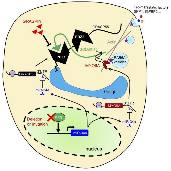
Abstract

Therapeutic strategies designed to target TP53-deficient cancer cells remain elusive. Here, we

showed that TP53 loss initiated a pharmacologically actionable secretory process that drove lung

adenocarcinoma (LUAD) progression. Molecular, biochemical, and cell biological studies showed

that TP53 loss increased the expression of Golgi reassembly and stacking protein 55 kD (G55),

a Golgi stacking protein that maintains Golgi organelle integrity and is part of a GOLGIN45/myosin

IIA-containing protein complex that activates secretory vesicle biogenesis in the Golgi. TP53 loss

activated G55-dependent secretion by relieving G55 and myosin IIA from miR-34a-dependent

silencing. G55-dependent secreted proteins enhanced the proliferative and invasive activities of

TP53-deficient LUAD cells and promoted angiogenesis and CD8+ T cell exhaustion in the tumor

microenvironment. A small molecule that blocks G55/G45 interactions impaired secretion and

reduced TP53-deficient LUAD growth and metastasis. These results identified a targetable

secretory vulnerability in TP53-deficient LUAD cells.

Introduction

The presence of a cytokine-rich, fibrotic stroma is correlated with advanced disease and poor

clinical outcome in multiple cancer types (1). In one working hypothesis, pro-metastatic features

of the tumor microenvironment are orchestrated by cancer cells that secrete cytokines, proteases,

and growth factors that inhibit anti-tumor immunity, promote angiogenesis, and facilitate cancer

cell dissemination (2-4). Transcriptomes activated by somatically acquired oncogenic mutations

are enriched in secreted proteins that promote tumor growth and metastasis (5). Thus, a better

understanding of the way in which oncogenic mutations activate secretion may identify

therapeutic vulnerabilities in cancer.

   Secreted proteins are transported as vesicular cargos from the endoplasmic reticulum to the

cell surface through defined intracellular pathways. Proteins that lack N-terminal signal peptides
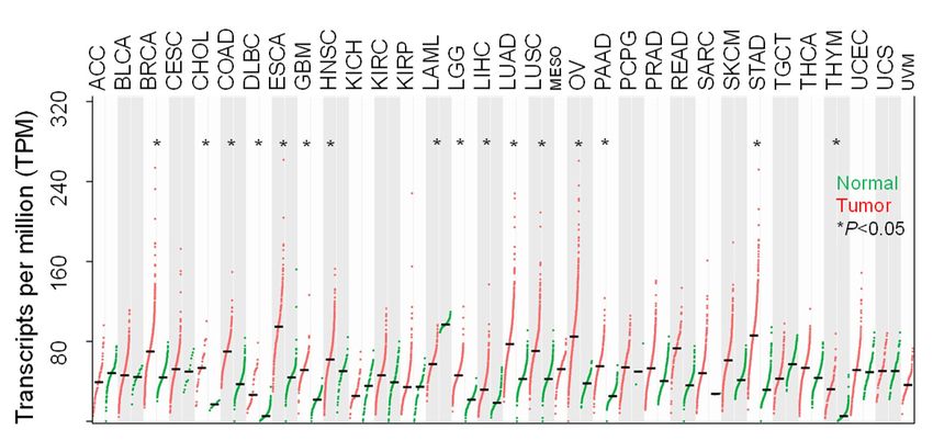
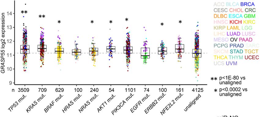
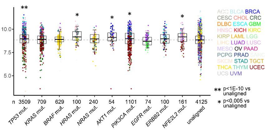
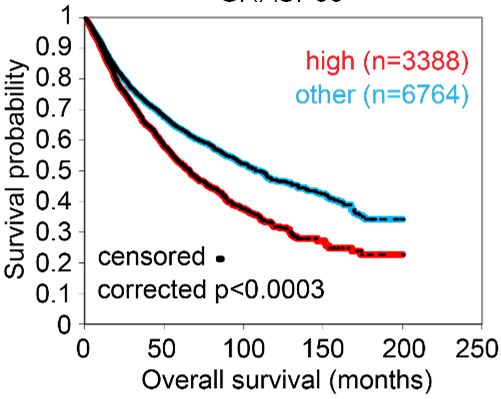
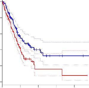
are transported via unconventional protein secretion (UPS), a stress-activated, autophagy-

dependent vesicular transport pathway that bypasses the Golgi (6). In autophagosomes, cargo

loading, autophagosome-lysosome fusion, and autophagosome maturation are facilitated by

Golgi reassembly and stacking protein 55 kD (G55), a Golgi stacking protein that translocates to

autophagosomes to facilitate UPS (7, 8). Proteins that contain signal peptides, however, are

transported to the plasma membrane via the Golgi apparatus, a process called conventional

protein secretion (CPS) (6). In the Golgi, vesicle budding, fission, and release are initiated by

Golgi phosphoprotein 3 (GOLPH3), which bridges Golgi membranes to F-actin fibers that convey

a tensile force required for secretory vesicle biogenesis (9, 10). On the basis of this conceptual

framework, here, we evaluated the molecular underpinnings of secretion in cancer and identified

a pro-tumorigenic, G55-dependent secretory process that is activated by TP53 deficiency.

Results

TP53 deficiency activates a G55-dependent secretory program. We queried the Gene

Ontology term “Golgi apparatus” (n=453 genes) in The Cancer Genome Atlas (TCGA) pan-cancer

cohort and found that, compared to matched normal tissues, lung adenocarcinoma (LUAD) and

other cancer types have higher expression of 201 genes (P < 0.001, fold-change > 1.4), including

G55, a Golgi stacking protein that promotes secretion (11, 12) (Figure 1, A and B). High G55

expression is correlated with multiple oncogenic driver mutations (TP53, KRAS, BRAF, NRAS,

AKT1, PIK3CA, ERBB2, NFE2L2) (Figure 1C) and shorter survival durations (Figure 1D). On the

basis of these findings, we delved into the role of G55 as an effector of TP53-mutant LUAD

progression.

   To investigate a causal relationship between p53 loss and increased G55 expression, we

subjected A549 LUAD cells, which are TP53-wild-type, to CRISPR/Cas9-mediated TP53

knockout (KO) and found that G55 expression was higher in TP53 KO cells (Figure 1E). Using a

microRNA-binding site prediction algorithm (www.targetscan.org), we identified a binding site for
miR-34a, which is a p53 transcriptional target (13), in the G55 3’-untranslated region (3’-UTR),

which led us to postulate that p53 loss upregulates G55 expression by silencing miR-34a. To test

this hypothesis, we subjected TP53-mutant H1299 cells and H2122 cells to ectopic miR-34a

expression and found that G55 levels were suppressed (Figure 1, F and G). Ectopic miR-34a

expression abrogated G55 upregulation induced by short hairpin RNA (shRNA)-mediated p53

deficiency (Figure 1, H and I), and miR-34a antagomiRs reversed ectopic p53-induced G55

suppression (Figure 1, J and K), indicating that G55 upregulation by TP53 deficiency is miR-34a-

dependent. To determine whether G55 is a miR-34a target, we performed G55 3’-UTR luciferase

reporter assays and found that ectopic miR-34a expression inhibited the activity of a wild-type

G55 3’-UTR luciferase reporter, whereas the activity of a mutant G55 3’-UTR reporter that lacks

the miR-34a binding site was unaffected (Figure 1L). Demonstrating the specificity of this

regulatory axis, ectopic miR-34a expression did not downregulate the G55 homologue GRASP65,

which lacks miR-34a binding sites, and ectopic expression of miR-200a, which is not predicted to

bind G55, did not inhibit G55 expression (Figure 1G). Chromatin immunoprecipitation assays to

quantify RNA polymerase II recruitment to the G55-encoding genomic locus (GORASP2)

demonstrated no significant difference between parental and TP53 KO cells (Figure 1M),

suggesting that p53 depletion did not increase GORASP2 gene transcription. These findings

indicate that p53 deficiency relieves G55 from miR-34a-dependent silencing.

   We postulated that high G55 expression is pro-tumorigenic in TP53-mutant LUAD and tested

this hypothesis in TP53-null LUAD cells (H1299, CALU-1) and LUAD cells that express mutant

p53 proteins that do (H441, 344SQ) or do not (H2122) have gain-of-function properties (14, 15).

Regardless of the type of TP53 mutation, small interfering RNA (siRNA)-mediated G55 depletion

impaired the proliferative, colony forming, migratory, and invasive activities of these cells (Figure

2, A-D) without inducing apoptosis (Supplemental Figure 1A). In colony formation assays, G55

siRNAs demonstrated greater inhibitory activity against TP53-mutant than -wild-type LUAD cell

lines (Figure 2, E-G, and Supplemental Figure 1B), suggesting a selective vulnerability of TP53-
mutant LUAD cells to G55 depletion. Conversely, ectopic G55 expression increased the

migratory, invasive, and colony forming activities of H1299 cells (Supplemental Figure 1, C-E)

and reconstituted these activities in HeLa cells that had been subjected to CRISPR/Cas9-

mediated GORASP2 KO (Supplemental Figure 1, F-H). Relative to G55-replete controls,

orthotopic LUADs generated by G55-deficient 344SQ cells were smaller, less metastatic, and

caused delayed mortality in syngeneic, immunocompetent mice (Figure 2, H-J). Conversely,

ectopic G55 expression accelerated the growth and enhanced the metastatic activity of

subcutaneous tumors generated by 344P cells (Figure 2, K and L, and Supplemental Figure 2I).

Thus, high G55 levels are required for, and may be sufficient to drive, LUAD progression.

Moreover, G55 siRNAs reduced the proliferative and invasive activities of TP53-mutant breast

and ovarian cancer cell lines (Supplemental Figure 1, J-N), suggesting a generalizable role for

G55 in TP53-mutant cancer cells.

   Given that G55 is a key secretory pathway component (8), we postulated that G55 drives

tumor progression by activating the secretion of pro-tumorigenic effector proteins. To test this

hypothesis, we performed conditioned medium (CM) transfer experiments and found that the

invasive and colony forming activities of G55-deficient cancer cells were rescued by treatment

with CM samples from G55-replete, but not –deficient, cancer cells (Figure 3, A and B, and

Supplemental Figure 2, A-C). To identify G55-dependent secreted proteins, we performed liquid

chromatography-mass spectrometry (LC-MS) analysis on CM samples from G55-deficient and –

replete H1299 cells and identified 925 proteins (≥ 2 peptides per protein, 1% false discovery rate),

18 of which were present at significantly lower concentrations (fold-change > 1.4, p < 0.05) in

G55-deficient cells (Figure 3C, and Supplemental Table 1), including the matricellular protein

osteopontin (SPP1), the insulin-like growth factor-binding protein IGFBP2, the lysosomal

prolylcarboxypeptidase PCRP, the serine peptidase HTRA1, the laminin receptor BCAM, and the

secreted neurosecretory protein VGF, which are pro-tumorigenic in cancer models (16-20).
Western blot analysis confirmed SPP1 and IGFBP2 down-regulation in CM samples, but not

lysates, from G55-deficient H1299, H2122 and 344SQ cells (Figure 3D, Supplemental Figure 2,

D-F). Following G55 reconstitution in GORASP2 KO HeLa cells, IGFBP2 and SPP1 secretion

were restored (Supplemental Figure 2, F and G), indicating that IGFBP2 and SPP1 are G55-

dependent secreted proteins. We assessed the functional roles of IGFBP2 and SPP1 by

performing siRNA- and neutralizing antibody-mediated depletion studies and found that they are

required to varying degrees for H1299 cells and H2122 cells to form colonies, migrate, and invade

(Figure 3, E and F, and Supplemental Figure 3, A-E) and for H1299 orthotopic LUADs to grow

and metastasize (Figure 3G, and Supplemental Figure 3F). Invasive activities of IGFBP2- and

SPP1-deficient H1299 cells were rescued by treatment with IGFBP2 and SPP1 recombinant

proteins (Supplemental Figure 3, G-I), demonstrating that colony forming activity decreased as a

result of on-target effects of the siRNAs.

   Proteins secreted by neuroendocrine and other secretory cells are stored in dense core

vesicles that degranulate in response to external cues (21). However, by immunofluorescence

staining, IGFBP2 and SPP1 did not localize in granules, which led us to speculate that secretion

draws from newly synthesized proteins, a conclusion supported by evidence that cycloheximide

treatment decreased IGFBP2 levels in cell lysates and CM medium samples and abrogated

ectopic G55-induced IGFBP2 secretion (Supplemental Figure 4A). To determine whether TP53

loss coordinately increases the expression and secretion of IGFBP2 and SPP1, we quantified

SPP1 and IGFBP2 levels in a panel of LUAD cell lines that are TP53-mutant or -wild-type and

found that SPP1 and IGFBP2 protein levels were higher in CM samples, but not lysates, from

TP53-mutant cells (Supplemental Figure 4, B and C). In gain- and loss-of-function studies, p53

and its downstream mediator miR-34a suppressed the secretion, but not the intracellular levels,

of IGFBP2 and SPP1 (Figure 3, H-J, Supplemental Figure 4, D-H). We conclude that p53-
dependent secretion draws on intracellular pools that are synthesized in a p53-independent

manner.

   Cancer cells secrete cytokines that drive inflammation, fibrosis, and angiogenesis in the tumor

microenvironment (4). To determine whether G55-dependent secretion governs these processes,

we performed flow cytometric analysis on flank tumors generated by G55-deficient or –replete

344SQ cells in syngeneic, immunocompetent mice and found that G55-deficient tumors had

reduced numbers of endothelial cells, increased numbers of effector/memory CD8+ T cells, and

reduced numbers of naïve and exhausted CD8+ T cells (Supplemental Figure 5, A-I). To identify

G55-dependent secreted cytokines that might mediate these activities, we performed multiplexed

antibody-based assays on CM samples from G55-deficient and –replete 344SQ cells and found

that concentrations of the pro-angiogenic cytokines interleukin-6 (IL-6), CXCL5/LIX, and VEGF-A

were reduced by ~ 50% in G55-deficient cells (Supplemental Figure 5J). Consistent with these

findings, G55 depletion in H1299 cells reduced IL-6 secretion (Supplemental Figure 5K) and

inhibited the migratory and tube-forming activities of human umbilical vein endothelial cells in co-

culture assays (Supplemental Figure 5, L and M). Thus, G55-dependent secreted proteins drive

pro-tumorigenic processes in the tumor microenvironment.

   A G55 antagonist inhibits secretion. Cartier-Michaud et al. used a virtual screening strategy

that combined high-throughput docking and pharmacophore filtering of chemical compounds to

identify a small molecule (GRASPIN) that competes with binding proteins for insertion into the N-

terminal PSD-95/Discs Large/ZO-1 (PDZ) domain of G55, which contains a surface groove and

a hydrophobic cavity that serve as docking sites for binding partners (22, 23). GRASPIN inhibits

G55-dependent spermatogenesis in mice and is under development as a male contraceptive (23).

We tested the selectivity of this compound in H1299 cells and found that 10 μM GRASPIN

treatment inhibited G55’s interactions with G45 (Figure 4A), a known G55 binding partner (22),

whereas binding of the G55 homologue GRASP65 to its binding partner GM130 was preserved
(Figure 4B). GRASPIN exerted anti-proliferative effects that were abrogated by shRNA-mediated

G55 depletion in H1299 cells and H2122 cells (Figure 4, C and D, and Supplemental Figure 6, A-

D). GRASPIN inhibited the colony forming activities of TP53-mutant and –wild-type LUAD cells

with 50% inhibitory concentration (IC 50 ) values of approximately 5 and 10 μM, respectively (Figure

4, E and F, and Supplemental Figure 6, E and F), demonstrating enhanced sensitivity to

GRASPIN in TP53-mutant LUAD cells. GRASPIN decreased IGFBP2 and SPP1 secretion (Figure

4G), and GRASPIN-dependent reductions in colony forming activity were partially rescued by

treatment with soluble IGFBP2 and SPP1 proteins (Supplemental Figure 6G), which had greater

effects in combination than they did alone, suggesting that GRASPIN suppressed colony

formation by inhibiting the secretion of IGFBP2, SPP1, and other proteins. Using a dose reported

to inhibit spermatogenesis in vivo (23), we treated syngeneic, immunocompetent mice bearing

subcutaneous 344SQ tumors for 3 weeks and found that GRASPIN treatment decreased

G55/G45 interactions in tumor lysates (Figure 4H) and reduced 344SQ tumor size and metastatic

activity (Figure 4, I and J). Anti-tumor activity was also observed in nu/nu mice bearing H1299

orthotopic lung tumors (Figure 4K) or TP53-mutant patient-derived LUAD xenografts (Figure 4L).

GRASPIN sharply reduced the enhanced metastatic tumor burden driven by TP53 KO in A549

orthotopic lung tumors (Figure 4M). GRASPIN caused no weight loss (Supplemental Figure 6, H

and I) or other signs of toxicity within the time frame of this study.

   G55 activates CPS through a GOLPH3-independent mechanism. We sought to elucidate

the molecular underpinnings of G55-dependent secretion. G55 is a key component of autophagy-

dependent UPS, a mechanism by which proteins that lack signal peptides are transported to the

cell surface (7, 8, 24). However, analysis of IGFBP2 and SPP1 coding sequences using prediction

algorithms (https://www.uniprot.org, https://www.nextprot.org, https://elm.eu.org)        identified

classical N-terminal signal peptides in both proteins. Furthermore, the autophagy inhibitors

wortmanin and 3’-methyladenine, which block UPS (25), had no detectable effect on IGFBP2 or
SPP1 secretion in H1299 cells (Figure 5A), whereas brefeldin-A, an ARF-1 inhibitor that blocks

CPS (26), suppressed IGFBP2 and SPP1 secretion (Figure 5B), suggesting involvement of a

CPS pathway.

   To elucidate how G55 activates CPS, we assessed whether G55-dependent secretion

requires a GOLPH3/myosin-18A/RAB1B-containing protein complex that executes vesicular

budding, fission, and release in the Golgi (9). Transfection of GOLPH3 or myosin-18A siRNAs in

H2122 cells substantially reduced the secretion of clusterin (CLU) and stanniocalcin 2 (STC2),

which are transported via CPS (25), but not IGFBP2 or SPP1 (Figure 5, C-E, and Supplemental

Figure 7, A and B), suggesting that G55-driven CPS activation is GOLPH3-independent. To

identify mediators of G55, we subjected H1299 cells to pull-down assays using GFP-tagged G55

as bait. Based on spectral counts of peptides identified by LC-MS analysis (Supplemental Figure

7C, and Supplemental Table 2), GOLGIN45 (G45) was the top hit among 88 proteins identified,

an indication that G45 is a physiologic binding partner of G55 in our model. G45 is a coiled-coil

domain protein that facilitates SNARE-mediated vesicle fusion with Golgi membranes (12, 27).

High G45 levels are correlated with shorter survival in the TCGA lung adenocarcinoma cohort

(Figure 5F), and G45 is a known driver of vesicle biogenesis in the Golgi (12), but the mechanism

by which G45 drives vesicle biogenesis and the biological significance of G45-dependent

secretion in cancer remain unclear. We hypothesized that G45 mediates G55-dependent

secretion and tested this hypothesis by performing siRNA-mediated G45 depletion studies in

LUAD cell lines. G45 depletion in TP53-mutant LUAD cells impaired IGFBP2 and SPP1 secretion

(Figure 5, G and H, and Supplemental Figure 7, D and E); reduced proliferative, migratory,

invasive, and colony forming activities (Supplemental Figure 7, F-H); and decreased HUVEC

chemoattraction (Supplemental Figure 7I). The colony forming activity of G45-deficient H1299

cells was rescued by treatment with CM from G45-replete, but not –deficient, H1299 cells (Figure

5I). G45 was required for ectopic G55 expression-driven 344P orthotopic lung tumor growth and
metastasis (Figure 5, J and K). To determine whether G45 promotes secretion through its

interactions with G55, we reconstituted G45-deficient H1299 cells with wild-type or mutant

(L400R) G45 that cannot interact with G55 (22) (Figure 6, A and B) and found that only wild-type

G45 reconstituted proliferative and colony forming activities (Figure 6C, and Supplemental Figure

8, A and B). Conversely, the secretory, colony forming, invasive, and metastatic activities of G55-

deficient cells could not be reconstituted with a G55 mutant (G97D) that cannot interact with G45

(22) (Figure 6, D-H, and Supplemental Figure 8, C-F). Finally, ectopic expression of a 30-amino

acid peptide that blocks G55/G45 interactions inhibited colony formation, reduced cell migration

and invasion, blocked HUVEC recruitment, reduced tumor growth and metastasis, and prolonged

the survival of tumor-bearing mice (Figure 6, I-L, and Supplemental Figure 8, G and H). Thus,

G45 is a key component of the G55-dependent secretory pathway.

   G55 and G45 lack contractile and GTPase activities required for vesicle biogenesis. To

identify proteins that execute these functions, we performed pull-down assays on H1299 cell

lysates using full-length HA-tagged G45 as bait. LC-MS of the immunoprecipitates identified 81

proteins (Supplemental Figure 8A, and Supplemental Table 3), including G55 and myosin IIA

(MYOIIA), a non-muscle myosin that executes RAB6A vesicle fission in the Golgi (28). G45 co-

immunoprecipitated with MYOIIA in cells transfected with tagged forms of MYOIIA and full-length,

but not N-terminally truncated, G45 (Figure 7, A and B). Depletion of G55, G45, or MYOIIA in

H1299 cells reduced RAB6A, IGFBP2, and SPP1 in vesicle-, but not Golgi-, enriched cellular

fractions (Figure 7, C-E). Depletion of G55 or MYOIIA decreased post-Golgi RAB6A vesicles and

increased unfissioned Golgi-associated tubules (Figure 7, F-J, and Supplemental Movie 1), an

indication of impaired vesicle fission. In line with these findings, depletion of MYOIIA or RAB6A in

LUAD cells decreased IGFBP2 and SPP1 secretion, colony formation, cell migration and invasion,

and HUVEC recruitment (Figure 7, K and L, and Supplemental Figure 9, B-K), whereas siRNA-

mediated depletion of other Golgi-associated RABs (RAB8, RAB10, RAB13) (29) did not inhibit
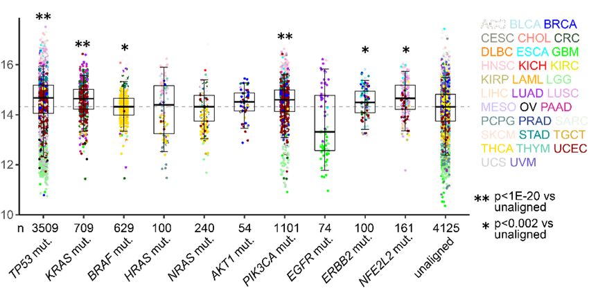
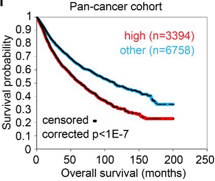
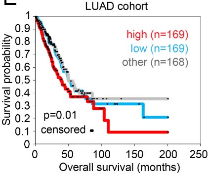
IGFBP2 or SPP1 secretion (Supplemental Figure 9, L and M). Ectopic expression of full-length,

but not N-terminally truncated, G45 enhanced H1299 cell colony formation, which was abrogated

by siRNA-mediated MYOIIA depletion (Supplemental Figure 9N). Thus, G45 and MYOIIA are key

mediators of G55.

   p53 governs a CPS regulatory axis. To determine whether G55, G45, and MYOIIA are

coordinately regulated in cancer, we queried the TCGA LUAD and pan-cancer cohorts and

identified positive correlations between G55, G45, and MYOIIA (Figure 8, A and B). Like G55,

G45 and MYOIIA expression were correlated with multiple oncogenic mutations (TP53, KRAS,

BRAF, PIK3CA, ERBB2, NFE2L2) (Figure 8, C and D). A 3-gene signature composed of G55,

G45, and MYOIIA was negatively correlated with survival duration (Figure 8, E and F).

   G45 protein expression was lower in G55-deficient than -replete LUAD cells (Supplemental

Figure 10A), but G45 mRNA expression was unchanged (Supplemental Figure 10B), which led

us to ask whether G55 regulates G45 protein stability. In the presence of cycloheximide, G45

protein stability was reduced by siRNA-mediated G55 depletion (Supplemental Figure 10C) or by

a G45 mutation (L400R) that disrupts interactions with G55 (Supplemental Figure 10D). G45

ubiquitination was increased by siRNA-mediated G55 depletion (Supplemental Figure 10E) or the

presence of L400R (Supplemental Figure 10F), and MG132 proteasome inhibitor treatment

restored G45 protein expression in GORASP2 KO HeLa cells (Supplemental Figure 10G). Thus,

G45 is degraded through the ubiquitin-mediated proteasomal pathway and is protected from

proteasomal degradation by interactions with G55.

   Based on the discovery of a predicted miR-34a binding site in the MYOIIA 3’-UTR

(www.targetscan.org), we postulated that p53 deficiency relieves MYOIIA from miR-34a-

dependent silencing. In A549 cells, shRNA-mediated p53 depletion increased MYOIIA

expression, which was abrogated by ectopic miR-34a expression (Figure 8G). Conversely,

ectopic p53 expression in H1299 cells downregulated MYOIIA expression, which was reversed
by co-transfecting miR-34a antagomiRs (Figure 8H). Demonstrating the specificity of this

regulatory axis, MYOIIA expression was downregulated by ectopic expression of miR-34a but not

miR-200a (Figure 8I), which is not predicted to bind the MYOIIA 3’-UTR. The functionality of the

miR-34a-binding site in the MYOIIA 3’-UTR was validated in H1299 cells co-transfected with a

miR-34a expression vector and luciferase reporters containing wild-type or mutant MYOIIA 3’-

UTRs or or a MYO18A 3’-UTR that lacks predicted miR-34a binding sites (Figure 8J). Given that

p53-dependent miR-34a silencing coordinately upregulates G55 and MYOIIA, we conclude that

miR-34a is a nodal factor in the p53-dependent CPS regulatory axis.

Discussion

   Here, we show that p53 loss activates the secretion of pro-tumorigenic effector proteins to

create a secretory vulnerability that is actionable using a small molecule inhibitor of G55 (Figure

8K). Our findings suggest that therapeutic strategies designed to suppress G55-dependent

secretion may be applicable to diverse oncogenic contexts in which G55 levels are upregulated.

For example, activating BRAF mutations, which were correlated with high G55 levels, are reported

to silence miR-34a (30) and may thereby activate G55-dependent secretion.

   The findings presented here build on a growing body of evidence that p53 is a key secretory

regulator in cancer. p53 loss enhances SDF-1 secretion by cancer-associated fibroblasts to

promote tumor growth and metastasis (31). p53 deficiency in hepatic stellate cells releases factors

that stimulate macrophage polarization into a tumor-promoting M2 state and enhances the

proliferation of premalignant hepatic epithelial cells (32). Transcriptomes activated by TP53 gain-

of-function mutations in cancer cells are enriched in secreted proteins, including heat shock

protein 90α, which promotes metastasis (14, 33, 34). Thus, secretion underlies p53’s contextual

cellular functions in cancer.

   The N-terminal PDZ domain of G55 has been the focus of intense study. The PDZ domain

facilitates the formation of G55 homodimers and trans-oligomers that maintain cisternal stacking
and Golgi organelle integrity (35) and is the insertion site for other binding partners that carry out

various cellular functions, including Junctional Adhesion Molecule-C, which is essential for

germ/Sertoli cell contacts and spermatogenesis (23), and the autophagy effector proteins

lysosomal membrane protein 2 and microtubule-associated protein 1A/1B-light chain 3, which

promote autophagosome-lysosome fusion and cargo transport via UPS (24). Here we show that

G55/G45 interactions are required for G55-dependent RAB6A vesicle formation, which extends

evidence that G55/G45 interactions generate a RAB2 effector complex that drives anterograde

vesicle transport (12). Thus, G55 influences cellular functions through PDZ domain-binding

proteins that play pleiotropic roles in coordinating vesicular transport through CPS and UPS

pathways.

   In the Golgi, proteins destined for secretion are sorted and loaded into vesicles through a

complex process involving secretory carrier membrane proteins (e.g., CARTS, LAMP1-RUSH,

RAB6, and ARF1) (36), transmembrane cargo receptors (e.g., Sortlin and Wntless), and cargo

adaptors (e.g., Golgi-localized, γ-ear containing, ADP-ribosylation factor binding proteins) (37).

However, the way in which oncogenic mutations regulate secretion at the level of cargo sorting

and loading remains unclear. Although our study does not directly address this question, several

findings presented here support the existence of an oncogene-regulated cargo sorting apparatus

in the Golgi. For example, in addition to decreasing the secretion of pro-tumorigenic effector

proteins, G55 depletion increased the secretion of other proteins, including cystatin-B, a potent

tumor suppressor (38). Furthermore, the G55-dependent secreted proteins identified here do not

overlap with GOLPH3-dependent secreted proteins in the same LUAD cell lines (39). Notably,

the cargo-sorting protein ARFGAP1 (40) was identified in G45 pull-down assays (Supplemental

Table 3).

   There are important limitations to our study. Our focus was on TP53 mutations, but the extent

to which co-occurring mutations influence p53-regulated secretion remains unclear. We

demonstrate enhanced vulnerability of TP53-mutant LUAD cells to GRASPIN, but determining
whether TP53 mutations predict enhanced clinical benefit from GRASPIN treatment awaits clinical

trials. Our biochemical, cellular, and in vivo studies with GRASPIN revealed no evidence of off-

target effects, but a broader analysis of GRASPIN’s selectivity, long-term toxicities, and

mechanisms of de novo and acquired resistance were not addressed.

   In conclusion, we have identified an actionable secretory vulnerability in p53-deficient

malignancies. Basket clinical trials are warranted to test the efficacy of G55 antagonists in TP53-

mutant malignancies and other conditions in which secretion plays a contributing role, including

acquired resistance to targeted therapies in receptor tyrosine kinase-mutant cancers and fibrotic

and inflammatory diseases (41-43).

Methods

Animal Husbandry

Mice received standard care and were euthanized according to the standards set forth by the

Institutional Animal Care and Use Committee, The University of Texas MD Anderson Cancer

Center. Syngeneic, immunocompetent mice (n = 8-10 per group) were injected with 106 murine

LUAD cells in the right flank and necropsied after 5 weeks. Primary tumors were weighed, and

lung metastases on the pleural surfaces were counted. To generate orthotopic lung tumors, we

subjected nu/nu mice (n=10 mice per cohort) to intrathoracic injection with 106 human LUAD cells,

necropsied them after 5 weeks, and measured primary tumor size and the number of metastases

on the contralateral lung surface. For GRASPIN treatment experiments, we injected syngeneic,

immunocompetent mice or nu/nu mice with 106 murine or human LUAD cells or lung cancer

patient-derived xenografts by subcutaneous or intra-thoracic approach and initiated treatment

with GRASPIN (25 mg/kg or 50 mg/kg) or vehicle (10% DMSO and 90% corn oil) one week after

tumor cell injection. Drugs were administered intraperitoneally every two days. The mice were

checked daily and necropsied at first sign of morbidity or after the last treatment to measure
primary tumor size, count metastases to the contralateral lung, and obtain tumor tissues for

analysis.

Cell lines

Murine LUAD cells generated in mice that express K-rasG12D and p53R172H (344P,344SQ, 531LN2,

and 393P) (44), human LUAD cells (A549, H1299, H460, H596, H23, H1792, H441, H358, H157,

H226, H2122, CALU-1, H292 and H520), and CAOV-4 human ovarian cancer cell lines were

cultured in RPMI 1640 containing 10% fetal bovine serum (FBS). MDA-MB-468 cell lines were

cultured in Dulbecco’s minimal essential medium (DMEM) containing 10% FBS. HUVECs were

cultured in EGMTM-2 Endothelial Cell Growth Medium-2 BulletKitTM (CC-5035, Lonza). Cells

were maintained at 37°C in an incubator with a humidified atmosphere containing 5% CO2. Cells

were transfected with jetPRIME® Versatile DNA/siRNA transfection reagent (Polyplus). Stable

cell transfectants were selected by using puromycin (for pLVX or pLKO.1 vectors) or G418 (for

pcDNA3.1 and pEGFP-C3 vectors). TP53 KO A549 cells (guide RNA sequence: 5’-

gagcgctgctcagatagcga-3’) were generated in the Cell-Based Assay Screening Service Core

Facility at Baylor College of Medicine. Parental and CRISPR/Cas9-mediated GORASP2 KO HeLa

cells were a gift from Dr. Yanzhuang Wang (U. Michigan) (45).

Reagents

We purchased SYBR Green, FBS, HEPES buffered media, DMEM, RPMI 1640, Alexa Fluor-

tagged secondary antibodies, and DAPI from Life Technologies; puromycin from InvivoGene;

paraformaldehyde from Electron Microscopy Sciences; Transwell and Matrigel-coated Boyden

chambers from BD Biosciences; G418 from Corning; GRASPIN (PubChem CID: 3113208) from

Vitas-M Laboratory (STK700118); Wortmannin (W1628), 3-Methyladenine (SAE0107), nutlin-3

(444151),    and   Brefeldin   A   (B7651)    from   Sigma;   shRNAs   against   murine   G55

(TRCN0000278363,TRCN0000077518,              and     TRCN0000077520)    and      murine   G45
(TRCN0000096503), shRNAs against human G55 (TRCN0000278363 and TRCN0000130898),

human    IGFBP2     (TRCN0000318660,      TRCN0000318716,       TRCN0000318659,      and

TRCN0000006576), human SPP1 (TRCN0000342616, TRCN0000342561, TRCN0000004878

and TRCN0000004875), human G45 (TRCN0000013243); siRNAs against human G55

(SASI_Hs01_00012371 and SASI_Hs02_00348040), human IGFBP2 (SASI_Hs02_00302878

and SASI_Hs01_00039595), human SPP1 (SASI_Hs01_00174866 and SASI_Hs01_00174867),

human    G45   (SASI_Hs01_00073080      and    SASI_Hs01_00073083),     human    MYOIIA

(SASI_Hs01_00197338 and SASI_Hs01_00197339), human MYO18A (SASI_Hs01_00224320

and SASI_Hs01_00224321), GOLPH3 (SASI_Hs02_00355527 and SASI_Hs01_00133692);

esiRNAs against human RAB6A (EHU113451), human RAB8A (EHU054251), human RAB10

(EHU131761), human RAB13 (EHU107931) and EGFP (EHUEGFP) from Sigma. We purchased

primary antibodies against G55 (NB100-74431) from Novus Biologicals; against GM130

(#560066) from BD Transduction Laboratories; against G45 (OAAN01805) from Aviva Systems

Biology; against GOLPH3 (ab98023) from Abcam; against IGFBP2 (11065-3-AP), SPP1 (22952-

1-AP), MYO18A (14611-1-AP), MAOA (10539-1-AP), and RAB6A (10187-2-AP) from

Proteintech; against α-tubulin (#T9026), EGFP (G6539) from Sigma; against PARP-1 (#9542),

hemagglutinin (#3724 and #2367), β-actin (#4970), p53 (#9282), GM130 (#12480), EEA1

(#3288), Ubiquitin (#43124), and MYOIIA (#3403) from Cell Signaling; against ACBD3 (sc-

101277), GRASP65 (sc-374423) and p53 (sc-126) from Santa Cruz Biotechnology; against

Calnexin (ab22595), RAB11 (ab95375) from Abcam; against RNA polymerase II (05-623, clone

CTD4H8) from Millipore. We purchased neutralizing antibodies against IGFBP2 (AF674) and

SPP1 (AF1433) from R&D systems. We purchased recombinant IGFBP2 (ab63223) and SPP1

(ab92964) proteins from Abcam. We purchased human IL-6 enzyme-linked immunosorbent assay

(ELISA) kit (ab178013) from Abcam. rG55-pEGFP-N3 is a gift from Yanzhuang Wang at

University of Michigan Medical School. Myosin-IIA-GFP was a gift from Matthew Krummel
(Addgene plasmid # 38297). EGFP-RAB6A was a gift from Marci Scidmore (Addgene plasmid #

49469).

Vector construction

The human G55 and G45 coding sequences were isolated by performing PCR on cDNA prepared

from H1299 cells, and then cloned into pLVX-Puro or pEGFP-C3 (Clontech) or pcDNA3.1

(Invitrogen). To generate a G45 BP expression vector for transfection studies in cells, the DNA

sequence encoding the last 30 amino acids of G45 C-terminus was amplified by PCR and cloned

into the pEGFP-C3 vector. The G55, MYH9 and MYO18A 3’-UTR sequences were amplified from

genomic DNA from H1299 cells by PCR and cloned into pCI-neo-hRL vector. Mutations were

introduced by PCR method. PCR primers are listed in Supplementary table 4.

Cell proliferation, colony formation, apoptosis, migration, and invasion assays.

Cell proliferation assays were performed using Cell Proliferation Reagent WST-1 (Roche)

according to the manufacturer's protocol. Colony formation at low density on plastic and in soft

agarose were performed as described previously (46). Apoptosis was determined by flow

cytometry using Dead Cell Apoptosis Kit (ThermoFisher Scientific, V13242) according to

manufacturer's instructions. Migration and invasion assays were performed in Transwell and

Matrigel-coated Boyden chambers, respectively, as we have described (47). For endothelial

recruitment assays, 2×10⁴ HUVEC cells seeded in the top chamber in Transwell plates, co-

cultured with H1299 cells for 12 h, and migrated HUVEC cells were stained with 0.1% crystal

violet, photographed and quantified.

Tube formation assays
As previously described (39), HUVECs (1×10⁵/well) were seeded into the lower chambers of

Transwell plates containing Matrigel-coated filters, and tumor cells (2×10⁴/well) were seeded in

the upper chambers. After 8 h, HUVECs were imaged and tube-like structures were quantified.

Chromatin immunoprecipitation assays

A549 cell lysates were formaldehyde fixed and digested using SimpleChIP Enzymatic Chromatin

IP Kit (#9002, Cell signaling) following manufacturer's instructions. Resulting chromatin was

immunoprecipitated with anti–RNA polymerase II or anti-rabbit IgG antibodies (Santa Cruz

Biotechnology). DNA was eluted and purified with the MinElute Reaction Cleanup Kit (Qiagen)

and subjected to quantitative PCR. PCR primers are listed in Supplemental table 4.

Luciferase reporter assays

3′-UTR luciferase reporters (10 ng), pGL3-enhancer control (100 ng), and miR mimics (20 nM)

were co-transfected into H1299 cells. After 24 h, luciferase activity was measured with the Dual-

Luciferase Reporter Assay System (Promega).

Western blot and immunoprecipitation assays

For Western blot analysis, protein lysates were separated on a 4–20% Bis-Tris gel and transferred

to PVDF membranes using Trans-Blot Turbo Transfer System (Bio-Rad). The membranes were

blocked in 5% milk and probed with primary antibodies following a standard protocol. For

immunoprecipitation followed by western blot analysis, H1299 cells were transfected with

indicated expression vectors, lysed in 1× radioimmunoprecipitation assay buffer (RIPA buffer, Cell

Signaling Technologies) 48 h later, and incubated with antibodies or control IgG at 4 °C overnight.

The immune complex was captured with protein G agarose beads (GE Healthcare Life Sciences),

washed with lysis buffer 4 times, and boiled in 1x sodium dodecyl sulfate loading buffer for 5 min.

The resulting samples were subjected to Western blot analysis using relevant antibodies.
Quantitative RT-PCR (qPCR) assays

Total RNA was isolated from cells using RNeasy Mini Kit (Qiagen, 74106) and subjected to

reverse transcription using the qScript cDNA superMix (Quanta Biosciences). mRNA levels were

determined using SYBR® Green Real-Time PCR Master Mixes (Thermo Fisher Scientific) and

normalized on the basis of ribosomal protein L32 (Rpl32) mRNA. PCR primers are listed in

Supplementary Table 4.

CM assays

As previously described (48), CM samples were isolated, filtered through a 0.45 μm filter, mixed

with an equal volume of complete growth medium, and applied to cells. For Transwell

experiments, cells that had been incubated in serum-free medium for 16 h were seeded in the top

chambers in serum-free medium, and CM samples were added to the bottom wells. For colony

formation assays, cells were incubated in CM samples that were refreshed every two days.

IL-6 ELISA

H1299 cells in 6-well plate were transfected with siRNAs against G55 or control siRNA. After 48

h, cells were incubated in free medium for 16 h. CM samples were collected and subjected to

ELISA assay to quantify IL-6 concentrations using Human IL-6 ELISA Kit (ab46027, Abcam)

according to the manufacturer’s instructions.

LC-MS

To identify G55- and G45-interacting proteins, H1299 cells were transfected with GFP-G55 or HA-

G45, lysed in 1× RIPA buffer, and subjected to immunoprecipitation with anti-GFP or -HA

antibodies, respectively, linked to agarose G beads. Immune complexes were subjected to LC-

MS. To identify G55-dependent secreted proteins, CM samples were collected, concentrated, and
subjected to LC-MS as previously described (49). Samples were solubilized with 5% SDS, 50 mM

TEAB, pH 7.55, final volume 25 µl. The sample was then centrifuged at 17,000 g for 10 minutes

to remove any debris. Proteins were reduced by making the solution 20 mM TCEP (Thermo,

#77720) and incubated at 65 °C for 30 min. The sample was cooled to RT and 1 µl of 0.5M

iodoacetamide acid added and allowed to react for 20 min in the dark. 2.75 µl of 12% phosphoric

acid was added to the protein solution. 165 µl of binding buffer (90% Methanol, 100 mM TEAB,

final pH 7.1) was then added to the solution. The resulting solution was added to S-Trap spin

column (protifi.com) and passed through the column using a bench top centrifuge (30 s spin at

4,000 g).   The spin column was washed with 400 µl of binding buffer and centrifuged. This is

repeated two more times. Trypsin was added to the protein mixture in a ratio of 1:25 in 50 mM

TEAB, pH=8, and incubated at 37 °C for 4 h. Peptides were eluted with 80 µl of 50 mM TEAB,

followed by 80 µl of 0.2% formic acid, and finally 80 uL of 50% acetonitrile, 0.2% formic acid. The

combined peptide solution was then dried in a speed vac and resuspended in 2% acetonitrile,

0.1% formic acid, 97.9% water and placed in an autosampler vial.

Cytokine and chemokine analysis

Cells were seeded (5x10⁵ per well) in triplicate in a 6-well plate. CM samples were collected and

analyzed using a multiplex magnetic bead-based assay (Luminex 200 System, Luminex, USA).

Isolation of Golgi and vesicle fractions

Minute™ Golgi Apparatus Enrichment Kit (GO-037, Invent biotechnologies) was used to enrich

cell lysates in Golgi and vesicle fractions following manufacturer's instructions. Briefly, 3 X 10⁷

cells were suspended in 550 µl buffer A. Cell suspension was loaded into a filter cartridge,

centrifuged at 16,000 g for 30 sec and then for 5 min at 5,000 g at 4oC without removing the filter.

After centrifugation, the filter was removed and the supernatants were transferred to a fresh 1.5

ml tube and centrifuged at 4oC for 30 min at 16,000 g. After centrifugation, 400 µl supernatant
was transferred to a fresh 1.5 ml tube. The pellet contains mainly mitochondria, endoplasmic

reticulum, lysosomes, and plasma membranes. 400 µl buffer B was added to the tube containing

400 µl supernatant. The tube was placed on ice for 10 min and centrifuged at 8,000 g for 5 min

at 4oC. Supernatants containing secretory vesicles associated with trans-Golgi membranes were

transferred to a new tube. The remaining pellet was resuspended in 200 µl cold buffer A and

centrifuged at 8,000 X g for 5 min. The supernatant was transferred to a fresh 1.5 ml tube and

100 µl cold buffer C was added to the tube and mixed by vortexing. The tube was incubated on

ice for 20 min and centrifuged at 8,000 g for 10 min to isolate the pellet, which constituted the

“Golgi fraction”. To isolate vesicle fractions, 100 µl buffer D was added to supernatants containing

secretory vesicles associated with trans-Golgi membranes, and the mixture was incubated on ice

for 20 min and centrifuged at 16,000 X g for 5 min at 4oC. The supernatant was removed to isolate

the pellet, which constituted the “vesicle fraction”. The pellets containing Golgi and vesicle

fractions were resuspended in 50 µl SDS loading buffer, heated for 10 min at 98 oC, and subjected

to western blot analysis.

RAB6A vesicle trafficking

Live-cell imaging was performed on H1299 cells stably expressing RAB6A-EGFP. Cells were

plated on 35mm glass-bottom plates (MatTek), transfected with siRNAs, and imaged 48-72 h later

using a Nikon A1+ confocal microscope equipped with 488nm laser line, GaAsP detectors, and

100X 1.45 NA objective. Live-cell imaging conditions (5% CO2, 37 oC temperature, and relative

humidity) were maintained on stage using an Okolab incubation chamber. For optimal spatial and

temporal resolution, Nyquist sampling was applied in bidirectional image acquisition mode without

averaging. Images were acquired at 1 fps for 2.5-3 min. Post-acquisition, images were bleach-

corrected (Huygens Professional). RAB6A vesicles were identified and tracked using the spots

module in Imaris (Bitplane). A spot diameter of 500 nm was applied to segment the vesicles before
tracking them using an autoregressive motion algorithm. To avoid unnecessary splitting of tracks

due to uneven illumination or out-of-plane movement, a gap-closing algorithm was used. As

described (28), unfissioned tubular structures were defined as RAB6A-expressing tubular

structures that are attached to the Golgi. For each cell, a mask that isolates the Golgi and

connected tubular structures was generated, and unfissioned tubules were counted and

measured.

Statistical analysis

For analysis of G55 mRNA levels in tumors annotated on the basis of somatic DNA mutations in

the TCGA pan-cancer cohort (50), oncogenic mutations were defined by Single Nucleotide

Variant or indel falling within a "hotspot" residue as defined by Chang et. al (51). For pan-cancer

TCGA cohort analyses, p-values are by univariate Cox, stratified by cancer type. For LUAD cohort

analyses, p-values are by univariate Cox. For computing multiple-gene signature scores, we first

normalized each gene to standard deviations from the median across the pan-cancer cohort

(using log2-transformed expression values), then took the average of the three normalized values.

Unless stated otherwise, the results shown are representative of replicated experiments and are

the means ± standard deviations from triplicate samples or randomly chosen cells within a field.

Statistical evaluations were carried out with Prism 6 (GraphPad Software, Inc.). Unpaired 2-tailed

Student t-tests were used to compare the mean values of 2 groups. One-way ANOVA with

Dunnett's test was used for comparing multiple treatments to a control. P values < 0.05 were

considered statistically significant. Plots were generated using software (http://gepia.cancer-

pku.cn/). Box plots: 5%, 25%, 50%, 75%, 95%. Tumor type abbreviations are described

(http://cancergenome.nih.gov).

Study approval
All mouse studies were approved by the Institutional Animal Care and Use Committee at The

University of Texas MD Anderson Cancer Center, Houston.

Author contributions

X.T. conceived, designed, executed, and interpreted cell culture and in vivo experiments. P.B.

conceived, designed, executed, and interpreted all of the experiments analyzing vesicular

trafficking by confocal microscopy. L.S. assisted X.T. with immunoprecipitation experiments and

performed the HUVEC tube formation assay. X.L. assisted X.T. with the in vivo experiments. J.Y.

bred mice for the in vivo studies. N.B-R. performed cell culture assays. H-F.G. assisted with G55

protein crystal structure analysis. B.L.R. and N.B.R. performed flow cytometric analysis of tumor

tissues. D.L.G. supervised the efforts of B.L.R. W.K.R. directed and interpreted mass

spectrometry experiments. C.J.C. directed and interpreted bioinformatic analyses. J.M.K.

conceived and supervised the project and contributed to the design and interpretation of all

experiments.

Acknowledgments

We thank Fengju Chen for technical assistance. Funding: This work was supported by the

National Institutes of Health (NIH) through R01 CA181184 (to J.M.K.), R01 CA2111125 (to

J.M.K.), P30 CA125123 (to C.J.C.), K99 CA225633 (to H.F.G.), and Lung Cancer Research

Foundation FP#00005299 (to X.T.). J.M.K. holds the Gloria Lupton Tennision Distinguished

Endowed Professorship in Lung Cancer. D.L.G. is an R. Lee Clark Fellow of the University of

Texas MD Anderson Cancer Center, supported by the Jeane F Shelby Scholarship Fund. The

work was also supported by the generous philanthropic contributions to The University of Texas
MD Anderson Lung Cancer Moon Shots Program. Data and materials availability: All data

associated with this study are present in the paper or the Supplementary Materials.

REFERENCES

1.     Werb Z, and Lu P. The Role of Stroma in Tumor Development. Cancer J.
       2015;21(4):250-3.
2.     Prager BC, Xie Q, Bao S, and Rich JN. Cancer Stem Cells: The Architects of the Tumor
       Ecosystem. Cell Stem Cell. 2019;24(1):41-53.
3.     Li J, and Stanger BZ. The tumor as organizer model. Science. 2019;363(6431):1038-9.
4.     Paltridge JL, Belle L, and Khew-Goodall Y. The secretome in cancer progression.
       Biochim Biophys Acta. 2013;1834(11):2233-41.
5.     Coussens LM, Fingleton B, and Matrisian LM. Matrix metalloproteinase inhibitors and
       cancer: trials and tribulations. Science. 2002;295(5564):2387-92.
6.     Kim J, Gee HY, and Lee MG. Unconventional protein secretion - new insights into the
       pathogenesis and therapeutic targets of human diseases. J Cell Sci. 2018;131(12).
7.     Chiritoiu M, Brouwers N, Turacchio G, Pirozzi M, and Malhotra V. GRASP55 and UPR
       Control Interleukin-1beta Aggregation and Secretion. Dev Cell. 2019;49(1):145-55 e4.
8.     Dupont N, Jiang S, Pilli M, Ornatowski W, Bhattacharya D, and Deretic V. Autophagy-
       based unconventional secretory pathway for extracellular delivery of IL-1beta. EMBO J.
       2011;30(23):4701-11.
9.     Halberg N, Sengelaub CA, Navrazhina K, Molina H, Uryu K, and Tavazoie SF. PITPNC1
       Recruits RAB1B to the Golgi Network to Drive Malignant Secretion. Cancer Cell.
       2016;29(3):339-53.
10.    Dippold HC, Ng MM, Farber-Katz SE, Lee SK, Kerr ML, Peterman MC, et al. GOLPH3
       bridges phosphatidylinositol-4- phosphate and actomyosin to stretch and shape the
       Golgi to promote budding. Cell. 2009;139(2):337-51.
11.    Kim J, Noh SH, Piao H, Kim DH, Kim K, Cha JS, et al. Monomerization and ER
       Relocalization of GRASP Is a Requisite for Unconventional Secretion of CFTR. Traffic.
       2016;17(7):733-53.
12.    Short B, Preisinger C, Korner R, Kopajtich R, Byron O, and Barr FA. A GRASP55-rab2
       effector complex linking Golgi structure to membrane traffic. J Cell Biol.
       2001;155(6):877-83.
13.    He L, He X, Lim LP, de Stanchina E, Xuan Z, Liang Y, et al. A microRNA component of
       the p53 tumour suppressor network. Nature. 2007;447(7148):1130-4.
14.    Yeudall WA, Vaughan CA, Miyazaki H, Ramamoorthy M, Choi MY, Chapman CG, et al.
       Gain-of-function mutant p53 upregulates CXC chemokines and enhances cell migration.
       Carcinogenesis. 2012;33(2):442-51.
15.    Lang GA, Iwakuma T, Suh YA, Liu G, Rao VA, Parant JM, et al. Gain of function of a
       p53 hot spot mutation in a mouse model of Li-Fraumeni syndrome. Cell.
       2004;119(6):861-72.
16. Drewniok C, Wienrich BG, Schon M, Ulrich J, Zen Q, Telen MJ, et al. Molecular
interactions of B-CAM (basal-cell adhesion molecule) and laminin in epithelial skin
cancer. Arch Dermatol Res. 2004;296(2):59-66.
17. Duan L, Motchoulski N, Danzer B, Davidovich I, Shariat-Madar Z, and Levenson VV.
Prolylcarboxypeptidase regulates proliferation, autophagy, and resistance to 4-
hydroxytamoxifen-induced cytotoxicity in estrogen receptor-positive breast cancer cells.
J Biol Chem. 2011;286(4):2864-76.
18. Shevde LA, and Samant RS. Role of osteopontin in the pathophysiology of cancer.
Matrix Biol. 2014;37:131-41.
19. Klose R, Adam MG, Weis EM, Moll I, Wustehube-Lausch J, Tetzlaff F, et al. Inactivation
of the serine protease HTRA1 inhibits tumor growth by deregulating angiogenesis.
Oncogene. 2018;37(31):4260-72.
20. Yao X, Sun S, Zhou X, Guo W, and Zhang L. IGF-binding protein 2 is a candidate target
of therapeutic potential in cancer. Tumour Biol. 2016;37(2):1451-9.
21. Revelo NH, Ter Beest M, and van den Bogaart G. Membrane trafficking as an active
regulator of constitutively secreted cytokines. J Cell Sci. 2019;133(5).
22. Zhao J, Li B, Huang X, Morelli X, and Shi N. Structural Basis for the Interaction between
Golgi Reassembly-stacking Protein GRASP55 and Golgin45. J Biol Chem.
2017;292(7):2956-65.
23. Cartier-Michaud A, Bailly AL, Betzi S, Shi X, Lissitzky JC, Zarubica A, et al. Genetic,
structural, and chemical insights into the dual function of GRASP55 in germ cell Golgi
remodeling and JAM-C polarized localization during spermatogenesis. PLoS Genet.
2017;13(6):e1006803.
24. Zhang X, Wang L, Lak B, Li J, Jokitalo E, and Wang Y. GRASP55 Senses Glucose
Deprivation through O-GlcNAcylation to Promote Autophagosome-Lysosome Fusion.
Dev Cell. 2018;45(2):245-61 e6.
25. Noh SH, Gee HY, Kim Y, Piao H, Kim J, Kang CM, et al. Specific autophagy and ESCRT
components participate in the unconventional secretion of CFTR. Autophagy.
2018;14(10):1761-78.
26. Orci L, Tagaya M, Amherdt M, Perrelet A, Donaldson JG, Lippincott-Schwartz J, et al.
Brefeldin A, a drug that blocks secretion, prevents the assembly of non-clathrin-coated
buds on Golgi cisternae. Cell. 1991;64(6):1183-95.
27. Tiwari N, Graham M, Liu X, Yue X, Zhu L, Meshram D, et al. Golgin45-Syntaxin5
Interaction Contributes to Structural Integrity of the Golgi Stack. Sci Rep.
2019;9(1):12465.
28. Miserey-Lenkei S, Chalancon G, Bardin S, Formstecher E, Goud B, and Echard A. Rab
and actomyosin-dependent fission of transport vesicles at the Golgi complex. Nat Cell
Biol. 2010;12(7):645-54.
29. Goud B, Liu S, and Storrie B. Rab proteins as major determinants of the Golgi complex
structure. Small GTPases. 2018;9(1-2):66-75.
30. Christoffersen NR, Shalgi R, Frankel LB, Leucci E, Lees M, Klausen M, et al. p53-
independent upregulation of miR-34a during oncogene-induced senescence represses
MYC. Cell Death Differ. 2010;17(2):236-45.
31. Addadi Y, Moskovits N, Granot D, Lozano G, Carmi Y, Apte RN, et al. p53 status in
stromal fibroblasts modulates tumor growth in an SDF1-dependent manner. Cancer Res.
2010;70(23):9650-8.
32. Lujambio A, Akkari L, Simon J, Grace D, Tschaharganeh DF, Bolden JE, et al. Non-cell-
autonomous tumor suppression by p53. Cell. 2013;153(2):449-60.
33. Solomon H, Buganim Y, Kogan-Sakin I, Pomeraniec L, Assia Y, Madar S, et al. Various
p53 mutant proteins differently regulate the Ras circuit to induce a cancer-related gene
signature. J Cell Sci. 2012;125(Pt 13):3144-52.34. Ubertini V, Norelli G, D'Arcangelo D, Gurtner A, Cesareo E, Baldari S, et al. Mutant p53
gains new function in promoting inflammatory signals by repression of the secreted
interleukin-1 receptor antagonist. Oncogene. 2015;34(19):2493-504.
35. Xiang Y, and Wang Y. GRASP55 and GRASP65 play complementary and essential
roles in Golgi cisternal stacking. J Cell Biol. 2010;188(2):237-51.
36. Stalder D, and Gershlick DC. Direct trafficking pathways from the Golgi apparatus to the
plasma membrane. Semin Cell Dev Biol. 2020.
37. Guo Y, Sirkis DW, and Schekman R. Protein sorting at the trans-Golgi network. Annu
Rev Cell Dev Biol. 2014;30:169-206.
38. Zhang J, Shi Z, Huang J, and Zou X. CSTB Downregulation Promotes Cell Proliferation
and Migration and Suppresses Apoptosis in Gastric Cancer SGC-7901 Cell Line. Oncol
Res. 2016;24(6):487-94.
39. Tan X, Banerjee P, Pham EA, Rutaganira FUN, Basu K, Bota-Rabassedas N, et al.
PI4KIIIbeta is a therapeutic target in chromosome 1q-amplified lung adenocarcinoma.
Sci Transl Med. 2020;12(527).
40. Lee SY, Yang JS, Hong W, Premont RT, and Hsu VW. ARFGAP1 plays a central role in
coupling COPI cargo sorting with vesicle formation. J Cell Biol. 2005;168(2):281-90.
41. Jones VS, Huang RY, Chen LP, Chen ZS, Fu L, and Huang RP. Cytokines in cancer
drug resistance: Cues to new therapeutic strategies. Biochim Biophys Acta.
2016;1865(2):255-65.
42. Gong B, Kiyotani K, Sakata S, Nagano S, Kumehara S, Baba S, et al. Secreted PD-L1
variants mediate resistance to PD-L1 blockade therapy in non-small cell lung cancer. J
Exp Med. 2019;216(4):982-1000.
43. Nilsson MB, Sun H, Diao L, Tong P, Liu D, Li L, et al. Stress hormones promote EGFR
inhibitor resistance in NSCLC: Implications for combinations with beta-blockers. Sci
Transl Med. 2017;9(415).
44. Gibbons DL, Lin W, Creighton CJ, Rizvi ZH, Gregory PA, Goodall GJ, et al. Contextual
extracellular cues promote tumor cell EMT and metastasis by regulating miR-200 family
expression. Genes Dev. 2009;23(18):2140-51.
45. Bekier ME, 2nd, Wang L, Li J, Huang H, Tang D, Zhang X, et al. Knockout of the Golgi
stacking proteins GRASP55 and GRASP65 impairs Golgi structure and function. Mol
Biol Cell. 2017;28(21):2833-42.
46. Ahn YH, Gibbons DL, Chakravarti D, Creighton CJ, Rizvi ZH, Adams HP, et al. ZEB1
drives prometastatic actin cytoskeletal remodeling by downregulating miR-34a
expression. J Clin Invest. 2012;122(9):3170-83.
47. Tan X, Banerjee P, Liu X, Yu J, Gibbons DL, Wu P, et al. The epithelial-to-mesenchymal
transition activator ZEB1 initiates a prometastatic competing endogenous RNA network.
J Clin Invest. 2018.
48. Brady JJ, Chuang CH, Greenside PG, Rogers ZN, Murray CW, Caswell DR, et al. An
Arntl2-Driven Secretome Enables Lung Adenocarcinoma Metastatic Self-Sufficiency.
Cancer Cell. 2016;29(5):697-710.
49. Johnson BA, Hage A, Kalveram B, Mears M, Plante JA, Rodriguez SE, et al.
Peptidoglycan-Associated Cyclic Lipopeptide Disrupts Viral Infectivity. J Virol.
2019;93(22).
50. Chen F, Zhang Y, Gibbons DL, Deneen B, Kwiatkowski DJ, Ittmann M, et al. Pan-
Cancer Molecular Classes Transcending Tumor Lineage Across 32 Cancer Types,
Multiple Data Platforms, and over 10,000 Cases. Clin Cancer Res. 2018;24(9):2182-93.
51. Chang MT, Asthana S, Gao SP, Lee BH, Chapman JS, Kandoth C, et al. Identifying
recurrent mutations in cancer reveals widespread lineage diversity and mutational
specificity. Nat Biotechnol. 2016;34(2):155-63.You can also read