ATTENUATING CD3 AFFINITY IN A PSMAXCD3 BISPECIFIC ANTIBODY ENABLES KILLING OF PROSTATE TUMOR CELLS WITH REDUCED CYTOKINE RELEASE
←
→
Page content transcription
If your browser does not render page correctly, please read the page content below
Open access Original research
Attenuating CD3 affinity in a
J Immunother Cancer: first published as 10.1136/jitc-2021-002488 on 4 June 2021. Downloaded from http://jitc.bmj.com/ on June 11, 2021 by guest. Protected by copyright.
PSMAxCD3 bispecific antibody enables
killing of prostate tumor cells with
reduced cytokine release
Kevin Dang ,1 Giulia Castello ,1 Starlynn C Clarke,1 Yuping Li,1
Aarti Balasubramani,1 Andrew Boudreau,1 Laura Davison,1 Katherine E Harris,1
Duy Pham,1 Preethi Sankaran,1 Harshad S Ugamraj,1 Rong Deng,1 Serena Kwek,2
Alec Starzinski,2 Suhasini Iyer,1 Wim van Schooten,1 Ute Schellenberger,1
Wenchao Sun,1 Nathan D Trinklein,1 Roland Buelow,1 Ben Buelow,1
Lawrence Fong,2 Pranjali Dalvi 1
To cite: Dang K, Castello G, ABSTRACT Conclusions Our data suggest that TNB-585, with its
Clarke SC, et al. Attenuating Background Therapeutic options currently available low-affinity anti-CD3, may be efficacious while inducing
CD3 affinity in a PSMAxCD3 for metastatic castration-resistant prostate cancer a lower incidence and severity of CRS in patients with
bispecific antibody enables prostate cancer compared with TCEs that incorporate
(mCRPC) do not extend median overall survival >6
killing of prostate tumor cells
months. Therefore, the development of novel and effective high-affinity anti-CD3 domains.
with reduced cytokine release.
Journal for ImmunoTherapy therapies for mCRPC represents an urgent medical need.
of Cancer 2021;9:e002488. T cell engagers (TCEs) have emerged as a promising
doi:10.1136/jitc-2021-002488 approach for the treatment of mCRPC due to their targeted INTRODUCTION
mechanism of action. However, challenges remain in the Prostate cancer (CaP) is the second most
►► Additional supplemental clinic due to the limited efficacy of TCEs observed thus far common cancer in men and the fifth leading
material is published online only. in solid tumors as well as the toxicities associated with cause of cancer death worldwide.1 It is esti-
To view, please visit the journal cytokine release syndrome (CRS) due to the usage of high- mated that there will be 191 930 new cases and
online (http://dx.d oi.org/10. affinity anti-CD3 moieties such as OKT3. 33 330 deaths from CaP in the USA in 2020.2
1136/j itc-2021-0 02488). Methods Using genetically engineered transgenic rats Although CaP is usually localized at presen-
(UniRat and OmniFlic) that express fully human IgG tation, it may progress to metastatic disease,
Accepted 29 April 2021 antibodies together with an NGS-based antibody discovery which is a major cause of death in patients
pipeline, we developed TNB-585, an anti-CD3xPSMA TCE for with advanced CaP.3 Androgen deprivation
the treatment of mCRPC. TNB-585 pairs a tumor-targeting
therapy in disseminated CaP is the first-line
anti-PSMA arm together with a unique, low-affinity anti-CD3
therapy, but patients invariably progress to
arm in bispecific format. We tested TNB-585 in T cell-
redirected cytotoxicity assays against PSMA+ tumor cells in metastatic castration-resistant prostate cancer
both two-dimensional (2D) cultures and three-dimensional (mCRPC).4 Therapeutic options currently
(3D) spheroids as well as against patient-derived available for mCRPC include non- steroidal
prostate tumor cells. Cytokines were measured in culture antiandrogens (abiraterone and enzalut-
supernatants to assess the ability of TNB-585 to induce amide), chemotherapy (docetaxel and caba-
tumor killing with low cytokine release. TNB-585-mediated T zitaxel) and sipuleucel- T, but none extend
© Author(s) (or their cell activation, proliferation, and cytotoxic granule formation median overall survival >6 months.5 Thus,
employer(s)) 2021. Re-use
were measured to investigate the mechanism of action. novel therapies for the treatment of mCRPC
permitted under CC BY-NC. No
commercial re-use. See rights Additionally, TNB-585 efficacy was evaluated in vivo against represent an urgent, unmet medical need.
and permissions. Published by C4-2 tumor-bearing NCG mice. In CaP, tumor cells express a number of
BMJ. Results In vitro, TNB-585 induced activation and prostate-specific surface proteins that repre-
1
Teneobio, Inc, Newark, proliferation of human T cells resulting in the killing of
sent promising targets for therapy including
California, USA PSMA+ prostate tumor cells in both 2D cultures and 3D
2 PSMA, a type II transmembrane protein
Department of Medicine, spheroids with minimal cytokine release and reduced
Division of Hematology/ regulatory T cell activation compared with a positive
expressed predominantly on prostate cells.6–8
Oncology, University of California control antibody that contains the same anti-PSMA arm PSMA is an attractive target due to its low
San Francisco, San Francisco, but a higher affinity anti-CD3 arm (comparable with OKT3). expression on non- prostatic tissue and its
California, USA overexpression in a majority of CaP tumors,
In addition, TNB-585 demonstrated potent efficacy against
Correspondence to patient-derived prostate tumors ex vivo and induced with expression level correlated to tumor
Dr Pranjali Dalvi; immune cell infiltration and dose-dependent tumor stage and aggressiveness.9–13 The overex-
pdalvi@teneobio.com regression in vivo. pression of PSMA in CaP has been shown to
Dang K, et al. J Immunother Cancer 2021;9:e002488. doi:10.1136/jitc-2021-002488 1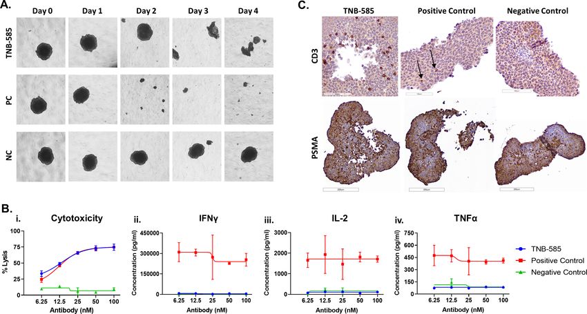
Open access
promote tumor progression through the aberrant acti- complex could enable T cell-mediated killing to be uncou-
J Immunother Cancer: first published as 10.1136/jitc-2021-002488 on 4 June 2021. Downloaded from http://jitc.bmj.com/ on June 11, 2021 by guest. Protected by copyright.
vation of PI3K-AKT signaling pathways.14 Therefore, a pled from cytokine release. Previous work from our lab
diverse array of PSMA-targeted therapies are in develop- identified such a low-affinity anti-CD3 (CD3_F2B), which
ment including radionuclides, antibody drug conjugates, induced tumor killing with minimal cytokine release
chimeric antigen receptor T cells and T cell engagers against hematologic tumors including multiple myeloma
(TCEs).15 (MM) and B cell non-Hodgkin’s lymphoma.25 26 To extend
TCEs are heterodimeric antibodies engineered to these favorable properties into the solid tumor setting, we
simultaneously bind to a tumor- associated antigen on developed TNB-585, a fully human anti-CD3xPSMA IgG4
cancer cells and to CD3 on T cells, forming an immu- TCE for the treatment of mCRPC. TNB-585 combines
nological synapse that promotes T cell redirected lysis of a tumor-targeting anti-PSMA arm together with our low
tumor cells in an MHC-independent manner.16 Although affinity anti-CD3_F2B to facilitate T cell redirected killing
T cell redirection is promising, all such approaches of prostate tumor cells with minimal cytokine release.
to date induce strong pan-T cell stimulation and toxic TNB-585 mediated killing of PSMA+ tumor cells (two-
immune activation culminating in the systemic release dimensional (2D) cultures and three-dimensional (3D)
of proinflammatory cytokines leading to cytokine release spheroids) in vitro and of patient-derived prostate tumors
syndrome (CRS). This major side effect of TCE therapy ex vivo. In addition, TNB-585 induced immune cell infil-
can potentially be attributed to the fact that a majority of tration and dose-dependent tumor regression in vivo in an
TCEs developed thus far incorporate strong binding/acti- NCG mouse xenograft model. Notably, TNB-585 induced
vating anti-CD3 moieties with affinities in the 1–200 nM substantially lower cytokine production and regulatory T
range such as OKT3 and UCHT1.17 Despite their effec- cell (Treg) activation while maintaining equivalent levels
tiveness in hematologic tumors, TCEs have also demon- of maximum tumor killing compared with a positive
strated limited success in solid tumors, and combination control antibody that contains the same anti-PSMA arm
treatments may be needed to maximize efficacy.18 19 but a higher affinity anti-CD3 arm. With potent antitumor
Currently, a number of TCEs targeting PSMA and CD3 activity combined with a favorable safety profile, TNB-585
are being investigated in early phase clinical studies for will potentially create a safer and more effective therapy
the treatment of mCRPC. Pasotuxizumab (AMG 212), for mCRPC and expand the opportunities for its use in
a PSMA- targeting bispecific T cell engager (BiTE), combination treatments.
demonstrated signs of clinical activity in a phase I study
as approximately one-third of patients in the 20, 40, and
80 µg/dose groups of the continuous intravenous infu- MATERIALS AND METHODS
sion (cIV) cohort exhibited a >50% decline in Prostate- Immunization, NGS, clonotype analysis, and cloning
specific antigen (PSA) levels.20 Due to a sponsor change, Using genetically engineered transgenic rats (UniRat
this study was terminated early, and the maximum toler- and OmniFlic) that express fully human IgG antibodies
ated dose for the cIV cohort was not determined. AMG together with an NGS-based antibody discovery pipeline
160 is a half-life extended BiTE also being investigated in (TeneoSeek),25–28 we have developed TNB-585. Methods
phase I clinical trials. In contrast to the high-frequency, for generating antibodies targeting CD3 in OmniFlic
once-daily dosing regimen of pasotuxizumab, AMG 160 animals have been previously described.25 For generating
is administered once every 2 weeks. Data from the phase heavy chain only antibodies against PSMA, 12 UniRats
I study indicate that 27.6% of patients had a confirmed were immunized with recombinant human PSMA protein
PSA response to AMG 160, although grade 2 and grade fused to a his-tag (R&D Systems, Minneapolis, Minnesota,
3 CRS occurred in 60.5% and 25.6% of patients, respec- USA) for up to 8 weeks using either Titermax/Ribi or
tively.21 For HPN424, a once-weekly dosed TriTAC mole- CFA/IFA adjuvant. Draining lymph nodes from all animals
cule targeting PSMA and CD3, clinical activity at the were then harvested, and total RNA was collected. cDNA
highest fixed dose tested (160 ng/kg) was demonstrated samples containing the full heavy chain variable domain
by PSA reduction in three out of seven patients with one (VH) underwent next-generation sequencing using the
confirmed partial response.22 Overall, these phase I clin- MiSeq platform (Illumina, San Diego, California, USA)
ical trial data suggest that, although promising, there with 2×300 paired-end reads. Data from all animals were
are shortcomings associated with current PSMA-targeted analyzed, and the most frequent 265 VH sequences were
TCE therapeutics, including either safety (polycytokine selected for cloning followed by expression in HEK 293
secretion and clinical CRS), efficacy, or dosing schedule, cells.
that could be addressed by an IgG TCE that facilitates
tumor killing with minimal cytokine release. Expression and purification of TNB-585, positive control (PC),
Given that T cells possess a dual activation threshold and negative control (NC) TCEs
(one for cytotoxicity and the other for cytokine produc- TNB-585 consists of two heavy and one light chain(s)
tion)23 and that the formation of a stable mature immu- paired using knobs- into-holes technology.29 30 Heavy
nological synapse is not required for T cell cytotoxicity,24 chain 1 (HC1) and the kappa light chain form the para-
we hypothesized that a low-affinity anti-CD3 more closely tope that binds to human CD3. Heavy chain 2 (HC2) is
resembling the low-affinity interaction of the TCR:pMHC composed of a single VH domain that targets PSMA. The
2 Dang K, et al. J Immunother Cancer 2021;9:e002488. doi:10.1136/jitc-2021-002488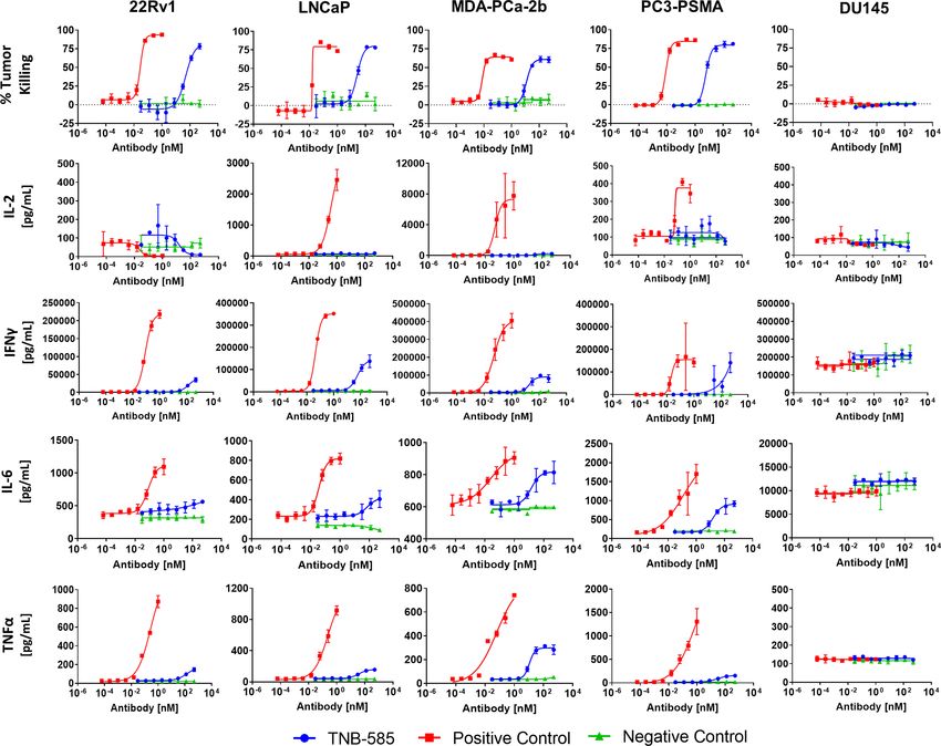
Open access
Fc contains a mutation that results in the loss of Fc-me- to each well. After a 6-hour incubation at 37°C, Bio-Glo
J Immunother Cancer: first published as 10.1136/jitc-2021-002488 on 4 June 2021. Downloaded from http://jitc.bmj.com/ on June 11, 2021 by guest. Protected by copyright.
diated effector functions in order to disable non-specific reagent was added to each well, and luminescence was
activation and depletion of T cells in patients. TNB-585 is measured on a SpectraMax i3x microplate reader (Molec-
further engineered to prevent arm-exchange with other ular Devices, San Jose, California, USA).
IgG4 molecules.
The positive control (PC) antibody is composed of an Assessment of pharmacokinetics in cynomolgus monkey
HC2 (anti-PSMA) and Fc region identical to TNB-585, Nine male cynomolgus monkeys were administered a
whereas the heavy chain and the kappa light chain on single intravenous bolus dose of 0.07, 0.97, or 10.39 mg/
the other arm combine to form a paratope that binds kg TNB-585 (Alta Science Preclinical Seattle, Everett,
to human CD3 with stronger affinity than TNB-585. The Washington, USA). Blood samples collected from a
affinity of the CD3 arm in the PC is similar to OKT3 peripheral vein were processed to serum on day −12
(muronomab-CD3). The NC antibody is composed of an (predose), and on days 1 (1 hour and 6 hours postdose),
HC1 (anti-CD3) and an Fc region identical to TNB-585. 2, 3, 5, 7, 15 and 21. Concentrations of TNB-585 in serum
However, the VH domain on the targeting arm of the NC were measured by ELISA. In brief, a rabbit anti-idiotype
is against an irrelevant, non-human target. antibody against the anti-PSMA arm was coated at 1 µg/
TNB-585, PC, and NC antibodies were expressed in mL onto 96-well plates and incubated overnight at 4°C.
ExpiCHO cells as per manufacturer’s instructions. Super- The plates were then washed three times with wash buffer
natants were harvested, clarified, and affinity purified (TBST +0.1% Tween-20) and blocked using 1% milk for
using an anti-CH1 resin (CaptureSelect CH1-XL, Ther- 1 hour at room temperature. Following incubation, the
moFisher, Waltham, Massachusetts, USA), followed by plates were washed, and 100 µL of diluted serum was
a polishing step with size exclusion or cation exchange added to each well and incubated for 2 hours at room
chromatography. temperature. After three washes, a rat anti-idiotype anti-
body against the anti-CD3 arm was added at 1 µg/mL for
SDS-PAGE and capillary gel electrophoresis
30 min. Binding was detected by incubation with anti-rat
Purified proteins were run on a 4%–12% Bis-Tris SDS-
IgG2a-HRP followed by addition of ELISA Ultra TMB
PAGE gel (NuPAGE, ThermoFisher) under non-reducing
substrate. After sufficient color developed, 100 µL of stop
conditions, followed by Coomassie Blue staining. Analysis
solution was added to each well. Absorbance was read
was performed on Image Lab (Bio- Rad Laboratories,
at 450 nm on a SpectraMax i3x plate reader. TNB-585
Hercules, California, USA) against a prestained protein
concentrations in serum were determined by interpola-
ladder (PageRuler, ThermoFisher) run on the same gel.
tion from the standard curve. Non-compartmental PK
Capillary gel electrophoresis was performed on Maurice
parameters were estimated using Phoenix WinNonlin
(ProteinSimple, San Jose, California, USA) using its
V.8.1 software (Certara USA, Inc, Princeton, New Jersey,
preassembled, prequalified CE- SDS cartridge. Purified
USA).
proteins were mixed with internal standard (Protein-
Simple) and iodoacetamide according to manufacturer’s
instructions, denatured at 70°C for 10 min, and separated Cell lines and cell culture
by electrophoresis for 35 min at 5750V. Prostate tumor cell lines LNCaP, 22Rv1, MDA-PCa- 2b,
PC3, and DU145 were purchased from ATCC (Manassas,
Size exclusion ultra-performance liquid chromatography Virginia, USA). LNCaP and 22Rv1 cells were cultured in
(UPLC) RPMI-1640 (Fisher Scientific, Waltham, Massachusetts,
Size exclusion UPLC was performed on a ThermoFisher USA) and 10% FBS (ThermoFisher). DU145 cells were
Ultimate 3000 UPLC with a UV/Vis detector set to 280 nm cultured in EMEM (ATCC) and 10% FBS. PC3 cells were
and 220 nm for monitoring. The SEC column (TSKgel cultured in F12K (ATCC) and 10% FBS. MDA-PCa-2b
UP-SW3000, 2 µm, 30 cm × 4.6 mm) was from Tosoh Biosci- cells were cultured in BRFF- HPC1 (Athena Enzyme
ence. The isocratic method was run in 100 mM Sodium Systems, Baltimore Maryland, USA) and 20% FBS. All cell
Citrate, 500 mM Sodium Chloride, 200 mM L-Arginine, lines were maintained in a humidified chamber at 37°C
pH 6.2 at a flow rate of 0.25 mL/min. and 8% CO2.
A stable PSMA- overexpressing PC3 cell line (PC3-
Potency assay PSMA) was developed at Antibody Solutions (Santa
Determination of TNB-585 potency before and after Clara, California, USA). PSMA cDNA was synthesized
heat stress was evaluated using a T cell activation bioassay and cloned into a stable integration expression vector
(Promega, Madison, Wisconsin, USA). In brief, 22Rv1 and transfected into PC3 cells using Lipofectamine 3000
cells were seeded in 96-well tissue culture plates and incu- (ThermoFisher). Cells transiently expressing the trans-
bated overnight at 37°C. Following incubation, culture fected DNA were selected using puromycin to generate
supernatant was aspirated and replenished with assay stable expressing pools. High- expressing clones were
media (RPMI 1640, 10% fetal bovine serum (FBS)) along then isolated by fluorescence-activated cell sorting. PSMA
with a dilution series of antibody. TCR/CD3 effector cells cells were cultured in F12K with 10% FBS and 1 µg/mL
were then thawed, resuspended in assay media, and added puromycin (ThermoFisher).
Dang K, et al. J Immunother Cancer 2021;9:e002488. doi:10.1136/jitc-2021-002488 3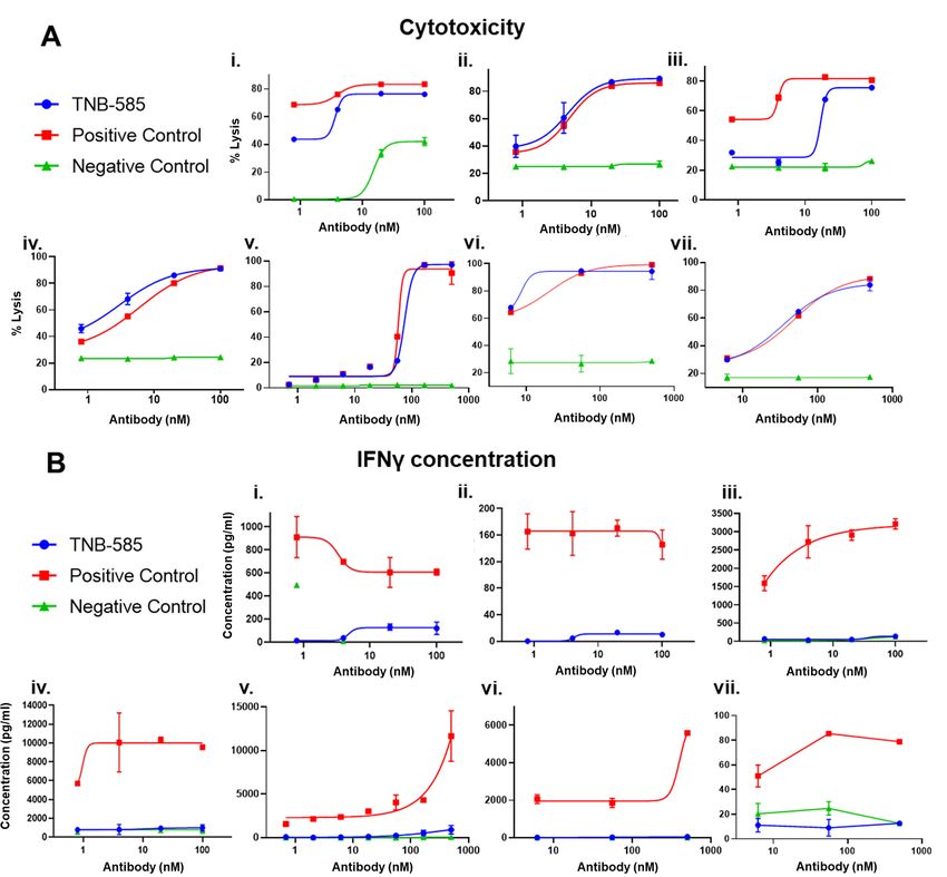
Open access
Cell binding and flow cytometry Measurement of T cell proliferation
J Immunother Cancer: first published as 10.1136/jitc-2021-002488 on 4 June 2021. Downloaded from http://jitc.bmj.com/ on June 11, 2021 by guest. Protected by copyright.
Binding of TNB-585 to PSMA+ and PSMA− cells was eval- 22Rv1 and DU145 cells were seeded (25,000 cells/well) in
uated by flow cytometry. In brief, tumor cells were incu- a 96-well plate and incubated overnight at 37°C and 8%
bated with a dilution series of antibody for 30 min at CO2. Following incubation, human T cells were labeled
4°C. After incubation, the cells were washed twice with with 2 µM carboxyfluorescein succinimidyl ester (CFSE,
flow cytometry buffer (PBS, 1% BSA, 0.1% NaN3), resus- ThermoFisher), as per manufacturer’s instructions, and
pended in a 1:100 diluted solution of PE-conjugated goat co-cultured with tumor cells at an E:T ratio of 4:1 in the
antihuman IgG secondary antibody (Southern Biotech, presence of antibody for 48 hours at 37°C and 8% CO2.
Birmingham, Alabama, USA), and incubated for 20 min The cells were then stained with fluorophore conjugated
at 4°C. The cells were then washed twice in flow cytom- anti-
CD4 and anti- CD8 antibodies (BioLegend) and
etry buffer, and the mean fluorescence intensity (MFI) analyzed by flow cytometry. Proliferation was calculated
was measured using either FACSCelesta (BD Biosciences, as the percent of T cells that showed a reduction in CFSE
San Jose, California, USA) or Guava easyCyte 8HT (EMD signal compared with untreated control.
Millipore, Hayward, California, USA).
To determine PSMA antigen density, the cells were Measurement of CD4, CD8, and regulatory T cell activation
stained with a saturating concentration of PE-conjugated LNCaP and DU145 cells were seeded (20,000 cells/well)
anti-PSMA antibody (BioLegend, San Diego, California, in a 96-well plate and incubated overnight at 37°C and 8%
USA), and the MFI was measured by flow cytometry. A CO2, followed by incubation with human T cells at an E:T
calibration curve was generated using BD Quantibrite of 5:1 in the presence of antibody for 48 hours at 37°C and
Beads (BD Biosciences), which are composed of distinct 8% CO2. The cells were then stained with fluorophore-
bead populations containing a known number of mole- conjugated antibodies against CD4, CD8, CD69, CD25,
cules of equivalent fluorochrome (MESF) to convert the and Foxp3 and analyzed by flow cytometry. Activation was
MFI of a sample to the number of PE molecules on the measured by the upregulation of CD69, a marker of T cell
cell surface. A linear regression of log10(MESF) against activation. The percentage of Tregs within the activated T
log10(MFI) was plotted to calculate the number of anti- cell population (CD69+) was determined by sub-gating on
bodies bound per cell (ABC) using a PE to mAb ratio of CD25+ and Foxp3+ cells.
1:1. The ABC was then multiplied by two to approximate
the antigen density assuming that, at saturation, one anti- Evaluation of TNB-585 efficacy in 3D tumor spheroid models
body binds to two antigen molecules. Ninety-six well round- bottom, non- treated microplates
(Corning, Barrington, New Jersey, USA) were coated with
Cytotoxicity and cytokine analysis antiadherence solution (STEMCELL Technologies, Kent,
Prostate tumor cells were labeled with 0.5 µM 1′-diocta- Washington, USA), centrifuged for 5 min at 500× g and
decyl-3,3,3′,3′-tetramethylindotricarbocyanine iodide washed once with spheroid growth media (RPMI-1640
(DiR’, ThermoFisher), as per manufacturer’s instruc- containing 10% FBS). LNCaP cells were seeded (5000
tions, and incubated with human T cells at an effector cells/well) and incubated at 37°C and 8% CO2. Half of
to target (E:T) ratio of 10:1 in the presence antibody the media was replenished with fresh media every 2 days.
for 48–72 hour at 37°C and 8% CO2. To measure tumor On day 7, antibody was added to each well together with
killing, the cells were stained with FITC- conjugated healthy donor PBMCs at an E:T of 1:1. Images were
Annexin-V (BioLegend) and analyzed by flow cytometry. taken daily using a Nikon TMS inverted phase-contrast
Alternatively, the cells were incubated with a 1:10 dilu- microscope connected to a 16MP digital camera (Boli-
tion of WST-1 for 90 min at 37°C, and absorbance was Optics, Rancho Cucamonga, California, USA) to capture
measured at 450 nm using a SpectraMax i3x microplate morphological changes in the spheroid. After 4 days of
reader to colorimetrically assess cell viability. antibody treatment, culture supernatant was harvested
For cytokine analysis, culture supernatant was harvested to measure Interleukin-2 (IL-2), interferon gamma
at 48 or 72 hours and used to quantify cytokines by ELISA (IFNγ), and tumor necrosis factor alpha (TNFα) by MSD.
(BioLegend) or Meso Scale Discovery technology (Meso Tumor killing was determined using CytoTox 96 Non-
Scale Diagnostics, Rockville, Maryland, USA) as per Radioactive Cytotoxicity Assay (Promega) per manufac-
manufacturer’s instructions. turer’s instructions. Percent cytotoxicity was determined
using the formula [(ODtest-OD“no antibody” control)/ODtest]×100.
Granzyme and perforin analysis To qualitatively evaluate cytotoxicity, the spheroids were
LNCaP cells (15,000 cells/well) were incubated with stained with trypan blue after 4 days of treatment followed
human T cells at an E:T ratio of 10:1 in the presence of by microscopic imaging at 4× magnification.
antibody for 48 hours at 37°C and 8% CO2. Following
incubation, culture supernatants were harvested, and Immunohistochemical staining of spheroids
perforin and granzyme concentrations were determined Immunohistochemical staining of the spheroids was
using human granzyme B ELISA (ThermoFisher) and performed to evaluate T cell infiltration. Histology, IHC
human perforin ELISA (Cell Sciences, Newburyport, staining, and whole slide scanning were performed by
Massachusetts, USA). NDB Bio, LLC (Baltimore, Maryland, USA). In brief,
4 Dang K, et al. J Immunother Cancer 2021;9:e002488. doi:10.1136/jitc-2021-002488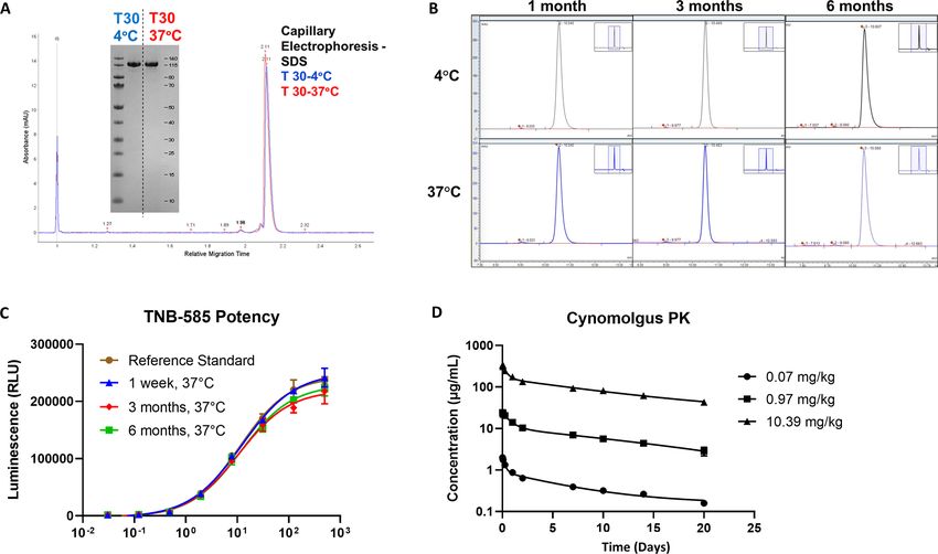
Open access
the spheroids were paraformaldehyde fixed, embedded 2×10⁶ C4-2 tumor cells. One day postimplantation, 10×106
J Immunother Cancer: first published as 10.1136/jitc-2021-002488 on 4 June 2021. Downloaded from http://jitc.bmj.com/ on June 11, 2021 by guest. Protected by copyright.
in Histogel, dehydrated in gradient concentrations of preactivated human PBMCs were administered intraperi-
ethanol, cleared in xylene, and embedded in paraffin wax. toneally. Treatment began 6 days post-tumor engraftment
Sections 4 µm thick were stained with antibodies against when the average tumor size was ~50 mm3. Mice were
PSMA (Clone 3E6; Dako) and CD8 (Clone C8/144B; randomized into groups and received TNB-585 treatment
Cell Marque). Whole slide scans were generated using (5.5, 16.6, 50, 150, or 450 µg per dose) via tail vein injec-
an Olympus PlanN 20× objective (N.A. 0.40). Scans were tion twice per week for a total of 8 treatments. Tumor
viewed and analyzed using ImageScopex64 software. burden was monitored biweekly, and the study was termi-
nated when the volumes in the control group exceeded
Ex vivo processing of patient prostate tumors 1000 mm3. The tumors were then harvested and stained
Tumor cells were either isolated from fresh prostate using an antihuman CD45 antibody to assess immune cell
tumor tissue procured from LF’s lab at UCSF (San Fran- infiltration into the tumor.
cisco, California, USA) or received as frozen dissociated
aliquots along with donor matched PBMCs from Discovery Statistics
Life Sciences (Newtown, Pennsylvania, USA). The fresh Statistical analysis was performed using one-way analysis of
prostate tissue was dissociated as previously described.31 variance (ANOVA) with a post hoc Tukey’s test for multiple
For the cytotoxicity assay, exogenous human PBMCs were comparisons. Two-sided p values were calculated for all in
co-cultured with dissociated tumor cells at an E:T of 1:2 vitro experiments using GraphPad Prism V.8. The results
for 24 hours at 37°C, 8% CO2. The death of PSMA+ cells were judged statistically significant when the Bartlett’s
was analyzed by flow cytometry on the BD FACSCelesta. corrected p values were less than 0.05. A non-parametric
An anti-CD45 antibody (BioLegend) was used to distin- Mann-Whitney test at a significance level of 0.05 was used
guish hematopoietic cells, and a non-epitope competing for the assays containing only two data sets. Data from in vivo
anti-PSMA antibody (clone LNI17, BioLegend) was used studies were analyzed in R language (V.3.3.1) using one-way
to identify PSMA+ cells. The percentage of PSMA+ live ANOVA.
cells was determined using eBioscience Fixable Viability
Dye eFluor 780 (Invitrogen, Carlsbad, California, USA).
Culture supernatant from each well was used to measure RESULTS
IL-2, IFNγ, and TNFα by MSD. Discovery of TNB-585, a PSMAxCD3 bispecific antibody
Discovery and characterization of lead anti- CD3 anti-
In vivo assessment of TNB-585 in an NCG mouse xenograft bodies have been previously described.25 To generate lead
model anti-
PSMA antibodies, 265 heavy chain only antibodies
In vivo antitumor activity of TNB-585 was evaluated in (UniAbs) identified using a custom NGS-based approach
murine models of CaP (Crown Bioscience, Taicang, on immunized UniRats were selected for gene assembly,
China). In the first study, immunodeficient NCG mice recombinant expression, and functional screening. High-
(n=3) were engrafted subcutaneously with 2×10⁶ C4-2 throughput ELISA and cell binding screens identified 40
tumor cells followed by implantation of 10×106 of either PSMA-specific binders from 12 different CDR3 clonotype
resting pan-T cells or preactivated PBMCs 1 day posten- families. An additional 117 VH sequences were identified
graftment. PBMCs were activated 3 days prior to implanta- by antibody repertoire analysis of four lead CDR3 clonotype
tion by incubation with anti-CD28 in tissue culture plates families that contain sequence variation in CDR1, CDR2,
coated with anti- CD3 (both at 1 µg/mL). Treatment and framework regions. Candidates were assessed for cell
began 9 days post-tumor engraftment when the average binding to PSMA+ cell lines, and the top binders from each
tumor volume was ~50 mm3. Mice were randomized into CDR3 family were scaled up for further studies. To identify
groups and received treatment of TNB-585, PC, or NC high-affinity anti-PSMA leads, cell binding dose curves were
(150 µg/dose) antibody via tail vein injection twice per performed with LNCaP cells, and the dissociation constants
week for a total of five doses. Tumor burden was moni- were determined by biolayer interferometry using an Octet
tored biweekly, and the study was terminated when the QK384. High-affinity anti-PSMA leads were combined into
tumor volumes in the control group exceeded 2000 mm3. TCEs using knobs- into-
holes technology with our previ-
After termination of the in-life portion of the study, the ously identified low-affinity anti-CD3_F2B arm that mediates
tumors were harvested, processed into formalin- fixed tumor killing with minimal cytokine secretion. The TCEs
paraffin-embedded sections, and stained using an anti- were evaluated in parallel for their functional characteristics,
human CD45 antibody following a standard protocol to including cytotoxicity and cytokine release, as well as their
visualize immune cell infiltration into the tumor. The developability characteristics. A final lead TCEs was selected
stained sections were scanned with NanoZoomer-HT 2.0 and designated TNB-585.
Image system (Hamamatsu, Japan) for 40× magnification,
and the images were analyzed using the HALO platform TNB-585 is a stable molecule with favorable developability
(Indica Labs, Albuquerque, New Mexico, USA). characteristics
In a follow-on study, six cohorts of immunodeficient TNB-585 is stable at 4°C and 37°C as represented by
NCG mice (n=4) were engrafted subcutaneously with a single band and a single peak on SDS- PAGE and
Dang K, et al. J Immunother Cancer 2021;9:e002488. doi:10.1136/jitc-2021-002488 5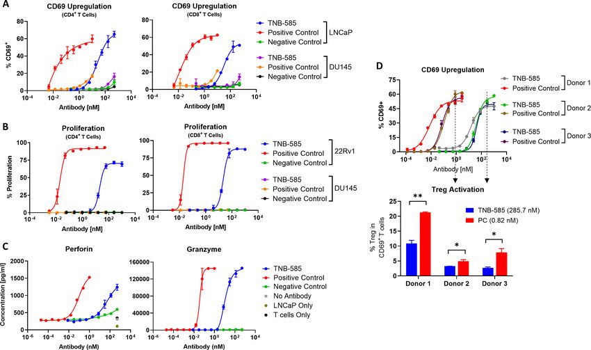
Open access
J Immunother Cancer: first published as 10.1136/jitc-2021-002488 on 4 June 2021. Downloaded from http://jitc.bmj.com/ on June 11, 2021 by guest. Protected by copyright.
Figure 1 TNB-585 is a stable molecule with favorable developability characteristics. (A) TNB-585 stability was assessed
following incubation at 37°C for 1 month. High and low molecular weight species were analyzed by CE-SDS and SDS-PAGE.
(B) SE-UPLC chromatograms of TNB-585 stressed at 37°C for 1, 3, and 6 months compared with 4°C are shown. (C) TNB-585
activity was measured using a T cell activation bioassay after heat stress at 37°C at 1 week, 3 months, and 6 months. (D) PK in
cynomolgus monkeys was evaluated by ELISA following a single dose intravenous administration of TNB-585 at 0.07, 0.97, or
10.39 mg/kg.
capillary electrophoresis, respectively (figure 1A). After observed in cynomolgus monkeys were consistent with
incubation at 37°C for 1, 3, and 6 months, no significant that of an IgG with linear PK, indicating stability of
increase in high molecular weight species was observed TNB-585 in serum in vivo.
by SEC- UPLC (figure 1B). Similarly, TNB-585 main-
tained potency after heat stress, as demonstrated in a TNB-585 binds to PSMA and CD3 and induces antigen-
T cell activation bioassay (figure 1C). Relative poten- dependent cytotoxicity and cytokine release
cies are listed in table 1. TNB-585 was also assessed in a Specific binding of TNB-585 to PSMA was confirmed by
single-dose Pharmacokinetics (PK) study in cynomolgus
flow cytometry on various PSMA+ and PSMA− cell lines
monkey. Group mean serum concentrations versus time
(figure 2A). TNB-585 demonstrated dose- dependent
profiles are shown in figure 1D. Group mean PK param-
binding to PSMA+ cells with EC50 values ranging from
eter estimates are provided in table 2. TNB-585 PK was
linear across the dose range of 0.1–10 mg/kg (nominal 24.1 nM to 38.4 nM (table 3). No binding of TNB-585
dose, actual doses were 0.07–10.39 mg/kg). Group to PSMA− cells was observed. To evaluate high, medium,
mean CL ranged from 4.16 to 6.78 mL/day/kg and and low PSMA-expressing cell lines, the antigen densi-
group mean t1/2 ranged from 9.75 to 11.0 days following ties were determined by flow cytometry using fluorescent
single intravenous doses ranging from 0.1 to 10 mg/ beads that allow for the conversion of MFI to the number
kg in cynomolgus monkeys. Since TNB-585 does not of antigen molecules on the surface of the cell. As shown
cross-react with CD3 or PSMA in non-human primates, in table 3, 22Rv1, LNCaP, MDA-PCa-2b, and PC3-PSMA
the observed linear PK is consistent with non-specific cells expressed a wide range of PSMA antigens on the
clearance mechanisms dominating PK. The PK profiles cell surface. PSMA expression on CaP cell lines such as
LNCaP has been demonstrated to be representative of
human CaP for preclinical studies.32 The PC3-PSMA cells,
Table 1 TNB-585 potency after heat stress
which expressed the highest levels of PSMA, were used
TNB-585 EC50 (nM) % relative potency
to assess initial proof of activity of TNB-585 in compar-
Reference standard 11.2 100 ison with non-transfected, PSMA− PC3 cells (PC3-WT).
1 week, 37°C 12.3 91 As shown in figure 2B, T cell redirected killing and cyto-
3 months, 37°C 10.9 102 kine release induced by TNB-585 is dependent on PSMA
6 months, 37°C 11.9 94 expression. Additionally, we observed TNB-585-mediated
cytotoxicity at multiple E:T ratios (online supplemental
EC50, Half maximal effective concentration.
file S1).
6 Dang K, et al. J Immunother Cancer 2021;9:e002488. doi:10.1136/jitc-2021-002488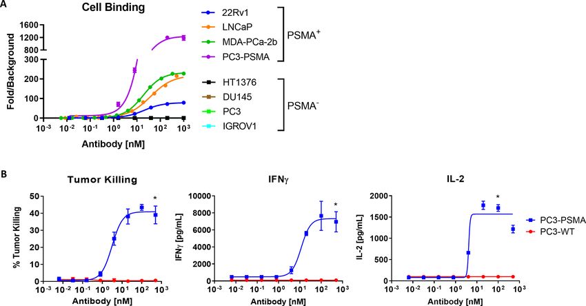
Open access
Table 2 PK parameters of TNB-585 in cynomolgus monkeys
J Immunother Cancer: first published as 10.1136/jitc-2021-002488 on 4 June 2021. Downloaded from http://jitc.bmj.com/ on June 11, 2021 by guest. Protected by copyright.
0.07 mg/kg 0.97 mg/kg 10.39 mg/kg
Parameter Units Mean SD Mean SD Mean SD
Cmax µg/mL 2.0 0.1 24.5 1.1 344 39
Cmax/D (µg/mL)/(mg/kg) 28.7 1.7 25.3 1.1 33.1 3.8
t1/2 Days 9.9 0.7 9.8 3.0 11 1.3
AUCinf µg/mL*days 10.4 0.8 178 48.9 2500 118
AUCinf/D (µg/mL*days)/(mg/kg) 148 10.8 184 50.4 241 11.3
% Extrap % 21.9 1.8 22.6 9.8 27.1 2.7
CL mL/day/kg 6.8 0.5 5.7 1.6 4.2 0.2
Vss mL/kg 86.8 1.3 72.2 2.6 62.7 2.5
AUCinf: area under the serum concentration-time curve from time zero to infinity; AUCinf/D, dose-adjusted area under the serum concentration-
time curve; Cmax: maximum serum drug concentration ; CL, serum clearance; Cmax/D, dose-adjusted maximum serum drug concentration;
% Extrap, area under the serum concentration-time curve extrapolated from time t to infinity as a percentage of total AUC; t1/2, half-life; Vss,
apparent volume of distribution at steady state.
Activation of T cells by TNB-585 induces CD69 upregulation, was approximately 0.01 nM, while the maximum percent
proliferation, and perforin and granzyme production activation ranged from 59.7% to 62.2%. Minimal CD69
To evaluate the mechanism of action of TNB-585, LNCaP upregulation was observed in the presence of DU145 cells
(PSMA+) or DU145 (PSMA−) cells were co-cultured with or NC.
T cells in the presence of increasing concentrations of TNB-585-mediated T cell proliferation was assessed by
TNB-585, PC, or NC and analyzed by flow cytometry to incubating CFSE-labeled T cells with 22Rv1 (PSMA+) or
measure the upregulation of CD69, a marker of T cell acti- DU145 (PSMA−) tumor cells and increasing concentra-
vation. TNB-585 induced antigen-dependent and dose- tions of TNB-585, PC, or NC. Following a 5-day incubation,
dependent activation of T cells comparable with the PC percent proliferation was measured by flow cytometry as
but with reduced potency (figure 3A). The EC50 values for the percent of T cells that showed a reduction in CFSE
TNB-585 mediated CD4+ and CD8+ T cell activation were signal compared with untreated control. The dose–
27.1 nM and 31.2 nM, respectively, while the maximum response curves in figure 3B demonstrate that TNB-585
percent activation ranged from 53.3% to 67.3%. For the induced antigen-dependent and dose-dependent prolif-
PC, the EC50 of both CD4+ and CD8+ T cell activation eration of CD4+ and CD8+ T cells. The EC50 values of
Figure 2 TNB-585 binds to PSMA and CD3 and induces antigen-dependent cytotoxicity and cytokine release. (A) Dose–
response curves of TNB-585 binding to multiple on-target (22RV1, LNCaP, MDA-PCa-2b and PC3-PSMA) and off-target
(HT1376, DU145, PC3 and IGROV-1) cell lines as assessed by flow cytometry are shown. (B) TNB-585-mediated cytotoxicity
and cytokine release was evaluated against either PSMA-transfected (PC3-PSMA) or non-transfected (PC3-WT) PC3 tumor
cells. Cytotoxicity was evaluated by annexin V staining using flow cytometry. IFNγ and IL-2 concentrations were measured by
ELISA. Data are reported as mean±SD. *pOpen access
after 48 hours of treatment with TNB-585, PC, or NC.
Table 3 PSMA antigen density and EC50 of TNB-585
J Immunother Cancer: first published as 10.1136/jitc-2021-002488 on 4 June 2021. Downloaded from http://jitc.bmj.com/ on June 11, 2021 by guest. Protected by copyright.
binding on prostate tumor cell lines As seen in figure 3C, TNB-585 mediated the release of
both perforin and granzyme from T cells at levels compa-
Cell line Antigen density EC50 (nM)
rable with the PC. Minimal levels were detected in the
22Rv1 33,000±60 24.1 NC-treated cells. These data are consistent with the tradi-
LNCaP 170,000±7000 38.4 tional view that the perforin and granzyme pathway is
MDA-PCa-2b 200,000±200 28 one of the primary mechanisms used by effector T cells to
PC3-PSMA 951,000±13 400 26.8 eliminate target cells.33
TNB-585 induces preferential activation of effector T cells
TNB-585 mediated proliferation were 20.4 nM and over Tregs
21.84 nM for CD4+ and CD8+ T cells, respectively, while Since the mechanism of action of TCEs is T cell redi-
the maximum percent proliferation ranged from 70.5% rected killing, overstimulation of Tregs, an immunosup-
to 87.8%. For the PC, the EC50 of both CD4+ and CD8+ pressive T cell subset, can potentially suppress antitumor
T cell proliferation was 0.02 nM, while the maximum immunity. Therefore, the relative ability of TNB-585 to
percent proliferation ranged from 91.4% to 96.0%. No T activate CD4+ and CD8+ effector T cells versus Tregs was
cell proliferation was observed in the presence of either compared with the PC. T cells isolated from three healthy
DU145 cells or NC, demonstrating target-specificity of donors were incubated with LNCaP cells and increasing
TNB-585. concentrations of TNB-585, PC, or NC for 48 hours. Acti-
To evaluate the mechanism of T cell mediated cytotox- vation was assessed by measuring CD69 upregulation by
icity by TNB-585, we analyzed the production of effector flow cytometry. To determine the relative activation of
molecules released from cytotoxic lymphocytes: the Tregs induced by TNB-585 and the PC, the percentage
pore-forming protein, perforin, and the serine protease, of Tregs within the activated T cell population (CD69+)
granzyme. Perforin and granzyme concentrations were was measured by subgating on CD25+Foxp3+ cells. While
measured from LNCaP and T cell co-culture supernatants TNB-585 stimulated equivalent levels of maximum T
Figure 3 Activation of T cells by TNB-585 induces CD69 upregulation, proliferation, and perforin and granzyme production.
(A) CD69 upregulation was assessed on CD4+ and CD8+ T cells by flow cytometry following incubation of LNCaP or DU145
tumor cells and T cells in the presence of increasing concentrations of TNB-585, PC, or NC for 48 hours at 37°C. (B) proliferation
of CFSE-labeled CD4+ and CD8+ T cells was assessed following incubation with 22RV1 or DU145 tumor cells in the presence
of increasing concentrations of TNB-585, PC, or NC for 48 hours at 37°C. Percent proliferation was measured by CFSE
dilution using flow cytometry. (C) Perforin and granzyme concentrations in culture supernatants were measured by ELISA after
48 hours incubation of TNB-585, PC, or NC with T cells and LNCaP tumor cells. (D) Upregulation of CD69 on T cells from three
healthy donors was measured by flow cytometry after 48 hours incubation with 22RV1 tumor cells and either TNB-585, PC,
or NC. Activated T cells (CD69+) were further subgated on CD25+Foxp3+ expression to evaluate the percentage of Tregs that
comprise the total activated T cell population. Data are reported as mean±SD. *POpen access
cell activation compared with the PC, the proportion of of 60.1%–80.6% and EC50 values ranging from 6.5 to
J Immunother Cancer: first published as 10.1136/jitc-2021-002488 on 4 June 2021. Downloaded from http://jitc.bmj.com/ on June 11, 2021 by guest. Protected by copyright.
Tregs that comprised the activated T cell population was 49.3 nM across the three donors (figure 4). In compar-
significantly lower across all three donors (figure 3D). On ison with the PC, TNB-585 induced similar levels of
average, the maximum percentage of Tregs within the maximum tumor killing but with substantially reduced
CD69+ T cell population was twofold lower in TNB-585- cytokine release. At concentrations sufficient to induce
treated cells compared with PC- treated cells, demon- maximum tumor killing, TNB-585 induced less IL-2, IL-6,
strating preferential activation of effector T cells over IFNγ, and TNFα production compared with the PC. No
Tregs by TNB-585. antibody-mediated tumor killing or cytokine release was
observed in the presence of either DU145 cells or NC.
TNB-585 mediates robust killing of prostate tumor cells with
reduced cytokine release TNB-585 demonstrates potent cytotoxicity of 3D tumor
TNB-585-mediated tumor killing and cytokine release spheroids
was evaluated in a co- culture assay using four PSMA+ Since TNB-585 is being developed for the treatment of
tumor cell lines (22Rv1, LNCaP, MDa-PCa-2b, and PC3- a solid tumor, its efficacy was tested against 3D tumor
PSMA) incubated with resting T cells isolated from three spheroids to better represent the solid tumor setting
healthy donors in the presence of TNB-585, PC, or NC. and to allow for the analysis of T cell infiltration by IHC
DU145 cells were used as an off- target, NC cell line. following treatment. Antitumor activity in a spheroid
Tumor cell killing was assessed using either flow cytom- culture model was assessed by incubating LNCaP spher-
etry (22Rv1, MDA-PCa-2b, PC3-PSMA, and DU145) or oids with healthy donor PBMCs at an E:T ratio of 1:1 in
WST-1 (LNCaP), and cytokine production was measured the presence of TNB-585, PC, or NC for 4 days at 37°C. As
by MSD. TNB-585 mediated robust killing of all 4 PSMA+ demonstrated in figure 5A, treatment with either TNB-585
tumor cell lines with an average maximum tumor killing or PC resulted in dissociation of spheroid integrity over
Figure 4 TNB-585 induces tumor cell killing with reduced cytokine release. TNB-585 mediated tumor killing and cytokine
release was evaluated against four PSMA+ cell lines (22RV1, LNCaP, MDA-PCa-2b, and PC3-PSMA) and one PSMA− cell line
(DU145) using T cells isolated from three healthy donors. T cells were incubated with prostate tumor cells at a 10:1 E:T ratio in
the presence of increasing concentrations of TNB-585, PC, or NC. Cytotoxicity was measured using either WST-1 (LNCaP) or
flow cytometry (22Rv1, MDA-PCa-2b, PC3-PSMA, and DU145). Cytokine (IL-2, IFNγ, IL-6, and TNFα) concentrations in culture
supernatants were measured using MSD technology. Representative dose response curves from a single donor are shown. Data
are reported as mean±SD. NC, negative control; PC, positive control.
Dang K, et al. J Immunother Cancer 2021;9:e002488. doi:10.1136/jitc-2021-002488 9Open access
J Immunother Cancer: first published as 10.1136/jitc-2021-002488 on 4 June 2021. Downloaded from http://jitc.bmj.com/ on June 11, 2021 by guest. Protected by copyright.
Figure 5 TNB-585 mediates cytotoxicity of PSMA+ LNCaP spheroids with minimal cytokine production. LNCaP spheroids
were incubated with huPBMCs at an E:T ratio of 1+1 in the presence of increasing concentrations of TNB-585, PC, or NC for
4 days at 37°C. (A) Images were taken daily to capture morphological changes in the spheroids (100 nM antibody concentration
is shown). Cytotoxicity (Bi) was assessed by LDH release, and cytokine concentrations (Bii–iv) were measured using MSD
technology. (C) Spheroids were analyzed by IHC using anti-PSMA and anti-CD3 antibodies to evaluate T cell infiltration. Scale:
top panel: 100 µm; bottom panel: 200 µm. IHC, Immunohistochemistry; LDH, lactate dehydrogenase; NC, negative control; PC,
positive control.
time, while treatment with NC showed no difference in killing of PSMA+ tumor cells similar to PC, as measured
size after 4 days of treatment. TNB-585 induced dose- by percent tumor cell death, in all tested conditions
dependent killing of LNCaP spheroids (figure 5Bi) including freshly dissociated tumor incubated with
with minimal cytokine release (figure 5Bii, iii and iv). TNB-585 in the absence of exogenous PBMCs (figure 6Ai
The maximum percent killing for TNB-585-treated and and ii). While the PC induced substantial secretion of all
PC-treated cells were comparable at approximately 80%. cytokines tested, TNB-585 induced cytokine secretion
However, the PC induced substantial secretion of IFNγ, comparable with the NC (figure 6B and online supple-
IL-2, and TNFα, while TNB-585 induced cytokine secre- mental figure S3), correlating with our in vitro results.
tion at levels similar to the NC. Tumor killing was also
assessed qualitatively by trypan blue staining. As seen in TNB-585 induces immune cell infiltration and dose-dependent
online supplemental figure S2, a higher staining intensity tumor regression in an NCG mouse xenograft model
as well as significant morphological changes in terms of The in vivo pharmacologic activity of TNB-585 was evalu-
compromised tumor integrity were observed in TNB-585- ated in NCG mouse xenograft models. In the first study,
and PC-treated spheroids compared with NC, indicating immunodeficient NCG mice were engrafted with C4-2
tumor death. tumor cells and either resting T cells or preactivated
To assess T cell infiltration within the 3D tumors, IHC PBMCs as indicated in the study design (figure 7A).
staining was performed. Representative IHC images Mice were administered (intravenous) TNB-585, PC, or
shown in figure 5C demonstrate that TNB-585 induced NC biweekly at 150 µg per dose, and tumor burden was
CD3+ T cell infiltration into the PSMA+ LNCaP spher- monitored twice a week by measuring tumor volume. As
oids, whereas no T cell infiltration was observed in the demonstrated in figure 6B, TNB-585 and PC treatment
NC-treated spheroids. resulted in comparable inhibition of tumor growth in
both conditions whether resting T cells or preactivated
TNB-585 is efficacious against ex vivo patient prostate tumors PBMCs were used as effector cells. After termination of the
We analyzed five prostate patient tumor samples either in-life portion of the study, the tumors were harvested and
freshly isolated (n=2) or previously frozen (n=3) for T cell stained using an anti-human CD45 antibody to visualize
mediated cytotoxicity and cytokine release by TNB-585. tumor infiltrating lymphocytes (TILs) by IHC. Greater
As shown in figure 6A, TNB-585 induced dose-dependent immune cell (CD45+) infiltration of tumors in TNB-585
10 Dang K, et al. J Immunother Cancer 2021;9:e002488. doi:10.1136/jitc-2021-002488Open access
J Immunother Cancer: first published as 10.1136/jitc-2021-002488 on 4 June 2021. Downloaded from http://jitc.bmj.com/ on June 11, 2021 by guest. Protected by copyright.
Figure 6 TNB-585 mediates lysis of patient-derived prostate tumors with minimal cytokine production. TNB-585, PC, or
NC were added to dissociated tumor cells from freshly procured prostatic adenocarcinoma tissue (i–iv) or thawed previously
dissociated prostatic adenocarcinoma (v–vii) and incubated without additional human PBMCs (i, ii) or with either unmatched
huPBMC (iii–iv) or donor matched huPBMC (v–vii) at an effector to target cell ratio (E:T=1:2) for 24 hours at 37°C and 8% CO2.
Percent cytotoxicity of PSMA-positive cells (A) and IFNγ concentration in the corresponding wells (B) are shown. NC, negative
control; PBMCs, Peripheral blood mononuclear cellsl; PC, positive control.
treated and PC-treated mice was observed compared with kill cancer cells. Following the Food and drug adminis-
tumors of NC-treated mice (online supplemental figure tration (FDA) approval of blinatumomab in 2014, TCEs
S4). have gained momentum in cancer immunotherapy, and
In a follow-on study, mice were administered preacti- currently, there are 38 TCEs being investigated in clin-
vated PBMCs and treated biweekly with TNB-585 at doses ical trials for patients with cancer.34 Although T cell redi-
ranging from 5.5 µg to 450 µg or vehicle. On day 28 post- recting approaches are promising, treatment in the clinic
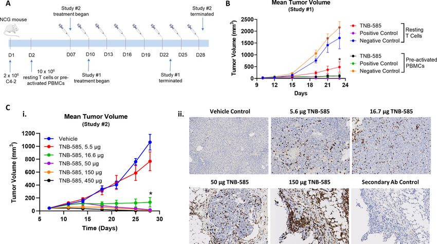
implantation, the tumor growth inhibition was 27.8%, has been hampered due to the toxicities associated with
87.4%, 98.4%, 99.2%, and 100% for dose groups 5.6, 16.7, CRS, an acute systemic inflammatory condition triggered
50, 150, and 450 µg, respectively, demonstrating dose- by elevated levels of proinflammatory cytokines. The
dependent tumor regression by TNB-585 (figure 7Ci). An
immunosuppressive microenvironment of solid tumors
IHC analysis of the harvested tumors revealed substantial
presents additional challenges to the clinical success of
immune cell infiltration in tumors of TNB-585-treated
TCEs, as the lack of effector T cell infiltration combined
mice, whereas the vehicle treated mice had no detectable
with an increased frequency of Tregs poses a significant
TILs (figure 7Cii).
barrier to efficacy against solid tumors.35 To overcome
these challenges, TNB-585 has been designed using a
DISCUSSION low-affinity anti-CD3 in order to induce tumor killing with
TCEs are an emerging class of antibody-based immuno- reduced cytokine release, promote immune cell infiltra-
therapies that redirect the cytolytic activity of T cells to tion into tumors, and minimize Treg activation thereby
Dang K, et al. J Immunother Cancer 2021;9:e002488. doi:10.1136/jitc-2021-002488 11Open access
J Immunother Cancer: first published as 10.1136/jitc-2021-002488 on 4 June 2021. Downloaded from http://jitc.bmj.com/ on June 11, 2021 by guest. Protected by copyright.
Figure 7 TNB-585 induces immune cell infiltration and dose-dependent tumor regression in an NCG mouse xenograft model.
(A) A schematic representation of the study designs is shown. NCG mice were engrafted with 2×10⁶ C4-2 tumor cells followed
by injection of either resting T cells or preactivated PBMCs. Biweekly treatment began when the average tumor volume was
~50 mm3. (B) Tumor growth inhibition was evaluated in C4-2 tumor bearing mice comparing treatment with TNB-585, PC,
and NC (150 µg/dose) using either resting T cells or preactivated PBMCs as effectors cells. (C) Tumor growth inhibition was
evaluated in C4-2 tumor bearing mice using preactivated PBMCs at TNB-585 doses ranging from 5.5 to 450 µg per dose. tumor
volumes were measured twice per week (i). At the end of study, tumors were harvested and stained by IHC using an anti-CD45
antibody to evaluate immune cell infiltration into the tumor. Representative images for each dose group are shown (ii). Tumor
volumes are reported as mean±SEM. *POpen access
clinic for the treatment of MM using TNB-383B, our anti- responsibility arising from any reliance placed on the content. Where the content
J Immunother Cancer: first published as 10.1136/jitc-2021-002488 on 4 June 2021. Downloaded from http://jitc.bmj.com/ on June 11, 2021 by guest. Protected by copyright.
BCMAxCD3 TCE.42 By incorporating the same anti-CD3_ includes any translated material, BMJ does not warrant the accuracy and reliability
of the translations (including but not limited to local regulations, clinical guidelines,
F2B, TNB-585 induced maximal killing of prostate tumor terminology, drug names and drug dosages), and is not responsible for any error
cells with reduced cytokine release compared with the PC and/or omissions arising from translation and adaptation or otherwise.
in all preclinical studies tested, suggesting that TNB-585 Open access This is an open access article distributed in accordance with the
may be efficacious while inducing a lower incidence and Creative Commons Attribution Non Commercial (CC BY-NC 4.0) license, which
severity of CRS in CaP patients compared with other TCEs permits others to distribute, remix, adapt, build upon this work non-commercially,
and license their derivative works on different terms, provided the original work is
that incorporate high-affinity anti-CD3s.
properly cited, appropriate credit is given, any changes made indicated, and the use
is non-commercial. See http://creativecommons.org/licenses/by-nc/4.0/.
CONCLUSION ORCID iDs
Kevin Dang http://orcid.org/0000-0001-5695-926X
In summary, TNB-585 is a novel CD3xPSMA TCE in Giulia Castello http://orcid.org/0000-0002-5647-178X
development for the treatment of mCRPC that warrants Pranjali Dalvi http://orcid.org/0000-0002-5247-9645
further investigation in clinical trials. Our data demon-
strate that TNB-585 induces antigen- dependent and
dose-dependent killing of PSMA+ tumor cells in vitro (2D
cultures and 3D spheroids), ex vivo, and in vivo. In all REFERENCES
preclinical models tested, TNB-585 stimulated signifi- 1 Rawla P. Epidemiology of prostate cancer. World J Oncol
2019;10:63–89.
cantly less cytokine production compared with a PC anti- 2 National Cancer Institute. Seer cancer STAT facts: prostate cancer.
body that contains the same anti-PSMA arm but a higher Available: https://seer.cancer.gov/statfacts/html/prost.html
3 Patrikidou A, Loriot Y, Eymard J-C, et al. Who dies from prostate
affinity anti-CD3 arm. Our results suggest that TNB-585, cancer? Prostate Cancer Prostatic Dis 2014;17:348–52.
with its low-affinity anti-CD3, may provide an efficacious 4 Karantanos T, Evans CP, Tombal B, et al. Understanding the
mechanisms of androgen deprivation resistance in prostate cancer at
approach to kill PSMA+ CaP cells while inducing a lower the molecular level. Eur Urol 2015;67:470–9.
incidence and severity of CRS in patients. 5 Teo MY, Rathkopf DE, Kantoff P. Treatment of advanced prostate
cancer. Annu Rev Med 2019;70:479–99.
Acknowledgements The authors would like to thank Heather Ogana for her 6 Chang SS. Overview of prostate-specific membrane antigen. Rev
Urol 2004;6 Suppl 10:S13–18.
contributions during the primary and diversity screens of the anti-PSMA antibody 7 Kiessling A, Wehner R, Füssel S, et al. Tumor-Associated
discovery phase. antigens for specific immunotherapy of prostate cancer. Cancers
Contributors KD, PD, GC, SI, and YL contributed to manuscript preparation. KD, 2012;4:193–217.
PD, GC, YL, and SCC contributed to the study design and analysis of results. NDT, 8 Caromile LA, Dortche K, Rahman MM, et al. Psma redirects cell
survival signaling from the MAPK to the PI3K-Akt pathways to
KEH, and AB performed the NGS-based repertoire analysis. KEH, LMD, and AB
promote the progression of prostate cancer. Sci Signal 2017;10.
completed the molecular biology. KD, SCC, and PS performed the ELISA and flow doi:10.1126/scisignal.aag3326. [Epub ahead of print: 14 Mar 2017].
cytometry lead discovery assays. DP characterized the anti-CD3 antibodies. YL 9 Kawakami M, Nakayama J. Enhanced expression of prostate-
and HSU conducted the expression, purification, and biophysical characterization specific membrane antigen gene in prostate cancer as revealed by in
of antibodies. WS contributed to the developability assessment. KD, PD, and GC situ hybridization. Cancer Res 1997;57:2321–4.
performed the in vitro functional assays. PD, KD, BB, and SCC contributed to the 10 Silver DA, Pellicer I, Fair WR, et al. Prostate-Specific membrane
in vivo study designs. RD performed the cynomolgus PK analysis. AS, SK and LF antigen expression in normal and malignant human tissues. Clin
provided the fresh patient tumors for ex vivo analysis. PD designed and performed Cancer Res 1997;3:81–5.
11 Mhawech-Fauceglia P, Zhang S, Terracciano L, et al. Prostate-
the ex vivo assays. SI, BB, US, WvS, and RB contributed to overall data review. All Specific membrane antigen (PSMA) protein expression in normal
authors reviewed and approved the final manuscript. and neoplastic tissues and its sensitivity and specificity in prostate
Funding The study was sponsored by Teneobio, Inc and funded in part by SBIR adenocarcinoma: an immunohistochemical study using mutiple
grant number 1R43CA232972-01 awarded to Teneobio, Inc. LF's lab is separately tumour tissue microarray technique. Histopathology 2007;50:472–83.
12 Hupe MC, Philippi C, Roth D, et al. Expression of prostate-specific
funded by the Prostate Cancer Foundation Challenge Grant and NIH R01CA223484. membrane antigen (PSMA) on biopsies is an independent risk
Competing interests All authors except AS, SK, and LF were employees of Stratifier of prostate cancer patients at time of initial diagnosis. Front
Teneobio, Inc with equity interests when the reported work was conducted. LF and Oncol 2018;8:623.
RD are consultants of Teneobio, Inc. 13 Bravaccini S, Puccetti M, Bocchini M, et al. Psma expression: a
potential ally for the pathologist in prostate cancer diagnosis. Sci
Patient consent for publication Not required. Rep 2018;8:4254.
14 Kaittanis C, Andreou C, Hieronymus H, et al. Prostate-Specific
Ethics approval Human PBMCs were collected by STEMCELL Technologies membrane antigen cleavage of vitamin B9 stimulates oncogenic
and AllCells in accordance with scientific, ethical, and regulatory guidelines. Rat signaling through metabotropic glutamate receptors. J Exp Med
maintenance and immunizations were performed by Antibody Solutions (Sunnyvale, 2018;215:159–75.
California, USA) with protocols reviewed by Institutional Animal Care and Use 15 Marshall CH, Antonarakis ES. Emerging treatments for metastatic
Committee (IACUC) boards. Mouse studies were reviewed and approved by the castration-resistant prostate cancer: immunotherapy, PARP
IACUC of CrownBio prior to execution, and studies were conducted in accordance inhibitors, and PSMA-targeted approaches. Cancer Treat Res
with the regulations of the Association for Assessment and Accreditation of Commun 2020;23:100164.
16 Sedykh SE, Prinz VV, Buneva VN, et al. Bispecific antibodies: design,
Laboratory Animal Care. therapy, perspectives. Drug Des Devel Ther 2018;12:195–208.
Provenance and peer review Not commissioned; externally peer reviewed. 17 Wu Z, Cheung NV. T cell engaging bispecific antibody (T-BsAb): from
technology to therapeutics. Pharmacol Ther 2018;182:161–75.
Data availability statement All data relevant to the study are included in the 18 Cruz E, Kayser V. Monoclonal antibody therapy of solid tumors:
article or uploaded as supplementary information. All data relevant to the study are clinical limitations and novel strategies to enhance treatment efficacy.
included in the article or uploaded as supplementary information. Biologics 2019;13:33–51.
19 Cha H-R, Lee JH, Ponnazhagan S. Revisiting immunotherapy: a
Supplemental material This content has been supplied by the author(s). It has focus on prostate cancer. Cancer Res 2020;80:1615–23.
not been vetted by BMJ Publishing Group Limited (BMJ) and may not have been 20 Hummel H-D, Kufer P, Grüllich C, et al. Pasotuxizumab, a BiTE®
peer-reviewed. Any opinions or recommendations discussed are solely those immune therapy for castration-resistant prostate cancer: Phase I,
of the author(s) and are not endorsed by BMJ. BMJ disclaims all liability and dose-escalation study findings. Immunotherapy 2021;13:125–41.
Dang K, et al. J Immunother Cancer 2021;9:e002488. doi:10.1136/jitc-2021-002488 13You can also read