RAF Kinase Activity Regulates Neuroepithelial Cell Proliferation and Neuronal Progenitor Cell Differentiation during Early Inner Ear Development
←
→
Page content transcription
If your browser does not render page correctly, please read the page content below
RAF Kinase Activity Regulates Neuroepithelial Cell
Proliferation and Neuronal Progenitor Cell
Differentiation during Early Inner Ear Development
Marta Magariños1,2,3*, Marı́a R. Aburto1,3, Hortensia Sánchez-Calderón1,3, Carmen Muñoz-Agudo1, Ulf R.
Rapp4, Isabel Varela-Nieto1,3
1 Instituto de Investigaciones Biomédicas ‘‘Alberto Sols’’, CSIC-UAM, Madrid, Spain, 2 Departamento de Biologı́a, Universidad Autónoma de Madrid, Madrid, Spain, 3 Unit
761, Centro de Investigación Biomédica en Red de Enfermedades Raras (CIBERER), Instituto de Salud Carlos III, Madrid, Spain, 4 Department of Molecular Biology, Max-
Planck-Institute of Biochemistry, Munich, Germany

     Abstract
     Background: Early inner ear development requires the strict regulation of cell proliferation, survival, migration and
     differentiation, coordinated by the concerted action of extrinsic and intrinsic factors. Deregulation of these processes is
     associated with embryonic malformations and deafness. We have shown that insulin-like growth factor I (IGF-I) plays a key
     role in embryonic and postnatal otic development by triggering the activation of intracellular lipid and protein kinases. RAF
     kinases are serine/threonine kinases that regulate the highly conserved RAS-RAF-MEK-ERK signaling cascade involved in
     transducing the signals from extracellular growth factors to the nucleus. However, the regulation of RAF kinase activity by
     growth factors during development is complex and still not fully understood.

     Methodology/Principal Findings: By using a combination of qRT-PCR, Western blotting, immunohistochemistry and in situ
     hybridization, we show that C-RAF and B-RAF are expressed during the early development of the chicken inner ear in
     specific spatiotemporal patterns. Moreover, later in development B-RAF expression is associated to hair cells in the sensory
     patches. Experiments in ex vivo cultures of otic vesicle explants demonstrate that the influence of IGF-I on proliferation but
     not survival depends on RAF kinase activating the MEK-ERK phosphorylation cascade. With the specific RAF inhibitor
     Sorafenib, we show that blocking RAF activity in organotypic cultures increases apoptosis and diminishes the rate of cell
     proliferation in the otic epithelia, as well as severely impairing neurogenesis of the acoustic-vestibular ganglion (AVG) and
     neuron maturation.

     Conclusions/Significance: We conclude that RAF kinase activity is essential to establish the balance between cell
     proliferation and death in neuroepithelial otic precursors, and for otic neuron differentiation and axonal growth at the AVG.

  Citation: Magariños M, Aburto MR, Sánchez-Calderón H, Muñoz-Agudo C, Rapp UR, et al. (2010) RAF Kinase Activity Regulates Neuroepithelial Cell Proliferation
  and Neuronal Progenitor Cell Differentiation during Early Inner Ear Development. PLoS ONE 5(12): e14435. doi:10.1371/journal.pone.0014435
  Editor: Rafael Linden, Universidade Federal do Rio de Janeiro, Brazil
  Received July 24, 2010; Accepted November 24, 2010; Published December 28, 2010
  Copyright: ß 2010 Magariños et al. This is an open-access article distributed under the terms of the Creative Commons Attribution License, which permits
  unrestricted use, distribution, and reproduction in any medium, provided the original author and source are credited.
  Funding: This work was supported by the Integrated Action HA2004-0096 to IVN and URR, Ministerio de Ciencia e Innovacion (SAF2008-00470) and Fundacion
  Mutua Madrilea to IVN. Marı́a R. Aburto and Hortensia Sánchez-Calderón hold contracts from Consejo Superior de Investigaciones Cientificas-FSE I3. The funders
  had no role in study design, data collection and analysis, decision to publish, or preparation of the manuscript.
  Competing Interests: The authors have declared that no competing interests exist.
  * E-mail: mmagarinos@iib.uam.es

Introduction                                                                           involved are specified in the otic epithelium and these neuroblasts
                                                                                       migrate from the neurogenic zone to a nearby area where, after an
   The vertebrate inner ear is responsible for the detection of                        intense period of proliferation, they differentiate into post-mitotic
sound and balance, and it contains two main functional parts, the                      neurons that extend their processes to the sensory epithelium in
auditory system dedicated to hearing and the vestibular system                         the brainstem nuclei through the VIIIth cranial nerve [1,2,4,5].
that controls balance. This complex sensory organ derives from an                         Otocysts can be explanted from the embryo and their ex vivo
ectodermic region adjacent to the hindbrain, the otic placode. As                      development can be followed in a defined culture medium to study
development proceeds, the otic placode thickens, invaginates and                       the molecular cues that instruct the cellular diversity found in vivo
forms the otic cup, which will then close to form an ectoderm-                         [4]. Through the combination of in vivo and organotypic culture
detached, pear-shaped structure: the otic vesicle or otocyst [1].                      studies, it has been shown that Wnt, fibroblast growth factors,
The otic vesicle is an autonomous structure that contains the                          neurotrophins and factors of the insulin family can reinitiate cell
genetic information required to generate most of the cell types and                    proliferation of quiescent otic vesicles, to drive morphogenesis,
structures of the adult inner ear, including the neurons of the                        determine cell fate specification, and promote migration or final
acoustic-vestibular ganglion (AVG) [2,3]. The AVG contains the                         differentiation [6–9].
neural precursors of the auditory and vestibular ganglia, which                           Insulin-like growth factor I (IGF-I) has been shown to modulate
form a single ganglion at this stage of development. The neurons                       otic development in evolutionary distant species [4] and indeed,

        PLoS ONE | www.plosone.org                                                 1                           December 2010 | Volume 5 | Issue 12 | e14435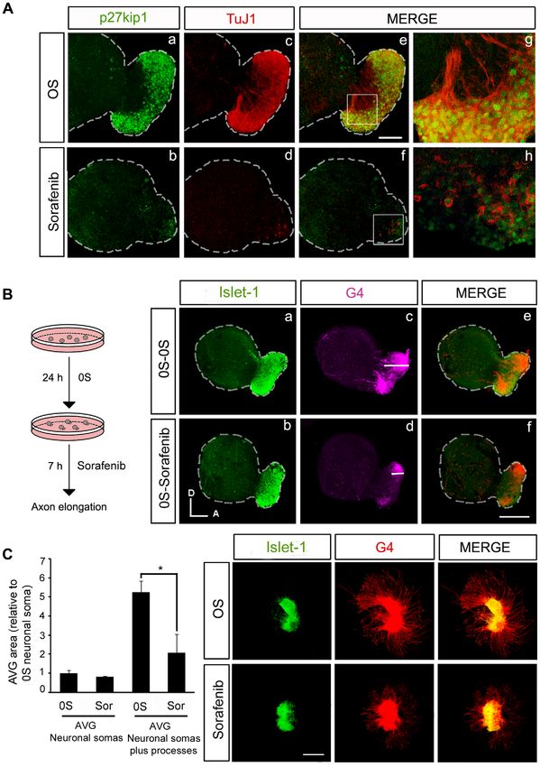
RAF in Inner Ear Neurogenesis

IGF-I deficit is associated to profound sensorineural deafness and           activate the RAF-MEK-ERK cascade in explanted otic vesicles
cochlear malformation in man and mice (MIM 147440) [10,11].                  and by blocking RAF kinases with Sorafenib, we show that RAF
IGF-I deficit in the mouse is associated with caspase-3-mediated             activity is essential for cell proliferation. By contrast, survival may
apoptosis of immature cochlear neurons [12] and with altered                 be recovered by IGF-I induction of the PI3K/Akt kinase pathway.
signaling pathways, including poor activation of Akt and ERK1/2,             Finally, our data show that the RAF-MEK-ERK cascade is an
and the up-regulation of p38 kinase pathways [13]. Cochlear                  important mediator of otic neuronal survival, migration and the
ganglion neurons have many immature traits including the                     outgrowth of neuronal processes.
aberrant expression of the MEF2A, MEF2D, SIX 6 and MASH1
transcription factors [13]. In the chicken inner ear, IGF-I drives           Materials and Methods
cellular programs that are important for specific events during otic
development, including proliferation, survival, metabolism and               Ethics Statement
differentiation [7]. Both IGF-I and its high affinity IGF1R                     All animals were handled in strict accordance with good animal
receptor are expressed during inner ear development [6].                     practice as defined in the European Council Directive (86/609/
Moreover, endogenous otic IGF-I activity is essential for the                EEC), and all animal work was approved by the Ethics Committee
survival and neurogenesis of otic precursors due to its activation of        of the UAM and the Bioethics Committee of the Consejo Superior de
the PI3K/Akt kinase pathway [6,14]. On the other hand,                       Investigaciones Cientificas.
exogenous IGF-I mimics morphogenetic traits in vivo, promoting
neurogenesis and axon sprouting, accelerating the rate of cell               Chicken embryos
proliferation and improving cell survival by inhibiting apoptosis of            Chicken embryos were obtained from fertilized eggs from a
both epithelial and neural progenitors [6]. IGF-I can activate the           local farm (Granja Santa Isabel, Cordoba, Spain) and they were
RAF-MEK-ERK cascade in the otic epithelium, and C-RAF is                     incubated in a humidified atmosphere at 37.8uC. Embryos were
essential for otic vesicle proliferation and morphogenesis [15].             staged as HH18, HH20, HH22, HH24, HH27 and HH34
However, it is not still fully clear how the strict balance between          according to Hamburger and Hamilton’s criteria [28].
signaling pathways is regulated by IGF-I during development.
   RAF kinases are serine/threonine kinases whose activity is                Isolation, organotypic culture and treatment of otic
modulated by growth factors and that play a central role in normal           vesicles and AVG
and pathologic cellular processes, including development, cell                  Embryos at stage HH18 (65 h of incubation) were obtained and
regeneration, cell senescence and cancer [16]. The first RAF                 the otic vesicles were dissected from the surrounding mesenchymal
kinase identified was the oncogenic product of mouse sarcoma                 tissue with sharpened tungsten needles, they were transferred into
virus 3611 [17] and since, the mammalian RAF kinases have been               four-well culture-plates (Nunc, Roskilde, Denmark) and then
shown to belong to a family that is formed by A-, B- and C-RAF.              incubated at 37uC in a water-saturated atmosphere containing 5%
In invertebrates only a single RAF kinase exists whereas the two             CO2, as described previously [7]. The standard culture medium
isoforms in birds are homologues of B- and C-RAF [18]. In                    consisted of M199 medium with Earle’s salts (Sigma-Aldrich, Saint
mammals, A-RAF is the less abundant kinase and it is expressed in            Louis, MO) supplemented with 2 mM glutamine (Gibco, Paisley,
the urogenital and gastrointestinal systems. By contrast, B-RAF is           UK) and antibiotics [50 IU/ml penicillin (Ern, Barcelona, Spain)
more abundant and it is found in the nervous system and gonads,              and 50 mg/ml streptomycin (CEPA, Madrid, Spain)]. AVG were
whereas C-RAF is ubiquitously expressed [19]. The study of                   obtained from stage HH19+ chicken embryos dissected out
knock-out mice lacking each of these kinases has revealed that they          aseptically and plated onto glass coverslips that had been
fulfill common and distinct functions [18,20], as well as shedding           previously coated with poly-D-lysine and fibronectin [5]. The
light on how they are regulated, their distinct intracellular                AVG was cultured in 0.25 ml F12/Dulbecco’s modified Eagle
localization [21] and association with scaffold proteins [22].               medium (Gibco) containing 100 mg/ml transferrin, 16 mg/ml
Indeed, B-RAF knock-out mice have defects in neural cell                     putrescine, 6ng/ml progesterone, 5.2 ng/ml sodium selenite (all
lineages, including reduced cell proliferation in the neocortex,             from Sigma), and antibiotics as above.
and impaired migration and dendrite formation associated with                   Explanted otic vesicles were treated with IGF-I (10 nM,
cortical neurons [20].                                                       Recombinant IGF-I Roche Molecular Biochemicals, Basel,
   RAF kinases transmit growth factor signals from the receptor/             Switzerland), various concentrations of Sorabenib (BAY 43-9006
RAS complex via the phosphorylation of MEK in the cytosol. This              1, 5 and 10 mM; Bayer HealthCare Pharmaceuticals, West Haven,
leads to the phosphorylation and activation of ERK that in turn,             CT, USA), the MEK inhibitor U0126 (50 mM; Promega,
can phosphorylate cytoplasmic and nuclear transcription factors              Madison, WI), the C-RAF inhibitor GW5074 (1 mM; Sigma-
that regulate gene expression and cellular responses. Bcl-2 family           Aldrich, Saint Louis, MO) the PI3K inhibitor LY294002 (25 mM;
members are targets of the RAF-MEK-ERK pathway [23], and                     Cell Signaling Boston, MA) or a pan-caspase inhibitor Boc-D-
therefore, RAF kinases are considered to be anti-apoptotic factors.          FMK (100 mM; Calbiochem La Jolla, CA) for the times indicated
The activation of the RAS-RAF-MEK-ERK cascade is essential                   in the text. The solvent used (DMSO) had no detectable effect on
for cellular proliferation during malignant transformation [16],             cultured otic vesicles when used at a final concentration of 0.01%
which has led to the synthesis of bi-aryl urea Sorafenib that inhibits       for LY294002, Boc-D-FMK and Sorafenib cultures, and 0.2% for
the catalytic activity of B-RAF and C-RAF and that also blocks               U0126 cultures. Otic vesicles cultured in medium without
proangiogenic-receptor-tyrosine kinases [24]. B-RAF is the most              additives were used as controls (0S). For immunostaining and
potent of the kinases that phosphorylates ERK [25] and it is                 TUNEL labeling otic vesicles were fixed for 2 h in 4% (w/v)
strongly expressed in the nervous system [26,27]. However, its               paraformaldehyde (Merck, Darmstadt, Germany) at 4uC. When
expression during inner ear development and the participation of             indicated, the otic vesicle and AVG areas were measured using
RAF kinases in AVG neurogenesis has not yet been explored.                   Image Analysis Software (Olympus, Tokyo, Japan). At least five
   Here we show that both B-RAF and C-RAF are present in the                 explants per condition were assayed from 2–6 independent
inner ear during its early development in vivo, and that B-RAF               experiments and the statistical significance was estimated using
expression becomes restricted as development proceeds. IGF-I can             the Student’s t-test.

       PLoS ONE | www.plosone.org                                        2                       December 2010 | Volume 5 | Issue 12 | e14435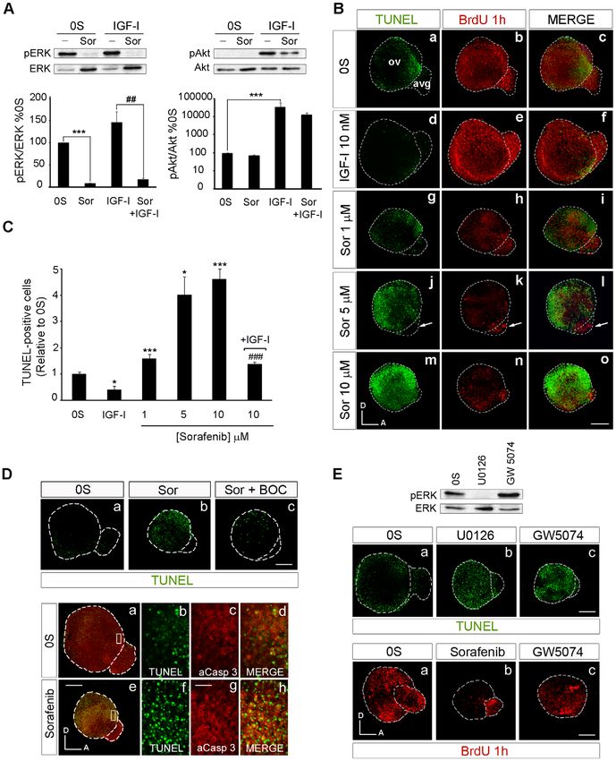
RAF in Inner Ear Neurogenesis

Quantitative RT-PCR                                                           antibodies were incubated for 2 h at room temperature. For dual-
   Inner ears from chicken embryos were pooled to obtain RNA at               fluorescence immunolabeling, otic vesicles were incubated with
different stages: HH18 (n = 40), HH22 (n = 25), HH24 (n = 20) and             Alexa Fluor 488 goat anti-mouse (1:200), Alexa Fluor 647 goat
HH27 (n = 10). Three independent RNA pools from each stage                    anti-rabbit and/or Alexa Fluor 546 goat anti-rabbit secondary
were isolated with Trizol (Invitrogen) following the manufacturer’s           antibodies (1:200; all from Molecular Probes, Eugene, OR).
instructions, and the integrity and concentration of the RNA was              TxRed-conjugated phalloidin was used to identify the apical actin-
assessed with an Agilent Bioanalyzer 2100 (Agilent Technologies).             containing structures of hair cells. Control experiments omitting
From this RNA, cDNA was generated by reverse transcription                    the primary antibody were carried out to confirm that the staining
(High Capacity cDNA Reverse Transcription Kit: Applied                        patterns were specific for antigen recognition and additionally
Biosystems). Real-Time PCR of each pool was performed in                      frozen sections from wild type or B-Raf2/2 null embryos were
triplicate using specific oligonucletides from ‘‘Quantitec Primer             included as negative controls (data not shown). The sections were
Assays’’ for chicken B-Raf and C-Raf (Gg_BRAF_1_SG                            mounted in Prolong Gold with DAPI (Invitrogen, Carlsbad, CA)
(QT01141413), Gg_RAF1_1_SG (QT00599123); Geneglobe, Qia-                      and visualized by fluorescence (Nikon 90i, Tokyo, Japan) or
gen) and using SYBR Green as the detection system. PCR was                    confocal microscopy (Leica TCS SP2, Wetzlar, Germany). For
performed on an Applied Biosystems 7900HT Real-Time PCR                       whole-mount immunofluorescence, otic vesicles were incubated
System using eukaryotic 18S rRNA as the endogenous housekeep-                 with the secondary antibodies for 3 h at room temperature, the
ing gene (Hs99999901_s1, TaqMan, Applied Biosystems). The                     otic vesicles were mounted in Vectashield with DAPI (Vector,
estimated gene expression was calculated as 22DDCt and statistical            Peterborough, UK) and the fluorescence was visualized by
significance was estimated using the Student’s t-test.                        confocal microscopy (Leica TCS SP2, Wetzlar, Germany). At
                                                                              least five to six otic vesicles/frozen sections were analyzed for each
Western blotting                                                              condition from at least two independent experiments.
   Otic vesicles (HH18) were isolated and cultured, 30 otic vesicles
from each condition were homogenized in ice cold Laemmli buffer               BrdU incorporation and immunodetection
with 50 mM dithiotreitol (DTT), Phosphatase Inhibitor Cocktail 2                 To study cell proliferation, otic vesicles were incubated with 5-
and Protease Inhibitor Cocktail (both 1:100, from Sigma-Aldrich).             Bromo-29-deoxyuridine (Sigma-Aldrich, Saint Louis, MO), a
The homogenized samples were heated at 95uC for 5 min and                     thymidine analogue that is incorporated into DNA during the S
frozen immediately. Gels were loaded with solutions containing                phase of the cell cycle. BrdU (10 mg/ml) was added to the culture
equal amounts of proteins and the otic vesicle protein extracts were          medium 1 h before the end of the incubation and its incorporation
resolved by SDS-PAGE on 8% or 12% polyacrylamide gels. The                    was detected with a specific antibody as above, but including a
proteins were transferred to nitrocellulose membranes and after               DNA denaturation step as recommended by the manufacturer
incubation with blocking solution (5% non-fat dry milk in TRIS-               (incubation in 50% (v/v) formamide-SSC, 40 minutes at 65uC and
buffered saline with 0.1% Tween-20: TBS-T), the membranes were                in HCl 2N, 30 minutes at 37uC, with a wash for 10 minutes in
probed overnight at 4uC with the appropriate specific primary                 Tris 0.1M [pH 8]). At least five to six otic vesicles were assayed per
antibodies to analyze the RAF kinases, pERK/ERK or pAkt/Akt                   condition in three independent experiments.
[14] (See Supplementary material Table S1). All antibodies were
diluted in blocking solution except anti-phospho-Akt antibody,                In situ hybridization
which was diluted in TBS-T and 5% bovine serum albumin (BSA:                     In situ hybridization was performed on cryostat sections (20 mM)
Sigma-Aldrich, Saint Louis, MO). The membranes were subse-                    of specimens from HH24 embryos essentially as described
quently washed and then incubated with the appropriate peroxi-                previously with only minor modifications [13]. Digoxigenin-
dase-conjugated secondary antibody (1:3000) for 1 h at RT.                    labeled sense and antisense RNA probes (1 mg/ml) were
Antibody binding was visualized by chemiluminiscence (GE                      hybridized overnight at 72uC and their binding was detected by
Healthcare, Buckinghamshire, UK) and exposed to X-ray film                    overnight incubation with an alkaline phosphatase-conjugated
(Konica Minolta, Wayne, NJ). The films were scanned and the                   anti-digoxigenin antibody (1:3500, Roche Applied Science), which
bands quantified by densitometry with Image J software (Wayne                 was visualised with 5-Bromo-4-chloro-3-indolyl phosphate, nitro
Rasband, National Institutes of Health, USA). At least three                  blue tetrazolium substrate (NTBT/BCIP, Roche Applied Science).
independent experiments were performed per condition and the                  The chicken C-Raf gene was amplified by PCR (C-Raf forward 59-
statistical significance was estimated using the Student’s t-test.            ACCTGCACGTTCAAGAGACC-39; C-Raf reverse 59-GCTAC-
                                                                              GAGCCTCTTCATTGC-39) and subsequently ligated into a
Inmunohistochemistry                                                          pGEM-T plasmid (Promega) to prepare the probe. Single-
   The sources, dilution, and cell specificities of the antibodies used       stranded sense (ApaI/T7) and antisense (PstI/Sp6) RNA probes
for immunofluorescent staining are shown in Supplementary                     were prepared by in vitro transcription and no specific signal was
material Table 1. Samples were washed and permeabilized in 1%                 obtained when control sense probes were used (data not shown).
PBS/Triton-X-100 (PBS-T), and they were exposed to the
primary antibodies overnight at 4uC. Non-specific binding sites               Analysis of programmed cell death
were blocked for 1 h in PBS-T, 3% (wt/vol) BSA (Sigma-Aldrich)                   The pattern of cell death in the otic vesicle was studied by Tdt-
and 5% (vol/vol) normal goat serum.                                           mediated dUTP nick-end labeling (TUNEL) of fragmented DNA
   For single immunostaining, sections were incubated for 2 h in a            using the kit Dead-EndTM Fluorometric TUNEL System (Promega,
biotinylated anti-rabbit secondary antibody (1:100, biotin-conju-             Madison, WI) essentially as described by the manufacturer and
gated anti-rabbit, Chemicon), processed with ExtrAvidin-peroxi-               adapted to whole organ labeling [14,29]. The otic vesicles were
dase conjugate solution (1:200, Sigma). Finally, antibody binding             mounted with Vectashield with DAPI (Vector) and visualized on a
was visualized using DAB as the chromogen and the sections                    confocal microscope (Leica, TCS SP2). TUNEL-positive cells
mounted in Mowiol for observation under a Nikon 90i microscope.               were counted using Image Analysis Software (Olympus, Tokyo,
   For immunofluorescent staining of frozen sections, the primary             Japan) attributing a value of 1 to the control condition (no
antibodies were used as described above and the secondary                     addition, 0S). At least five otic vesicles were assayed per condition

       PLoS ONE | www.plosone.org                                         3                       December 2010 | Volume 5 | Issue 12 | e14435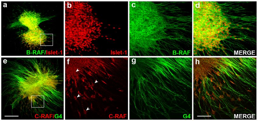
RAF in Inner Ear Neurogenesis

in three independents experiments. The data are presented as the                present in inner ear extracts from stage HH18 embryos, the stage at
mean 6 SEM and the statistical significance was estimated with                  which otic vesicles can be explanted and cultured ex vivo (Figure 1
the Student’s t-test.                                                           C). Indeed, there was a significant amount of phosphorylated ERK,
                                                                                a read out of RAF activity [30]. The presence of RAF kinases was
Results                                                                         further confirmed by immunohistochemistry for B-RAF and in situ
                                                                                hybridization for C-Raf (Figure 1 D) and the AVG was strongly
RAF kinases are expressed during early inner ear                                stained for both RAF kinases. In addition, B-RAF expression was
development                                                                     also evident in the otic epithelium (arrowheads in Figure 1 D).
   The expression of chicken B-Raf and C-Raf at selected stages of
otic development was studied by quantitative RT-PCR (Figure 1 A                 Spatiotemporal expression of B-RAF during inner ear
and B). The expression of transcripts encoding these RAF isoforms               development
was comparable from HH18 to HH24, although the RNA                                 In inner ear sections from HH24, HH27 and HH34 embryos,
transcripts for both these RAF kinases were strongly downregulated              the distribution of B-RAF was determined by immunohistochem-
at stage HH27. Moreover, B-RAF and C-RAF proteins were both                     istry (Figure 2). The different cell types labeled were identified by

Figure 1. Expression of the B-RAF and C-RAF kinases during otic development. (A) Schematic drawings showing the development of the
chicken inner ear at Hamburger and Hamilton stages HH18, HH24 and HH27. (B) Expression of inner ear B-Raf and C-Raf mRNA analyzed by qRT-PCR
at different stages using Eukaryotic 18S rRNA as the endogenous housekeeping control gene. Gene expression was calculated as 22DDCt and
normalized to the levels at HH18. The results are expressed as the mean 6 SEM of at least three independent experiments performed in triplicate.
Statistical significance was estimated with the Student’s t-test: ***P,0.005 versus HH18, ##P,0.01 versus HH24 and ###P,0.005 versus HH24. (C)
HH18 otic vesicle lysates analyzed in western blots to determine the levels of B-RAF, C-RAF and phosphorylated ERK (pERK). ß-Tubulin (ß-Tub) was
used as a loading control. A representative blot of three independent experiments is shown and the average densitometric measurements of the B-
RAF and C-RAF bands are plotted as bars. The results are given as the mean 6 SEM of three independent experiments. (D) Immunofluorescence of B-
RAF and in situ hybridization of C-Raf at HH22 and HH24, respectively showing their location in the otic epithelium and acoustic-vestibular ganglion
(arrowheads). Abbreviations: bp, basilar papilla; ed, endolymphatic duct; es, endolymphatic sac; lc, lateral crista; ml, macula lagena; ms, macula
sacculi; mu, macula utriculi; pc, posterior crista; sc, superior crista; ssc, superior semicircular canal. Orientation: C, caudal; D, dorsal; M, medial; R,
rostral.
doi:10.1371/journal.pone.0014435.g001

       PLoS ONE | www.plosone.org                                           4                         December 2010 | Volume 5 | Issue 12 | e14435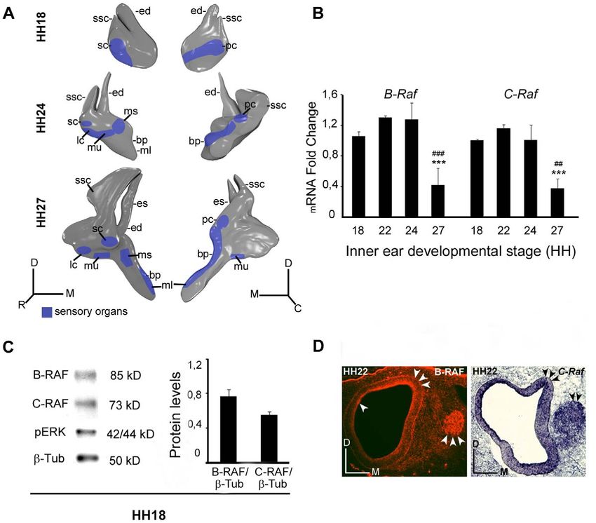
RAF in Inner Ear Neurogenesis

Figure 2. Spatiotemporal expression of B-RAF in the developing inner ear. a–h) At stage HH24, B-RAF (green throughout the figure) is
abundantly expressed in the macula sacculi (ms) and in the acoustic-vestibular ganglion (avg), which is labelled red due to the expression of the
axonal marker 3A10 (a–d). B-RAF is expressed strongly in the basilar papilla (bp, e, arrow). B-RAF and SOX2 (red), a transcription factor essential for
the self-renewal of undifferentiated otic progenitors, are expressed in non-overlapping regions of the avg (e–h). i–p) At HH27, B-RAF is expressed

       PLoS ONE | www.plosone.org                                         5                        December 2010 | Volume 5 | Issue 12 | e14435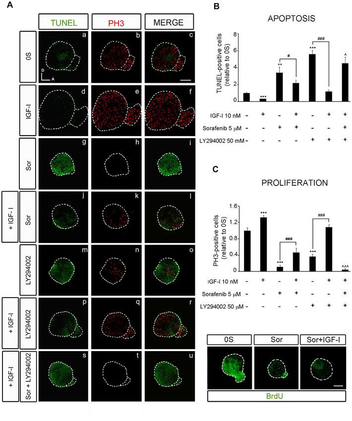
RAF in Inner Ear Neurogenesis

strongly in the internal cell layers of the basilar papilla (bp, i–l), whereas SOX2 is present in more external cell layers (m–p). q–w) At HH34, SOX2 is
expressed in supporting cells of the ms whereas B-RAF labels the hair cells (hc: q–t). TxRed-phalloidin staining (red) labels actin in the hc stereocilia of
the macula utriculi (mu), and B-RAF is evident in the cytoplasm (u–w). B-RAF is also expressed in the outer (OHC) and inner hair cells (IHC) of E18.5
mouse embryos (x). x9 shows a higher magnification of the sensory region in x. Schematic drawings of HH24 and HH27 inner ears are shown. The
boxed areas show higher magnifications of the selected regions. Abbreviations: avg, acoustic-vestibular ganglion; bp, basilar papilla; ed,
endolymphatic duct; es, endolymphatic sac; hc, hair cells; IHC, inner hair cells; OHC, outer hair cells; lc, lateral crista; ml, macula lagena; ms, macula
sacculi; mu, macula utriculi; sc, superior crista; ssc, superior semicircular canal; vp, vertical canal pouch. Orientation: D, dorsal; M, medial; R, rostral.
Scale bars: 100 mm.
doi:10.1371/journal.pone.0014435.g002

staining for the neurofilament-related 3A10 protein [6] and                      compare a with j and m). At the highest concentration of Sorafenib
SOX2, a transcription factor associated to immature pluripotent                  tested (10 mM), most cells in the otocyst were TUNEL positive
precursors. In mammals, SOX2 participates in the specification of                (Figure 3 B, m). It is worth noting, that Sorafenib induced less
the otic prosensory domain [31,32] and the generation of cochlear                programmed cell death in the AVG than in the otic vesicle
neurons [33], while it is required for hair cell survival and                    epithelium, even though the size of the AVG decreased dramatically
regeneration in the inner ear of the zebrafish [34]. B-RAF was                   (Figure 3 B, arrow in j). The cell death was caspase-dependent as it
expressed homogenously in the otic epithelia and in the AVG at                   was blocked by the pan-caspase inhibitor BOC (Figure 3 D, upper
HH18 and HH22 (Figure 1 and data not shown), while its                           panels, a-c, and compare b with c). Apoptosis was further studied by
expression became more restricted as development proceeded. At                   combining TUNEL staining with the immunodetection of active
HH24, B-RAF was abundantly expressed in the macula sacculi                       caspase-3 (Figure 3 D, lower panels, a–h). The Sorafenib-treated otic
(Figure 2 a–d) and it was also expressed in the AVG, where a                     vesicles showed areas of apoptotic cell death where TUNEL-labeled
subset of neuroblasts was more strongly stained for B-RAF                        apoptotic nuclei were surrounded by cytoplasm containing active
(Figure 2 e–h). B-RAF and SOX2 were expressed in adjacent                        caspase-3 (Figure 3 D, lower panels, e–h).
regions and B-RAF expression did not appear to overlap with that                    As RAF activation leads to cell proliferation, we assessed
of SOX2. A similar situation was also observed in the basilar                    bromodeoxyuridine (BrdU) uptake in cultured otic vesicles to
papilla where even though both proteins appeared to overlap in                   measure the rate of proliferation. IGF-I promoted otic prolifer-
some cells, the internal cells most strongly expressed B-RAF,                    ation when compared with control cultures (Figure 3 B, compare b
whereas SOX2 expression was observed more laterally (Figure 2,                   and e), while Sorafenib impaired BrdU incorporation in a dose
arrow in e). At HH27, B-RAF expression was restricted to the                     dependent manner. As a consequence, the size of Sorafenib-
internal layer of the otic epithelia and the cells of the AVG                    treated otic vesicles was severely reduced (Figure 3 B, b, h, k, n).
(Figure 2, i–p). At HH34, SOX2 was strongly expressed by                         AVG size was also reduced, even though cell proliferation at the
supporting cells, whereas B-RAF was expressed strongly in the                    AVG was not strongly affected by Sorafenib (Figure 3 B, arrow in
macula sacculi, limiting the region of SOX2 expression and                       k), suggesting that neuronal cells that escaped from the RAF
suggesting that B-RAF is expressed by hair cells (Figure 2, q–t). To             blockage are resistant to Sorafenib, possibly because RAF kinase
further explore the expression of B-RAF in hair cells, we used                   activity is no longer required.
TxRed-phalloidin to label the actin in the stereocilia of hair cells                To confirm that the effects observed were a consequence of the
(u, w). B-RAF expression was observed in the cytoplasm of both                   inhibition of the RAF-MEK-ERK cascade a MEK inhibitor [35],
auditory and vestibular hair cells, and in AVG neurons (Figure 2,                U0126, was used (Figure 3 E). The effects of blocking RAF catalytic
u–w, and data not shown). Specific hair cell expression of B-RAF                 activity with Sorafenib were emulated by U0126, which abolished
was confirmed in E18.5 wild type mice, as was the specificity B-                 ERK phosphorylation (Figure 3 E), reduced cell proliferation (data
RAF by labeling null B-Raf mouse embryos (Figure 2, x–x9:                        not shown) and increased apoptosis (Figure 3 E, upper panels,
Magariños, Rapp and Varela-Nieto, manuscript in preparation).                   compare a with b). In contrasts, treatment with the C-RAF highly
                                                                                 specific inhibitor GW5074 [36] showed reduced AVG size,
The activity of RAF kinases is required for the                                  undifferentiated-rounded shape OV with reduced size and
proliferation and survival of otic neuroepithelial cells                         increased TUNEL positive cells (Figure 3 E, upper panels, compare
   The RAF-MEK-ERK phosphorylation cascade can be specifically                   c with a), but not evident changes in cell proliferation (Figure 3 E,
inhibited by Sorafenib, an inhibitor of RAF kinase activity developed            lower panels, compare c with a). GW5074-treatment slightly
to treat B-RAF-associated cancer [16]. Otic vesicles were explanted              reduced BrdU incorporation when compared to the 0S condition,
and cultured ex vivo in the presence of Sorafenib to further                     whereas Sorafenib showed a more dramatic reduction on BrdU
understand the role of RAF activation in early inner ear development.            levels (lower panels in Figure 3 E, compare c with b). Furthermore,
This compound totally abolished ERK phosphorylation, both the                    treatment with GW5074 increased ERK phosphorylation (Figure 3
basal phosphorylation and that induced by IGF-I (Figure 3 A, left                E), suggesting that actions of C-RAF on apoptosis are, at least in
panels). The specificity of Sorafenib to the RAF-MEK-ERK cascade                 part, independent of the activation of MEK and ERK.
was witnessed by its failure to effect Akt phosphorylation, both basal              These results show that inhibition of the RAF-MEK-ERK
and IGF-I induced (right panels). Further insight into the actions of            cascade caused a reduction in the rate of cell proliferation and an
Sorafenib was obtained by studying its effects on cell proliferation and         increase in the number of caspase-dependent apoptotic cells,
apoptosis in organotypic cultures of explanted HH18 otic vesicles.               without affecting Akt activity, thereby leading to a decrease in the
When cultured otic vesicles were exposed to Sorafenib, cell                      number of neural progenitor cells.
proliferation was reduced and apoptosis was induced in a dose-
dependent manner (Figure 3 B). The number of apoptotic TUNEL                     The activity of RAF kinases is required for otic
positive cells found in the control otic vesicles (0S) was 2.5-fold higher       neurogenesis
than that found when IGF-I was added to the medium (Figure 3 C:                     To further explore the regulation by IGF-I and the role of the
[14]). However, the addition of increasing concentrations of                     RAF-MEK-ERK cascade in otic neurogenesis, the expression of
Sorafenib (1, 5 and 10 mM) significantly increased the number of                 the neuroblast markers Islet-1 and TuJ1 was analyzed in cultured
apoptotic cells by 1.8-, 4- and 4.6-fold, respectively (Figure 3 C and B,        otic vesicles treated with Sorafenib in the presence or absence of

       PLoS ONE | www.plosone.org                                            6                         December 2010 | Volume 5 | Issue 12 | e14435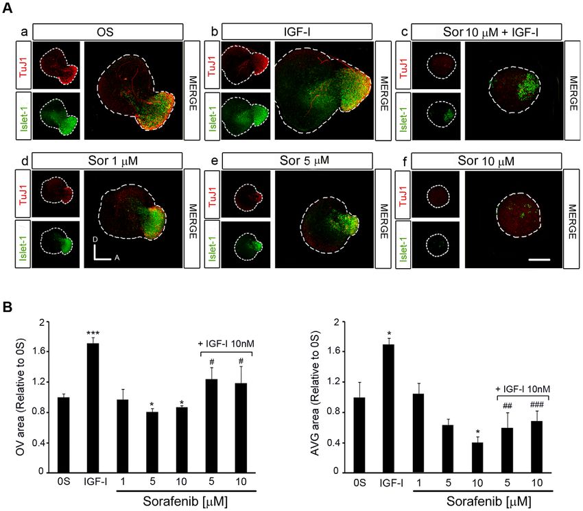
RAF in Inner Ear Neurogenesis

Figure 3. Selective inhibition of the RAF-MEK-ERK cascade blocks proliferation and promotes apoptosis. (A) Sorafenib inhibits the
RAF-MEK-ERK pathway. Otic vesicles were explanted from stage HH18 chicken embryos and incubated for 24 h in serum-free medium (0S). The
explants were then incubated for 1 h in serum-free medium without additives (0S), with IGF-I (10 nM), Sorafenib (Sor; 5 mM) or a combination of both
IGF-I and Sorafenib. Otic vesicles were lysed and the levels of phosphorylated and unphosphorylated ERK and Akt kinases were quantified in Western
blots by densitometry, as described in Materials and Methods. Representative blots are shown in the upper row. The results are expressed relative to
the control value (0S), which was given an arbitrary value of 100, as the mean 6 SEM of three independent experiments. Statistical significance was
estimated with the Student’s t-test: ***P,0.005 versus 0S and ##P,0.01 versus IGF-I. (B) Apoptosis and proliferation in Sorafenib-treated
cultures of otic vesicles. Apoptotic cell death was visualized by TUNEL (green) in cultured otic vesicles. Proliferation was measured by the

       PLoS ONE | www.plosone.org                                        7                       December 2010 | Volume 5 | Issue 12 | e14435
RAF in Inner Ear Neurogenesis
incorporation of BrdU (red) over 1 h. Otic vesicles were isolated from HH18 chicken embryos, made quiescent and cultured for 24 h in serum-free
culture medium without additives (0S), with IGF-I (10 nM), Sorafenib (Sor; 1, 5 or 10 mM) or a combination of both IGF-I and Sorafenib. Scale bars,
150 mm. (C) Cell death quantification of B. The TUNEL positive nuclei were quantified relative to the 0S condition, which was given an arbitrary
value of 1. The bars show the mean 6 SEM of at least five otic vesicles from any of the conditions shown in B. Statistical significance was estimated
with the Student’s t-test: *P,0.05 versus control, ***P,0.005 versus control, ###P,0.005 versus Sorafenib 10 mM. (D) Sorafenib increases cell
death through a caspase-dependent mechanism. Otic vesicles were isolated from HH18 chicken embryos and cultured for 24 h in serum-free
culture medium without stimuli (0S; a upper panel), or cultured in the presence of Sorafenib 5 mM (Sor; b upper panel) or in combination with the
pan-caspase inhibitor Boc-D-FMK 50 mM (Sor+BOC; c upper panel) and cell death was visualized using the TUNEL technique. Lower panel shows
apoptotic cell death visualized by TUNEL staining (green) and immunostaining for activated-caspase-3 (red) of otic vesicles cultured in free serum (0S,
a–d) or in the presence of Sorafenib 5 mM (e–h). Boxed areas in a and e are shown at a higher magnification to show the TUNEL-positive nuclei (b,f
and merge) surrounded by activated caspase-3 (c,g and merge). Scale bar, 150 mm (a,e); 20 mm (b–d and f–h). (E) Treatment of cultured otic
vesicles with the MEK inhibitor U0126 and with the C-RAF inhibitor GW5074. A representative blot of the effects of U0126 (50 mM) and
GW5074 (1 mM) on ERK phosphorylation is shown. Apoptosis in cultured otic vesicles was visualized with TUNEL (upper panels, a–c). Proliferation was
measured by the incorporation of BrdU (red) over 1 h in otocysts cultured with no additives (0S), with Sorafenib (5 mM) or with GW5074 (1 mM) (lower
panels, a–c). Scale bar: 150 mm. Compiled projections of confocal microscopy images from otic vesicles are shown. A, anterior; D, dorsal.
Abbreviations: AVG, acoustic-vestibular ganglion; OV, otic vesicle. The images shown are representative of at least three independent experiments,
using five to six otic vesicles per condition.
doi:10.1371/journal.pone.0014435.g003
IGF-I [Figure 4, A; [6]]. The expression of Islet-1 and TuJ1 was otic vesicles was similar to that of the controls (Figure 5 A, p–r, and
reduced in a dose-dependent manner in the presence of Sorafenib, bars in B and C). In combination, Sorafenib and LY294002
indicating that inactivation of the RAF kinase caused a loss of completely abolished the effects of IGF-I on proliferation and
neuroblasts (Figure 4 A, compare a with d, e, f). Indeed, in otocysts apoptosis (Figure 5 A, s–u). TUNEL staining was stronger in
exposed to Sorafenib, and hence with impaired RAF catalytic vesicles exposed to Sorafenib and LY294002 than in those treated
activity, there was a dose-dependent 20 and 40% reduction in the with Sorafenib alone (1.3-fold) or those exposed to Sorafenib and
otic vesicle epithelia and AVG size, respectively (Figure 4 A) when IGF-I (2.0-fold: Figure 5 B). The rate of proliferation was also
compared to otocysts cultured under control conditions (Figure 4 reduced 3-fold in the presence of Sorafenib plus LY294002 when
B). The impact of RAF inactivation on the proliferation of otic compared to Sorafenib alone, and 12-fold when compared to
precursors was further confirmed by studying the number of cells Sorafenib plus IGF-I (Figure 5 C). These results indicate that the
in mitosis and the incorporation of BrdU (Figure 5 A, C). protective effects of IGF-I on survival when the RAF-MEK-ERK
IGF-I promoted cell proliferation in the AVG neuroblast cascade is inhibited are mediated by the induction of the PI3K/
population, as witnessed by the increased number of Islet-1 positive Akt kinase pathway. In the presence of IGF-I, Sorafenib did not
cells and the 160% increase of the AVG area (Figure 4 A, compare a completely abolish mitosis (Figure 5 C, quantification of PH3
with b; Figure 4 B, bars on the right). TuJ1 labels neuroblasts at a positive cells) but it did abolish the incorporation of BrdU (Figure 5
more mature stage [4] and hence, TuJ1 positive cells are diminished C, lower panels), suggesting that IGF-I can partially overcome the
or unaffected by the addition of IGF-I (Figure 4 A, compare a with b effects of RAF inhibition. These data possibly reflect the capacity
and c). IGF-I also promoted cell proliferation in the otic epithelium of IGF-I to sustain progenitors that are already in the M-phase of
(Figure 5 A, compare b with e). In the presence of IGF-I, the number the cell cycle but not to promote cell cycle entry.
of Islet-1 positive cells and mitosis increased in Sorafenib-treated otic
vesicles (Figure 4 A, compare c with f), while apoptosis was reduced RAF activity is required for neuronal progenitor cell
(Figure 5 A–C), leading to a small but significant recovery of the OV differentiation and the outgrowth of processes from
and AVG (Figure 4 B). Because Sorafenib completely blocks IGF-I sensory otic neurons
activation of the RAF-MEK-ERK pathway (Figure 3), these results B-RAF has previously been reported to play a role in sensory
suggest that the rescue of the neuroblast population by IGF-I is axon and dendrite growth. Figure 6 shows B-RAF and C-RAF
mediated by an alternative pathway. expression in explanted AVG cultured in serum-free medium. B-
RAF was highly expressed in the cytoplasm and in the neural
IGF-I-induction of the PI3K/Akt kinase pathway rescues processes (Figure 6, a–d), whereas C-RAF showed a more
otic progenitors from apoptosis restricted cytoplasmic expression (Figure 6, e–h, arrowheads). To
IGF-I activates the Akt pathway even in the presence of study the role of RAF kinases in neural process outgrowth we
Sorafenib and the total inactivation of the RAF-MEK-ERK examined the differentiation state of AVG cultured explants.
cascade (Figure 3). To further define the roles of the RAF-MEK- Postmitotic otic neurons were identified by labeling with the
ERK and PI3K/Akt kinase pathways in otic neurogenesis, we nuclear cyclin-dependent kinase inhibitor p27kip1 [13] and with
studied the proliferation and apoptosis of explanted otic vesicles TuJ1, a neural tubulin that is found in processes (Figure 7 A).
treated with combinations of RAF and Akt inhibitors in culture. AVG neurons that have exited the cell cycle and that are located
Cell death was studied by detecting the TUNEL positive cells more distally with respect to the neurogenic zone of the otic vesicle
(TUNEL) and cell proliferation was detected by following the epithelium expressed p27kip1 (Figure 7 A, a), and their TuJ1
nuclei labeled with phospho-histone-3 (PH3), which identifies cells staining indicated that they had begun to extend axons towards
in the M phase of the cell cycle (Figure 5 A), or by BrdU the otic vesicle (Figure 7 A, c, e, g). Sorafenib treatment of cultured
incorporation (Figure 5 C). otic vesicles reduced the number of neuroblasts and mature
LY294002 is a well-characterized inhibitor of the PI3K/Akt neurons (Figure 7 A, compare a and b) and more interestingly,
kinase pathway that specifically impairs Akt phosphorylation [37]. these mature neurons did not develop axons since their TuJ1
Inhibition of the PI3K/Akt kinase pathway increased the number expression remained surrounding the cytoplasm (Figure 7 A, d, f,
of apoptotic cells (5.5-fold) and reduced the amount of cells h). These results suggested that RAF-MEK-ERK signaling is
expressing the mitotic marker PH3 (0.7-fold: Figure 5 A, m–o, and necessary to initiate axonal growth.
bars in B and C). The presence of IGF-I partially impaired the To determine whether RAF activity is required for axonal
effects of LY294002 as the number of proliferating cells in treated growth once differentiation has been initiated, axonal growth was
PLoS ONE | www.plosone.org 8 December 2010 | Volume 5 | Issue 12 | e14435RAF in Inner Ear Neurogenesis
Figure 4. Inhibition of the RAF-MEK-ERK cascade impairs AVG formation. (A) Otic vesicles were isolated from HH18 chicken embryos and
incubated for 24 h in serum-free culture medium without additives (0S), with IGF-I (10 nM), Sorafenib, (Sor;1, 5 or 10 mM) or a combination of Sor
(10 mM) and IGF-I. Whole otic vesicles were then immunostained for the ganglion neuroblast nuclei marker Islet-1 (green) and for the marker of neural
processes, TuJ1 (red). Fluorescence images were obtained from the compiled projections of confocal images of otic vesicles. Representative images
of at least five to six otic vesicles per condition and from at least three independent experiments are shown. Orientation: A, anterior; D, dorsal. Scale
bar: 150 mm. (B) The otic vesicles (OV) and the acoustic-vestibular ganglia (AVG) areas were measured with Image Analysis Software (Olympus, Tokyo,
Japan). The data are expressed as the mean 6 SEM relative to the control value (0S) and they were compiled from the analysis of at least five to six
otic vesicles per condition. Statistical significance was estimated with the Student’s t-test: *P,0.05, ***P,0.005 versus 0S; #P,0.05, ##P,0.01 and
###
P,0.005 versus IGF-I.
doi:10.1371/journal.pone.0014435.g004
followed with the immature neuroblast marker Islet-1 and the neuronal differentiation. Again, exposure to Sorafenib (2.5 mM)
axonal glycoprotein G4 [6]. Otic vesicles were cultured for 24 h to caused a dramatic reduction in the number and length of processes
allow AVG formation, and the elongation of the processes was (Figure 7 C), despite affecting the size of the AVG. When the areas
allowed to continue for 7 h in the presence (0S- Sorafenib) or of the AVG with neuronal soma alone or plus processes were
absence of Sorafenib (0S-0S: Figure 7, B). After 24 h, mature otic quantified it was confirmed that RAF activity is required for otic
neurons have already started to extend axons to innervate the neuron maturation and the outgrowth of processes.
dorsal (vestibular) and ventral (auditory) sensory epithelia (Figure 7
B, c and e). However, subsequent exposure to Sorafenib impaired Discussion
this process and the axons remained shorter than those of controls
(Figure 7 B, compare white bar in c with that in d), both in the otic Inner ear organogenesis requires strict spatial and temporal
vesicle and AVG areas (Figure 7 B, compare a and e with b and f). regulation of cellular proliferation, death and differentiation to
Similar experiments were then performed on isolated explanted generate the appropriate number of different cell types and their
AVG to further study the role of RAF-MEK-ERK signaling in otic interconnections [2]. IGF-I drives cell proliferation and the
PLoS ONE | www.plosone.org 9 December 2010 | Volume 5 | Issue 12 | e14435RAF in Inner Ear Neurogenesis
Figure 5. IGF-I partially rescues the effects of inhibiting RAF activity through the PI3K/Akt kinase pathway. (A) Apoptotic cell death
was visualized by TUNEL (green) in cultured otic vesicles and proliferation was detected with the mitosis marker Phospho-Histone 3 (PH3, red). Otic
vesicles were isolated from HH18 chicken embryos and cultured for 24 h in serum-free medium without additives (0S, a–c), with IGF-I (10 nM, d–f),
Sorafenib, Sor, (5 mM, g–i), LY294002 (50 mM, m–o), a combination of IGF-I and Sor (j–l), IGF-I and LY294002 (p–r) or IGF-I, Sor and LY294002 (s–u).
(B) TUNEL positive or (C) proliferative PH3-labeled cells were quantified as described in at least 5 otic vesicles per condition. The results are shown as
the mean 6 SEM relative to the 0S condition. Statistical significance was estimated with the Student’s t-test: *P,0.05, ***P,0.005 versus 0S; #P,0.05
and ###P,0.005 versus the indicated inhibitors; ‘P,0.05, ‘‘‘P,0.005 versus Sorafenib +IGF-I. Lower panels in C show BrdU (green) incorporation
into cultured otic vesicles incubated for 24 h in the following conditions: 0S, with Sor (5 mM), or a combination of Sor and IGF-I (10 nM). Compiled
projections of confocal images from otic vesicles are shown, and are representative of at least five to six otic vesicles per condition from three
different experiments. Scale bar, 150 mm.
doi:10.1371/journal.pone.0014435.g005
PLoS ONE | www.plosone.org 10 December 2010 | Volume 5 | Issue 12 | e14435RAF in Inner Ear Neurogenesis
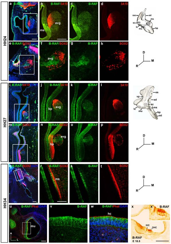
Figure 6. RAF proteins show different subcellular distribution in the acoustic- vestibular ganglion. AVG explants were obtained from
stage HH19 chicken embryos and cultured in serum-free medium for 20 h with no additives (0S). (a–d) Whole AVG explants were immunostained for
B-RAF (green) and Islet-1 (red) or (e–h) for C-RAF (red) and G4 (green), The cytoplasmatic distribution of C-RAF is shown (arrowheads). Fluorescence
images were obtained from compiled projections of confocal images of AVG. Scale bar, 350 mm (a, e); 75 mm (b–d, f–h).
doi:10.1371/journal.pone.0014435.g006
survival of otic progenitors, and it is essential for neuronal are down-regulated and the strongest B-RAF expression is
differentiation in the time window between neuronal cell fate associated to sensory hair and neuronal cells. Indeed, RAF kinases
specification and neurotrophin dependence [4]. RAF proteins are participate in late differentiation processes of other cell types such
serine/threonine kinases that regulate the RAF-MEK-ERK as T-cells [41] and sensory cells [42], as well as in post-
signaling pathway involved in the transduction of extracellular differentiation events, such as cortical neuron migration [20] or
stimuli into cellular responses [16]. Three RAF kinase isoforms the modulation of synaptic plasticity [43].
exist in mammals, A-, B- and C-RAF, whose activity is exquisitely Due to the established role of RAF kinases in cancer, the search
regulated at the post-transcriptional level by a number of different for inhibitors of RAF-MEK-ERK signaling has been intense [16].
mechanisms [38]. C-RAF activation is regulated by IGF-I and it is Sorafenib 43-9006 is a potent small-molecule that inhibits RAF
essential for cell proliferation in the otic vesicle [15]. However, no kinases, and its use has been approved for renal carcinoma
studies have been conducted on B-RAF even though this isoform is therapy, as well as in clinical trials for melanoma and thyroid
more active as a protein kinase and more abundantly expressed in cancer [44]. Sorafenib was primarily identified as a C-RAF
the nervous system, where it is fundamental for axonal and inhibitor but upon further characterization, it was shown to inhibit
dendrite growth [25,27,39,40]. B-RAF and other kinases involved in angiogenesis [24]. We have
Here we show that B-RAF and C-RAF transcripts are expressed used Sorafenib to study the influence of RAF kinases on otic
during inner ear development in a specific spatiotemporal pattern progenitors and their regulation by IGF-I. Our results show that
and that B-RAF is expressed specifically in neurosensorial Sorafenib effectively inhibits basal and IGF-I induced ERK
components of the inner ear at later stages of development. phosphorylation, without affecting Akt phosphorylation. Inhibition
RAF transcripts are translated into proteins that phosphorylate of RAF catalytic activity by Sorafenib also caused an increase in
ERK, and RAF activity is regulated in the inner ear by IGF-I. caspase-dependent apoptosis. Interestingly, in some melanoma cell
Such RAF-MEK-ERK signaling is required for neuroepithelial lines this is not the case and Sorafenib-dependent apoptosis is
cell proliferation and survival in this structure, although IGF-I can caspase independent [45]. RAF activation of mitochondrial targets
restore cell survival by activating the PI3K/Akt kinase pathway such as BAD [21,46], ASK-1 [47], and MST-2 [48] has an anti-
when RAF-MEK-ERK is inhibited by Sorafenib. However, IGF-I apoptotic effect and therefore, RAF inactivation can provoke cell
it is not able to restore cell proliferation in these conditions. death. RAF inactivation also annuls the activity of the MEK-ERK
Finally, we demonstrate that RAF kinase activity is required for module, which along with the lack of activation of cell cycle
neuronal progenitor cell differentiation and for the outgrowth of proteins and transcription factors, such as retinoblastoma [49],
sensory otic neuron processes. Cdc25 [50] or AP1 [51,52], may also cause apoptosis of
B-RAF and C-RAF are expressed in a similar temporal pattern proliferating cells. MEK inhibition by U0126 [35] and C-RAF
from HH18 to HH27, when the striking reduction in both inhibition by GW5074 [36] also caused apoptosis but presented
transcripts suggests that the transcription of both RAF isoforms is different traits. The MEK inhibitor completely abolished ERK
developmentally regulated. Up to stage HH24 these kinases are phosporylation and, in contrast, GW5074 induced it. Paradoxical
expressed homogeneously in the otic epithelia and AVG, and actions of C-RAF inhibitors have been reported in other neuronal
during these stages RAF kinases support the basic cellular contexts [53,54] and elimination of C-RAF activity by either
programmes of otic progenitors. As development proceeds, the knocking it out or siRNA targeting did not altered ERK
activity of RAF kinases may be more directly related to phosphorylation [55,56]. Therefore, RAF quinases inactivation
differentiation and post-differentiation events, and accordingly could promote apoptosis of otic progenitor cells by both ERK
they become more spatially restricted. At HH27 when auditory dependent and independent mechanisms, as reported in other cell
and vestibular hair cells have just differentiated, both RAF kinases types [57].
PLoS ONE | www.plosone.org 11 December 2010 | Volume 5 | Issue 12 | e14435RAF in Inner Ear Neurogenesis
Figure 7. RAF kinase activity is required for the correct outgrowth of sensory otic neuron processes. (A) Otic vesicles were isolated from
HH18 chicken embryos and incubated for 24 h either in serum-free medium without additives (0S, a,c,e,g) or in the presence of Sorafenib (2.5 mM)
(b,d,f,h). Immunohistochemistry of whole otic vesicles was carried out by double-staining for the nuclear cyclin-dependent kinase inhibitor p27kip1
(green) and for the marker of neural processes, TuJ1 (red). The boxed areas in panels e and f, correspond to the enlarged images in panels g and h
respectively. Scale bar: 75 mm. (B) Otic vesicles were isolated from HH18 chicken embryos and incubated for 24 h in serum-free medium as in the 0S
condition in A, and they were then incubated for a further 7 h without additives (0S-0S, a,c,e) or with Sorafenib (2.5 mM: 0S-Sorafenib, b,d,f). Whole
PLoS ONE | www.plosone.org 12 December 2010 | Volume 5 | Issue 12 | e14435RAF in Inner Ear Neurogenesis
otic vesicles were immunostained for the ganglion neuroblast nuclei marker, Islet-1 (green), and for the G4-glycoprotein marker of neuronal
processes (G4, magenta). Note the differences in the magnitude of the white bars in the region in the acoustic-vestibular ganglia corresponding to
the staining of neural processes in panels c and d. Scale bar: 150 mm. (C) Acoustic-vestibular ganglia (AVG) explants were obtained from stage HH19
chicken embryos and cultured in serum-free medium for 20 h with no additives (0S) or with Sorafenib (2.5 mM). Whole AVG explants were
immunostained for G4 (red) and Islet-1 (green). Sorafenib-treated AVG have shorter processes. Scale bar: 300 mm. Fluorescence images were obtained
from compiled projections of confocal images of otic vesicles and AVG. Bar graph on the left shows the quantification of the neuronal soma area in
the AVG, which does not vary following Sorafenib treatment. In contrast, there is a statistically significant difference in the area of the AVG covered by
processes (*P,0.05, Sorafenib versus 0S). Representative images of three independent experiments using five to six otic vesicles or AVG per condition
are shown. Orientation: A, anterior; D, dorsal.
doi:10.1371/journal.pone.0014435.g007
The inactivation of the RAF pathway with Sorafenib also observation suggested that the RAF-MEK-ERK cascade may be
produced a dramatic decrease in cell proliferation since the RAF- involved in differentiation of neural cells in the AVG, as seen in the
MEK-ERK cascade plays a fundamental role in the G1/S differentiation of cortical and dorsal neurons [42,40]. Otic neuronal
transition, where its signaling induces cyclin D1 and down-regulates identity and axonal growth are determined by various factors once
many other antiproliferative genes [52]. Accordingly, exposing the cell has exited the cell cycle [4]. Axonal growth in post-mitotic
cultured otic vesicles to Sorafenib caused a dose-dependent decrease p27kip1 positive AVG neurons was almost completely inhibited in
of BrdU incorporation at S phase. U0126 treatment also reduced the presence of Sorafenib, indicating that RAF kinase activation
proliferation in the otic vesicle; however, the specific inhibition of C- plays a fundamental role in the late differentiation of otic neurons.
RAF with GW5074 did not show the striking reduction on In summary, we show here that B-RAF and C-RAF are
proliferation observed with the other inhibitors. In mice, of the expressed during chicken inner ear development in specific
three RAF isoforms, C-RAF appears to be preferentially involved in spatiotemporal patterns, and that RAF-MEK-ERK signaling is
promoting survival, rather than controlling proliferation [55,58]. In required for neuroepithelial cell proliferation and otic neuronal
contrast, B-RAF is essential for ERK activation [25] that in turn differentiation.
triggers cell proliferation [59]. These data suggest that RAF kinases
also have distinct roles during chicken inner ear development. C- Supporting Information
RAF would preferentially promote anti-apoptotic signaling whilst B-
RAF, through the MEK-ERK module, would modulate prolifer- Table S1 Primary antibodies. 1: Antibody type: RbP, rabbit
ation of neuroepithelial progenitors. polyclonal; MouM mouse monoclonal; GtP goat polyclonal. 2:
Experiments with IGF-I showed that RAF activity is essential for Technique: IHF, Immunohistofluorescence. WB, Western Blotting
the progression of cell proliferation but not for cell survival. Indeed, 3: Monoclonal antibody developed by Thomas Jessell and Jane
IGF-I was even able to rescue otic progenitors by activating the Dodd were obtained from the Developmental Studies Hybridoma
PI3K/Akt pathway in the presence of Sorafenib. Blockage of the Bank developed under the auspices of the NICHD and maintained
PI3K/Akt kinase pathway with LY294002 indicated that IGF-I is by the University of Iowa, Department of Biological Sciences,
dependent on Akt activation for cell survival. Therefore, IGF-I Iowa City, IA 52242.
orchestrates cell proliferation and survival in the otic vesicle through Found at: doi:10.1371/journal.pone.0014435.s001 (0.04 MB
distinct pathways, although cross-talk between signaling pathways DOC)
also occurs, as reported in other cell contexts [60].
The AVG is generated from a pool of neuroepithelial progenitors Acknowledgments
that when specified in the otic vesicle epithelia, migrate from the
neurogenic zone and form the ganglia [4]. C-RAF and B-RAF are We appreciate the useful comments of Dr Guadalupe Camarero and
Lourdes Rodriguez de la Rosa. We thank Ricardo Ramos (Genomic Unit,
expressed in otic neurons but they exhibit distinct subcellular PCM, Madrid), Ricardo Uña (Image Unit, IIB, Madrid) for their technical
distribution, as reported in the rat brain [43]. B-RAF is abundantly support and Jose Sanchez-Calderon for the schematic drawings of the
expressed in cell bodies and neuronal processes, while C-RAF developing inner ear. The anti-3A10 and anti-Islet-1 monoclonal
expression is more restricted to the cytoplasmic compartment. antibodies were developed by Drs T.M. Jessel and J. Dodd and they were
Exposure to Sorafenib caused a dramatic decrease in the area of the obtained from the Developmental Studies Hybridoma Bank, maintained at
AVG, although inhibiting RAF kinase activity did not appear to the Iowa University, Department of Biological Sciences.
affect the population of neuronal cells in the AVG, which continued
to proliferate and exhibited little apoptosis. These data suggest that Author Contributions
mature neurons do not require RAF kinases for survival but that Conceived and designed the experiments: MM MRA HSC IVN.
RAF activity is essential for early neurogenesis. Accordingly, Performed the experiments: MM MRA CMA. Analyzed the data: MM
Sorafenib caused a clear reduction of mature neurons as indicated MRA HSC IVN. Contributed reagents/materials/analysis tools: URR
by the reduced levels of axonal outgrowth markers. This IVN. Wrote the paper: MM URR IVN.
References
1. Kelley MW (2006) Regulation of cell fate in the sensory epithelia of the inner 6. Camarero G, Leon Y, Gorospe I, De Pablo F, Alsina B, et al. (2003) Insulin-like
ear. Nat Rev Neurosci 7: 837–849. growth factor 1 is required for survival of transit-amplifying neuroblasts and
2. Rubel EW, Fritzsch B (2002) Auditory system development: primary auditory differentiation of otic neurons. Dev Biol 262: 242–253.
neurons and their targets. Annu Rev Neurosci 25: 51–101. 7. León Y, Vazquez E, Sanz C, Vega JA, Mato JM, et al. (1995) Insulin-like growth
3. León Y, Sánchez-Galiano S, Gorospe I (2004) Programmed cell death in the factor-I regulates cell proliferation in the developing inner ear, activating
development of the vertebrate inner ear. Apoptosis 9: 255–264. glycosyl-phosphatidylinositol hydrolysis and Fos expression. Endocrinology 136:
4. Sanchez-Calderon H, Milo M, Leon Y, Varela-Nieto I (2007) A network of 3494–3503.
growth and transcription factors controls neuronal differentation and survival in 8. Ladher RK, Wright TJ, Moon AM, Mansour SL, Schoenwolf GC (2005) FGF8
the developing ear. Int J Dev Biol 51: 557–570. initiates inner ear induction in chick and mouse. Genes Dev 19: 603–613.
5. Davies D (2007) Temporal and spatial regulation of alpha6 integrin expression 9. Sienknecht UJ, Fekete DM (2009) Mapping of Wnt, frizzled, and Wnt inhibitor
during the development of the cochlear-vestibular ganglion. J Comp Neurol 502: gene expression domains in the avian otic primordium. J Comp Neurol 517:
673–682. 751–764.
PLoS ONE | www.plosone.org 13 December 2010 | Volume 5 | Issue 12 | e14435You can also read