Covalent linkage of the DNA repair template to the CRISPR-Cas9 nuclease enhances homology-directed repair - eLife
←
→
Page content transcription
If your browser does not render page correctly, please read the page content below
SHORT REPORT

                                    Covalent linkage of the DNA repair
                                    template to the CRISPR-Cas9 nuclease
                                    enhances homology-directed repair
                                    Natasa Savic1†, Femke CAS Ringnalda1†, Helen Lindsay2,3, Christian Berk4,
                                    Katja Bargsten5, Yizhou Li4, Dario Neri4, Mark D Robinson2,3, Constance Ciaudo1,
                                    Jonathan Hall4, Martin Jinek5, Gerald Schwank1*
                                    1
                                     The Institute of Molecular Health Sciences, ETH Zurich, Zurich, Switzerland; 2The
                                    Institute of Molecular Life Sciences, University of Zurich, Zurich, Switzerland; 3SIB
                                    Swiss Institute of Bioinformatics, Zurich, Switzerland; 4Institute for Pharmaceutical
                                    Sciences, ETH Zurich, Zurich, Switzerland; 5Department of Biochemistry, University
                                    of Zurich, Zurich, Switzerland

                                    Abstract The CRISPR-Cas9 targeted nuclease technology allows the insertion of genetic
                                    modifications with single base-pair precision. The preference of mammalian cells to repair Cas9-
                                    induced DNA double-strand breaks via error-prone end-joining pathways rather than via homology-
                                    directed repair mechanisms, however, leads to relatively low rates of precise editing from donor
                                    DNA. Here we show that spatial and temporal co-localization of the donor template and Cas9 via
                                    covalent linkage increases the correction rates up to 24-fold, and demonstrate that the effect is
                                    mainly caused by an increase of donor template concentration in the nucleus. Enhanced correction
                                    rates were observed in multiple cell types and on different genomic loci, suggesting that covalently
                                    linking the donor template to the Cas9 complex provides advantages for clinical applications where
*For correspondence:                high-fidelity repair is desired.
schwankg@ethz.ch                    DOI: https://doi.org/10.7554/eLife.33761.001
†
 These authors contributed
equally to this work

Competing interests: The            Introduction
authors declare that no
                                    The CRISPR-Cas9 system is a versatile genome-editing tool that enables the introduction of site-spe-
competing interests exist.
                                    cific genetic modifications (Jinek et al., 2012). In its most widespread variant a programmable chi-
Funding: See page 14                meric single guide RNA (sgRNA) directs the Cas9 nuclease to the genomic region of interest, where
Received: 23 November 2017          it generates a site-specific DNA double-strand break (DSB) (Mali et al., 2013). In mammalian cells
Accepted: 26 May 2018               the repair of DSBs by different end-joining (EJ) pathways, such as classical non-homologous end join-
Published: 29 May 2018              ing (c-NHEJ), alternative non-homologous end-joining (a-NHEJ), or single-strand annealing (SSA)
                                    often leads to the formation of insertions or deletions (indels) (Shalem et al., 2014; Ceccaldi et al.,
Reviewing editor: Bernard de
                                    2016). Alternatively, when a donor template is provided, mammalian cells can also resolve DSBs via
Massy, Institute of Human
Genetics, CNRS UPR 1142,
                                    homology-directed repair (HDR) mechanisms, such as the classical homologous recombination (HR)
France                              pathway (Mao et al., 2008) and the Fanconi Anemia (FA) repair pathway (Richardson, 2017). While
                                    the formation of indels allows the elimination of gene function, repair from an ectopic donor oliogo-
   Copyright Savic et al. This
                                    nucleotide (oligo) via HDR mechanisms enables the introduction of DNA modifications with single
article is distributed under the
                                    base pair precision (van den Bosch et al., 2002).
terms of the Creative Commons
Attribution License, which              Therapeutic applications of CRISPR-Cas9 generally require the precise correction of pathogenic
permits unrestricted use and        mutations using donor templates. However, DSBs introduced in mammalian cells are predominantly
redistribution provided that the    repaired by error-prone EJ pathways. As the resulting indels inhibit the CRISPR-Cas9 complex from
original author and source are      retargeting the locus, error-prone repair indirectly competes with HDR, and therefore reduces the
credited.                           rates of precise correction from donor templates. Furthermore, if the targeted allele is a hypomorph

Savic et al. eLife 2018;7:e33761. DOI: https://doi.org/10.7554/eLife.33761                                                          1 of 18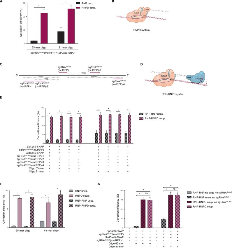
Short report                                                          Chromosomes and Gene Expression Human Biology and Medicine

                                    eLife digest Genome editing allows scientists to change an organism’s genetic information by
                                    adding, replacing or removing sections of its DNA sequence. The CRISPR-Cas9 system is a genome-
                                    editing tool that has had a large impact on biological research in recent years, and also shows
                                    promise for the treatment of patients with genetic disorders.
                                       The tool works as follows: a small piece of RNA (a close cousin to DNA) is used to guide an
                                    enzyme called the Cas9 endonuclease to the desired region of the genome. Then, like a pair of
                                    molecular scissors, the enzyme cuts the DNA, breaking both strands of its double helix. The cell
                                    naturally starts to repair the damaged DNA, and one way to do this is to use another similar piece of
                                    intact DNA as a template. Scientists can exploit this repair mechanism (known as homology-directed
                                    repair) by giving the cell extra DNA that carries their desired sequence change, with the hope that
                                    the cell will use it as a template and edit its own genome in precisely the same way. However, it
                                    turns out that mammalian cells rarely use the template DNA to repair the damage. Instead,
                                    mammals tend to fix double-stranded breaks in DNA by simply joining the broken ends together, a
                                    method that is prone to errors.
                                       To overcome this specific issue, Savic, Ringnalda et al. tested the effect of physically linking the
                                    template DNA to the Cas9 enzyme, so that the DNA was already nearby when the enzyme made
                                    the cut. Experiments with human cells confirmed that this new approach increased the frequency of
                                    homology-directed repair up to 24-fold compared to leaving the enzyme and the template DNA
                                    separate. Improving the CRISPR-Cas9 system in this manner makes it more likely that genome
                                    editing may one day become a routine treatment for patients with genetic disorders. But first, more
                                    preclinical studies are needed to assess the safety of the CRISPR-Cas9 technology for gene editing
                                    in patients.
                                    DOI: https://doi.org/10.7554/eLife.33761.002

                                    with residual gene function, the generated indels can further worsen the clinical phenotype of the
                                    disease (Chu et al., 2015). In recent years, several attempts have therefore been made to enhance
                                    HDR-mediated correction of CRISPR-Cas9-induced DSBs from donor oligos. Based on the knowl-
                                    edge that HDR pathways are primarily active during the S/G2 phase of the cell cycle, cells have been
                                    synchronized prior to CRISPR-Cas9 delivery (Lin et al., 2014a), and Cas9 expression has been lim-
                                    ited to the S/G2/M phase of the cell cycle (Gutschner et al., 2016; Howden et al., 2016). Other
                                    studies have increased HDR by chemically modulating the EJ and HDR pathways (Chu et al., 2015;
                                    Maruyama et al., 2015; Yu et al., 2015; Song et al., 2016), and by rationally designing DNA repair
                                    templates with optimal homology arm lengths (Richardson et al., 2016). In addition, it has been
                                    proposed that the availability of the DNA repair template might present a rate-limiting factor for

Figure 1. Schematic overview of the workflow for linking the DNA repair template to the Cas9 RNP complex. O6-benzylguanine (BG)-labeled DNA
oligos are covalently linked to Cas9-SNAP fusion proteins. The DNA-Cas9 molecules are then complexed with the specific sgRNAs to form the
functional ribonucleoprotein-DNA (RNPD) complexes.
DOI: https://doi.org/10.7554/eLife.33761.003

Savic et al. eLife 2018;7:e33761. DOI: https://doi.org/10.7554/eLife.33761                                                               2 of 18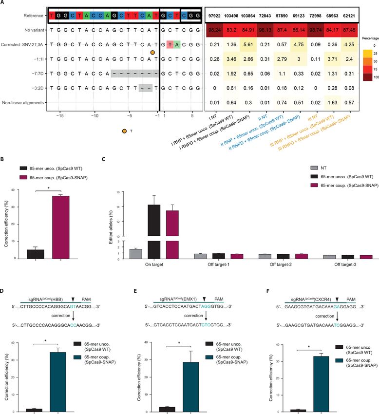
Short report Chromosomes and Gene Expression Human Biology and Medicine
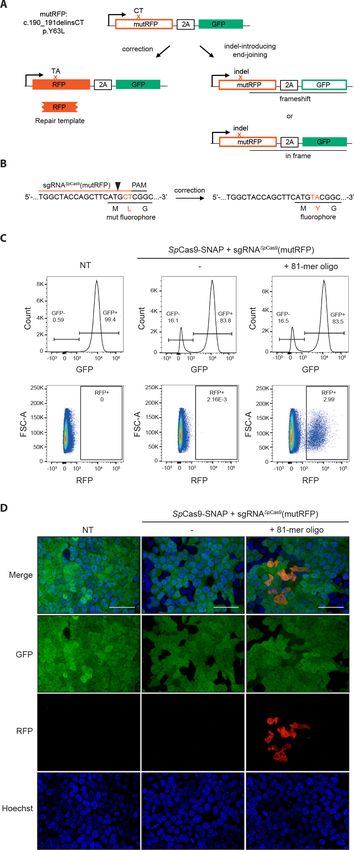
Figure 2. Fluorescent reporter system for high-throughput analysis of DSB repair rates. (a) Schematic overview of
the HEK293T fluorescent reporter system. The RFP fluorophore carries a c.190_191delinsCT mutation that
substitutes two nucleotides TA at the positions 190 and 191 in the RFP sequence to CT. This leads to the
inactivation the RFP fluorophore by the substitution of tyrosine at the position 63 with leucine (p.Y63L). Repair of
the mutation via donor oligos generates RFP/GFP double positive cells; indel mutations generate RFP/GFP double
negative cells if they induce a frame shift. Of note, analysis of the reporter locus by next generation sequencing
(NGS) demonstrated that 20 percent of indels did not lead to a frame shift (Supplementary file 4). Nevertheless,
Figure 2 continued on next page
Savic et al. eLife 2018;7:e33761. DOI: https://doi.org/10.7554/eLife.33761 3 of 18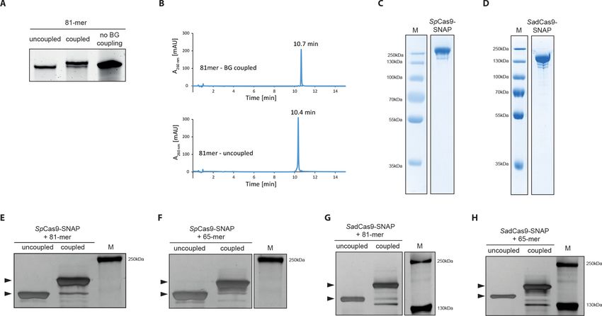
Short report                                                          Chromosomes and Gene Expression Human Biology and Medicine

                                    Figure 2 continued
                                    although FACS analysis thereby underestimates the absolute number of edited cells, it allows to accurately
                                    compare the correction efficiencies of different Cas9 systems. ‘X’ labels the mutation in RFP. 2A stands for 2A ‘self-
                                    cleaving’ peptide (Kim et al., 2011). (b) Schematic overview of the Streptococcus pyogenes sgRNA targeting
                                    mutRFP fluorophore and the corresponding PAM site. Black arrow indicates the introduced DSB site. The two
                                    nucleotides in the sgRNA seed sequence as well as the amino acid in the fluorophore region that are changed
                                    upon of precise repair (CT >TA and L > Y) are shown in orange. (c) Correction and indel rates can be quantified by
                                    FACS. The panels show FACS plots for gating GFP negative cells (upper panel) and the RFP positive cells (lower
                                    panel). (d) Representative confocal microscopy images. Scale bar: 50 mm, magnification 20x. Live cell nuclei were
                                    stained with Hoechst 33342. The efficiency of the sgRNA (sgRNASpCas9(mutRFP)) is shown in Figure 3—figure
                                    supplement 1b.
                                    DOI: https://doi.org/10.7554/eLife.33761.004

Figure 3. Covalent linkage of the DNA repair template to the Cas9 RNP complex. (a) Band shift of the 81-mer amino-modified oligo after coupling to
BG-GLA-NHS shown on a denaturing PAGE gel. Amino modified oligos were mixed with amine-reactive BG building blocks and the samples were
taken prior to the reaction (uncoupled) and after the reaction (coupled). No BG coupling: no amine-reactive BG building block was added to the amino
modified oligos. (b) LC-MS analysis of HPLC-purified BG-coupled and uncoupled DNA repair templates. (c,d) Coomassie Blue stained SDS-PAGE gels
of the purified SpCas9-SNAP and the SadCas9-SNAP fusion proteins (functionality of the SNAP-tags is shown in Figure 3—figure supplement 1e,f). (e-
h) Silver stained SDS-PAGE gels. Band shifts confirm covalent linkage of Cas9-SNAP proteins to BG-coupled 81-mers. Lower arrowheads: unbound
Cas9-SNAP. Upper arrowheads: Cas9-SNAP covalently bound to oligos.
DOI: https://doi.org/10.7554/eLife.33761.005
The following source data and figure supplement are available for figure 3:
Source data 1. Numerical data and the exact p values for all graphs in Figure 3—figure supplement 1.
DOI: https://doi.org/10.7554/eLife.33761.007
Figure supplement 1. Covalent linkage of the DNA repair template to the Cas9 RNP complex.
DOI: https://doi.org/10.7554/eLife.33761.006

Savic et al. eLife 2018;7:e33761. DOI: https://doi.org/10.7554/eLife.33761                                                                        4 of 18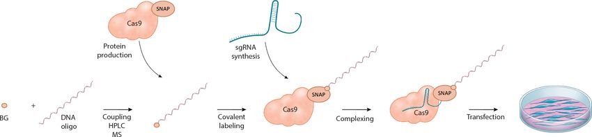
Short report                                                          Chromosomes and Gene Expression Human Biology and Medicine

Figure 4. Linking the repair template to the Cas9 RNP complex enhances correction efficiency in a fluorescent reporter cell line. (a) Comparison
between the control Cas9 system (RNP unco.: SpCas9-SNAP plus unlabeled donor oligo) and our novel system (RNPD coup.: SpCas9-SNAP conjugated
to BG-labeled donor oligo). Cells were analyzed 5 days after transfection by FACS. Results are presented as correction efficiency (percentage of
correction in edited cells). (b) Illustration of our novel Cas9 system, in which the repair template is covalently bound to SpCas9-SNAP (RNPD coup.). (c)
Schematic overview of the binding positions of different SadCas9 sgRNAs (sgRNAsSadCas9(mutRFP)-1-4) in comparison to the SpCas9 sgRNA targeting
the mutRFP fluorophore (sgRNAsSpCas9(mutRFP)). (d) Illustration of the two-component system, where the repair template is linked to the catalytically
inactive SadCas9-SNAP (RNP-RNPD coup.). (e) Comparison between the two-component system (RNP-RNPD coup.: SpCas9-SNAP + SadCas9-
Figure 4 continued on next page

Savic et al. eLife 2018;7:e33761. DOI: https://doi.org/10.7554/eLife.33761                                                                        5 of 18
Short report                                                        Chromosomes and Gene Expression Human Biology and Medicine

Figure 4 continued
SNAP bound to BG-labeled donor oligo) and the corresponding control Cas9 system (RNP-RNP unco.: SpCas9-SNAP + SadCas9-SNAP + unlabeled
repair oligo). (f) Transfection of the one component system (grey and pink panels) and two component system (black/grey and black/pink panels) into
reporter cells at a 5-time lower concentrations. In the two component system sgRNASadCas9(mutRFP)-3 was used. (g) Comparison of two component
systems with and without the sgRNA for the SadCas9 complex. RNP unco. (SpCas9-SNAP + uncoupled oligo + sgRNASpCas9(mutRFP)); RNPD coup.
(SpCas9-SNAP-coupled BG-oligo + sgRNASpCas9(mutRFP)); RNP-RNP unco. (SpCas9-SNAP + sgRNASpCas9(mutRFP) + SadCas9-SNAP + uncoupled
oligo + sgRNASadCas9(mutRFP)); RNP-RNPD coup. (SpCas9-SNAP + sgRNASpCas9(mutRFP) + SadCas9-SNAP-coupled BG-oligo +
sgRNASadCas9(mutRFP)). All values are shown as mean ±s.e.m of biological replicates; *p
Short report                                                          Chromosomes and Gene Expression Human Biology and Medicine

Figure 5. Linking the repair template to the Cas9 RNP complex enhances correction efficiency at endogenous loci. (a,b,c) Upper panels: Schematic
overview of the target genomic regions of the Streptococcus pyogenes gRNAs. Black arrow indicates the introduced DSB site. The nucleotides that are
exchanged in case of precise repair are shown in blue. Lower panels: NGS data quantification: Correction efficiency of the control Cas9 system (RNP
unco.: SpCas9-SNAP plus unlabeled donor oligo) compared to our novel system (RNPD coup.: SpCas9-SNAP bound to BG-labeled donor oligo) is
shown. (a) The HBB locus was targeted in a K562 cell line. The (b) Rosa26 and (c) Pcsk9 loci were targeted in mouse ESCs. All values are shown as
mean ±s.e.m of biological replicates; *p
Short report                                                           Chromosomes and Gene Expression Human Biology and Medicine

Figure 6. Direct comparison of the Cas9 RNPD system to the classical Cas9 complex. Classical Cas9 system (wild type SpCas9 plus unlabeled donor
oligo); Our novel RNPD system (SpCas9-SNAP conjugated to BG-labeled donor oligo). (a) Targeting of the reporter locus in HEK293T cells. Illustration
of the most frequent variants found by NGS in untreated samples (NT), in samples transfected with the classical Cas9 system (65-mer unco. (SpCas9
WT)), and in our engineered system (65-mer coup. (SpCas9-SNAP)). Alleles with a frequency above 0.5% in any of the nine samples are shown. For
alleles with lower frequencies see Supplementary file 3. Abbreviations: Insertion (I), Deletion (D), Single nucleotide variant (SNV). Different colours in
the x-axis indicate the three experimental replicates. A detailed description of the plot labels can be found in the Supplementary File 2 legend. (b,d,e,f)
Figure 6 continued on next page

Savic et al. eLife 2018;7:e33761. DOI: https://doi.org/10.7554/eLife.33761                                                                          8 of 18
Short report                                                           Chromosomes and Gene Expression Human Biology and Medicine

Figure 6 continued
NGS data quantification of the (b) reporter locus, (d) HBB locus, (e) EMX1, and (f) CXCR4 locus targeted in HEK293T cells. In (a,b,c) the mutRFP sgRNA
(see Figure 2b) was used. (c) Off target analysis for sgRNASpCas9(mutRFP): the percentage of edited alleles detected using NGS in untreated samples,
in samples transfected with the classical Cas9 system, and in our engineered system. Information on the off target loci can be found in
Supplementary file 1 – Supplementary Table 1. (d,e,f) Upper panels: Schematic overview of the target genomic regions of the gRNAs. Black arrow
indicates the introduced DSB site. The nucleotides that are exchanged in case of precise repair are shown in blue. Lower panels: NGS data
quantification. Correction efficiency of the classical Cas9 system compared to our novel system is shown. All values are shown as mean ±s.e.m of
biological replicates. *p
Short report                                                      Chromosomes and Gene Expression Human Biology and Medicine

                                        In the previous experiments the RNPD system was always compared to Cas9 SNAP-tag fusion
                                    proteins with uncoupled donor oligos. To also directly compare the engineered RNPD system to the
                                    classical CRISPR-Cas9 system, we performed experiments where we used wild-type Cas9 with the
                                    uncoupled donor oligos as a control. We first targeted the fluorescent reporter locus and analyzed it
                                    by NGS. We found that while the mean percentage of corrected loci increased from 0.8% with the
                                    classical Cas9 system to 4.9% with the RNPD system, the number of incorrectly edited loci slightly
                                    decreased from 12.6% to 9.3% (Figure 6a, Supplementary file 2,3,4). This corresponds to a 7-fold
                                    increase in correction efficiency (Figure 6b). In addition, the analysis of three computationally pre-
                                    dicted off-target sites (Lin et al., 2014b; Cradick et al., 2014) of the reporter locus, suggests that
                                    the risk for generating off-target mutations is not enhanced with the RNPD system (Figure 6c,
                                    Supplementary file 2,3,4). In the next step we also targeted and analyzed the endogenous loci
                                    HBB, empty spiracles homeobox 1 (EMX1), and C-X-C chemokine receptor type 4 (CXCR4) in
                                    HEK293T cells. NGS analysis revealed that in all three loci the mean correction efficiency of the
                                    RNPD system was markedly increased to: 34,4% at the HBB locus, 28.6% at the EMX1 locus and
                                    33.1% at the CXCR4 locus (Figure 6d,e,f, Supplementary file 2,3,4). Compared to the classical
                                    CRISPR-Cas9 system this represents a 20-fold, a 10-fold, and a 24-fold increase, respectively
                                    (Figure 6d,e,f).
                                        Direct delivery of Cas9 RNP complexes into tissues promises great potential for therapeutic appli-
                                    cations. Compared to genetically encoded systems, RNPs avoid the danger of genomic integration,
                                    and due to their limited lifetime, the risk of off-target activities is low (Kim et al., 2014). In addition,
                                    procedures for large-scale production of recombinant proteins for clinical use are well established,
                                    and several recently developed protocols enable in vivo delivery of Cas9 RNP complexes in animal
                                    models (Wang et al., 2016; Zuris et al., 2015; Staahl et al., 2017; Lee et al., 2017). Here, we pres-
                                    ent a method where we enhance correction efficiency of Cas9-induced DSBs by conjugating the
                                    donor oligo to the Cas9 complex. Our data suggests that the increase in HDR efficiency is caused by
                                    enhanced nuclear concentration of the repair template. Unlike previous approaches that increase
                                    HDR rates by chemically modulating DNA repair pathways, our approach does not alter endogenous
                                    cellular processes, thus reducing risk of potential negative side effects. In addition, covalent linkage
                                    of the repair template to the Cas9 RNP complex also addresses another central challenge of in vivo
                                    gene editing therapies – namely that simultaneous delivery of the RNP complex and the repair tem-
                                    plate needs to be ensured. Taken together, we suggest that covalently linking the DNA repair tem-
                                    plate to the Cas9 RNP complex is poised to further drive the CRISPR/Cas technology towards
                                    clinical translation.

                                    Materials and methods
                                    Generation of CRISPR-Cas9 complexes with covalently bound repair templates is described in more
                                    detail at Bio-protocol (Savić et al., 2019).

Key resources table
Reagent type (species)
or resource                      Designation                 Source or reference                Identifiers           Additional information
Recombinant protein              SpCas9-SNAP                 This paper                                               Schwank and Jinek lab
(Streptococcus pyogenes)
Recombinant protein              SadCas9-SNAP                This paper                                               Schwank and Jinek lab
(Staphylococcus aureus)
Genetic reagent                  NH2-modified oligo          Integrated DNA Technologies        -                     Custom DNA oligos/ ’5 C6
                                                                                                                      NH2 modif.
Chemical compound                BG-GLA-NHS                  New England Biolabs                ID_NEB:S9151S

                                        Please see Supplementary file 1-Supplementary Tables 1–6 for a list of the DNA sequences used
                                    in this manuscript.

                                    Plasmids
                                    All plasmids used in this study (listed in Supplementary file 1-Supplementary Table 6) have been
                                    deposited for the TULIPs system, along with maps and sequences, in Addgene.

Savic et al. eLife 2018;7:e33761. DOI: https://doi.org/10.7554/eLife.33761                                                              10 of 18
Short report Chromosomes and Gene Expression Human Biology and Medicine
Cloning of pNS19-LV-mutRFP-2A-GFP: pEGIP (addgene plasmid #26777) was mutagenized using
QuikChange Lightning Multi Site-Directed Mutagenesis Kit (Agilent Technologies) to destroy the
start codon of eGFP. Next the vector was linearized with BamHI and In-Fusion HD Cloning Plus CE
(Takara) was used to insert the mutRFP-2A gBlocks Gene Fragment (Integrated DNA Technologies).
Cloning of pNS20-SpCas9-SNAP: pMJ922-SpyCas9-GFP bacterial expression vector was a kind
gift from Prof. Martin Jinek. GFP was digested using BamHI and KpnI, and SNAPtag-NLS gBlocks
(Integrated DNA Technologies) were integrated using In-Fusion HD Cloning Plus CE (Takara).
Cloning of pNS38-SadCas9-SNAP: pAD-SaCas9-GFP was generated by replacing the SpCas9
coding sequence in pMJ922 with SaCas9 sequence using Gibson cloning (Keppler et al., 2003).
QuikChange Lightning Multi Site-Directed Mutagenesis Kit (Agilent Technologies) was used to
remove the stop codon and to introduce the D10A and N580A mutations into the SaCas9 (SadCas9)
gene. Subsequently, GFP was cut out using BamHI and KpnI, and replaced by a SNAP-tag-NLS
gBlock (Integrated DNA Technologies) using In-Fusion HD Cloning Plus CE (Takara).
Plasmid pMJ806 was a gift from Jennifer Doudna (Addgene plasmid # 39312) (Jinek et al.,
2012).
Benzylguanine coupling reaction
Synthetic oligonucleotides with a 50 -Amino Modifier C6 functional group (100 mM) (Integrated DNA
Technologies) were incubated with benzylguanine-GLA-NHS (1 mM) (NEB) and Hepes pH8.5
(200mM) for 60 min at 30˚C. Coupling reactions were performed in following ratios: 30:1, 60:1 and
100:1 BG-GLA-NHS: amino modified oligo. After the coupling reaction all oligos were purified by
ethanol precipitation. Repair oligo sequences can be found in Supplementary file 1-Supplementary
Table 4.
Denaturating PAGE
The benzylguanine (BG) coupled reactions were run on 20% polyacrylamide TBE gel containing 8M
urea at 200 V for 60 min. The gel was stained for 30 min in 1x TBE containing Sybr Gold (Invitrogen),
and imaged with a UV transilluminator (Biorad).
HPLC purification and LC-MS analysis of the repair oligos
Benzylguanine coupled oligos were purified on an Agilent 1200 series preparative HPLC fitted with a
Waters XBridge Oligonucleotide BEH C18 column, 10 50 mm, 2.5 mm at 65˚C using a gradient of
5–25% buffer B over 8 min, flow rate = 5 ml min-1. Buffer A was 0.1 M triethylammonium acetate,
pH 8.0. Buffer B was methanol. Fractions were pooled, dried in a speedvac and dissolved in H2O.
Analysis of the purified BG-oligonucleotide was conducted on an Agilent 1200/6130 LC-MS system
fitted with a Waters Acquity UPLC OST C18 column (2.1 50 mm, 1.7 mm) at 65˚C, with a gradient
of 5–35% buffer B in 14 min with a flowrate of 0.3 mL min 1. Buffer A was aqueous hexafluoroiso-
propanol (0.4 M) containing triethylamine (15 mM). Buffer B was methanol.
Expression and purification of Cas9-SNAP
Snap-tagged Streptococcus pyogenes Cas9 (SpCas9-SNAP), Staphylococcus aureus dCas9 (Sad-
Cas9-SNAP) and Wild Type Streptococcus pyogenes Cas9 (SpCas9 WT) proteins were expressed in
Escherichia coli BL21 (DE3) Rosetta 2 (Novagen) fused to an N-terminal fusion protein containing a
hexahistidine affinity tag, the maltose binding protein (MBP) polypeptide sequence, and the tobacco
etch virus (TEV) protease cleavage site. The cells were lysed in 20 mM Tris pH 8.0, 500 mM NaCl, 5
mM Imidazole pH 8.0. Clarified lysate was applied to a 10 ml Ni-NTA (Qiagen) affinity chromatogra-
phy column. The column was washed by increasing the imidazole concentration to 10 mM and
bound protein was eluted in 20 mM Tris pH 8.0, 250 mM NaCl, 100 mM Imidazole pH 8.0. To
remove the His6-MBP affinity tag, the eluted protein was incubated overnight in the presence of TEV
protease. The cleaved protein was further applied to a heparin column (HiTrap Heparin HP, GE
Healthcare) and eluted with a linear gradient of 0.1–1.0 KCl. The eluted protein was further purified
by size exclusion chromatography using a Superdex 200 16/600 (GE Healthcare) equilibrated in 20
mM HEPES pH 7.5, 500 mM KCl.
Savic et al. eLife 2018;7:e33761. DOI: https://doi.org/10.7554/eLife.33761 11 of 18Short report Chromosomes and Gene Expression Human Biology and Medicine
Covalent binding of Cas9-SNAP protein and BG-coupled
oligonucleotide
Repair oligo templates coupled to BG were incubated with Cas9-SNAP proteins on the same day
when the transfection is performed. BG-coupled oligos (2.2 pmols) were mixed with either SpCas9-
SNAP or SadCas9-SNAP (2.2 pmols) and incubated for 60 min at 30˚C. The negative controls (wild-
type Cas9 +BG oligo or Cas9-SNAP + unlabeled oligo) were treated in the same way.
SDS-PAGE gels
For confirming successful labeling of the Cas9-SNAP proteins with the BG-coupled oligonucleotides,
BG-coupled and uncoupled oligonucleotides were mixed with either SpCas9-SNAP, SadCas9-SNAP
or only the Cas9-SNAP proteins alone, reactions were incubated for one hour at 30˚C. For the
SNAP-Vista Green (NEB) substrate, the protein was incubated for 30 min on 30˚C in the dark. After
incubation, reactions (300 ng) were loaded on 6% SDS-PAGE gel and run at 80V for 160 min. Gels
that were containing BG-Vista Green (NEB, SNAP-Vista Green), were imaged prior to silver staining.
The green fluorescence signal of the SNAP-tag was detected with a UV transilluminator (Biorad).
Subsequently, silver staining was completed using the Pierce Silver Stain Kit (Thermo Scientific)
according to manufacturer instructions, and imaged with a UV transilluminator (Biorad).
Production of sgRNAs
sgRNAs were generated from DNA templates using the T7 RNA Polymerase (Roche) in vitro tran-
scription (IVT) kit. In short, sgRNA specific primers that also contain the T7 sequence were annealed
with a common reverse primer that contains the sequence of the sgRNA scaffold (final concentra-
tions 10 mM). DNA was purified with the QIAquick purification (Qiagen) kit and eluted in DEPC-
treated water. PCR products were run on agarose to estimate concentration and to confirm ampli-
con size. In vitro transcription was performed at 37˚C overnight. For purification, DNase I was added
to the sgRNAs and incubated for 15 min at 37˚C, and subsequently ethanol precipitation was per-
formed overnight at 20˚C. The sgRNAs were then further purified using RNA Clean and Concentra-
tors (Zymo Research). Before use, all sgRNAs were checked on denaturing 2% MOPS gels.
Complete sequences for all sgRNA protospacers, IVT primers and crRNAs can be found in
Supplementary file 1-Supplementary Table 1, 2 and 3, respectively.
Lentivirus production
HEK293T were PEI transfected with following plasmids: pNS19-LV-mutRFP-2A-eGFP, Pax2 and VSV-
G. After 12 hr, the supernatant was discarded and changed to DMEM plus 10% FBS. 24 and 72 hr
post-transfection, the media was collected and filtered through 0.45 mm filter and centrifuged at 20
000 G for 2:00 hr at 4˚C. The pellet was then resuspended in 1 ml of DMEM and stored at 80˚C.
Fluorescent reporter generation
HEK293T cells were transduced with a lentiviral vector carrying the fluorescent reporter construct.
Serial virus dilutions were used to isolate clonal populations using Puromycine selection (2 mg/ml) for
2 weeks.
Cell culture and reagents
HEK293T cells were obtained from ATCC and verified mycoplasma free (GATC Biotech). The
HEK293T reporter line was maintained in DMEM with GlutaMax (Gibco). Media was supplemented
with 10% FBS (Sigma), and 100 mg/mL Penicillin-Streptomycin (Gibco). K562 cells were obtained
from Sigma Aldrich, verified mycoplasma free and were maintained in RPMI 1640 medium with Glu-
taMax. Additional the medium supplemented with 10% FBS, and 100 mg/mL Penicillin-Streptomycin.
Cells were passaged three times per week. Cells were grown at 37˚C in a humidified 5% CO2 envi-
ronment. WT E14 mESC line (ATCC CRL-1821) was cultured in Dulbecco’s Modified Eagle Media
(DMEM) (Sigma-Aldrich), containing 15% of fetal bovine serum (FBS; Life Technologies), 100 U/mL
LIF (Millipore), 0.1 mM 2-ß-mercaptoethanol (Life Technologies) and 1% Penicillin/Streptomycin
(Gibco), on 0.2% gelatin-coated support in absence of feeder cells. The culture medium was
changed daily. Cells were grown at 37˚C in 8% CO2.
Savic et al. eLife 2018;7:e33761. DOI: https://doi.org/10.7554/eLife.33761 12 of 18Short report Chromosomes and Gene Expression Human Biology and Medicine
Transfection reactions
HEK293T cells were seeded in 24-well plates at 120.000–140.000 cells per well, 1 day prior to trans-
fection. K562 cells were 6 hr prior to transfection distributed in 24 well plates at a density of
220.000–240.000 cells per well. On the day of transfection, RNP and RNPD complexes (2.2 pmols)
were complexed with sgRNA (3.88 pmols) in Opti-MEM (Invitrogen) and briefly vortexed, followed
by adding 3 ml the Lipofectamine 2000 reagent (Invitrogen) with Opti-MEM. The resulting mixture
was incubated for 15 min at room temperature to allow lipid particle formation. After 15 min of incu-
bation at room temperature, the mixture was dropped slowly into the well. One day post-transfec-
tion, cells were transferred to an 10 cm dish. mESCs were transfected into 6-well plate using
Lipofectamine 2000. Cells were plated 24 hr before transfection at a density of 20,000 cells/cm2 per
well and cultured in culture medium without streptomycin and penicillin. The medium was changed
to mESC culture medium 8 hr after transfection. Cells were collected 48 hr post-transfection.
Flow cytometry analysis
For flow cytometry analysis, HEK293T reporter cells were analysed 5 days after transfection. Cells
were trypsinized with TrypLE Express Enzym (Gibco), and resuspended in FACS buffer containing
PBS/1% FBS/1% EDTA. Sytox Red was added for the exclusion of dead cells. Data were acquired on
a BD LSR Fortessa cell analyser (Becton-Dickinson) and were further analysed using FlowJo software
(FlowJo 10.2). In all experiments, a minimum of 200.000 cells were analysed. Gating strategy: For-
ward versus side scatter (FSC-A vs SSC-A) gating was used to identify cells of interest. Doublets
were excluded using the forward scatter height versus forward scatter area density plot (FSC-H vs.
FSC-A). Live cells were gated based on Sytox-Red-negative staining. Live-gated cells were further
used to quantify the percentage of eGFP negative and turboRFP positive populations. Correction
efficiency (%) or (percentage of corrections in edited cells) was culculated as 100 * (eGFP/turboRFP
double positive population / (eGFP/turboRFP double negative population + eGFP/turboRFP double
positive population)).
Next Generation Sequencing
Transfected cells were collected by trypsinisation and were washed with PBS. PBS was discarded
and DNA extraction was preformed using DNeasy Blood and Tissue kit (Qiagen) following manufac-
turer’s protocol. The PCR amplicons flanking the targeted site were generated using NEBNext High-
Fidelity 2X PCR Master Mix (NEB), primers that were used are listed in Supplementary file 1-Sup-
plementary Table 5. PCR cycling conditions used were as follows: 1 98˚C for 3 min; 27 95˚C for
15 s, 65˚C for 15 s, 72˚C for 30 s; 1 72˚C for 5 min. Annealing temperature was optimized for each
primer set to ensure that a single amplicon was produced. PCR amplicons were purified by solid
phase reversible immobilization (SPRI) bead cleanup using Agencourt AMPure XP reagent (#A63881,
Beckmann-Coulter, Indianapolis, IN, USA), per the manufacturer’s instructions. For the generation of
the pooled sequencing libraries, the TruSeq (Illumina) Index Adaptor Sequences were added at the
second amplification step. The resulting Illumina libraries with Index Adaptors were purified with
AMPure XP reagent. Quality control for the final library was performed using the High sensitivity
D1000 ScreenTape at Agilent 2200 TapeStation. The libraries were sequenced using an Illumina
MiSeq sequencer (MiSeq Reagent Kit v2 (15M, 500 cycle kit) or MiSeq Reagent Micro Kit v2 (4M,
500 cycle kit), Illumina, San Diego, CA, USA). Sequences were received in the format of demulti-
plexed FASTQ files produced by Illumina’s bcl2fastq software (v2.19.0). Reads were merged with
Pear v0.9.8 (Zhang et al., 2014) and merged reads mapped to the amplicon sequences with BWA-
MEM v0.7.13-r1126 (Li, 2010). Unmerged reads were discarded. Sequence analysis was performed
in R using CrispRVariants v1.9.0 (Lindsay et al., 2016). Variants within the protospacer +PAM region
were analysed. Reads that align linearly and do not match the guide sequence are considered
‘Edited’. Percentage of edited alleles is calculated as 100* Edited reads/Total reads (excluding non-
linear alignments). Correction efficiency is 100* Perfectly corrected/Edited reads. The Scripts for
mapping sequencing data, counting mutations and generating plots are available at https://github.
com/HLindsay/Savic_CRISPR_HDR (Lindsay, 2018; copy archived at https://github.com/elifescien-
ces-publications/Savic_CRISPR_HDR) Fastq files have been uploaded to ArrayExpress
(Brazma et al., 2003), the accession number is E-MTAB-6808.
Savic et al. eLife 2018;7:e33761. DOI: https://doi.org/10.7554/eLife.33761 13 of 18Short report Chromosomes and Gene Expression Human Biology and Medicine
Fluorescence microscopy
HEK293T reporter cells were imaged 7 days after transfection. Transfected cells were grown on
Poly-L-lysine coated 8-well glass chamber slides (Vitaris) to 80–90% confluence. Hoechst 33342
(Thermo Scientific, Pierce) was added in the cell culture media to a final concentration of 0.1 mg/ml,
and cells were incubated for 10 min at 37˚C, 5% CO2, prior to the image session. Confocal imaging
was performed using a Leica DMI8-CS (ScopeM) with a sCMOS camera (Hamamatsu Orca Flash 4.0).
The laser unit for confocal acquisition (AOBS system) contains 458, 477, 488, 496, 514 nm (Argon
laser), 405 nm, 561 nm, 633 nm. Images were acquired using Leica LAS X SP8 Version 1.0 software,
through using a 20 0.75 NA HC PLAN APO CS2 objective. Imaging conditions and intensity scales
were matched for images presented together. Images were analysed using the Leica LAS AF (Lite)
software version 3.3. Confocal images were processed using ImageJ software (Version 1.51 n).
Statistical analyses
Statistical analyses were conducted using Graphpad’s Prism7 software. A Mann-Whitney T-test was
conducted for two-sample analyses (P value style: 0.1234(ns), 0.0332(*), 0.0021(**)). All values are
shown as mean ± s.e.m of biological replicates. The number of biological replicates for each experi-
ment was detailed in the Figure Legends. Numerical data and the exact p values for all graphs have
been included in the Source data files.
Data availability
The data that support the findings of this study are available within the paper, Supplementary files,
Source data and NGS Fastq files have been uploaded to ArrayExpress.
Acknowledgements
This work has been funded by the Swiss National Science Foundation PMPDP3_171388 (to NS),
31003A_160230 (to GS), 31003A_149393 (to MJ) and by the Vallee Scholar Award from The Bert L
and N Kuggie Vallee Foundation (to MJ). The FACS analysis were performed at the ETH Flow
Cytometry Core Facility (E-FCCF). Cell imaging was performed at the Scientific Center for Optical
and Electron Microscopy (ScopeM) of the ETH Zurich. The NGS was performed at the Functional
Genomics Center Zurich (FGCZ) of the ETH Zurich an the University of Zurich and Genetic Diversity
Centre (GDC) of the ETH Zurich. We want to thank J. Huotari for critical feedback on the
manuscript.
Additional information
Funding
Funder Grant reference number Author
Schweizerischer Nationalfonds PMPDP3_171388 Natasa Savic
zur Förderung der Wis-
senschaftlichen Forschung
Schweizerischer Nationalfonds 31003A_160230 Gerald Schwank
zur Förderung der Wis-
senschaftlichen Forschung
Schweizerischer Nationalfonds 31003A_149393 Martin Jinek
zur Förderung der Wis-
senschaftlichen Forschung
Vallee Foundation Martin Jinek
The funders had no role in study design, data collection and interpretation, or the
decision to submit the work for publication.
Author contributions
Natasa Savic, Conceptualization, Data curation, Formal analysis, Supervision, Funding acquisition,
Validation, Investigation, Visualization, Methodology, Writing—original draft, Project administration,
Savic et al. eLife 2018;7:e33761. DOI: https://doi.org/10.7554/eLife.33761 14 of 18Short report Chromosomes and Gene Expression Human Biology and Medicine
Writing—review and editing, Conceived and designed the study, performed the experiments, Wrote
the paper; Femke CAS Ringnalda, Data curation, Formal analysis, Validation, Investigation, Visualiza-
tion, Methodology, Writing—original draft, Writing—review and editing, Performed the experi-
ments, Wrote the paper; Helen Lindsay, Data curation, Software, Formal analysis, Investigation,
Visualization, Writing—review and editing; Christian Berk, Investigation, Writing—review and edit-
ing, Performed HPLC and MS analysis; Katja Bargsten, Investigation, Methodology, Writing—review
and editing, Produced the recombinant proteins; Yizhou Li, Investigation, Performed HPLC and MS
analysis; Dario Neri, Methodology, Provided technical advice; Mark D Robinson, Software, Supervi-
sion, Writing—review and editing; Constance Ciaudo, Investigation, Writing—review and editing;
Jonathan Hall, Methodology, Writing—review and editing, Provided technical advice; Martin Jinek,
Supervision, Methodology, Writing—review and editing, Provided technical advice; Gerald Schwank,
Conceptualization, Supervision, Funding acquisition, Validation, Investigation, Visualization, Method-
ology, Writing—original draft, Project administration, Writing—review and editing, Conceived and
designed the study, Supervised the experiments, Wrote the paper
Author ORCIDs
Natasa Savic https://orcid.org/0000-0003-3110-5780
Femke CAS Ringnalda http://orcid.org/0000-0002-0684-4613
Constance Ciaudo http://orcid.org/0000-0002-0857-4506
Jonathan Hall http://orcid.org/0000-0003-4160-7135
Martin Jinek http://orcid.org/0000-0002-7601-210X
Gerald Schwank http://orcid.org/0000-0003-0767-2953
Decision letter and Author response
Decision letter https://doi.org/10.7554/eLife.33761.031
Author response https://doi.org/10.7554/eLife.33761.032
Additional files
Supplementary files
. Supplementary file 1. This file contains Supplementary Tables 1-6 (referenced in the Materials and
methods). Supplementary Table 1 contains the guide protospacer sequences used in this study. Sup-
plementary Table 2 contains the primer sequences for IVT of guides used in this study. Supplemen-
tary Table 3 contains the crRNA sequences of guides used in this study. Supplementary Table 4
contains the repair oligo sequences used in this study. The nucleotide substitution introduced by
precise correction using repair template is shown in lowercase. Supplementary Table 5 contains the
NGS primers used in this study. The target specific part of the primer is shown in uppercase, and the
Illumina adapter is shown in lowercase. Supplementary Table 6 contains the plasmids used in this
study.
DOI: https://doi.org/10.7554/eLife.33761.018
. Supplementary file 2. This file contains the allele plots for the loci analyzed by NGS. Allele plots
show insertion/deletion variant alleles with frequency of at least 0.01%, and non-indel variants with
frequency of at least 0.05% in any sample. When more than 50 variants passed these criteria, the
top 50 alleles according to their maximum frequency in any sample are shown. From top to bottom,
the consensus sequences for variant alleles are displayed in the order: no variant, precisely corrected
allele, insertions (I) and deletions (D), single nucleotide variants (SNVs) and non-linear alignments.
SNVs are only shown for non-indel variants and appear in color. In the y-axis labels, nucleotide num-
bers indicate the distance to the cut site. Variants are labelled with respect to the leftmost base. For
example 5:9D is a 9 base pair deletion starting 5 bases upstream of the cut site. SNV labels show
the bases that differ between the non-indel reads and the reference. The most common inserted
sequences with less than 20 base pairs are shown in full in the legend. For longer and less frequent
insertions the length is indicated. In the heatmap at right, the header shows the number of merged
read pairs with alignments spanning the guide sequence. The x-axis is coloured according to experi-
mental replicate.
DOI: https://doi.org/10.7554/eLife.33761.019
Savic et al. eLife 2018;7:e33761. DOI: https://doi.org/10.7554/eLife.33761 15 of 18Short report Chromosomes and Gene Expression Human Biology and Medicine
.Supplementary file 3. This file contains complete variant count tables for the genomic loci analyzed
by NGS. Variants are labeled as in Supplementary file 2.
DOI: https://doi.org/10.7554/eLife.33761.020
. Supplementary file 4. This file contains categorized variant count tables for the genomic loci ana-
lyzed by NGS. Reads were classified as ‘indel’ if any insertions or deletions were present in the guide
region, as ‘no variant’ if they perfectly matched the guide reference, (for on-target loci) ‘corrected’ if
the targeted bases were changed as expected, and ‘mismatch’ if any other nucleotide changes were
present.
DOI: https://doi.org/10.7554/eLife.33761.021
. Transparent reporting form
DOI: https://doi.org/10.7554/eLife.33761.022
Data availability
The data that support the findings of this study are available within the paper and its Supplementary
files. Source data files have been provided for Figure 4, Figure 5, Figure 6, Figure 3-Figure Supple-
ment 1, Figure 4-Figure Supplement 1 and Figure 4-Figure Supplement 2. Scripts for mapping
sequencing data, counting mutations and generating plots are available at https://github.com/
HLindsay/Savic_CRISPR_HDR (copy archived at https://github.com/elifesciences-publications/Savic_
CRISPR_HDR). Fastq files have been uploaded to ArrayExpress, and accession number is E-MTAB-
6808.
The following dataset was generated:
Database and
Author(s) Year Dataset title Dataset URL Identifier
Helen Lindsay, Na- 2018 Fastq file from Covalent linkage of https://www.ebi.ac.uk/ar- EMBL-EBI Array
tasa Savic the DNA repair template to the rayexpress/experiments/ Express, E-MTAB-680
CRISPR-Cas9 nuclease enhances E-MTAB-6808/ 8
homology-directed repair
References
Aronsohn AI, Hughes JA. 1998. Nuclear localization signal peptides enhance cationic liposome-mediated gene
therapy. Journal of Drug Targeting 5:163–169. DOI: https://doi.org/10.3109/10611869808995871, PMID:
9606006
Brazma A, Parkinson H, Sarkans U, Shojatalab M, Vilo J, Abeygunawardena N, Holloway E, Kapushesky M,
Kemmeren P, Lara GG, Oezcimen A, Rocca-Serra P, Sansone SA. 2003. ArrayExpress–a public repository for
microarray gene expression data at the EBI. Nucleic Acids Research 31:68–71. DOI: https://doi.org/10.1093/
nar/gkg091, PMID: 12519949
Carlson-Stevermer J, Abdeen AA, Kohlenberg L, Goedland M, Molugu K, Lou M, Saha K. 2017. Assembly of
CRISPR ribonucleoproteins with biotinylated oligonucleotides via an RNA aptamer for precise gene editing.
Nature Communications 8:1711. DOI: https://doi.org/10.1038/s41467-017-01875-9, PMID: 29167458
Ceccaldi R, Rondinelli B, D’Andrea AD. 2016. Repair pathway choices and consequences at the Double-Strand
break. Trends in Cell Biology 26:52–64. DOI: https://doi.org/10.1016/j.tcb.2015.07.009, PMID: 26437586
Chu VT, Weber T, Wefers B, Wurst W, Sander S, Rajewsky K, Kühn R. 2015. Increasing the efficiency of
homology-directed repair for CRISPR-Cas9-induced precise gene editing in mammalian cells. Nature
Biotechnology 33:543–548. DOI: https://doi.org/10.1038/nbt.3198, PMID: 25803306
Cradick TJ, Qiu P, Lee CM, Fine EJ, Bao G. 2014. COSMID: a Web-based tool for identifying and validating
CRISPR/Cas Off-target sites. Molecular Therapy - Nucleic Acids 3:e214. DOI: https://doi.org/10.1038/mtna.
2014.64, PMID: 25462530
Gutschner T, Haemmerle M, Genovese G, Draetta GF, Chin L. 2016. Post-translational regulation of Cas9 during
G1 enhances Homology-Directed repair. Cell Reports 14:1555–1566. DOI: https://doi.org/10.1016/j.celrep.
2016.01.019, PMID: 26854237
Howden SE, McColl B, Glaser A, Vadolas J, Petrou S, Little MH, Elefanty AG, Stanley EG. 2016. A Cas9 variant
for efficient generation of Indel-Free knockin or Gene-Corrected human pluripotent stem cells. Stem Cell
Reports 7:508–517. DOI: https://doi.org/10.1016/j.stemcr.2016.07.001, PMID: 27499201
Jinek M, Chylinski K, Fonfara I, Hauer M, Doudna JA, Charpentier E. 2012. A programmable dual-RNA-guided
DNA endonuclease in adaptive bacterial immunity. Science 337:816–821. DOI: https://doi.org/10.1126/science.
1225829, PMID: 22745249
Keppler A, Gendreizig S, Gronemeyer T, Pick H, Vogel H, Johnsson K. 2003. A general method for the covalent
labeling of fusion proteins with small molecules in vivo. Nature Biotechnology 21:86–89. DOI: https://doi.org/
10.1038/nbt765, PMID: 12469133
Savic et al. eLife 2018;7:e33761. DOI: https://doi.org/10.7554/eLife.33761 16 of 18Short report Chromosomes and Gene Expression Human Biology and Medicine
Kim JH, Lee SR, Li LH, Park HJ, Park JH, Lee KY, Kim MK, Shin BA, Choi SY. 2011. High cleavage efficiency of a
2A peptide derived from porcine teschovirus-1 in human cell lines, zebrafish and mice. PLoS One 6:e18556.
DOI: https://doi.org/10.1371/journal.pone.0018556, PMID: 21602908
Kim S, Kim D, Cho SW, Kim J, Kim JS. 2014. Highly efficient RNA-guided genome editing in human cells via
delivery of purified Cas9 ribonucleoproteins. Genome Research 24:1012–1019. DOI: https://doi.org/10.1101/
gr.171322.113, PMID: 24696461
Lee K, Conboy M, Park HM, Jiang F, Kim HJ, Dewitt MA, Mackley VA, Chang K, Rao A, Skinner C, Shobha T,
Mehdipour M, Liu H, Huang W-chin, Lan F, Bray NL, Li S, Corn JE, Kataoka K, Doudna JA, et al. 2017.
Nanoparticle delivery of Cas9 ribonucleoprotein and donor DNA in vivo induces homology-directed DNA
repair. Nature Biomedical Engineering 1:889–901. DOI: https://doi.org/10.1038/s41551-017-0137-2
Li H. 2010. Aligning New-Sequencing Reads by BWA: Broad Institute. https://www.broadinstitute.org/files/
shared/mpg/nextgen2010/nextgen_li.pdf.
Lin S, Staahl BT, Alla RK, Doudna JA. 2014a. Enhanced homology-directed human genome engineering by
controlled timing of CRISPR/Cas9 delivery. eLife 3:e04766. DOI: https://doi.org/10.7554/eLife.04766,
PMID: 25497837
Lin Y, Cradick TJ, Brown MT, Deshmukh H, Ranjan P, Sarode N, Wile BM, Vertino PM, Stewart FJ, Bao G. 2014b.
CRISPR/Cas9 systems have off-target activity with insertions or deletions between target DNA and guide RNA
sequences. Nucleic Acids Research 42:7473–7485. DOI: https://doi.org/10.1093/nar/gku402, PMID: 24838573
Lindsay H, Burger A, Biyong B, Felker A, Hess C, Zaugg J, Chiavacci E, Anders C, Jinek M, Mosimann C,
Robinson MD. 2016. CrispRVariants charts the mutation spectrum of genome engineering experiments. Nature
Biotechnology 34:701–702. DOI: https://doi.org/10.1038/nbt.3628, PMID: 27404876
Lindsay H. 2018. Github. https://github.com/HLindsay/Savic_CRISPR_HDR
Ludtke JJ, Zhang G, Sebestyén MG, Wolff JA. 1999. A nuclear localization signal can enhance both the nuclear
transport and expression of 1 kb DNA. Journal of cell Science 112:2033–2041. PMID: 10341220
Mali P, Yang L, Esvelt KM, Aach J, Guell M, DiCarlo JE, Norville JE, Church GM. 2013. RNA-guided human
genome engineering via Cas9. Science 339:823–826. DOI: https://doi.org/10.1126/science.1232033, PMID: 232
87722
Mao Z, Bozzella M, Seluanov A, Gorbunova V. 2008. Comparison of nonhomologous end joining and
homologous recombination in human cells. DNA Repair 7:1765–1771. DOI: https://doi.org/10.1016/j.dnarep.
2008.06.018, PMID: 18675941
Maruyama T, Dougan SK, Truttmann MC, Bilate AM, Ingram JR, Ploegh HL. 2015. Increasing the efficiency of
precise genome editing with CRISPR-Cas9 by inhibition of nonhomologous end joining. Nature Biotechnology
33:538–542. DOI: https://doi.org/10.1038/nbt.3190, PMID: 25798939
Richardson CD, Ray GJ, DeWitt MA, Curie GL, Corn JE. 2016. Enhancing homology-directed genome editing by
catalytically active and inactive CRISPR-Cas9 using asymmetric donor DNA. Nature Biotechnology 34:339–344.
DOI: https://doi.org/10.1038/nbt.3481, PMID: 26789497
Richardson CD. 2017. CRISPR-Cas9 genome editing in human cells works via the fanconi Anemia pathway.
bioRxiv . DOI: https://doi.org/10.1101/136028
Ruff P, Koh KD, Keskin H, Pai RB, Storici F. 2014. Aptamer-guided gene targeting in yeast and human cells.
Nucleic Acids Research 42:e61. DOI: https://doi.org/10.1093/nar/gku101, PMID: 24500205
Savić N, Ringnalda FC, Berk C, Bargsten K, Hall J, Jinek M, Schwank G. 2019. In vitro generation of CRISPR-Cas9
complexes with covalently bound repair templates for genome editing in mammalian cells. Bio-Protocol 9.
DOI: https://doi.org/10.21769/BioProtoc.3136, PMID: 30675496
Shalem O, Sanjana NE, Hartenian E, Shi X, Scott DA, Mikkelson T, Heckl D, Ebert BL, Root DE, Doench JG,
Zhang F. 2014. Genome-scale CRISPR-Cas9 knockout screening in human cells. Science 343:84–87.
DOI: https://doi.org/10.1126/science.1247005, PMID: 24336571
Song J, Yang D, Xu J, Zhu T, Chen YE, Zhang J. 2016. RS-1 enhances CRISPR/Cas9- and TALEN-mediated knock-
in efficiency. Nature Communications 7:10548. DOI: https://doi.org/10.1038/ncomms10548, PMID: 26817820
Staahl BT, Benekareddy M, Coulon-Bainier C, Banfal AA, Floor SN, Sabo JK, Urnes C, Munares GA, Ghosh A,
Doudna JA. 2017. Efficient genome editing in the mouse brain by local delivery of engineered Cas9
ribonucleoprotein complexes. Nature Biotechnology 35:431–434. DOI: https://doi.org/10.1038/nbt.3806,
PMID: 28191903
Subramanian A, Ranganathan P, Diamond SL. 1999. Nuclear targeting peptide scaffolds for lipofection of
nondividing mammalian cells. Nature Biotechnology 17:873–877. DOI: https://doi.org/10.1038/12860,
PMID: 10471928
van den Bosch M, Lohman PH, Pastink A. 2002. DNA double-strand break repair by homologous recombination.
Biological Chemistry 383:873–892. DOI: https://doi.org/10.1515/BC.2002.095, PMID: 12222678
Wang M, Zuris JA, Meng F, Rees H, Sun S, Deng P, Han Y, Gao X, Pouli D, Wu Q, Georgakoudi I, Liu DR, Xu Q.
2016. Efficient delivery of genome-editing proteins using bioreducible lipid nanoparticles. PNAS 113:2868–
2873. DOI: https://doi.org/10.1073/pnas.1520244113, PMID: 26929348
Yu C, Liu Y, Ma T, Liu K, Xu S, Zhang Y, Liu H, La Russa M, Xie M, Ding S, Qi LS. 2015. Small molecules enhance
CRISPR genome editing in pluripotent stem cells. Cell Stem Cell 16:142–147. DOI: https://doi.org/10.1016/j.
stem.2015.01.003, PMID: 25658371
Zanta MA, Belguise-Valladier P, Behr JP. 1999. Gene delivery: a single nuclear localization signal peptide is
sufficient to carry DNA to the cell nucleus. PNAS 96:91–96. DOI: https://doi.org/10.1073/pnas.96.1.91, PMID:
9874777
Savic et al. eLife 2018;7:e33761. DOI: https://doi.org/10.7554/eLife.33761 17 of 18Short report Chromosomes and Gene Expression Human Biology and Medicine
Zhang J, Kobert K, Flouri T, Stamatakis A. 2014. PEAR: a fast and accurate Illumina Paired-End reAd mergeR.
Bioinformatics 30:614–620. DOI: https://doi.org/10.1093/bioinformatics/btt593, PMID: 24142950
Zuo E, Cai YJ, Li K, Wei Y, Wang BA, Sun Y, Liu Z, Liu J, Hu X, Wei W, Huo X, Shi L, Tang C, Liang D, Wang Y,
Nie YH, Zhang CC, Yao X, Wang X, Zhou C, et al. 2017. One-step generation of complete gene knockout mice
and monkeys by CRISPR/Cas9-mediated gene editing with multiple sgRNAs. Cell Research 27:933–945.
DOI: https://doi.org/10.1038/cr.2017.81, PMID: 28585534
Zuris JA, Thompson DB, Shu Y, Guilinger JP, Bessen JL, Hu JH, Maeder ML, Joung JK, Chen ZY, Liu DR. 2015.
Cationic lipid-mediated delivery of proteins enables efficient protein-based genome editing in vitro and in vivo.
Nature Biotechnology 33:73–80. DOI: https://doi.org/10.1038/nbt.3081, PMID: 25357182
Savic et al. eLife 2018;7:e33761. DOI: https://doi.org/10.7554/eLife.33761 18 of 18You can also read