Advances in Modeling the Immune Microenvironment of Colorectal Cancer
←
→
Page content transcription
If your browser does not render page correctly, please read the page content below
MINI REVIEW
                                                                                                                                              published: 10 February 2021
                                                                                                                                         doi: 10.3389/fimmu.2020.614300

                                             Advances in Modeling the Immune
                                             Microenvironment of
                                             Colorectal Cancer
                                             Paul Sukwoo Yoon 1†, Nuala Del Piccolo 2†, Venktesh S. Shirure 2, Yushuan Peng 2,
                                             Amanda Kirane 1, Robert J. Canter 1, Ryan C. Fields 3, Steven C. George 2
                                             and Sepideh Gholami 1*
                                             1 Department of Surgery, University of California, Davis, Sacramento, CA, United States, 2 Department of Biomedical

                                             Engineering, University of California, Davis, Davis, CA, United States, 3 Department of Surgery, The Alvin J. Siteman Cancer
                                             Center, Washington University School of Medicine, St. Louis, MO, United States

                                             Colorectal cancer (CRC) is the third most common cancer and second leading cause of
                                             cancer-related death in the US. CRC frequently metastasizes to the liver and these
                                             patients have a particularly poor prognosis. The infiltration of immune cells into CRC
                                             tumors and liver metastases accurately predicts disease progression and patient survival.
                           Edited by:
                        Benjamin Frey,       Despite the evident influence of immune cells in the CRC tumor microenvironment (TME),
University Hospital Erlangen, Germany        efforts to identify immunotherapies for CRC patients have been limited. Here, we argue
                        Reviewed by:         that preclinical model systems that recapitulate key features of the tumor
              Nathalie Britzen-Laurent,
University Hospital Erlangen, Germany
                                             microenvironment—including tumor, stromal, and immune cells; the extracellular matrix;
                          Laura Bracci,      and the vasculature—are crucial for studies of immunity in the CRC TME and the utility of
 National Institute of Health (ISS), Italy
                                             immunotherapies for CRC patients. We briefly review the discoveries, advantages, and
                   *Correspondence:
                                             disadvantages of current in vitro and in vivo model systems, including 2D cell culture
                      Sepideh Gholami
                 sgholami@ucdavis.edu        models, 3D culture systems, murine models, and organ-on-a-chip technologies.
     †
      These authors have contributed         Keywords: colorectal cancer, tumor microenvironment, cancer immunology, tissue engineering, organ-on-a-
                 equally to this work        chip (OOC)

                     Specialty section:
           This article was submitted to
                        Cancer Immunity      INTRODUCTION
                    and Immunotherapy,
                 a section of the journal    In the US, colorectal cancer (CRC) is the third most common cancer and second leading cause of
                Frontiers in Immunology      cancer-related death (1). CRC is largely asymptomatic until it has progressed to advanced stages (2),
           Received: 05 October 2020         with 5 year survival rates of 90% and 14% for localized and metastatic cases, respectively (1).
         Accepted: 29 December 2020          Population-wide screening campaigns in the last two decades have led to earlier diagnoses and
          Published: 10 February 2021        boosted the overall 5 year survival rate to ~65% (1). Due to anatomical proximity, CRC often
                            Citation:        metastasizes to the liver: 20%–25% of patients present with colorectal liver metastases (CRLM) at
  Yoon PS, Del Piccolo N, Shirure VS,        initial diagnosis and 50-60% of CRC patients will develop CRLM at some point (2–4). Hepatectomy
         Peng Y, Kirane A, Canter RJ,        is currently the best course of action for CRLM patients, offering a 5 year survival rate of up to 60%
Fields RC, George SC and Gholami S
                                             (3–5). Unfortunately, only 20%–25% of CRLM patients are eligible for resection at time of diagnosis,
     (2021) Advances in Modeling the
         Immune Microenvironment of
                                             leaving a large majority of patients to succumb to progressive metastatic cancer (3, 5).
                   Colorectal Cancer.            Recent research has demonstrated the role of immunity on CRC progression, prognosis, and
          Front. Immunol. 11:614300.         response to therapy. For example, immune cell infiltration into tumors correlates with clinical
    doi: 10.3389/fimmu.2020.614300           outcomes: T cells (6–10), Tregs (11), and NK cells (10) in primary CRC or CRLM lesions correlate

Frontiers in Immunology | www.frontiersin.org                                        1                                       February 2021 | Volume 11 | Article 614300
Yoon et al. Advances in Modeling Colorectal Cancer
with better prognoses, while the presence of tumor-associated patients. Pre-clinical work addressing this gap is focused on
macrophages (TAMs) has been alternately associated with pro- adoptive cell therapies, vaccines, immunostimulatory cytokines,
(12, 13) and anti-tumor (13–15) effects. In 2006, Galon and and combinations thereof, and early studies have produced
colleagues introduced the ImmunoScore. This measure of the promising results (19–21).
density of immune cells in the invasive margin and core of a The development of more efficacious cancer therapeutics is
lesion (9, 16–18) provides more accurate predictions of hindered by the limitations of current preclinical model systems,
recurrence, overall survival, and disease-free survival than which do not recapitulate the whole tumor microenvironment
traditional TNM staging for both CRC (7, 16, 18) and CRLM (TME) (Table 1) (24, 25). The TME is crucial for investigating
patients (6, 8, 11, 16). tumor-immune cell crosstalk, modeling tumor heterogeneity
Based on ImmunoScore’s prognostic success, clinicians are within and between patients, recapitulating events in the
actively pursuing immunotherapies for CRC and CRLM patients. metastatic cascade, and simulating responses to therapeutics
Checkpoint blockade therapies have shown particular promise (24–28). 2D in vitro models of cells growing in tissue culture
for mismatch repair deficient (dMMR)/microsatellite instability- plates lend themselves to the study of tumor growth and cell
high (MSI-H) CRC tumors (19–21). In a 2015 Phase II clinical migration, but lack complex tissue features like the vasculature
trial, dMMR/MSI-H patients treated with pembrolizumab (PD-1 and extracellular matrix (ECM) (24, 25, 28–31). In 3D in vitro
inhibitor) exhibited a 40% response rate and 78% 12-month models, multiple cell types can be co-cultured in ECM scaffolds,
progression free survival (22); the FDA approved this course of enabling the study of cell-cell interactions and nutrient/waste
treatment in 2017 (19). More recent work has probed the utility transport over small distances; however, these models lack key
of combining nivolumab (PD-1 inhibitor) with ipilimumab biomechanical features of the TME, including vascular and
(CTLA-4 inhibitor) (23). Results from this Phase II trial are interstitial perfusion (24, 25, 28–31). Animal models are
still maturing, but preliminary results suggest a response rate as capable of simulating the dynamic, multi-cellular/organ nature
high as 55% (19, 23). Unfortunately, only 15% of CRC tumors of the TME, but are expensive, difficult to manipulate, and
are classified as dMMR/MSI-H (19), and there are currently no limited in their ability to recreate human immunobiology (24,
immunotherapies available to the remaining 85% of CRC 32–34).
TABLE 1 | Advances in modeling colorectal cancer.
Model Application Advantages Disadvantages
2D In vitro • Adhesion • Simple • Low predictive power
Culture plate • Gene expression • Low cost • Lack of native architecture
• Drug screening • High throughput • Loss of tumor heterogeneity
Wound healing • Migration
3D In vitro • Proliferation • Retain native tumor geometry • Avascular
Organoid/ • Migration • Cell-cell/ECM interaction • High cost
Spheroid • Gene expression • Tumor heterogeneity • Low scalability
• Drug screening • Low reproducibility
Co-culture • Stromal crosstalk
• Immune crosstalk
In vivo • Tumor heterogeneity • High cost
Patient-derived xenografts • Laborious
• Low predictive power
• Proliferation • Immunocompromised
• Migration • Limited metastasis
Humanized mice • Invasion • Tumor microenvironment • High cost
• Angiogenesis • Tumor heterogeneity • Laborious
• Gene expression • Immunocompetent • Incomplete immune function
• Drug screening • Engraftment difficulties
Genetically engineered mice • Tumor microenvironment • High cost
• Tumor heterogeneity • Laborious
• Immunocompetent • Time consuming
• Natural disease progression
Organ-on-a-chip • Proliferation • Tumor microenvironment • Lack of standardization
Migration • Tumor heterogeneity • High cost
• Intravasation • Vascular • Laborious
Extravasation • Hydrodynamic properties • Low reproducibility
• Invasion • Biochemical gradient
• Angiogenesis • Precise control
• Stromal crosstalk • Easy visualization
• Immune crosstalk
• Gene expression
• Drug screening
Frontiers in Immunology | www.frontiersin.org 2 February 2021 | Volume 11 | Article 614300Yoon et al. Advances in Modeling Colorectal Cancer
Model systems that combine tissue engineering with vitro systems more accurately model in vivo biochemical factor
microfluidic technology represent a new frontier for the study of distribution and transport (28, 30); cell morphology, polarity, and
cancer development, progression, immunity, and metastasis. gene expression (61–66); heterogeneity in cell types (62, 64, 67); and
Dubbed “organ-on-a-chip” (OOC) systems, these models sensitivity to cancer therapeutics (61, 64, 67, 68). This accuracy is
incorporate many features of the TME, including multiple cell more pronounced under perfused culture conditions (61, 67–69)
types, matrix components, biochemical cues, spatiotemporal (Table 1). The challenges facing 3D cell culture systems include: i)
distribution of soluble mediators and oxygen, and perfusable uncertainty introduced by the underdefined, variable composition
vascular networks (24, 26, 35–37). Thus, OOC platforms offer of popular scaffold materials (including the gold standard Matrigel);
great potential as a preclinical tool for precision therapy. This ii) the absence of vascular flow, which is responsible for cancer cell
review will highlight recent advances in the utility of OOC dissemination, trafficking of some immune cells, and delivery of
devices to model immunity in the CRC/CRLM TME and therapeutics; iii) the inability to replicate the long-range interactions
compare this work with conventional model systems (Figure 1A). between tumors and other organs in the body that govern
metastasis and the immune response (24, 29, 35, 70); and iv)
limited reproducibility, scalability, and ease of use.
A handful of recent reports demonstrate the utility of 3D models
TWO-DIMENSIONAL (2D) CULTURES systems for the study of immunity in the CRC TME. In a 2018
Cell cultures in 2D (Figure 1A) are a standard and well-established paper, Dijkstra et al. co-cultured organoids from dMMR CRC
model system because they are simple, inexpensive, and easy to patients with autologous peripheral blood lymphocytes (71). In
manipulate, and enable imaging with high spatiotemporal this novel culture system, the team generated patient-specific,
resolution (Table 1) (24, 31). 2D cultures rely on cells adhering to cancer-reactive T cells from 4 of 8 patients, characterized the
a flat surface—generally a flask or plate—which does not reflect the specificity of T cells for tumor versus healthy tissue, and
natural 3D architecture of tissues or tumors. Furthermore, cells in measured the efficiency of T cell mediated tumor cell killing. A
2D cultures receive relatively uniform and often excessive levels of 2019 report by Courau and colleagues demonstrated that primary T
oxygen, nutrients, and growth factors, compromising their ability to and NK cells infiltrate into cell line-derived CRC spheroids, where
faithfully capture the in vivo TME (28–31). Despite these they kill tumor cells and degrade the 3D structure of the spheroid,
drawbacks, 2D experiments have revealed multiple mechanisms and that these effects can be enhanced by stimulating the immune
driving the behavior of epithelial sheets of cells (30) and epithelial- response with IL-15 plus anti-NKG2D and/or anti-MICA/B
derived tumors like CRC and CRLM (40–50). antibodies (72). The authors also showed that stimulation of the
2D cultures are conducive to studies of tumor-immune cell immune response is necessary for infiltration of autologous T and
crosstalk in the TME. For example, 2D in vitro systems have been NK cells into patient-matched CRC spheroids. Another recent study
used to examine the role of the CRC TME’s atypically high found that CAR-NK-92 cells engineered to recognize the universal
number of macrophages, a topic of active debate. These studies antigen EPCAM, the neoantigen EGFRvIII, or the tumor-associated
show that macrophages differentiate towards an M2-like antigen FRIZZLED can identify and lyse cells in murine- and
phenotype in response to tumor cells or tumor cell-conditioned patient-derived normal colon and CRC organoids, but the effects
media (51–55) and migrate towards tumor cells (54, 55). TAMs in are reduced by limited immune cell infiltration into organoids (73).
CRC have also been shown to modify the tumor cell response to Further, a 2019 report showed that primary CRC samples cultured
chemotherapy (56, 57); support tumor cell proliferation, under perfused conditions retained native tissue architecture, tumor
migration, and invasion (53–55); and limit tumor cell survival in cell density, and immune and stromal cell viability better than
a cell contact-dependent manner (13). Additionally, Yu and co- samples cultured under static conditions (69).
authors showed that mast cells migrate towards CRC tumor cell-
conditioned media in a Transwell assay and that co-culture of
mast and tumor cells increases tumor cell proliferation; the results
of these 2D culture experiments were verified in a 3D spheroid
IN VIVO MODELS
model (58). Studies with primary patient samples have In vivo models (Figure 1A) are integral tools in cancer research
demonstrated i) an HLA-mediated T cell response to the because they recapitulate several features of the TME not available
survivin protein in CRC tumor cells (59), and ii) NK cell in in vitro models, including vascular flow and communication
cytotoxicity directed against CRC tumor cells following immune between the tumor and distant organs (74–79). There are five types
cell activation or tumor cell priming (60). of mouse models of cancer: 1) xenograft, 2) allograft, 3) patient-
derived xenograft (PDX), 4) humanized, and 5) genetically modified
mouse (GEMM). Though murine models are labor intensive,
THREE-DIMENSIONAL (3D) CULTURES expensive, low-throughput, and susceptible to cross-species
incompatibilities, they have produced numerous insights into
3D cell cultures (Figure 1A) are comprised of cells distributed in CRC response to drug treatment (76, 77, 80–87) and metastasis
synthetic or naturally-occurring scaffolds or hydrogels to mimic in (85, 88–90) (Table 1).
vivo tissue architecture and can be cultured under static or perfused Though transplant mouse models (xenograft, allograft,
conditions (28–30). Compared to 2D cell culture systems, 3D in and PDX) accurately replicate the response to therapeutics
Frontiers in Immunology | www.frontiersin.org 3 February 2021 | Volume 11 | Article 614300Yoon et al. Advances in Modeling Colorectal Cancer
A
B
C
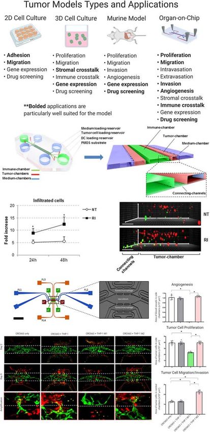
FIGURE 1 | (A) Colorectal liver metastasis models. Bolded applications are particularly well suited for the model. Even in the same category of model, constituent
models can vary greatly, based on design, method, and study goals. (B, C) Colorectal cancer (CRC) “organ-on-a-chip” (OOC) platforms can model the immune
response to tumors. (B) CRC cells (red) and IFN-DCs (green) are cultured in an OOC device (see cartoon) to simulate immune crosstalk. IFN-DCs migrate towards
and phagocytose CRC cells following treatment with interferon-a and romidepsin. Images have been adapted from Parlato et al’s 2017 Scientific Reports article (38).
(C) M1 and M2 macrophages (red) cultured with CRC cell lines (not shown) in a vascularized OOC platform (vessels shown in green) display anti- and pro-tumor
effects, respectively. Figure was originally published in Bi et al’s 2020 Integrative Biology report (39).
Frontiers in Immunology | www.frontiersin.org 4 February 2021 | Volume 11 | Article 614300Yoon et al. Advances in Modeling Colorectal Cancer
(76, 77, 80–84), they struggle to retain the genetic and cellular levels of pro-tumor immune cells, including myeloid-derived
features of native tumors (76, 79), recreate the metastatic suppressor cells, granulocytes, neutrophils, TAMs, and M2-like
cascade (with the possible exception of orthotopic transplant macrophages. Some CRC cases are associated with colitis, a state
models) (89), and mimic the immune response to a tumor (note of constant inflammation in the colon; colitis can be modeled in
that xenograft and PDX models are both necessarily mice through treatment with azoxymethane and/or dextran
immunocompromised to enable inoculation with human cell sodium sulfate. Through comparisons of wild-type and
lines and primary human tumor cells, respectively) (76, 79). knockout GEMM colitis mouse models, researchers have
Hence, humanized mouse models and GEMMs are more useful demonstrated the critical role of p53 (97), IL-6 and Stat3 (98),
for studies of immunity in the TME. Humanized mice are TLR4 (99), Pycard, Casp1, and Nlrp3 (100), and Nod1 (101) on
generated by engrafting specific mouse strains with human tumor formation and growth; all of these factors are implicated in
leukocytes (hematopoietic stem cells or peripheral blood regulation of the immune response.
mononuclear cells). These mice produce a human immune
response and are available commercially (91, 92), but
sometimes suffer from xenoreactive complications and do not ORGAN-ON-A-CHIP MODELS
mount a full humoral immune response (74). In the context of
CRC, humanized mouse models have been used to study tumor OOC models (Figure 1A) utilize microfluidic technology and
response to checkpoint blockade therapies (93, 94). In a 2015 tissue engineering to mimic and monitor dynamic 3D tissue
report, humanized mice engrafted with a CRC cell line and microenvironments, including epithelial barriers, parenchymal
treated with urelumab (CD137 inhibitor), nivolumab (PD-1 tissues, perfused microvasculature, multiple organ interactions,
inhibitor), or a combination of the two demonstrated limited and the immune response (24, 37). An OOC platform consists of
tumor growth and high infiltration of tumors by lymphocytes an interconnected series of 3D channels and chambers filled with
(93). Capasso and co-authors created humanized PDX mouse cells suspended in hydrogels. The geometry of these channels
models by implanting patient-derived MSI-H or microsatellite and chambers can be precisely selected to match a variety of
stable (MSS) tumor cells and then treated the mice with tissue architectures and mechanical forces, has a scale of tens to
nivolumab (PD-1 inhibitor) (94). Mice bearing MSI-H hundreds of microns, and is carved into an optically clear
tumors showed high T cell infiltration into tumors and polymer using microfabrication or 3D printing (24, 102).
inhibited tumor growth compared to mice bearing MSS Strengths of OOC systems include the ability to incorporate
tumors; these results match clinical observations. multiple human cell types at physiologically-relevant ratios;
GEMMs are created by activating or deactivating specific control hydrogel composition and spatial distribution;
genes using genome editing technology (75, 89, 95). These customize the physiochemical properties of the tissue
models retain a natural murine immune system; can simulate microenvironment; and image tissues with high spatiotemporal
the natural development of CRC tumors from adenoma to resolution. Drawbacks of this emerging technology include
carcinoma to metastasis (85, 88, 90); and can reproduce tumor difficulties transferring technology between labs, a lack of
response to therapy (75, 85–87, 95). Drawbacks to GEMMs standardized benchmarks of success, and low-throughput
include that they are time consuming and expensive to generate experiments (Table 1).
and characterize, and have a long time course of disease Recent studies in CRC OOC models have successfully
progression compared to other model systems (75, 89, 95). reproduced disease progression (103–106), immunity (38, 107–
Tauriello and colleagues reported a set of GEMMs with 109), metastasis (110–112), and response to therapy (38, 103,
mutations in one or more of the CRC-associated genes Apc, 105–107, 110, 113). Biochemical gradients of growth factors,
Kras, Tgfbr2, and Trp53 (87); these models recreate many features cytokines, and chemokines influence cell migration, tissue
of the human TME, including well-differentiated cancer cells, phenotype, and angiogenesis in the TME (114), and can be
desmoplasia, and metastasis to the lung and liver. In subsequent established, monitored, and perturbed using OOC technologies
experiments, the research team transplanted organoids from (24, 114–116). Emerging methods also enable the manipulation
these CRC GEMMs into C57BL/6J mice to produce a model of of hypoxia in OOC devices (117, 118); this property regulates
advanced disease characterized by immune cell exclusion, gene transcription and alters physiological and pathological
increased TGFb activity, and metastasis. Treatment with immunity (119, 120). Our group has also pioneered methods
galunisertib (TGFBR1 inhibitor) reduced tumor growth and to vascularize tissues, including CRC, in OOC devices (39, 103,
metastasis, increased immune cell infiltration and activation, 106, 113, 121, 122). These blood vessel networks self-assemble
and rendered tumors more responsive to anti-PD-L1 when endothelial cells and stromal cells are mixed, suspended in
immunotherapy. Kostic et al. used a CRC GEMM model with a hydrogels, and cultured under perfusion conditions. These
mutation in one copy of the Apc gene to explore the idea that the microvasculature models mimic transport of cells, nutrients,
microbiome plays a role in CRC development (96). Mice were fed waste, and therapies through tissues; and can be engineered
either a Streptococcus species or Fusobacterium nucleatum. The from autologous cell sources.
latter bacteria is found at higher levels in CRC tumor tissue than OOC platforms can mimic the immune-tumor cell crosstalk
healthy colon tissue; indeed, mice fed F. nucleatum developed found in the CRC TME. For example, Parlato et al. monitored
tumors more quickly and these tumors were infiltrated with high the interactions between untreated and treated CRC tumor cells
Frontiers in Immunology | www.frontiersin.org 5 February 2021 | Volume 11 | Article 614300Yoon et al. Advances in Modeling Colorectal Cancer
and interferon-a-conditioned dendritic cells (IFN-DCs)—a metastasis; further, successful transition of therapeutics from
potential cancer therapeutic with the ability to uptake cancer murine studies to clinical practice remains quite limited. OOC
antigens, stimulate a T cell response, and phagocytose tumor platforms are capable of recapitulating the CRC TME,
cells—in a 3D microfluidic model (Figure 1B). They observed characterizing tumor-immune cell crosstalk, and mimicking
that IFN-DCs preferentially migrate towards and phagocytose patient-specific tumor response to therapy, but remain limited
tumor cells that have been treated with interferon-a and in their ability to model metastasis.
romidepsin, thereby demonstrating the utility of the model In contrast to the extensively characterized and utilized 2D
for tracking immune-tumor cell interactions in real time and culture, 3D culture, and mouse model systems, OOC platforms
examining novel combination therapies. In a series of papers, remain in early-stage development with untapped potential.
an interdisciplinary team reported that patient- and murine- Future work with OOC technology should focus on recreating
derived organotypic tumor spheroids cultured in microfluidic colon-specific biological and physiochemical features of the
devices retain the tumor, stromal, and immune cell populations primary CRC and metastatic CRLM TME. In particular, these
for multiple cancers, including CRC (107–109). The team also models should seek to: i) incorporate tumor, stromal, and
demonstrated that this model system recreates the tumor immune cells at the ratios found in the native TME; ii) mimic
response to checkpoint blockade therapy more accurately both MSS and MSI-H tumors; iii) utilize patient-specific cell
than 3D in vitro systems and can be used to screen novel sources; and iv) recreate the metastatic cascade by connecting
therapeutics for efficacy. A 2020 report from our group probed CRC tissue models to liver tissue models using microfluidics.
the role of M1 and M2 macrophages in the TME using a These advances in experimental modeling, especially when
vascularized CRC OOC model (39) (Figure 1C). Our results coupled with unforeseen progress, will produce additional
showed that M1 macrophages inhibit angiogenesis and tumor knowledge regarding immunity in the CRC and CRLM TMEs
cell growth and migration, while M2 macrophages have the and tumor response to immunotherapies, which may inform
reverse effect. Further, we showed that these outcomes are future clinical strategies and patient outcomes.
mediated by macrophage-derived soluble factors, suggesting
new therapeutic targets and demonstrating the utility of the
OOC platform to characterize the CRC TME. AUTHOR CONTRIBUTIONS
PSY, NDP, VSS, and YP performed the literature review. PSY
and NDP wrote the manuscript. VSS, YP, AK, RJC, RCF, SCG,
FUTURE DIRECTIONS
and SG critically reviewed and edited the manuscript. YP,
Improvement of CRC and CRLM patient outcomes requires the VSS, and NDP created the figure. PSY compiled the table.
development of efficacious, targeted therapies. Immune- RCF, SCG, and SG received the funding sources. All authors
mediated therapeutic strategies are particularly promising but contributed to the article and approved the submitted version.
remain unrealized, which can be partially attributed to the
inability of current in vitro and in vivo models to fully
recapitulate immunity in the TME. 2D culture experiments FUNDING
provide an informative picture of tumor-immune cell crosstalk,
but are limited in the number of cell types that can be examined This work is supported in part by grants from the National
simultaneously and cannot mimic in vivo transport of cells and Institutes of Health (R21 CA223836, Fields and George), the
secreted factors. 3D culture systems can support multiple cell Cancer Research Coordinating Committee (CRCC, University of
types, mimic transport of biochemical factors through tumor California System, Gholami and George).
tissue, and reproduce tumor response to immunotherapy, but
lack the vascular supply necessary to mimic in vivo transport of
immune cells to and through the tumor. Murine models have ACKNOWLEDGMENTS
been critical to characterizing the immunobiology of the CRC
TME, but these models struggle to accurately recapitulate Figure 1 was created with Biorender.com.
REFERENCES 4. Misiakos EP, Karidis NP, Kouraklis G. Current treatment for colorectal liver
metastases. World J Gastroenterol (2011) 17(36):4067–75. doi: 10.3748/
1. Siegel RL, Miller KD, Goding Sauer A, Fedewa SA, Butterly LF, Anderson wjg.v17.i36.4067
JC, et al. Colorectal cancer statistics, 2020. CA Cancer J Clin (2020) 70 5. Pathak S, Jones R, Tang JM, Parmar C, Fenwick S, Malik H, et al. Ablative
(3):145–64. doi: 10.3322/caac.21601 therapies for colorectal liver metastases: a systematic review. Colorectal Dis
2. Dekker E, Tanis PJ, Vleugels JLA, Kasi PM, Wallace MB. Colorectal cancer. (2011) 13(9):e252–65. doi: 10.1111/j.1463-1318.2011.02695.x
Lancet (2019) 394(10207):1467–80. doi: 10.1016/S0140-6736(19)32319-0 6. Katz SC, Pillarisetty V, Bamboat ZM, Shia J, Hedvat C, Gonen M, et al. T cell
3. Chow FC, Chok KS. Colorectal liver metastases: An update on infiltrate predicts long-term survival following resection of colorectal cancer
multidisciplinary approach. World J Hepatol (2019) 11(2):150–72. doi: liver metastases. Ann Surg Oncol (2009) 16(9):2524–30. doi: 10.1245/
10.4254/wjh.v11.i2.150 s10434-009-0585-3
Frontiers in Immunology | www.frontiersin.org 6 February 2021 | Volume 11 | Article 614300Yoon et al. Advances in Modeling Colorectal Cancer
7. Pages F, Galon J, Dieu-Nosjean MC, Tartour E, Sautes-Fridman C, Fridman 26. Kumar V, Varghese S. Ex Vivo Tumor-on-a-Chip Platforms to Study
WH. Immune infiltration in human tumors: a prognostic factor that should Intercellular Interactions within the Tumor Microenvironment. Adv
not be ignored. Oncogene (2010) 29(8):1093–102. doi: 10.1038/onc.2009.416 Healthc Mater (2019) 8(4):e1801198. doi: 10.1002/adhm.201801198
8. Maker AV, Ito H, Mo Q, Weisenberg E, Qin LX, Turcotte S, et al. Genetic 27. Binnewies M, Roberts EW, Kersten K, Chan V, Fearon DF, Merad M, et al.
evidence that intratumoral T-cell proliferation and activation are associated with Understanding the tumor immune microenvironment (TIME) for effective
recurrence and survival in patients with resected colorectal liver metastases. therapy. Nat Med (2018) 24(5):541–50. doi: 10.1038/s41591-018-0014-x
Cancer Immunol Res (2015) 3(4):380–8. doi: 10.1158/2326-6066.CIR-14-0212 28. Edmondson R, Broglie JJ, Adcock AF, Yang L. Three-dimensional cell
9. Mlecnik B, Tosolini M, Kirilovsky A, Berger A, Bindea G, Meatchi T, et al. culture systems and their applications in drug discovery and cell-based
Histopathologic-based prognostic factors of colorectal cancers are biosensors. Assay Drug Dev Technol (2014) 12(4):207–18. doi: 10.1089/
associated with the state of the local immune reaction. J Clin Oncol (2011) adt.2014.573
29(6):610–8. doi: 10.1200/JCO.2010.30.5425 29. Asghar W, El Assal R, Shafiee H, Pitteri S, Paulmurugan R, Demirci U.
10. Donadon M, Hudspeth K, Cimino M, Di Tommaso L, Preti M, Tentorio P, Engineering cancer microenvironments for in vitro 3-D tumor models.
et al. Increased Infiltration of Natural Killer and T Cells in Colorectal Liver Mater Today (Kidlington) (2015) 18(10):539–53. doi: 10.1016/
Metastases Improves Patient Overall Survival. J Gastrointest Surg (2017) 21 j.mattod.2015.05.002
(8):1226–36. doi: 10.1007/s11605-017-3446-6 30. Duval K, Grover H, Han LH, Mou Y, Pegoraro AF, Fredberg J, et al.
11. Nakagawa K, Tanaka K, Homma Y, Nojiri K, Kumamoto T, Takeda K, et al. Modeling Physiological Events in 2D vs. 3D Cell Culture. Physiology
Low infiltration of peritumoral regulatory T cells predicts worse outcome (Bethesda) (2017) 32(4):266–77. doi: 10.1152/physiol.00036.2016
following resection of colorectal liver metastases. Ann Surg Oncol (2015) 22 31. Kapałczyń ska M, Kolenda T, Przybyła W, Zajączkowska M, Teresiak A, Filas V,
(1):180–6. doi: 10.1245/s10434-014-3974-1 et al. 2D and 3D cell cultures - a comparison of different types of cancer cell
12. Grossman JG, Nywening TM, Belt BA, Panni RZ, Krasnick BA, DeNardo DG, cultures. Arch Med Sci (2018) 14(4):910–9. doi: 10.5114/aoms.2016.63743
et al. Recruitment of CCR2(+) tumor associated macrophage to sites of liver 32. Mestas J, Hughes CC. Of mice and not men: differences between mouse and
metastasis confers a poor prognosis in human colorectal cancer. human immunology. J Immunol (2004) 172(5):2731–8. doi: 10.4049/
Oncoimmunology (2018) 7(9):e1470729. doi: 10.1080/2162402X.2018.1470729 jimmunol.172.5.2731
13. Forssell J, Oberg A, Henriksson ML, Stenling R, Jung A, Palmqvist R. High 33. Frese KK, Tuveson DA. Maximizing mouse cancer models. Nat Rev Cancer
macrophage infiltration along the tumor front correlates with improved (2007) 7(9):645–58. doi: 10.1038/nrc2192
survival in colon cancer. Clin Cancer Res (2007) 13(5):1472–9. doi: 10.1158/ 34. Guerin MV, Finisguerra V, Van den Eynde BJ, Bercovici N, Trautmann A.
1078-0432.CCR-06-2073 Preclinical murine tumor models: a structural and functional perspective.
14. Cavnar MJ, Turcotte S, Katz SC, Kuk D, Gonen M, Shia J, et al. Tumor- Elife (2020) 9:e50740. doi: 10.7554/eLife.50740
Associated Macrophage Infiltration in Colorectal Cancer Liver Metastases is 35. Caballero D, Kaushik S, Correlo VM, Oliveira JM, Reis RL, Kundu SC.
Associated With Better Outcome. Ann Surg Oncol (2017) 24(7):1835–42. Organ-on-chip models of cancer metastasis for future personalized
doi: 10.1245/s10434-017-5812-8 medicine: From chip to the patient. Biomaterials (2017) 149:98–115. doi:
15. Zhou Q, Peng RQ, Wu XJ, Xia Q, Hou JH, Ding Y, et al. The density of 10.1016/j.biomaterials.2017.10.005
macrophages in the invasive front is inversely correlated to liver metastasis 36. Huang YL, Segall JE, Wu M. Microfluidic modeling of the biophysical
in colon cancer. J Transl Med (2010) 8:13. doi: 10.1186/1479-5876-8-13 microenvironment in tumor cell invasion. Lab Chip (2017) 17(19):3221–33.
16. Galon J, Costes A, Sanchez-Cabo F, Kirilovsky A, Mlecnik B, Lagorce-Pages doi: 10.1039/C7LC00623C
C, et al. Type, density, and location of immune cells within human colorectal 37. Heylman C, Sobrino A, Shirure VS, Hughes CC, George SC. A strategy for
tumors predict clinical outcome. Science (2006) 313(5795):1960–4. doi: integrating essential three-dimensional microphysiological systems of
10.1126/science.1129139 human organs for realistic anticancer drug screening. Exp Biol Med
17. Galon J, Pages F, Marincola FM, Thurin M, Trinchieri G, Fox BA, et al. The (Maywood) (2014) 239(9):1240–54. doi: 10.1177/1535370214525295
immune score as a new possible approach for the classification of cancer. 38. Parlato S, De Ninno A, Molfetta R, Toschi E, Salerno D, Mencattini A, et al.
J Transl Med (2012) 10:1. doi: 10.1186/1479-5876-10-1 3D Microfluidic model for evaluating immunotherapy efficacy by tracking
18. Pages F, Mlecnik B, Marliot F, Bindea G, Ou FS, Bifulco C, et al. dendritic cell behaviour toward tumor cells. Sci Rep (2017) 7(1):1093. doi:
International validation of the consensus Immunoscore for the 10.1038/s41598-017-01013-x
classification of colon cancer: a prognostic and accuracy study. Lancet 39. Bi Y, Shirure VS, Liu R, Cunningham C, Ding L, Meacham JM, et al. Tumor-
(2018) 391(10135):2128–39. doi: 10.1016/S0140-6736(18)30789-X on-a-chip platform to interrogate the role of macrophages in tumor
19. Ganesh K, Stadler ZK, Cercek A, Mendelsohn RB, Shia J, Segal NH, et al. progression. Integr Biol (Camb) (2020) 12(9):221–32. doi: 10.1101/
Immunotherapy in colorectal cancer: rationale, challenges and potential. Nat Rev 2020.05.27.119636
Gastroenterol Hepatol (2019) 16(6):361–75. doi: 10.1038/s41575-019-0126-x 40. Pouliot N, Connolly LM, Moritz RL, Simpson RJ, Burgess AW. Colon cancer
20. Kalyan A, Kircher S, Shah H, Mulcahy M, Benson A. Updates on cells adhesion and spreading on autocrine laminin-10 is mediated by
immunotherapy for colorectal cancer. J Gastrointest Oncol (2018) 9 multiple integrin receptors and modulated by EGF receptor stimulation.
(1):160–9. doi: 10.21037/jgo.2018.01.17 Exp Cell Res (2000) 261(2):360–71. doi: 10.1006/excr.2000.5065
21. Tintelnot J, Stein A. Immunotherapy in colorectal cancer: Available clinical 41. Bartolomé RA, Barderas R, Torres S, Fernandez-Aceñero MJ, Mendes M,
evidence, challenges and novel approaches. World J Gastroenterol (2019) 25 Garcı́a-Foncillas J, et al. Cadherin-17 interacts with a2b1 integrin to regulate
(29):3920–8. doi: 10.3748/wjg.v25.i29.3920 cell proliferation and adhesion in colorectal cancer cells causing liver
22. Le DT, Uram JN, Wang H, Bartlett BR, Kemberling H, Eyring AD, et al. PD- metastasis. Oncogene (2014) 33(13):1658–69. doi: 10.1038/onc.2013.117
1 Blockade in Tumors with Mismatch-Repair Deficiency. N Engl J Med 42. Barbazan J, Alonso-Alconada L, Elkhatib N, Geraldo S, Gurchenkov V,
(2015) 372(26):2509–20. doi: 10.1056/NEJMoa1500596 Glentis A, et al. Liver Metastasis Is Facilitated by the Adherence of
23. Overman MJ, Lonardi S, Wong KYM, Lenz HJ, Gelsomino F, Aglietta M, et al. Circulating Tumor Cells to Vascular Fibronectin Deposits. Cancer Res
Durable Clinical Benefit With Nivolumab Plus Ipilimumab in DNA Mismatch (2017) 77(13):3431–41. doi: 10.1158/0008-5472.CAN-16-1917
Repair-Deficient/Microsatellite Instability-High Metastatic Colorectal Cancer. 43. Wang H, Wang HS, Zhou BH, Li CL, Zhang F, Wang XF, et al. Epithelial-
J Clin Oncol (2018) 36(8):773–9. doi: 10.1200/JCO.2017.76.9901 mesenchymal transition (EMT) induced by TNF-a requires AKT/GSK-3b-
24. Hachey SJ, Hughes CCW. Applications of tumor chip technology. Lab Chip mediated stabilization of snail in colorectal cancer. PloS One (2013) 8(2):
(2018) 18(19):2893–912. doi: 10.1039/C8LC00330K e56664. doi: 10.1371/journal.pone.0056664
25. Stock K, Estrada MF, Vidic S, Gjerde K, Rudisch A, Santo VE, et al. 44. Kahlert C, Lahes S, Radhakrishnan P, Dutta S, Mogler C, Herpel E, et al.
Capturing tumor complexity in vitro: Comparative analysis of 2D and 3D Overexpression of ZEB2 at the invasion front of colorectal cancer is an
tumor models for drug discovery. Sci Rep (2016) 6:28951. doi: 10.1038/ independent prognostic marker and regulates tumor invasion in vitro. Clin
srep28951 Cancer Res (2011) 17(24):7654–63. doi: 10.1158/1078-0432.CCR-10-2816
Frontiers in Immunology | www.frontiersin.org 7 February 2021 | Volume 11 | Article 614300Yoon et al. Advances in Modeling Colorectal Cancer
45. Deng JJ, Zhang W, Xu XM, Zhang F, Tao WP, Ye JJ, et al. Twist mediates an stroma co-cultures in vitro and in vivo. Sci Rep (2020) 10(1):9832. doi:
aggressive phenotype in human colorectal cancer cells. Int J Oncol (2016) 48 10.1038/s41598-020-66785-1
(3):1117–24. doi: 10.3892/ijo.2016.3342 63. Tsunoda T, Takashima Y, Yoshida Y, Doi K, Tanaka Y, Fujimoto T, et al.
46. Jackstadt R, Röh S, Neumann J, Jung P, Hoffmann R, Horst D, et al. AP4 is a Oncogenic KRAS regulates miR-200c and miR-221/222 in a 3D-specific
mediator of epithelial-mesenchymal transition and metastasis in colorectal manner in colorectal cancer cells. Anticancer Res (2011) 31(7):2453–9.
cancer. J Exp Med (2013) 210(7):1331–50. doi: 10.1084/jem.20120812 64. Zoetemelk M, Rausch M, Colin DJ, Dormond O, Nowak-Sliwinska P. Short-
47. Han X, Fang X, Lou X, Hua D, Ding W, Foltz G, et al. Silencing SOX2 term 3D culture systems of various complexity for treatment optimization of
induced mesenchymal-epithelial transition and its expression predicts liver colorectal carcinoma. Sci Rep (2019) 9(1):7103. doi: 10.1038/s41598-019-
and lymph node metastasis of CRC patients. PloS One (2012) 7(8):e41335. 42836-0
doi: 10.1371/journal.pone.0041335 65. Stankevicius V, Vasauskas G, Noreikiene R, Kuodyte K, Valius M, Suziedelis
48. Dai X, Ge J, Wang X, Qian X, Zhang C, Li X. OCT4 regulates epithelial- K. Extracellular Matrix-dependent Pathways in Colorectal Cancer Cell Lines
mesenchymal transition and its knockdown inhibits colorectal cancer cell Reveal Potential Targets for Anticancer Therapies. Anticancer Res (2016) 36
migration and invasion. Oncol Rep (2013) 29(1):155–60. doi: 10.3892/ (9):4559–67. doi: 10.21873/anticanres.11004
or.2012.2086 66. Luca AC, Mersch S, Deenen R, Schmidt S, Messner I, Schäfer KL, et al.
49. Cui YM, Jiao HL, Ye YP, Chen CM, Wang JX, Tang N, et al. FOXC2 Impact of the 3D microenvironment on phenotype, gene expression, and
promotes colorectal cancer metastasis by directly targeting MET. Oncogene EGFR inhibition of colorectal cancer cell lines. PloS One (2013) 8(3):e59689.
(2015) 34(33):4379–90. doi: 10.1038/onc.2014.368 doi: 10.1371/journal.pone.0059689
50. Bartolome RA, Pintado-Berninches L, Jaen M, de Los Rios V, Imbaud JI, 67. Devarasetty M, Wang E, Soker S, Skardal A. Mesenchymal stem cells support
Casal JI. SOSTDC1 promotes invasion and liver metastasis in colorectal growth and organization of host-liver colorectal-tumor organoids and
cancer via interaction with ALCAM/CD166. Oncogene (2020) 39(38):6085– possibly resistance to chemotherapy. Biofabrication (2017) 9(2):021002.
98. doi: 10.1038/s41388-020-01419-4 doi: 10.1088/1758-5090/aa7484
51. Lundholm M, Hagglof C, Wikberg ML, Stattin P, Egevad L, Bergh A, et al. 68. Wan X, Li Z, Ye H, Cui Z. Three-dimensional perfused tumour spheroid
Secreted Factors from Colorectal and Prostate Cancer Cells Skew the model for anti-cancer drug screening. Biotechnol Lett (2016) 38(8):1389–95.
Immune Response in Opposite Directions. Sci Rep (2015) 5:15651. doi: doi: 10.1007/s10529-016-2035-1
10.1038/srep15651 69. Manfredonia C, Muraro MG, Hirt C, Mele V, Governa V,
52. Edin S, Wikberg ML, Rutegard J, Oldenborg PA, Palmqvist R. Phenotypic Papadimitropoulos A, et al. Maintenance of Primary Human Colorectal
skewing of macrophages in vitro by secreted factors from colorectal cancer Cancer Microenvironment Using a Perfusion Bioreactor-Based 3D Culture
cells. PloS One (2013) 8(9):e74982. doi: 10.1371/journal.pone.0074982 System. Adv Biosyst (2019) 3(4):e1800300. doi: 10.1002/adbi.201800300
53. Patel SA, Gooderham NJ. IL6 Mediates Immune and Colorectal Cancer Cell 70. Fang Y, Eglen RM. Three-Dimensional Cell Cultures in Drug Discovery and
Cross-talk via miR-21 and miR-29b. Mol Cancer Res (2015) 13(11):1502–8. Development. SLAS Discov (2017) 22(5):456–72. doi: 10.1177/
doi: 10.1158/1541-7786.MCR-15-0147 1087057117696795
54. Zhang Y, Sime W, Juhas M, Sjolander A. Crosstalk between colon cancer 71. Dijkstra KK, Cattaneo CM, Weeber F, Chalabi M, van de Haar J, Fanchi LF,
cells and macrophages via inflammatory mediators and CD47 promotes et al. Generation of Tumor-Reactive T Cells by Co-culture of Peripheral
tumour cell migration. Eur J Cancer (2013) 49(15):3320–34. doi: 10.1016/ Blood Lymphocytes and Tumor Organoids. Cell (2018) 174(6):1586–98.e12.
j.ejca.2013.06.005 doi: 10.1016/j.cell.2018.07.009
55. Wei C, Yang C, Wang S, Shi D, Zhang C, Lin X, et al. Crosstalk between 72. Courau T, Bonnereau J, Chicoteau J, Bottois H, Remark R, Assante Miranda
cancer cells and tumor associated macrophages is required for mesenchymal L, et al. Cocultures of human colorectal tumor spheroids with immune cells
circulating tumor cell-mediated colorectal cancer metastasis. Mol Cancer reveal the therapeutic potential of MICA/B and NKG2A targeting for cancer
(2019) 18(1):64. doi: 10.1186/s12943-019-0976-4 treatment. J Immunother Cancer (2019) 7(1):74. doi: 10.1186/s40425-019-
56. Kaminski BM, Weigert A, Scherzberg MC, Ley S, Gilbert B, Brecht K, et al. 0553-9
Resveratrol-induced potentiation of the antitumor effects of oxaliplatin is 73. Schnalzger TE, de Groot MH, Zhang C, Mosa MH, Michels BE, Roder J,
accompanied by an altered cytokine profile of human monocyte-derived et al. 3D model for CAR-mediated cytotoxicity using patient-derived
macrophages. Apoptosis (2014) 19(7):1136–47. doi: 10.1007/s10495-014- colorectal cancer organoids. EMBO J (2019) 38(12):e100928. doi:
0988-x 10.15252/embj.2018100928
57. Yin Y, Yao S, Hu Y, Feng Y, Li M, Bian Z, et al. The Immune- 74. Morton JJ, Bird G, Refaeli Y, Jimeno A. Humanized Mouse Xenograft
microenvironment Confers Chemoresistance of Colorectal Cancer Models: Narrowing the Tumor-Microenvironment Gap. Cancer Res (2016)
through Macrophage-Derived IL6. Clin Cancer Res (2017) 23(23):7375– 76(21):6153–8. doi: 10.1158/0008-5472.CAN-16-1260
87. doi: 10.1158/1078-0432.CCR-17-1283 75. Kersten K, de Visser KE, van Miltenburg MH, Jonkers J. Genetically
58. Yu Y, Blokhuis B, Derks Y, Kumari S, Garssen J, Redegeld F. Human mast engineered mouse models in oncology research and cancer medicine.
cells promote colon cancer growth via bidirectional crosstalk: studies in 2D EMBO Mol Med (2017) 9(2):137–53. doi: 10.15252/emmm.201606857
and 3D coculture models. Oncoimmunology (2018) 7(11):e1504729. doi: 76. Yoshida GJ. Applications of patient-derived tumor xenograft models and
10.1080/2162402X.2018.1504729 tumor organoids. J Hematol Oncol (2020) 13(1):4. doi: 10.1186/s13045-019-
59. Casati C, Dalerba P, Rivoltini L, Gallino G, Deho P, Rini F, et al. The 0829-z
apoptosis inhibitor protein survivin induces tumor-specific CD8+ and CD4+ 77. Izumchenko E, Paz K, Ciznadija D, Sloma I, Katz A, Vasquez-Dunddel D,
T cells in colorectal cancer patients. Cancer Res (2003) 63(15):4507–15. et al. Patient-derived xenografts effectively capture responses to oncology
60. Turin I, Delfanti S, Ferulli F, Brugnatelli S, Tanzi M, Maestri M, et al. In therapy in a heterogeneous cohort of patients with solid tumors. Ann Oncol
Vitro Killing of Colorectal Carcinoma Cells by Autologous Activated NK (2017) 28(10):2595–605. doi: 10.1093/annonc/mdx416
Cells is Boosted by Anti-Epidermal Growth Factor Receptor-induced ADCC 78. Morton CL, Houghton PJ. Establishment of human tumor xenografts in
Regardless of RAS Mutation Status. J Immunother (2018) 41(4):190–200. immunodeficient mice. Nat Protoc (2007) 2(2):247–50. doi: 10.1038/
doi: 10.1097/CJI.0000000000000205 nprot.2007.25
61. Hirt C, Papadimitropoulos A, Muraro MG, Mele V, Panopoulos E, 79. Zhong W, Myers JS, Wang F, Wang K, Lucas J, Rosfjord E, et al. Comparison
Cremonesi E, et al. Bioreactor-engineered cancer tissue-like structures of the molecular and cellular phenotypes of common mouse syngeneic
mimic phenotypes, gene expression profiles and drug resistance patterns models with human tumors. BMC Genomics (2020) 21(1):2. doi: 10.1186/
observed “in vivo”. Biomaterials (2015) 62:138–46. doi: 10.1016/ s12864-019-6344-3
j.biomaterials.2015.05.037 80. Bertotti A, Migliardi G, Galimi F, Sassi F, Torti D, Isella C, et al. A
62. Devarasetty M, Dominijanni A, Herberg S, Shelkey E, Skardal A, Soker S. molecularly annotated platform of patient-derived xenografts
Simulating the human colorectal cancer microenvironment in 3D tumor- (“xenopatients”) identifies HER2 as an effective therapeutic target in
Frontiers in Immunology | www.frontiersin.org 8 February 2021 | Volume 11 | Article 614300Yoon et al. Advances in Modeling Colorectal Cancer
cetuximab-resistant colorectal cancer. Cancer Discov (2011) 1(6):508–23. 99. Fukata M, Chen A, Vamadevan AS, Cohen J, Breglio K, Krishnareddy S,
doi: 10.1158/2159-8290.CD-11-0109 et al. Toll-like receptor-4 promotes the development of colitis-associated
81. Kavuri SM, Jain N, Galimi F, Cottino F, Leto SM, Migliardi G, et al. HER2 colorectal tumors. Gastroenterology (2007) 133(6):1869–81. doi: 10.1053/
activating mutations are targets for colorectal cancer treatment. Cancer j.gastro.2007.09.008
Discov (2015) 5(8):832–41. doi: 10.1158/2159-8290.CD-14-1211 100. Allen IC, TeKippe EM, Woodford RM, Uronis JM, Holl EK, Rogers AB, et al.
82. Nunes M, Vrignaud P, Vacher S, Richon S, Lièvre A, Cacheux W, et al. The NLRP3 inflammasome functions as a negative regulator of
Evaluating patient-derived colorectal cancer xenografts as preclinical models tumorigenesis during colitis-associated cancer. J Exp Med (2010) 207
by comparison with patient clinical data. Cancer Res (2015) 75(8):1560–6. (5):1045–56. doi: 10.1084/jem.20100050
doi: 10.1158/0008-5472.CAN-14-1590 101. Chen GY, Shaw MH, Redondo G, Nunez G. The innate immune receptor
83. Zanella ER, Galimi F, Sassi F, Migliardi G, Cottino F, Leto SM, et al. IGF2 is Nod1 protects the intestine from inflammation-induced tumorigenesis.
an actionable target that identifies a distinct subpopulation of colorectal Cancer Res (2008) 68(24):10060–7. doi: 10.1158/0008-5472.CAN-08-2061
cancer patients with marginal response to anti-EGFR therapies. Sci Transl 102. Low LA, Mummery C, Berridge BR, Austin CP, Tagle DA. Organs-on-chips: into
Med (2015) 7(272):272ra12. doi: 10.1126/scitranslmed.3010445 the next decade. Nat Rev Drug Discov (2020). doi: 10.1038/s41573-020-0079-3
84. Bardelli A, Corso S, Bertotti A, Hobor S, Valtorta E, Siravegna G, et al. 103. Shirure VS, Bi Y, Curtis MB, Lezia A, Goedegebuure MM, Goedegebuure SP,
Amplification of the MET receptor drives resistance to anti-EGFR therapies et al. Tumor-on-a-chip platform to investigate progression and drug
in colorectal cancer. Cancer Discov (2013) 3(6):658–73. doi: 10.1158/2159- sensitivity in cell lines and patient-derived organoids. Lab Chip (2018) 18
8290.CD-12-0558 (23):3687–702. doi: 10.1039/C8LC00596F
85. Hung KE, Maricevich MA, Richard LG, Chen WY, Richardson MP, Kunin 104. Jeong SY, Lee JH, Shin Y, Chung S, Kuh HJ. Co-Culture of Tumor Spheroids
A, et al. Development of a mouse model for sporadic and metastatic colon and Fibroblasts in a Collagen Matrix-Incorporated Microfluidic Chip
tumors and its use in assessing drug treatment. Proc Natl Acad Sci U S A Mimics Reciprocal Activation in Solid Tumor Microenvironment. PloS
(2010) 107(4):1565–70. doi: 10.1073/pnas.0908682107 One (2016) 11(7):e0159013. doi: 10.1371/journal.pone.0159013
86. Coffee EM, Faber AC, Roper J, Sinnamon MJ, Goel G, Keung L, et al. 105. Carvalho MR, Barata D, Teixeira LM, Giselbrecht S, Reis RL, Oliveira JM,
Concomitant BRAF and PI3K/mTOR blockade is required for effective et al. Colorectal tumor-on-a-chip system: A 3D tool for precision onco-
treatment of BRAF(V600E) colorectal cancer. Clin Cancer Res (2013) 19 nanomedicine. Sci Adv (2019) 5(5):eaaw1317. doi: 10.1126/sciadv.aaw1317
(10):2688–98. doi: 10.1158/1078-0432.CCR-12-2556 106. Sobrino A, Phan DT, Datta R, Wang X, Hachey SJ, Romero-Ló pez M, et al.
87. Tauriello DVF, Palomo-Ponce S, Stork D, Berenguer-Llergo A, Badia- 3D microtumors in vitro supported by perfused vascular networks. Sci Rep
Ramentol J, Iglesias M, et al. TGFb drives immune evasion in genetically (2016) 6:31589. doi: 10.1038/srep31589
reconstituted colon cancer metastasis. Nature (2018) 554(7693):538–43. doi: 107. Aref AR, Campisi M, Ivanova E, Portell A, Larios D, Piel BP, et al. 3D
10.1038/nature25492 microfluidic ex vivo culture of organotypic tumor spheroids to model
88. Boutin AT, Liao WT, Wang M, Hwang SS, Karpinets TV, Cheung H, et al. immune checkpoint blockade. Lab Chip (2018) 18(20):3129–43. doi:
Oncogenic Kras drives invasion and maintains metastases in colorectal 10.1039/C8LC00322J
cancer. Genes Dev (2017) 31(4):370–82. doi: 10.1101/gad.293449.116 108. Deng J, Wang ES, Jenkins RW, Li S, Dries R, Yates K, et al. CDK4/6
89. Burtin F, Mullins CS, Linnebacher M. Mouse models of colorectal cancer: Inhibition Augments Antitumor Immunity by Enhancing T-cell
Past, present and future perspectives. World J Gastroenterol (2020) 26 Activation. Cancer Discov (2018) 8(2):216–33. doi: 10.1158/2159-
(13):1394–426. doi: 10.3748/wjg.v26.i13.1394 8290.CD-17-0915
90. Romano G, Chagani S, Kwong LN. The path to metastatic mouse models of 109. Jenkins RW, Aref AR, Lizotte PH, Ivanova E, Stinson S, Zhou CW, et al. Ex
colorectal cancer. Oncogene (2018) 37(19):2481–9. doi: 10.1038/s41388-018-0155-x Vivo Profiling of PD-1 Blockade Using Organotypic Tumor Spheroids.
91. Ishikawa F, Yasukawa M, Lyons B, Yoshida S, Miyamoto T, Yoshimoto G, Cancer Discov (2018) 8(2):196–215. doi: 10.1158/2159-8290.CD-17-0833
et al. Development of functional human blood and immune systems in 110. Skardal A, Devarasetty M, Forsythe S, Atala A, Soker S. A reductionist
NOD/SCID/IL2 receptor {gamma} chain(null) mice. Blood (2005) 106 metastasis-on-a-chip platform for in vitro tumor progression modeling and
(5):1565–73. doi: 10.1182/blood-2005-02-0516 drug screening. Biotechnol Bioeng (2016) 113(9):2020–32. doi: 10.1002/
92. Humanized Mice Services. Bar Harbor, ME: The Jackson Laboratory. Available at: bit.25950
https://www.jax.org/jax-mice-and-services/in-vivo-pharmacology/humanized- 111. Aleman J, Skardal A. A multi-site metastasis-on-a-chip microphysiological
mice#. system for assessing metastatic preference of cancer cells. Biotechnol Bioeng
93. Sanmamed MF, Rodriguez I, Schalper KA, Onate C, Azpilikueta A, (2019) 116(4):936–44. doi: 10.1002/bit.26871
Rodriguez-Ruiz ME, et al. Nivolumab and Urelumab Enhance Antitumor 112. Sung JH, Shuler ML. A micro cell culture analog (microCCA) with 3-D
Activity of Human T Lymphocytes Engrafted in Rag2-/-IL2Rgammanull hydrogel culture of multiple cell lines to assess metabolism-dependent
Immunodeficient Mice. Cancer Res (2015) 75(17):3466–78. doi: 10.1158/ cytotoxicity of anti-cancer drugs. Lab Chip (2009) 9(10):1385–94. doi:
0008-5472.CAN-14-3510 10.1039/b901377f
94. Capasso A, Lang J, Pitts TM, Jordan KR, Lieu CH, Davis SL, et al. Characterization 113. Weng KC, Kurokawa YK, Hajek BS, Paladin JA, Shirure VS, George SC.
of immune responses to anti-PD-1 mono and combination immunotherapy in Human Induced Pluripotent Stem-Cardiac-Endothelial-Tumor-on-a-Chip
hematopoietic humanized mice implanted with tumor xenografts. J Immunother to Assess Anticancer Efficacy and Cardiotoxicity. Tissue Eng Part C Methods
Cancer (2019) 7(1):37. doi: 10.1186/s40425-019-0518-z (2020) 26(1):44–55. doi: 10.1089/ten.tec.2019.0248
95. Oh BY, Hong HK, Lee WY, Cho YB. Animal models of colorectal cancer 114. Oudin MJ, Weaver VM. Physical and Chemical Gradients in the Tumor
with liver metastasis. Cancer Lett (2017) 387:114–20. doi: 10.1016/ Microenvironment Regulate Tumor Cell Invasion, Migration, and
j.canlet.2016.01.048 Metastasis. Cold Spring Harb Symp Quant Biol (2016) 81:189–205. doi:
96. Kostic AD, Chun E, Robertson L, Glickman JN, Gallini CA, Michaud M, 10.1101/sqb.2016.81.030817
et al. Fusobacterium nucleatum potentiates intestinal tumorigenesis and 115. Shirure VS, Lezia A, Tao A, Alonzo LF, George SC. Low levels of physiological
modulates the tumor-immune microenvironment. Cell Host Microbe (2013) interstitial flow eliminate morphogen gradients and guide angiogenesis.
14(2):207–15. doi: 10.1016/j.chom.2013.07.007 Angiogenesis (2017) 20(4):493–504. doi: 10.1007/s10456-017-9559-4
97. Schwitalla S, Ziegler PK, Horst D, Becker V, Kerle I, Begus-Nahrmann Y, et al. 116. Hwang PY, Brenot A, King AC, Longmore GD, George SC. Randomly
Loss of p53 in enterocytes generates an inflammatory microenvironment Distributed K14(+) Breast Tumor Cells Polarize to the Leading Edge and
enabling invasion and lymph node metastasis of carcinogen-induced colorectal Guide Collective Migration in Response to Chemical and Mechanical
tumors. Cancer Cell (2013) 23(1):93–106. doi: 10.1016/j.ccr.2012.11.014 Environmental Cues. Cancer Res (2019) 79(8):1899–912. doi: 10.1158/
98. Grivennikov S, Karin E, Terzic J, Mucida D, Yu GY, Vallabhapurapu S, et al. 0008-5472.CAN-18-2828
IL-6 and Stat3 are required for survival of intestinal epithelial cells and 117. Lam SF, Shirure VS, Chu YE, Soetikno AG, George SC. Microfluidic device to
development of colitis-associated cancer. Cancer Cell (2009) 15(2):103–13. attain high spatial and temporal control of oxygen. PloS One (2018) 13(12):
doi: 10.1016/j.ccr.2009.01.001 e0209574. doi: 10.1371/journal.pone.0209574
Frontiers in Immunology | www.frontiersin.org 9 February 2021 | Volume 11 | Article 614300Yoon et al. Advances in Modeling Colorectal Cancer
118. Shirure VS, Lam SF, Shergill B, Chu YE, Ng NR, George SC. Quantitative Conflict of Interest: SCG is co-founder of Aracari Biosciences, a start-up
design strategies for fine control of oxygen in microfluidic systems. Lab Chip company focused on the commercialization of vascularized OOC technology.
(2020) 20(16):3036–50. doi: 10.1039/D0LC00350F
The remaining authors declare that the research was conducted in the absence of
119. Semenza GL. Hypoxia-inducible factors in physiology and medicine. Cell
any commercial or financial relationships that could be construed as a potential
(2012) 148(3):399–408. doi: 10.1016/j.cell.2012.01.021
conflict of interest.
120. Taylor CT, Colgan SP. Regulation of immunity and inflammation by hypoxia
in immunological niches. Nat Rev Immunol (2017) 17(12):774–85. doi: Copyright © 2021 Yoon, Del Piccolo, Shirure, Peng, Kirane, Canter, Fields, George
10.1038/nri.2017.103 and Gholami. This is an open-access article distributed under the terms of the Creative
121. Moya ML, Alonzo LF, George SC. Microfluidic device to culture 3D in vitro human Commons Attribution License (CC BY). The use, distribution or reproduction in other
capillary networks. Methods Mol Biol (2014) 1202:21–7. doi: 10.1007/7651_2013_36 forums is permitted, provided the original author(s) and the copyright owner(s) are
122. Hsu YH, Moya ML, Hughes CC, George SC, Lee AP. A microfluidic platform for credited and that the original publication in this journal is cited, in accordance with
generating large-scale nearly identical human microphysiological vascularized accepted academic practice. No use, distribution or reproduction is permitted which
tissue arrays. Lab Chip (2013) 13(15):2990–8. doi: 10.1039/c3lc50424g does not comply with these terms.
Frontiers in Immunology | www.frontiersin.org 10 February 2021 | Volume 11 | Article 614300You can also read