Whole-body magnetic resonance imaging (WB-MRI) for cancer screening in asymptomatic subjects of the general population: review and recommendations ...
←
→
Page content transcription
If your browser does not render page correctly, please read the page content below
Zugni et al. Cancer Imaging     (2020) 20:34
https://doi.org/10.1186/s40644-020-00315-0

 REVIEW                                                                                                                                              Open Access

Whole-body magnetic resonance imaging
(WB-MRI) for cancer screening in
asymptomatic subjects of the general
population: review and recommendations
Fabio Zugni1* , Anwar Roshanali Padhani2, Dow-Mu Koh3, Paul Eugene Summers1, Massimo Bellomi1,4 and
Giuseppe Petralia4,5

  Abstract
  Background: The number of studies describing the use of whole-body magnetic resonance imaging (WB-MRI) for
  screening of malignant tumours in asymptomatic subjects is increasing. Our aim is to review the methodologies
  used and the results of the published studies on per patient and per lesion analysis, and to provide
  recommendations on the use of WB-MRI for cancer screening.
  Main body: We identified 12 studies, encompassing 6214 WB-MRI examinations, which provided the rates of
  abnormal findings and findings suspicious for cancer in asymptomatic subjects, from the general population. Eleven
  of 12 studies provided imaging protocols that included T1- and T2-weighted sequences, while only five included
  diffusion weighted imaging (DWI) of the whole body. Different categorical systems were used for the classification
  and the management of abnormal findings.
  Of 17,961 abnormal findings reported, 91% were benign, while 9% were oncologically relevant, requiring further
  investigations, and 0.5% of lesions were suspicious for cancer.
  A per-subject analysis showed that just 5% of subjects had no abnormal findings, while 95% had abnormal findings.
  Findings requiring further investigation were reported in 30% of all subjects, though in only 1.8% cancer was
  suspected. The overall rate of histologically confirmed cancer was 1.1%.
  Conclusion: WB-MRI studies of cancer screening in the asymptomatic general population are too heterogeneous
  to draw impactful conclusions regarding efficacy. A 5-point lesion scale based on the oncological relevance of
  findings appears the most appropriate for risk-based management stratification. WB-MRI examinations should be
  reported by experienced oncological radiologists versed on WB-MRI reading abnormalities and on onward referral
  pathways.
  Keywords: Whole-body imaging, Whole body screening, Magnetic resonance imaging, MRI, Incidental findings,
  Cancer screening

* Correspondence: fabio.zugni@ieo.it
1
 Division of Radiology, IEO European Institute of Oncology IRCCS, Via
Giuseppe Ripamonti 435, 20141 Milan, Italy
Full list of author information is available at the end of the article

                                         © The Author(s). 2020 Open Access This article is licensed under a Creative Commons Attribution 4.0 International License,
                                         which permits use, sharing, adaptation, distribution and reproduction in any medium or format, as long as you give
                                         appropriate credit to the original author(s) and the source, provide a link to the Creative Commons licence, and indicate if
                                         changes were made. The images or other third party material in this article are included in the article's Creative Commons
                                         licence, unless indicated otherwise in a credit line to the material. If material is not included in the article's Creative Commons
                                         licence and your intended use is not permitted by statutory regulation or exceeds the permitted use, you will need to obtain
                                         permission directly from the copyright holder. To view a copy of this licence, visit http://creativecommons.org/licenses/by/4.0/.
                                         The Creative Commons Public Domain Dedication waiver (http://creativecommons.org/publicdomain/zero/1.0/) applies to the
                                         data made available in this article, unless otherwise stated in a credit line to the data.
Zugni et al. Cancer Imaging (2020) 20:34 Page 2 of 13
Background serum paraprotein levels. Additionally, more regular use
Whole-body magnetic resonance imaging (WB-MRI) has of WB-MRI is recommended for the follow-up of oligo-
become established for the management of patients with secretory/non-secretory disease and for patients with
multiple epithelial and non-epithelial cancers, and extramedullary disease (Level of Evidence 1B, LE 1B) [4].
recently its use has been extended to early cancer detec- Guidelines have been published for the use of WB-MRI
tion in subjects with cancer predisposition syndromes [1, in multiple myeloma (Myeloma Response Assessment
2]. However, there is increasing interest in applying WB- and Diagnosis System, MY-RADS) [5], including
MRI to detect cancers in the general population given standardized acquisition protocols, which rely on both
the high sensitivity of the method that is free from ionis- morphological and diffusion weighted imaging (DWI)
ing radiation. The premise being that earlier detection sequences..
and appropriate targeted interventions can modify the In light of the good diagnostic performances for the
risk of disease development and so promote precision detection of metastases in several articles [6–8] the
health. In this setting, imaging modalities can be com- German Dermatology Society, the Dermatologic
bined with other molecular diagnostics, such as genomic Cooperative Oncology Group and the updated Swiss
profiling, biochemical tests and circulating cell-free Guidelines suggested the use of WB-MRI as an alterna-
DNA. Highly sensitive molecular diagnostics can be used tive to 18-flurodeoxyglucose (FDG) PET/CT for the
to stratify each subjects’ risk of developing malignant staging of high-risk and metastatic (stage III or IV) mel-
cancer. Thereafter, highly specific imaging tests such as anoma (LE 1A, GR B), and for the follow-up of stage IIC
WB-MRI are used to detect and characterise abnormal- or higher melanoma patients (LE 4) [9, 10].
ities in these subjects, allowing both early diagnosis of WB-MRI is also being increasingly used for the man-
malignant tumours for which interventions or surveil- agement of patients where there is a propensity for
lance is warranted. This use of WB-MRI here is distinct tumour spread to the bone marrow including prostate
to its current role for promoting precision oncology and breast cancers [1]. The European Association of
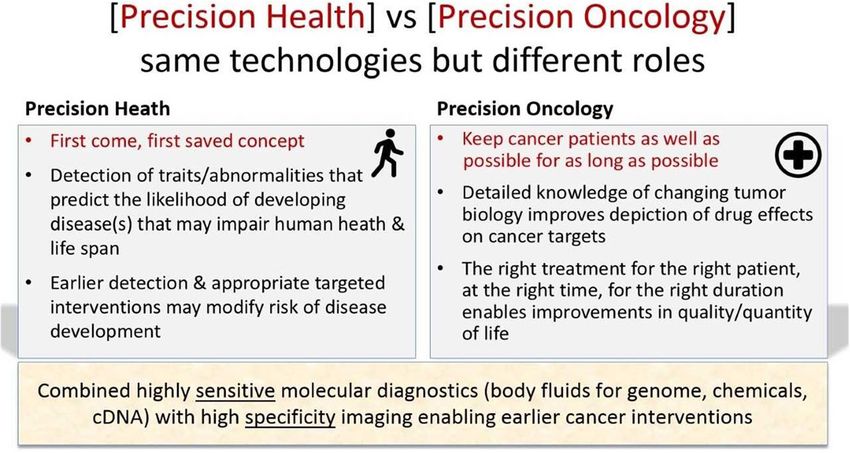
(Fig. 1). In this review, we first summarise the roles of Urology (EAU) recognized that WB-MRI is more sensi-
WB-MRI in oncology and cancer predisposition syn- tive than choline PET/CT and bone scan for detecting
dromes, before examining the feasibility of using this bone metastases in high-risk prostate cancer patients
technique to more general population screening. [11], but acknowledges the limited availability of the
technique [12]. The Advanced Prostate Cancer Consen-
Guideline recommendations and key uses in known sus Conference (APCCC) noted that WB-MRI, although
cancers less widely used, is more sensitive for detecting bone
The International Myeloma Working Group and the metastases than conventional techniques such as com-
British society of Haematology recommend the use of puted tomography and planar bone scans [13]. Recently,
WB-MRI for the detection and staging of multiple mye- an ASCO consensus guideline outlined a number of
loma (Grade A recommendation, GR A) [3], as well as clinical scenarios where next-generation imaging includ-
for the detection of relapsed disease prompted by rising ing PET/CT, PET/MRI, or WB-MRI could have
Fig. 1 [Precision Health] vs [Precision Oncology] same technologies but different rolesZugni et al. Cancer Imaging (2020) 20:34 Page 3 of 13
management impacts in men with advanced prostate of paediatric and adult subjects [27, 28]. Screening pro-
cancer [14]. Metastasis Reporting and Data System for tocols that include WB-MRI for subjects with LFS have
Prostate Cancer (MET-RADS-P) [15] guidelines pro- been also proposed by Australian and Canadian re-
vided a standardization of acquisition protocols, based searchers [29, 30].
on morphological and DWI sequences, and a guidance For children and adults with hereditary paraganglioma
for image interpretation and structured reporting. and pheochromocytoma syndromes, the AACR also rec-
The application of WB-MRI in breast cancer (BC) ommends biennial screening using WB-MRI [31].
patients can be applied to two specific clinical subgroups In patients with neurofibromatosis, WB-MRI showed
[16]. The first comprises BC patients with bone- good sensitivity in detecting the number, volume, and
predominant or bone-only metastatic disease, where WB- distribution of neurofibromata in a study of 247 subjects
MRI is able to show progressive disease earlier than by Plotkin et al. [32]. In light of these results, the NCCN
computed tomography (CT) and bone scans, enabling recently suggested the development of practical guide-
treatment changes at lower burdens of progressing disease lines to introduce WB-MRI for the detection of malig-
[17, 18]. The second comprises women who develop BC nant peripheral nerve sheath tumours and to establish a
during pregnancy. As a radiation-free imaging technique standardized, cost-efficient WB-MRI protocol for image
requiring no contrast medium administration, WB-MRI acquisition [33].
has been proposed as the technique of choice for systemic In subjects with constitutional mismatch repair defi-
staging of pregnant women developing BC [19, 20]. ciency syndrome (CMMRD), a consensus statement by
There is growing use of WB-MRI for the follow-up of the Care for CMMRD Consortium and by the
lymphoma patients with non-avid or variable FDG PET/ International Biallelic Mismatch Repair Deficiency
CT avidity where WB-MRI has superior diagnostic per- Consortium recommends yearly WB-MRI from the age
formance to FDG-PET/CT [21]. Furthermore, WB-MRI of six [34] to screen for development of cancers.
has a diagnostic performance comparable to FDG-PET/
CT in FDG avid lymphoma patients [22]. The enthusi- Cancer screening in the general population
asm for using WB-MRI as a surveillance method in chil- A meta-analysis [35] and systematic review [36] have re-
dren and younger patients is motivated by the clinical cently summarized the diagnostic yields of WB-MRI in
need to minimise radiation exposure following the the population screening context, with particular focus
ALARA (As Low As Reasonably Achievable) principles on the prevalence of relevant and indeterminate findings.
of radioprotection [23]. However, there are no evidence-based recommendations
Finally, two large multicentre prospective studies have on the key issues such as imaging protocols and strat-
been recently published, comparing the diagnostic ac- egies for classifying and/or managing findings.
curacy and efficiency of WB-MRI-based staging path- To address this short coming, we identified using
ways with standard pathways in colorectal and lung PubMed searches and cross-checking of citations, 14
cancer [24, 25]. In both studies, WB-MRI staging path- studies published between 2005 and 2020 describing the
ways had similar accuracy to standard pathways and re- use of WB-MRI for cancer screening in asymptomatic
duced staging time and costs. subjects in the general population. For 12 of the 14 stud-
ies (6423 subjects) the intended purpose was or included
Guideline recommendations in Cancer predisposition cancer screening [37–48]. In the remaining two studies,
syndromes the main purpose was the mapping of body fat (148
Several international guidelines recommend WB-MRI subjects) [49], or cardiovascular disease screening (138
for the early cancer detection in individuals with cancer subjects) [50], with any lesion suspicious for cancer de-
predisposition syndromes where regular surveillance is scribed as incidental findings. These two studies were
necessary. These recommendations are underpinned by not considered for this review. We note that the 209
the lack of ionizing radiation exposure using WB-MRI subjects included in the pilot study by Perkins et al. [39]
and the good diagnostic performance for disease detec- were included also in the larger study by Hou et al. [40].
tion, with a sensitivity ranging from 50 to 90%, and a Therefore, this was considered in the overall count of
specificity ranging from 93 to 95%, as described in the screened subjects, as reported in Table 1.
largest studies available [24–26].
In the setting of Li-Fraumeni syndrome (LFS), guide- Imaging protocol
lines developed by the National Comprehensive Cancer
Network (NCCN) and by the American Association for a) Literature review
Cancer Research (AACR) indicate annual WB-MRI
along with brain MRI with contrast (and breast MRI in In all 12 studies for cancer screening, the anatomical
women) as the techniques of choice for the surveillance coverage included head, neck, chest, abdomen andTable 1 overview of the 12 studies reporting WB-MRI in asymptomatic subjects of the general population
WB-MRI protocol Per-lesion analysis
Author Year Country Magnet T1W T1W T2W T2W DWI Contrast Cardiovascular Classification method All Not ≥ Highly
strenght TSE GE FS sub-protocol findings relevant Relevant relevant
37
Goehde 2005 GER 1.5 WB S S Y cardiac, binary: 1 (non-relevant), 2 (relevant) 329 233 70, 96 29,
WB-MRA 8% 2%
Baumgart 38 2007 GER 1.5 WB S Y cardiac, –
WB-MRA
Zugni et al. Cancer Imaging
Lo 41 2008 HKG 3.0 WB S WB binary: 1 (non-relevant), 2 (relevant) 234 210 89, 24 10,
7% 3%
Takahara 42 2008 NED 1.5 WB WB WB –
Hegenscheid 2013 GER 1.5 WB WB S S optional cardiac, categorical system: 1(non-relevant), 2 (relevant 13,455 12, 92, 990 7, 62 0,5%
43
WB-MRA benign), 3 (relevant unclear), 4 (relevant malignant) 403 2% 4%
(2020) 20:34
Cieszanowski 2014 PLN 1.5 WB WB categorical system: 1 (non-relevant), 2 (moderately 3375 2997 88, 378 11, 15 0,4%
44
or potentially relevant), 3 (relevant) 8% 2%
Tarnoki 45 2015 GER 3.0 WB WB WB WB Y WB-MRA categorical system: 1 (non-relevant), 2 (requiring 68 44 64, 24 35, 1 1,5%
further evaluation), 3 (relevant) 7% 3%
Ulus 46 2016 TUR 1.5 WB WB S optional –
47
Saya 2017 UK 1.5 WB WB WB categorical system: 1 (definitely benign), 2 (likely to
be benign), 3 (equivocal), 4 (likely to be malignant),
5 (definitely malignant)
Lee 48 2018 KOR 1.5 WB WB categorical system: 1 (benign), 2 (requiring further 500 290 58, 210 42, 6 1,2%
evaluation), 3 (malignant) 0% 0%
Perkins* 39 2018 USA 3.0 WB WB WB N cardiac –
Hou 40 2020 USA 3.0 WB WB WB N cardiac –
Total 17,961 16, 91, 1625 9, 84 0.5%
336 0% 0%
Abbreviations: T1W = T1 weighted; T2W = T2 weighted; TSE = turbo spin echo; GE = gradient echo; FS = fat saturated; DWI = diffusion weighted imaging; S = single anatomic segment; WB = whole-body; WB-MRA =
whole-body magnetic resonance angiography
* subjects included in this study are also included in the study by Hou et al. Therefore, they were not counted in the overall number of WB-MRI examinations, nor in the overall number of confirmed malignant cancers
Page 4 of 13Table 1 overview of the 12 studies reporting WB-MRI in asymptomatic subjects of the general population (Continued)
Per-subject analysis Cancer detection
Author n° of WB-MRI examinations Entirely normal (no Abnormal findings With relevant findings (requiring further Suspected malignant Confirmed malignant
findings) reported evaluation) cancers cancers
37
Goehde 298 82 31.0% 1 0,3% 1 0.3%
38
Baumgart 1007 4 0,4% 4 0.4%
Lo 41 132 8 6,1% 124 93,9% 24 18.2% 4 3,0% 4 3.0%
Zugni et al. Cancer Imaging
42
Takahara 10 1 10.0% 1 10,0% 1 10.0%
Hegenscheid 43 2500 787 31.5% 62 2,5%
Cieszanowski 44 666 7 1,1% 659 98,9% 7 1,1% 7 1.1%
Tarnoki 45 22 2 9,1% 20 90,9% 15 68.0% 1 4,5%
(2020) 20:34
Ulus 46 116 33 28,4% 83 71,6% 12 10.3% 3 2,6% 2 1.7%
Saya 47 44 8 18.2% 0 0,0% 0 0%
Lee 48 229 16 7,0% 213 93,0% 6 2,6% 2 0.9%
Perkins* 39 209* 70 33.5% 4 1,9% 4* 1.9%
Hou 40 1190 20 1.7%
Total 6214 66 / 1165 5,7% 1099 / 1165 94,3% 999 / 3331 30,0% 93 / 5233 1.8% 41 / 3692 1.1%
Page 5 of 13Zugni et al. Cancer Imaging (2020) 20:34 Page 6 of 13 pelvis; however, the lower limbs were included in nine b) Evidence Synthesis and recommendations studies (Supplementary Fig. 1). For all 12 studies it was possible to obtain detailed information regarding the WB-MRI scanning protocols for cancer screening are orientation of the acquired images and the types of the analogous to protocols laid out for metastasis detec- sequences used in the WB-MRI protocol, which are tion in advanced prostate cancer (MET-RADS-P) [15] summarized in supplementary Table 1 and supplemen- and multiple myeloma (MY-RADS) [5], with minor tary Fig. 1. In nine [39–41, 43–45, 47, 48], both T1 and modifications. Morphologic imaging forms the basis of T2 weighted images were acquired across the whole WB-MRI protocols in MET-RADS-P and MY-RADS body, while in the remaining three studies, just one guidelines, with GRE T1-weighted images in axial or morphological sequence was acquired (Table 1 and coronal orientation considered mandatory from head to supplementary Table 1). Whole body DWI sequences mid-thigh for MET-RADS and to the knee for MY- were utilized in just five studies [39, 40, 42, 45, 47]. All RADS, while axial TSE T2-weighted images without fat studies provided detailed information regarding the WB- suppression are considered optional. For cancer screen- MRI protocol used for cancer screening. This illustration ing protocols, both T1-weighted and T2-weighted im- provides a synthesis of the anatomical coverage and the ages without fat suppression are required for the optimal image orientation used for the standard unenhanced localization and characterisation of findings. T1- examination, in the different body regions [Additional weighted imaging can be performed using a GRE Dixon Fig. 1]. Additional sub-protocols for the evaluation of sequence, allowing fat-only, water-only and relative fat- specific organs were performed in six studies. fraction images to be derived [51]. While T2-weighted Whole-body T1-weighted images were acquired in 11 sequences with fat suppression have traditionally been studies [37–45, 47, 48], always using Gradient Echo used in musculoskeletal studies, T2-weighted sequences (GRE) sequences, while Turbo Spin-Echo (TSE) se- without fat suppression seem more suitable for onco- quences were used only in one of them, in addition to logical studies and more time efficient, as recommended GRE. Whole-body T2-weighted images were acquired in by MET-RADS-P and MY-RADS guidelines, and are eight studies using TSE sequences: with fat-suppression therefore suggested for WB-MRI cancer screening.. via Inversion Recovery techniques in five, with both fat- Inclusion of the lower limbs is mandatory in WB-MRI suppressed and unsuppressed acquisitions in one, and protocols for cancer screening in subjects with cancer without fat suppression in two. Whole body DWI was predisposition syndromes, such as Li-Fraumeni syn- performed in five studies [39, 40, 42, 45, 47], always in drome [30], due to high incidence of soft tissue cancers. addition to the morphological T1 and/or T2-weighted Since malignant lesions in the lower limbs have not been imaging. reported in any studies of WB-MRI for cancer screening Additional regional oncologic MRI sub-protocols were in the general population, a protocol that covers from performed in six out of 11 studies (Supplementary head to mid-thigh is sufficient for cancer screening. Table 1), including comprehensive multi-sequence brain While the use of gadolinium-based contrast agents MRI in four studies [37, 39, 40, 43], MR colonography in can increase the diagnostic performance of WB-MRI in two [37, 38], MRI mammography in one [43] and pros- some body regions (particularly the brain), it also repre- tate MRI in two [39, 40] (Supplementary Fig. 1). Six sents a more invasive approach to imaging with unclear studies made use of sub-protocols for the non-oncologic benefits in asymptomatic subjects [52]. The largest evaluation of the cardiovascular system [37–40, 43, 45]. study included in our review (2500 subjects) highlights Supplementary Table 1 provides further details regard- the low diagnostic yield of contrast enhanced sub- ing the protocols used in each study. protocols, with only three tumours diagnosed by MRI Intravenous contrast agent was administered in three mammography and no tumours detected on post- studies where WB-MRI was performed for cancer contrast T1-weighted imaging performed for whole- screening. However, its use was motivated by additional body MRI angiography (WB-MRA) [43]. In fact, most sub-protocols requiring contrast administration per- authors have avoided the use of contrast agent in gen- formed in the same sitting, including cardiac MRI, MR eral cancer screening, except when cardiovascular risk angiography and MR colonography [37, 38, 45]. In a is also being assessed or when abnormalities are seen fourth study, contrast was administered in those patients during WB-MRI examinations requiring supplementary who accepted to undergo optional cardiac MRI, whole- contrast enhancement to arrive at a diagnosis. The is- body MR angiography or MR mammography [43]. In sues of gadolinium deposition in the brain and other one study, intravenous contrast agent was administered body tissues [53], and the discomfort related to intra- in a minority of subjects (12 out of 116) to further char- venous puncture, represent further disincentives for its acterise suspicious findings detected by the unenhanced use in general cancer screening, therefore the use of sequences [46]. contrast agents is not recommended.
Zugni et al. Cancer Imaging          (2020) 20:34                                                                                                 Page 7 of 13

  Diffusion sequences have shown high sensitivity for                             Reading and reporting
cancer detection across multiple body regions; however,                           In a study on the diagnostic performance of WB-MRI
only seven studies included in our review made use of                             for cancer screening in subjects with LFS, Anupindi
this technique. Outside the brain, DWI sequences were                             et al. proposed that the examinations must be reported
limited to the upper abdomen in two studies and used                              by radiologists with experience in oncologic WB-MRI
for whole-body evaluation in five studies [39, 40, 42, 45,                        [55]. We suggest extending this recommendation to
47]. Notably, the studies including DWI were published                            WB-MRI for cancer screening also, where it is extremely
after year 2009, whereas three out of five studies not                            important that readers are experienced enough to avoid
using DWI were published before 2009. It is interesting                           harms through unnecessary additional testing on the
to note that recognition of the usefulness of DWI for                             one hand, and to have detailed knowledge of common
cancer imaging emerged from a consensus conference of                             cancer guidelines and of best practice recommendations,
the International Society for Magnetic Resonance in                               to appropriately advise subjects with relevant findings.
Medicine [54] published in 2009. Progress in MRI tech-                            To date, the number of WB-MRI examinations a radi-
nology has both improved DWI image quality and re-                                ologist should report to gain enough expertise is not
duced acquisition times, making this technique highly                             known, as no study has formally addressed this issue.
suitable for whole-body imaging. Therefore, DWI should                            However, it is likely that the required expertise can be
be used, pending future studies investigating WB-MRI                              most readily be reached by oncological radiologists, who
with DWI for general cancer screening.                                            routinely report WB-MRI examinations in cancer pa-
  With existing commercial MR hardware and se-                                    tients. Where this may not be possible or practical,
quences, the proposed mandatory components could be                               Greer et al. have suggested that centres with a low
acquired in under thirty minutes (Table 2). Additional                            volume of WB-MRI examinations could benefit from
regional assessments with specific sequences, for ex-                             central review of such examinations by more experi-
ample brain examinations with FLAIR sequences and                                 enced readers [56].
lungs evaluation with short echo-time GRE. Additional
T1 weighted, and T2 weighted images with fat suppres-                             Strategies for the classification of WB-MRI findings
sion of the spine, are recommended for metastasis detec-
tion by MET-RADS-P and MY-RADS guidelines, but                                       a) Literature review
this may not be necessary in the setting of cancer
screening; in fact, only four screening studies include sa-                         Seven studies reported the use of categorical systems
gittal imaging of the spine.                                                      for the classification of findings. Two studies made use
  To avoid errors and reduce the demands on radiogra-                             of a binary classification distinguishing between non-
phers, we strongly recommend the composing of con-                                relevant (benign and not requiring further evaluation) or
tiguous imaging blocks for each sequence, as well as the                          relevant findings (requiring further imaging or diagnostic
automated calculation of derived images (e.g. water, fat                          workup) [37, 41]. Three studies classified findings into
and fat fraction from Dixon images, and reconstruction                            three categories, as either non-relevant (benign, not sig-
of maximum intensity projections of the high b-value                              nificant), relevant (requiring further evaluation) or highly
DWI images), when possible.                                                       relevant (malignant, highly significant) [44, 45, 48]. One

Table 2 proposed WB-MRI protocol for cancer screening in asymptomatic subjects of the general population
          Sequence description                                                             Characteristics                                    Recommendation
1         Whole-body (head to mid-thigh) T1W GRE, Dixon technique                          Axial or coronal (5 mm slice thickness)            Mandatory
2         Whole-body (head to mid-thigh) T2W, TSE without fat- suppression                 Axial or coronal (5 mm slice thickness)            Mandatory
3         Whole-body (head to mid-thigh) DWI, STIR fat suppression,                        Axial (5 mm slice thickness)                       Mandatory
          contiguous slicing, multiple stations                                            2 b-values:
          • ADC calculations with mono-exponential data fitting                            • b50–100 s/mm2
          • 3D-MIP reconstructions of highest b-value images*                              • b800–1000 s/mm2)
Additional regional assessments:
4         • Brain: T2W FLAIR                                                               Brain: Axial (5 mm slice thickness)                Optional
5         • Lung: T1 GRE short echo-time single breath hold                                Lung: Axial (< 3 mm slice thickness)               Optional
6         • Whole spine T1W, TSE                                                           Sagittal (4–5 mm slice thickness)                  Optional
7         • Whole spine STIR (preferred) or fat suppressed T2W                             Sagittal (4–5 mm slice thickness)                  Optional
W = weighted; TSE = turbo spin echo; STIR = short tau inversion recovery; GRE = gradient echo; DWI = diffusion weighted imaging; ADC = apparent diffusion
coefficient; MIP = maximum intensity projection; FLAIR = FLuid Attenuated Inversion Recovery
* Whole-body rotational 3D MIP images rotating along the cranio-caudal axis (≤3 degrees of rotation per frame), displayed using an inverted grey scale
Zugni et al. Cancer Imaging (2020) 20:34 Page 8 of 13
study classified findings into four categories (non-rele- Strategies for the management of WB-MRI findings
vant, relevant benign, relevant unclear, relevant malig-
nant) [43], while the remaining study used five a) Literature review
categories (definitely benign, likely to be benign, equivo-
cal, likely to be malignant, definitely malignant) [47]. The management of relevant findings was only de-
Findings related to cardiovascular diseases were reported scribed in five studies, representing less than half of the
in a separate section for the six studies that included reviewed papers. In three, detailed descriptions of the
cardiac or angiographic imaging sub-protocols, but these management of relevant findings was reported: Lo et al.
are not relevant to the current discussion, which is fo- [41] made use of additional imaging evaluations for spe-
cused on oncologic findings. cific body regions (ultrasound for thyroid nodules, CT
for lung nodules, pancreatic and retroperitoneal lesions,
b) Evidence synthesis and recommendations contrast enhanced MRI for liver, kidney and prostate le-
sions, plain radiograph for long bones focal lesions);
Strategies adopted for classification of findings differed Ulus et al. [46] performed dedicated contrast enhanced
widely, rendering systematic comparison between stud- MRI studies in the same sitting of WB-MRI for the ma-
ies difficult. For example, the binary classifications jority of suspicious findings and used CT for lung nod-
adopted in two studies [37, 41] does not describe the ules; Goehde et al. [37] made use of region specific
number of subjects with a strong suspicion for tumour, imaging modalities (CT scans for lung nodules, MRI for
therefore reducing the interpretability of the results. brain, liver, kidney and bone lesions, sonography for thy-
Similarly, in one study [44] where three categories were roid nodules) and direct histopathological verification
used, the rate of highly relevant findings (0.4%) also in- for clearly malignant masses (kidney). In the remaining
cluded non-neoplastic findings requiring immediate re- two studies [43, 47], further management was discussed
ferral, implying that the rate of oncologically relevant by a multidisciplinary board, but provided no descrip-
findings was lower. This difference may not be clear to tions of additional examinations undertaken.
subjects willing to undergo the examination, creating er-
roneous expectations regarding the performance of WB- b) Evidence synthesis and recommendations
MRI for cancer screening in the general population.
The adoption of a standardized structured report akin The adoption of a standardised management of rele-
to disease specific MET-RADS-P and MY-RADS tem- vant findings represents a critical gap for the general use
plates adapted for screening applications will likely im- of WB-MRI for cancer screening. Given the high sensi-
prove reporting repeatability, as well as provide greater tivity of the technique, successful adoption of WB-MRI
reproducibility and comparability across studies. Such a depends on having the means and methods to manage
reporting template has yet to emerge for general popula- the entire range of findings generated by a single WB-
tion screening. We believe that a classification system MRI examination. Management should follow estab-
based on five categories should be adopted at a lesion lished guidelines for incidental findings in the different
level to indicate the likelihood of malignancy in cancer body regions as far as possible, such as those for lung
screening setting. Category 1 and 2 for normal and be- nodules [57], renal cysts [58], pancreatic cysts [59] and
nign findings, and categories 3, 4 and 5 for findings with the Radiology White Papers for Managing Incidental
increasing oncological relevance (Table 3). Stratification Findings on Abdominal and Pelvic CT and MRI [60],
of the oncological relevance of findings would allow the also requiring the establishment of specific onward refer-
application of different strategies for investigations and ral pathways for all findings observed.
patient management.
Abnormal findings in WB-MRI: per-finding and per-subject
analysis
Table 3 proposed classification system for findings detected by
WB-MRI A per-finding analysis of the outcome of WB-MRI was
possible in six studies (Table 1), which reported a total
Category Likelihood of cancer
of 17,961 findings. From a per-finding perspective, 91%
1 Normal
of reported findings were non-relevant and 9% were
2 Benign oncologically relevant (i.e. requiring further investiga-
3 Equivocal tion). In the four studies that also provided the rate of
4 Suspicious highly relevant findings (i.e. suspicious for malignancy),
5 Very suspicious this proportion reached 0.5% of all findings. The number
1–2 = no follow-up
of findings suspicious for malignancy reported in each
3–4-5 = follow-up or further investigation triggered by WB-MRI study across the different body regions are summarizedZugni et al. Cancer Imaging (2020) 20:34 Page 9 of 13
in Table 4. Notably, no suspicious tumours were re- of WB-MRI and from the prevalence of such disease in
ported in the lower limbs in the general population, des- the population being evaluated. A meta-analysis by Li
pite coverage across 4800 examinations. et al., including 1067 patients with different tumour
A per-subject analysis of the outcome of the WB-MRI types from 13 studies, calculated a pooled per-patient
was possible in five studies (Table 1). From a per-subject sensitivity and specificity for the detection of primary
perspective, 94% of the WB-MRI examinations were re- and/or metastatic lesions by WB-MRI with DWI of 90
ported to show some abnormal findings while 6% were and 95%, respectively [26]: from these results we calcu-
entirely normal. Nearly 30% of all WB-MRI yielded lated a NPV of 96%. Considering the lower prevalence of
oncologically relevant findings, while highly relevant malignant tumours (reported average < 2%) in asymp-
findings arose in only 1.8% of people. Despite the high tomatic subjects of the general population undergoing
number of findings detected by WB-MRI, the rate of ex- WB-MRI for cancer screening, as a consequence we
aminations that potentially lead to further diagnostic would expect even higher NPV values for WB-MRI in
evaluations, such as further imaging studies, remains cancer screening, also emphasising the need to adjust
relatively low, around 30%. This highlights the ability of the threshold for prompting further investigations of in-
WB-MRI not only for lesion detection but also for the cidental findings. Therefore, given the low probability of
characterization of potential abnormalities. diagnosing malignant cancer, a high threshold should be
applied when requiring additional diagnostic tests for
Cancer detection abnormal findings in the general population, to avoid
over-investigations. In-depth investigations should be
a) Literature review considered only for definite abnormalities, for which on-
ward diagnostic pathways should be planned according
On a per-subject basis, across eleven studies [37–39, to existing guidelines and good practises.
41–48], a total of 93 WB-MRI examinations out of 5233
were reported as positive for malignancy (1.8%). Notably
however, in the 10 studies [37–42, 44, 46–48] that re- Patient acceptability
ported the number of confirmed malignant cancers, Given the high frequency of “abnormal” findings at WB-
these were ultimately established in 41 out of 3692 ex- MRI screening, importance should be given to the pos-
aminations (1.1%) (Table 1). sible repercussions on quality of life and patient anxiety.
In 2013, Schmidt et al. published the results of a survey
a) Evidence synthesis conducted on 471 subjects from the SHIP study, who
had been notified of the presence of potentially relevant
The cancer detection rate of WB-MRI in the general findings [63]. Among these subjects, 10% reported
population is comparable to those observed in other strong distress while awaiting for WB-MRI results (six
cancer screenings. In a meta-analysis by Blanks et al. weeks) and 29% reported moderate to severe distress
[61] showed a detection rate of 7.59 per 1000 subjects after receiving the results. The same authors examined
(0.8%) for breast cancer at prevalent screening with the long-term impact on quality of life and depressive
digital mammography. Notably, a meta-analysis by Bal- symptoms [64] by surveying 2188 subjects 2.5 years after
linger et al. [62] conducted in subjects with Li-Fraumeni WB-MRI and 2232 individuals who had not undergone
Syndrome undergoing surveillance with WB-MRI re- WB-MRI. The survey did not detect significant differ-
ported a much higher cancer detection rate of 7%. ences in quality of life and depressive symptoms between
Therefore, WB-MRI for screening in the general popula- the two groups, or between the subjects who had been
tion should be assessed keeping in mind that the likely notified with potentially relevant findings and those who
low prevalence of malignant tumours in these subjects had not. The authors concluded that, while WB-MRI
will influence the negative predictive value (NPV) of the can generate distress and anxiety in the short term, it is
examination. On the other hand, the presence of risk generally well accepted in the long term, with quality of
factors and relevant family history for cancer should be life and subjective stress-levels comparable to those of
carefully collected, to allow personalised stratification of other already existing cancer screening programs.
the subjects’ cancer risk.
By the same measure, before WB-MRI examination,
subjects from the general population should be informed Conclusions
about both the low pre-test probability of detecting ma- Despite the heterogeneous methodology and the variable
lignant cancer and the high likelihood of findings requir- results of WB-MRI studies performed for cancer screen-
ing follow-up investigations. The NPV for the presence ing in the general population, we can make a few gener-
of a malignant tumour will depend upon the sensitivity alised conclusions:Table 4 Suspicious malignant cancers detected by WB-MRI in the published studies
Author Head Neck Chest Abdomen Pelvis Lower Spine MRI
limbs mammography
37
Goehde 1 NC brain 1 NC lung suspicious 1 H renal cell carcinoma
suspicious lesion
lesion
Baumgart 38 2 H bronchial 5 H renal cell carcinomas
adenocarcinoma
Zugni et al. Cancer Imaging
Lo 41 1 H thyroid 1 H bronchial 1 H renal cell carcinoma
follicular adenocarcinoma
carcinoma
1 H neuronedocrine tumor
42
Takahara 1 H lung cancer
(2020) 20:34
Hegenscheid 1 NC malignant 3 NC malignant 21 NC malignant urinary tract 17 NC female genital 2 NA malignant 3 NA malignant
43
head neck lesions lesions malignant vertebral lesions
lesion lesions lesions
7 NC malignant abdominal 8 NA metastases
organs lesions (site not
specified)
Cieszanowski 1 H glioma 1 H bronchial 2 H renal cell carcinomas 1 H ovarian tumor
44
adenocarcinoma
1 H metastases 1 H testicular
tumor
Tarnoki 45 1 NC pararectal
suspicious
lesion
Ulus 46 1 H renal cell carcinoma
1 H adrenal carcinoma
1 H pancreatic cystadenoma
Saya 47
Lee 48 1 I Tongue 3 NC suspicious renal lesions
cancer
1 I renal cell carcinoma
Hou 40 1 I optic 2 H papillary 2 H lymphoma 5 H renal cell carcinomas 6 H prostate
nerve thyroid cancers
glioma carcinoma
1 H thymoma 1 H low-grade intraductal 2 H urinary bladder
papillary mucinous carcinomas
neoplasm
Total 4 6 10 51 28 0 10 3
H = histologically confirmed, I = confirmed by further imaging, NC = non confirmed, NA = not investigated
Page 10 of 13Zugni et al. Cancer Imaging (2020) 20:34 Page 11 of 13
The typical imaging protocol comprises T1- Authors’ contributions
weighted GRE, T2-weighted FSE (fast spin-echo) FZ: literature search, data analysis, writing, figure editing. AP, DMK: writing,
revision, figure editing. PS: writing, revision. MB: revision. GP: study design,
and diffusion weighted sequences, extending from writing, revision. The author(s) read and approved the final manuscript.
the head to mid-thigh, with optional additional re-
gional assessments. The administration of intraven- Funding
ous contrast agent is not recommended. None.
Abnormal findings are expected in about 95% of
Availability of data and materials
screened subjects, about 30% of subjects would
The datasets during and/or analysed during the current study available from
require further investigations but less than 2% would the corresponding author on reasonable request.
be reported as suspicious for malignant cancers.
Findings should be classified using a categorical Ethics approval and consent to participate
system, based on their likelihood of malignancy. It is Not applicable.
important to set high thresholds for further
Consent for publication
investigations to minimize harms from diagnostic Not applicable.
testing.
Subject counselling on the high likelihood of Competing interests
incidental findings and the low likelihood of cancer The authors declare that they have no competing interests.
detection together with established onward referral
Author details
pathways are needed. 1
Division of Radiology, IEO European Institute of Oncology IRCCS, Via
Training is needed for reporting WB-MRI examina- Giuseppe Ripamonti 435, 20141 Milan, Italy. 2Paul Strickland Scanner Centre,
Mount Vernon Cancer Centre, Rickmansworth Rd, Northwood HA6 2RN, UK.
tions; however, the number of examinations a radi- 3
Department of Radiology, The Royal Marsden Hospital (Surrey), Downs Rd,
ologist should report to acquire this expertise still Sutton SM2 5PT, UK. 4Department of Oncology and Hemato-Oncology,
has to be investigated. University of Milan, Via S. Sofia, 9/1, 20122 Milan, Italy. 5Precision Imaging
and Research Unit, Department of Medical Imaging and Radiation Sciences,
IEO European Institute of Oncology IRCCS, Via Giuseppe Ripamonti 435,
Guidelines are needed to establish common strategies 20141 Milan, Italy.
for the classification and management of abnormal find-
Received: 26 March 2020 Accepted: 3 May 2020
ings in studies using WB-MRI for cancer screening. The
current experience is still too heterogeneous to draw
meaningful conclusions regarding general efficacy. Fu- References
ture multisite studies should aim to provide the evidence 1. Petralia G, Padhani AR, Pricolo P, Zugni F, Martinetti M, Summers PE, et al.
that may pave the way to guidelines and recommenda- Whole-body magnetic resonance imaging (WB-MRI) in oncology:
recommendations and key uses. Radiol Med. 2018.
tions for asymptomatic population screening. 2. Morone M, Bali MA, Tunariu N, Messiou C, Blackledge M, Grazioli L, et al.
Whole-body MRI: current applications in oncology. Am J Roentgenol.
American Roentgen Ray Society. 2017;209:W336–49.
Supplementary information
3. Dimopoulos MA, Hillengass J, Usmani S, Zamagni E, Lentzsch S, Davies
Supplementary information accompanies this paper at https://doi.org/10.
FE, et al. Role of magnetic resonance imaging in the Management of
1186/s40644-020-00315-0.
Patients with Multiple Myeloma: a consensus statement. J Clin Oncol.
2015;33:657–64.
Additional File 1: Table 1. summary of MR technology and protocols 4. Chantry A, Kazmi M, Barrington S, Goh V, Mulholland N, Streetly M, et al.
used for WB-MRI. This table provides a detailed overview of the types of Guidelines for the use of imaging in the management of patients with
sequences used by the 12 studies included in this review. For each body myeloma. Br J Haematol Wiley. 2017;178:380–93.
region, the different types of sequence performed are annotated, with 5. Messiou C, Hillengass J, Delorme S, Lecouvet FE, Moulopoulos LA, Collins
reference to the anatomical orientation of the planes. Additional sub pro- DJ, et al. Guidelines for acquisition, interpretation, and reporting of whole-
tocols are also described. body MRI in myeloma: myeloma response assessment and diagnosis system
Additional file 2: Figure 1. Summary of body regions and imaging (MY-RADS). Radiology. Radiological Society of North America. 2019;291:5–13.
planes covered by the WB-MRI core protocols of the 12 studies included 6. Müller-Horvat C, Radny P, Eigentler TK, Schäfer J, Pfannenberg C, Horger M,
in this review. Additional dedicated sub-protocols performed for the et al. Prospective comparison of the impact on treatment decisions of
evaluation of specific organs are shown on the right. Sub protocols re- whole-body magnetic resonance imaging and computed tomography in
quiring administration of contrast agents are marked by an asterisk. patients with metastatic malignant melanoma. Eur J Cancer. 2006;42:342–50.
7. Pfannenberg C, Aschoff P, Schanz S, Eschmann SM, Plathow C, Eigentler TK,
et al. Prospective comparison of 18F-fluorodeoxyglucose positron emission
Abbreviations tomography/computed tomography and whole-body magnetic resonance
WB-MRI: Whole-body magnetic resonance imaging; GR: Grade of imaging in staging of advanced malignant melanoma. Eur J Cancer. 2007;
recommendation; LE: Level of evidence; PET/CT: Positron emission 43:557–64.
tomography / computed tomography; CT: Computed tomography; LFS: Li 8. Petralia G, Padhani A, Summers P, Alessi S, Raimondi S, Testori A, et al.
Fraumeni syndrome; CMMRD: Constitutional mismatch repair deficiency; Whole-body diffusion-weighted imaging: is it all we need for detecting
GRE: Gradient echo; TSE: Turbo spin echo; DWI: Diffusion weighted imaging; metastases in melanoma patients? Eur Radiol. 2013;23:3466–76.
FLAIR: Fluid attenuated inversion recovery; NPV: Negative predictive value 9. Pflugfelder A, Kochs C, Blum A, Capellaro M, Czeschik C, Dettenborn T, et al.
Malignant Melanoma S3-Guideline “Diagnosis, Therapy and Follow-up of
Acknowledgements Melanoma.” JDDG J der Dtsch Dermatologischen Gesellschaft
Not applicable. 2013;11:1–116.Zugni et al. Cancer Imaging (2020) 20:34 Page 12 of 13
10. Dummer R, Siano M, Hunger RE, Lindenblatt N, Braun R, Michielin O, et al. 30. Villani A, Shore A, Wasserman JD, Stephens D, Kim RH, Druker H, et al.
The updated Swiss guidelines 2016 for the treatment and follow-up of Biochemical and imaging surveillance in germline TP53 mutation carriers
cutaneous melanoma. Swiss Med Wkly. 2016;146. with Li-Fraumeni syndrome: 11 year follow-up of a prospective
11. Shen G, Deng H, Hu S, Jia Z. Comparison of choline-PET/CT, MRI, SPECT, and observational study. Lancet Oncol Elsevier. 2016;17:1295–305.
bone scintigraphy in the diagnosis of bone metastases in patients with 31. Rednam SP, Erez A, Druker H, Janeway KA, Kamihara J, Kohlmann WK, et al.
prostate cancer: a meta-analysis. Skeletal Radiol Springer Nature. 2014;43: Von Hippel–Lindau and Hereditary Pheochromocytoma/Paraganglioma
1503–13. Syndromes: Clinical Features, Genetics, and Surveillance Recommendations
12. Mottet N, Bellmunt J, Briers E, Bolla M, Cornford P, De Santis M, et al. EAU in Childhood. Clin Cancer Res. American Association for Cancer Research
-ESTRO -SIOG guidelines on prostate Cancer. 2016. (AACR); 2017;23:e68–75.
13. Gillessen S, Attard G, Beer TM, Beltran H, Bjartell A, Bossi A, et al. 32. Plotkin SR, Bredella MA, Cai W, Kassarjian A, Harris GJ, Esparza S, et al.
Management of Patients with advanced prostate Cancer: report of the Quantitative Assessment of Whole-Body Tumor Burden in Adult Patients
advanced prostate Cancer consensus conference 2019. Eur Urol. Elsevier B. with Neurofibromatosis. PLoS One. Public Library of Science (PLoS);
V.; 2020. 2012;7:e35711.
14. Trabulsi EJ, Rumble RB, Jadvar H, Hope T, Pomper M, Turkbey B, et al. 33. Reilly KM, Kim A, Blakely J, Ferner RE, Gutmann DH, Legius E, et al.
Optimum Imaging Strategies for Advanced Prostate Cancer: ASCO Neurofibromatosis Type 1–Associated MPNST State of the Science: Outlining
Guideline. J Clin Oncol. American Society of Clinical Oncology (ASCO); 2020; a Research Agenda for the Future. JNCI J Natl Cancer Inst. Oxford University
JCO.19.02757. Press (OUP); 2017;109.
15. Padhani AR, Lecouvet FE, Tunariu N, Koh D-M, De Keyzer F, Collins DJ, et al. 34. Tabori U, Hansford JR, Achatz MI, Kratz CP, Plon SE, Frebourg T, et al. Clinical
METastasis reporting and data system for prostate Cancer: practical Management and Tumor Surveillance Recommendations of Inherited
guidelines for acquisition, interpretation, and reporting of whole-body Mismatch Repair Deficiency in Childhood. Clin Cancer Res. American
magnetic resonance imaging-based evaluations of multiorgan involvement Association for Cancer Research; 2017;23:e32–e37.
in advanced prostate Cancer. Eur Urol Elsevier. 2017;71:81–92. 35. Gibson LM, Paul L, Chappell FM, Macleod M, Whiteley WN, Salman RA-S,
16. Petralia G, Padhani AR. Whole-Body Magnetic Resonance Imaging in et al. Potentially serious incidental findings on brain and body magnetic
Oncology: Uses and Indications. Magn. Reson. Imaging Clin. N. Am. W.B. resonance imaging of apparently asymptomatic adults: systematic review
Saunders; 2018. p. 495–507. and meta-analysis. BMJ. BMJ Publishing Group; 2018;363:k4577.
17. Kosmin M, Makris A, Joshi PV, Ah-See M-L, Woolf D, Padhani AR. The 36. Kwee RM, Kwee TC. Whole-Body MRI for Preventive Health Screening: A
addition of whole-body magnetic resonance imaging to body Systematic Review of the Literature. J Magn Reson Imaging. John Wiley &
computerised tomography alters treatment decisions in patients with Sons, Ltd; 2019;jmri.26736.
metastatic breast cancer. Eur J Cancer. 2017;77:109–16. 37. Goehde SC, Hunold P, Vogt FM, Ajaj W, Goyen M, Herborn CU, et al. Full-
18. Zugni F, Ruju F, Pricolo P, Alessi S, Iorfida M, Colleoni MA, et al. The added Body Cardiovascular and Tumor MRI for Early Detection of Disease:
value of whole-body magnetic resonance imaging in the management of Feasibility and Initial Experience in 298 Subjects. 2005;18418405:598–611.
patients with advanced breast cancer. PLoS One. 2018;Article in. 38. Baumgart D, Egelhof T. Präventives Ganzkörperscreening unter
19. Peccatori FA, Codacci-Pisanelli G, Del Grande M, Scarfone G, Zugni F, Einbeziehung moderner Bildgebung mit Hilfe der
Petralia G. Whole body MRI for systemic staging of breast cancer in Magnetresonanztomographie. Herz Kardiovaskuläre Erkrankungen Urban &
pregnant women. Breast. 2017;35. Vogel. 2007;32:387–94.
20. Montagna E, Peccatori F, Petralia G, Tomasi Cont N, Iorfida M, Colleoni M. 39. Perkins BA, Caskey CT, Brar P, Dec E, Karow DS, Kahn AM, et al. Precision
Whole-body magnetic resonance imaging, metastatic breast cancer and medicine screening using whole-genome sequencing and advanced
pregnancy: A case report. Breast. Churchill Livingstone; 2014. p. 295–296. imaging to identify disease risk in adults. Proc Natl Acad Sci U S A. National
21. Mayerhoefer ME, Karanikas G, Kletter K, Prosch H, Kiesewetter B, Skrabs Academy of Sciences; 2018;115:3686–3691.
C, et al. Evaluation of diffusion-weighted MRI for pretherapeutic 40. Hou YCC, Yu HC, Martin R, Cirulli ET, Schenker-Ahmed NM, Hicks M, et al.
assessment and staging of lymphoma: results of a prospective study in Precision medicine integrating whole-genome sequencing, comprehensive
140 patients. Clin Cancer Res. American Association for Cancer metabolomics, and advanced imaging. Proc Natl Acad Sci U S A. National
Research; 2014;20:2984–2993. Academy of Sciences; 2020;117:3053–3062.
22. Mayerhoefer ME, Karanikas G, Kletter K, Prosch H, Kiesewetter B, Skrabs C, 41. Lo GG, Ai V, Au-Yeung KM, Chan JKF, Li KW, Chien D. Magnetic resonance
et al. Evaluation of Diffusion-Weighted Magnetic Resonance Imaging for whole body imaging at 3 Tesla: feasibility and findings in a cohort of
Follow-up and Treatment Response Assessment of Lymphoma: Results of asymptomatic medical doctors. Hong Kong Med J = Xianggang yi xue za
an 18F-FDG-PET/CT-Controlled Prospective Study in 64 Patients. Clin Cancer zhi. 2008;14:90–6.
Res. American Association for Cancer Research (AACR); 2015;21:2506–13. 42. Takahara T, Kwee T, Kibune S, Ochiai R, Sakamoto T, Niwa T, et al. Whole-
23. Hendee WR, Marc EF. ALARA and an integrated approach to radiation body MRI using a sliding table and repositioning surface coil approach. Eur
protection. Semin Nucl Med Elsevier BV. 1986;16:142–50. Radiol Springer. 2010;20:1366–73.
24. Taylor SA, Mallett S, Beare S, Bhatnagar G, Blunt D, Boavida P, et al. 43. Hegenscheid K, Seipel R, Schmidt CO, Völzke H, Kühn J-P, Biffar R, et al. Potentially
Diagnostic accuracy of whole-body MRI versus standard imaging pathways relevant incidental findings on research whole-body MRI in the general adult
for metastatic disease in newly diagnosed colorectal cancer: the prospective population: frequencies and management. Eur Radiol. 2013;23:816–26.
streamline C trial. Lancet Gastroenterol Hepatol Elsevier Ltd. 2019;4:529–37. 44. Cieszanowski A, Maj E, Kulisiewicz P, Grudzinski IP, Jakoniuk-Glodala K,
25. Taylor SA, Mallett S, Ball S, Beare S, Bhatnagar G, Bhowmik A, et al. Diagnostic Chlipala-Nitek I, et al. Non-Contrast-Enhanced Whole-Body Magnetic
accuracy of whole-body MRI versus standard imaging pathways for metastatic Resonance Imaging in the General Population: The Incidence of Abnormal
disease in newly diagnosed non-small-cell lung cancer: the prospective Findings in Patients 50 Years Old and Younger Compared to Older Subjects.
streamline L trial. Lancet Respir Med Lancet Publishing Group. 2019;7:523–32. Villa E, editor. PLoS One. Public Libr Sci; 2014;9:e107840.
26. Li B, Li Q, Nie W, Liu S. Diagnostic value of whole-body diffusion-weighted 45. Tarnoki DL, Tarnoki AD, Richter A, Karlinger K, Berczi V, Pickuth D. Clinical
magnetic resonance imaging for detection of primary and metastatic value of whole-body magnetic resonance imaging in health screening of
malignancies: a meta-analysis. Eur J Radiol Elsevier BV. 2014;83:338–44. general adult population. Radiol Oncol De Gruyter Open. 2015;49:10–6.
27. NCCN. NCCN Clinical Practice Guidelines in Oncology (NCCN Guidelines®) 46. Ulus S, Suleyman E, Ozcan UA, Karaarslan E. Whole-body MRI screening in
for Genetic/Familial High-Risk Assessment [Internet]. 2018. Available from: asymptomatic subjects; preliminary experience and long-term follow-up
http://bit.ly/1Nubll0. findings. Polish J Radiol Termedia Publishing. 2016;81:407–14.
28. Kratz CP, Achatz MI, Brugières L, Frebourg T, Garber JE, Greer M-LC, et al. 47. Saya S, Killick E, Thomas S, Taylor N, Bancroft EK, Rothwell J, et al. Baseline
Cancer Screening Recommendations for Individuals with Li-Fraumeni results from the UK SIGNIFY study: a whole-body MRI screening study in
Syndrome. Clin Cancer Res. American Association for Cancer Research TP53 mutation carriers and matched controls. Fam Cancer Springer.
(AACR); 2017;23:e38–45. 2017;16:433–40.
29. McBride KA, Ballinger ML, Killick E, Kirk J, Tattersall MHN, Eeles RA, et al. Li- 48. Lee SY, Park HJ, Kim MS, Rho MH, Han CH. An initial experience with the
Fraumeni syndrome: cancer risk assessment and clinical management. Nat use of whole body MRI for cancer screening and regular health checks.
Rev Clin Oncol. 2014;11:260–71. Chen X, editor. PLoS One. Public Libr Sci; 2018;13:e0206681.Zugni et al. Cancer Imaging (2020) 20:34 Page 13 of 13
49. Morin SHX, Cobbold JFL, Lim AKP, Eliahoo J, Thomas EL, Mehta SR, et al.
Incidental findings in healthy control research subjects using whole-body
MRI. Eur J Radiol. Elsevier BV. 2009;72:529–33.
50. Laible M, Schoenberg SO, Weckbach S, Lettau M, Winnik E, Bischof J, et al.
Whole-body MRI and MRA for evaluation of the prevalence of
atherosclerosis in a cohort of subjectively healthy individuals. Insights
Imaging Springer. 2012;3:485–93.
51. Dixon WT. Simple proton spectroscopic imaging. Radiology. 1984;153:189–94.
52. Ballinger ML, Best A, Mai PL, Khincha PP, Loud JT, Peters JA, et al. Baseline
Surveillance in Li-Fraumeni Syndrome Using Whole-Body Magnetic
Resonance Imaging. JAMA Oncol. American Medical Association;
2017;3:1634.
53. Guo BJ, Yang ZL, Zhang LJ. Gadolinium Deposition in Brain: Current
Scientific Evidence and Future Perspectives. Front Mol Neurosci. Frontiers
Media SA; 2018;11:335.
54. Padhani AR, Liu G, Koh DM, Chenevert TL, Thoeny HC, Takahara T, et al.
Diffusion-weighted magnetic resonance imaging as a cancer biomarker:
consensus and recommendations. Neoplasia Neoplasia Press. 2009;11:
102–25.
55. Anupindi SA, Bedoya MA, Lindell RB, Rambhatla SJ, Zelley K, Nichols KE,
et al. Diagnostic performance of whole-body MRI as a tool for Cancer
screening in children with genetic Cancer-predisposing conditions. Am J
Roentgenol American Roentgen Ray Society. 2015;205:400–8.
56. Greer M-LC, Voss SD, States LJ. Pediatric Cancer predisposition imaging:
focus on whole-body MRI. Clin Cancer Res American Association for Cancer
Research. 2017;23:e6–13.
57. MacMahon H, Naidich DP, Goo JM, Lee KS, Leung ANC, Mayo JR, et al.
Guidelines for Management of Incidental Pulmonary Nodules Detected on
CT images: from the Fleischner society 2017. Radiology. Radiological Society
of North America. 2017;284:228–43.
58. Silverman SG, Pedrosa I, Ellis JH, Hindman NM, Schieda N, Smith AD, et al.
Bosniak classification of cystic renal masses, version 2019: an update
proposal and needs assessment. Radiology Radiological Society of North
America. 2019;292:475–88.
59. Tanaka M, Fernández-del Castillo C, Kamisawa T, Jang JY, Levy P,
Ohtsuka T, et al. Revisions of international consensus Fukuoka
guidelines for the management of IPMN of the pancreas.
Pancreatology. Elsevier B.V.; 2017. p. 738–53.
60. Incidental Findings | American College of Radiology [Internet]. [cited
2019 Nov 30]. Available from: https://www.acr.org/Clinical-Resources/
Incidental-Findings.
61. Blanks RG, Wallis MG, Alison R, Kearins O, Jenkins J, Patnick J, et al. Impact of
Digital Mammography on Cancer Detection and Recall Rates: 11.3 Million
Screening Episodes in the English National Health Service Breast Cancer
Screening Program. Radiology. Radiological Society of North America; 2019;
290:629–37.
62. Ballinger ML, Best A, Mai PL, Khincha PP, Loud JT, Peters JA, et al. Baseline
surveillance in Li-Fraumeni syndrome using whole-body magnetic
resonance imaging. JAMA Oncol. 2017;3:1634.
63. Schmidt CO, Hegenscheid K, Erdmann P, Kohlmann T, Langanke M, Völzke
H, et al. Psychosocial consequences and severity of disclosed incidental
findings from whole-body MRI in a general population study. Eur Radiol
Springer-Verlag. 2013;23:1343–51.
64. Schmidt CO, Sierocinski E, Hegenscheid K, Baumeister SE, Grabe HJ, Völzke
H. Impact of whole-body MRI in a general population study. Eur J Epidemiol
Springer Netherlands. 2016;31:31–9.
Publisher’s Note
Springer Nature remains neutral with regard to jurisdictional claims in
published maps and institutional affiliations.You can also read