Giant Mesozoic coelacanths (Osteichthyes, Actinistia) reveal high body size disparity decoupled from taxic diversity - Nature
←
→
Page content transcription
If your browser does not render page correctly, please read the page content below
www.nature.com/scientificreports
OPEN Giant Mesozoic coelacanths
(Osteichthyes, Actinistia) reveal
high body size disparity decoupled
from taxic diversity
Lionel Cavin1*, André Piuz1, Christophe Ferrante1,2 & Guillaume Guinot3
The positive correlation between speciation rates and morphological evolution expressed by body
size is a macroevolutionary trait of vertebrates. Although taxic diversification and morphological
evolution are slow in coelacanths, their fossil record indicates that large and small species coexisted,
which calls into question the link between morphological and body size disparities. Here, we describe
and reassess fossils of giant coelacanths. Two genera reached up to 5 m long, placing them among
the ten largest bony fish that ever lived. The disparity in body size adjusted to taxic diversity is much
greater in coelacanths than in ray-finned fishes. Previous studies have shown that rates of speciation
and rates of morphological evolution are overall low in this group, and our results indicate that these
parameters are decoupled from the disparity in body size in coelacanths. Genomic and physiological
characteristics of the extant Latimeria may reflect how the extinct relatives grew to such a large size.
These characteristics highlight new evolutionary traits specific to these “living fossils”.
Abbreviations
DGM Divisão de Geológia e Mineralogia, Departamento Nacional da Produção, Mineral, Rio de
Janeiro, Brazil
CCK Columbus (Georgia) College, USA
MHNG GEPI Natural history Museum of Geneva, Switzerland (palaeontological collection)
MPV Paléospace, palaeontological museum of Villers-sur-Mer, France
SMC Sedgwick Museum, Oxford, UK
UFMA Coleção Paleontológica da Universidade Federal do Maranhão, Brazil
UMI University Moulay Ismail of Meknès, Morocco
Body size is often used as a proxy for analyzing morphological disparity, and this element is one of the main
evolutionary traits discussed by biologists and paleontologists in order to decipher macroevolutionary processes.
For example, a general increase in body size over time within animal lineages was one of the earliest nomological
law in biology raised by Cope and Depéret1, and has subsequently been regularly confirmed for various c lades2.
Recent studies have shown positive correlations between speciation rates and morphological evolution expressed
in body size3,4. However, body size is only one of the many traits that characterize morphological disparity,
which can be measured by many other parameters5, and the assumption that body size disparity directly reflects
morphological disparity can be questioned.
Coelacanths form a depauperate group of sarcopterygian fish with only one genus today but with a long evo-
lutionary history. These fish are nicknamed "living fossils" because they possess characteristics used by Darwin
to characterize this ill-defined concept, in particular "new forms … have been more slowly formed"6 (Darwin,
however, did not cite the coelacanths, known only from fossils at that time, as examples of "living fossils").
Indeed, the clade exhibits low taxic diversity since its origins in the Devonian (ca 420 Mya) with approximately
63 genera in total. Only three weak successive peaks of higher taxic diversity are recorded in the Upper Devonian,
in the Early Carboniferous and in the Middle Triassic7. Huxley (1866)8 has already noticed the low anatomical
disparity of coelacanths throughout their history, and this observation has been confirmed by most subsequent
1
Department of Geology and Palaeontology, Natural History Museum of Geneva, Geneva,
Switzerland. 2Department of Earth Sciences, University of Geneva, Rue des Maraîchais 13, 1205 Geneva,
Switzerland. 3Institut des Sciences de L’Evolution de Montpellier (Université de Montpellier, CNRS, IRD, EPHE),
Montpellier, France. *email: lionel.cavin@ville-ge.ch
Scientific Reports | (2021) 11:11812 | https://doi.org/10.1038/s41598-021-90962-5 1
Vol.:(0123456789)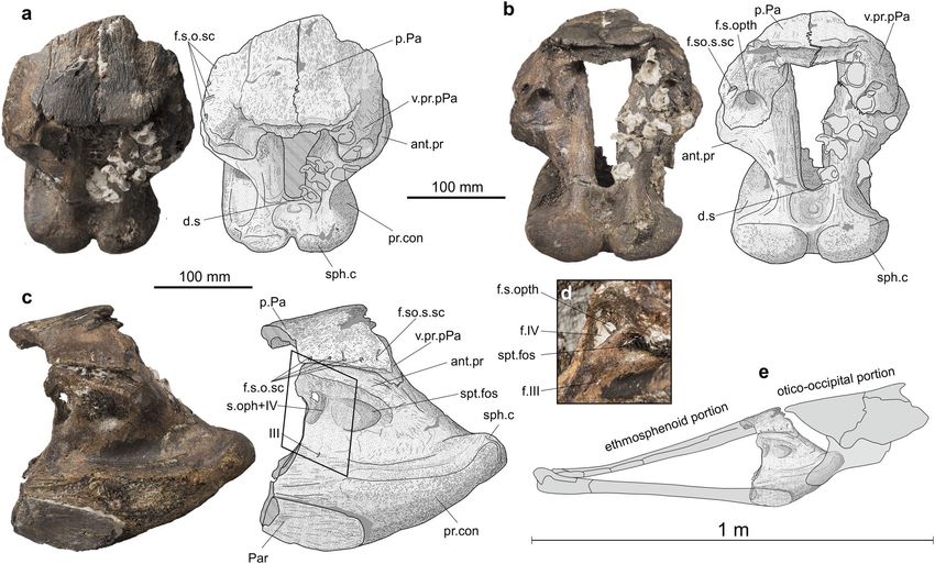
www.nature.com/scientificreports/
studies9–15. This monotonic rate of evolution is interrupted by at least three episodes of increased morphological
disparity, with forms presenting a different Bauplan, roughly contemporary with peaks of taxic d iversity9,16,17.
The rate of genetic evolution within the coelacanth lineage is found by most studies to be slower than that of
other vertebrate lineages in the mitochondrial genome18–20 as well as in the nuclear genome21, at least for the
protein-coding genes22,23.
Extinct giant coelacanths, i.e. fish several meters long, have long been described among the mawsoniids with
Mawsonia gigas by Woodward in 1 90724, from the Early Cretaceous of Brazil, then with Axelrodichthys lavocati
from the ‘mid’ Cretaceous of North Africa25,26, with Trachymetopon sp.27 from the Middle Jurassic of Europe,
but also among the latimeriids with Megalocoelacanthus from the Late Cretaceous of North America28,29. The
fossil record of coelacanths reveals a relative abundance of large-sized species as previously observed by Wenz25
and Dutel et al.27,30.
Here we describe new fossil remains from the Middle Jurassic of Normandy, France, representing one of the
largest known coelacanths ever reported. The specimen, a piece of a braincase, was found in the same deposits
as fragmentary fossils interpreted as possible pups of the same species. We further reassess the Mesozoic fossil
record of giant coelacanths and provide a large-scale comparison of body size disparity versus taxic diversity
between coelacanths (Actinistia) and ray-finned fishes (Actinopterygii) over the Devonian–Paleocene time
interval. We show that the per genus coefficient of body size variance is higher in coelacanths than in ray-finned
fishes. This result calls into question the positive correlation between speciation rates and body size found in most
vertebrate lineages, and more generally questions the use of body size as a valid proxy for anatomical disparity3,4.
Results
New material, geographical and stratigraphic provenances. A large, almost complete, basisphe-
noid of a coelacanth with the posterior end of the parasphenoid sutured ventrally and the posterior part of the
posterior parietals sutured dorsally (MHNG GEPI 5778) has been spotted in the paleontological collections of
the Geneva Natural History Museum, Switzerland. No labels nor information were associated with this speci-
men. A search in the museum’s archives to trace the origin of the specimen was unsuccessful. The fossil was
mechanically and chemically prepared, with 10% diluted HCl. The sediment recovered during the preparation of
specimen was prepared by acetolysis in order to extract microfossils. The material recovered includes vertebrae
and teeth of small fish, diverse micro-gastropods, as well as micro-bivalves, crinoids (roveacrinids), bryozoans,
and foraminifera. The diversity of foraminifera is relatively low, consisting of moderately preserved epistominids
and vaginulinids. The recognized species (Fig. 1) are Epistomina ex. gr. mosquensis Uhlig 1883, Epistomina ex.
gr. uhligi Mjatliuk 1953, Lenticulina quenstedti (Gümbel 1862), L. muensteri (Roemer 1839), L. subalata (Reuss
1854) and Planularia beierana (Gümbel 1862). These taxa have only moderate biostratigraphic value, being
mainly widespread in the upper Middle to Upper Jurassic (Supplementary Fig. S1). The presence of a modern
barnacle shell on the fossil (Supplementary Fig. S2A), an evidence of its discovery from a locality situated near
the seashore, associated to the general type of preservation of the specimen and to the presence of vaginulinids
and epistominids associated with vertebrate fossils are strong indications that this specimen probably comes
from the late Callovian “Marnes de Dives”, probably from the well-exposed cliffs of the “Vaches Noires”, Villers-
sur-Mer, Normandy, F rance31. This facies generally contains an abundance of encrusted gryphaeid oysters as
seen on the skull of the coelacanth bearing several of these shells on one of its sides (Supplementary Video,
Fig. 2, Supplementary Fig. S2B). In addition to these shells, the matrix of the specimen bore the imprint of an
ammonite, reminiscent of Heticoceras (Christian Meister, Antoine Pictet, personal communication 2014) and
a gastropod shell (Supplementary Fig. S2C, D). The “Marnes de Dives” are the equivalent of the lower part of
the “Oxford Clay” of Dorset, UK. It is assumed that the specimen reached the Geneva Natural History Museum
near its inception in 1820 (it was then called the Academic Museum), along with other fossil vertebrates from
Normandy, France.
Recently, two small basisphenoids of coelacanths (MPV 2020.1.13) were discovered by Elisabeth and Gérard
Pénnetier from a Callovian strata of the foreshore at the foot of the cliffs of the “Vaches Noires”, Villers-sur-Mer,
Normandy, France, and therefore come from the same formation as the large basisphenoid.
Morphological description and comparisons. The large specimen (MHNG GEPI V5778) consists of
a complete basisphenoid, in connection dorsally with the posterior part of the skull roof and ventrally with a
fragment of the parasphenoid (Fig. 2). The processus connectens are well developed, slightly curved in lateral
view and extend ventrally to the level of the parasphenoid. The dorsum sellae is proportionally short and forms
anteriorly a shallow wall that constricts ventrally the entrance to the cranial cavity. On the posterior side of the
bone, the well-developed and closely spaced sphenoid condyles are separated from each other by a marked
notch and from the paired processus connectens by shallow depressions. The opening of the cranial cavity is
much deeper than wide, with its dorsal part slightly wider than its ventral part. The antotic processes protrude
laterally and suture dorsally to the ventral processes of the posterior parietal. The surfaces of contact between
both processes are large and oval. The suprapterygoid fossa is well marked but shallow. Anteriorly, a large fora-
men opens oriented frontwards. Within the ossification, just behind the opening, the canal is divided by a thin
horizontal lamina, which separates a larger ventral canal from a smaller dorsal one (Fig. 2d). Based on the paths
of the nerves in Latimeria11, the small dorsal canal may have accommodated the superficial ophthalmic nerve
and the ventral canal the trochlear nerve (IV). In Latimeria, but also in most post-Paleozoic taxa in which this
part of the braincase is preserved, such as Megalocoelacanthus29 and Trachymetopon30, the nerves exit the cra-
nial cavity in the interorbital cartilage. In the Devonian genera Diplocercides and Euporosteus11, the nerves exit
through bone-enclosed foramens. We interpret the occurrence of this large foramen in MHNG GEPI V5778
as a consequence of the large size of the individual and its high degree of ossification rather than affinities with
Scientific Reports | (2021) 11:11812 | https://doi.org/10.1038/s41598-021-90962-5 2
Vol:.(1234567890)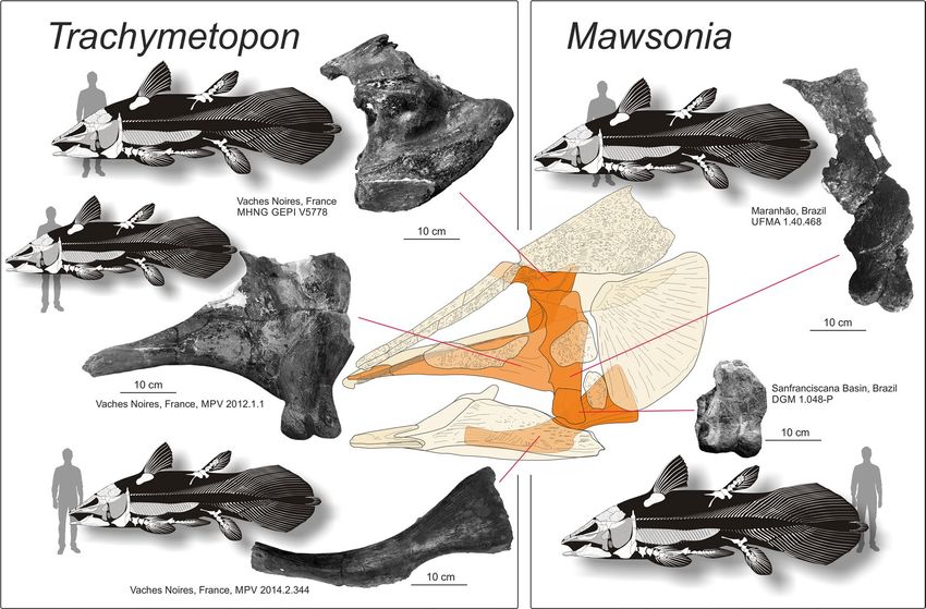
www.nature.com/scientificreports/
Figure 1. Foraminifera found in the matrix containing the fragment of the coelacanth skull (MHNG GEPI
V5778). (a–d) Epistomina ex. gr. mosquensis Uhlig 1883, umbilical, apertural, carinal and spiral views; (e–h)
Epistomina ex. gr. uhligi Mjatliuk 1953, spiral, apertural and umbilical views; (i–k) Lenticulina muensteri
(Roemer 1839), apertural and lateral views; (l–m) Lenticulina quenstedti (Gümbel 1862), apertural and lateral
views; (n) Lenticulina subalata (Reuss 1854), apertural and lateral views; (o) Planularia beierana (Gümbel 1862),
lateral and apertural views.
Paleozoic forms. Ventral to the large foramen, and present on both sides of the basisphenoid, opens the small
oculomotor foramen. The angle between the posteroventral surface of the basisphenoid and the ventral surface
of the parasphenoid is approximately 135°. The basipterygoid and suprapterygoid processes are absent.
The posterior parts of both posterior parietals are still sutured to the basisphenoid. The portion exposed to
the surface of the skull roof, ornamented with faint anastomosed longitudinal ridges, is divided in two parts: a
horizontal median part and an inclined lateral part on each side, forming an angle of approximately 120° with
the horizontal part in posterior view. At the posterolateral edge of the inclined part of the postparietal opens a
large foramen for the entry of supraorbital sensory canal. Along the lateral margin of the left preserved part of
the posterior parietal open four small pores for the supraorbital sensory canal. The descending process of the
posterior parietal extends ventroposteriorly from the inclined part of the bone. Posteriorly opens a large rounded
foramen for the superficial ophthalmic nerve.
Although no diagnostic characters of Trachymetopon liassicum identified by Dutel et al.30 are observable on
the specimen, except its large size, several features allow referring this material to this species: in lateral view the
basisphenoid is triangular in shape with a curved lateral margin and a short dorsum sellae; the antotic process
and processus connectens are well developed, the latter reaching the parasphenoid; the opening for the cranial
cavity is deeper than wide and its outline is quadrangular (slightly wider dorsally than ventrally in our specimen);
a marked notch separates the short and divergent sphenoid condyles; and the angle between the posteroventral
surface of the basisphenoid and the ventral surface of the parasphenoid is approximately 135°. The only significant
difference between the material of T. liassicum described by Dutel et al.30 and ours is that the anterior margin
of the intracranial joint is straight in the former, while it has two marked notches in the latter. We notice, how-
ever, that the material of T. liassicum from Holzmaden figured by H ennig32 and Dutel et al.30 is apparently not
very well preserved in this area. Consequently, MHNG GEPI V5778 is referred here to Trachymetopon sp. The
general morphology of the specimen is also very similar to those of other mawsoniids, in particular Mawsonia
and Axelrodichthys. Estimation of the total length of this individual based on proportions of the type specimen
of T. liassicus is 5 m (see below).
Scientific Reports | (2021) 11:11812 | https://doi.org/10.1038/s41598-021-90962-5 3
Vol.:(0123456789)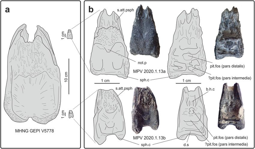
www.nature.com/scientificreports/

                                            Figure 2.  MHNG GEPI V5778. Trachymetopon sp. Basisphenoid with fragments of the posterior parietals
                                            and parasphenoid. Dorsal (a), dorsoposterior (b) and left lateral views (c). d, detail of exits of the nerve in
                                            anterolaterodorsal view (corresponding approximately to the frame in c); e, position of the fossil in a schematic
                                            reconstruction of the braincase of a mawsoniid coelacanth (modified from Maisey, 1986). Abbreviations:
                                            d.s, dorsum sellae; f.s.o.sc, foramen for the supraorbital sensory canal; f.s.opth, foramen for the superficial
                                            ophthalmic nerve; s.oph + IV, opening for the superficial ophthalmic nerve and the trochlear nerve; III, foramen
                                            for the oculomotor nerve; f.IV, foramen for the trochlear nerve (IV); ant. pr, antotic process; Par, parasphenoid;
                                            pr.con, processus conectens; p.Pa, posterior parietal; sph.c, sphenoid condyle; spt.fos, suprapterygoid fossa; v.pr.
                                            pPa, ventral process of the parietal posterior.

                                            Evidence of potential pups of Trachymetopon sp. Two small basisphenoids of coelacanth (Fig. 3 and
                                            Supplementary Fig. S3; MPV 2020.1.13a & b) were found by Elisabeth and Gérard Pennetier in the Callovian
                                            beds from the Vaches Noires. These specimens were donated to the Paléospace Muséum, France.
                                                Both specimens consist of ventral part of the basisphenoids only, i.e. the paired processus connectens con-
                                            nected via a bony surface against which abutted the notochord, the paired sphenoid condyles and the dorsum
                                            sellae in one specimen (Fig. 3b). In the largest specimen (MPV 00.1.13a), the sphenoid condyles are widely
                                            separated and not very protruding, probably for preservational reasons. The slightly smaller specimen (MPV
                                            00.1.13b) is better preserved than the larger one. The internal side of the both specimens shows well-marked
                                            reliefs. On the anterior part of the bone, the ventral side and the lateral sides bear strong grooves for suturing
                                            with the parasphenoid.
                                                Although both specimens are very incomplete, we tentatively refer them to Trachymetopon sp. because a
                                            marked notch separates the short and divergent sphenoid condyles and the angle between the posteroventral
                                            surface of the basisphenoid and the ventral surface of the parasphenoid is approximately 135° (slightly more
                                            in MPV 2020.1.13.a). Specimen MPV 2020.1.13.a retained the base of the pila antotica, which marks with the
                                            horizontal line an angle similar to that of the large specimen (approximately 150°, Supplementary Fig. S3). In
                                            ventral view, the length-to-width ratios are roughly similar for large and small specimens. This ratio is equivalent
                                            to nearly 1.8 in our material, while it is significantly lower in most other coelacanths.
                                                If this identification is confirmed, both small specimens obviously belong to two young individuals by com-
                                            parisons of their size with MHNG GEPI V5778. In MPV 2020.1.13a, a shallow notochordal pit is still visible
                                            ventrally between the two processus connectens (Figs. 3 and Supplementary Fig. S3). In the fetus of Latimeria,
                                            the tip of the notochord passes through the basisphenoid in the notochordal foramen, which closes during
                                            development and leaves a notochord pit present in young individuals. Moreover, while the inner surface of the
                                            basisphenoid is generally smooth in adult individuals of various species of coelacanths, with only a pituitary
                                            notch between the antotic processes and overhung by the dorsum sellae (e.g. Moenkopia wellesi33; Macropoma
                                            lewesiensis11), it is carved by strong cavities and ridges in the two small specimens described here. The fossae
                                            probably indicate the presence of a compact pituitary gland by comparison with the development of brain in
                                            fetus and pups of Latimeria34.

          Scientific Reports |   (2021) 11:11812 |               https://doi.org/10.1038/s41598-021-90962-5                                                    4

Vol:.(1234567890)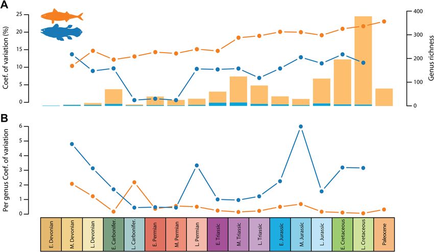
www.nature.com/scientificreports/
Figure 3. Basisphenoids of embryos or newborns of Trachymetopon sp. from Callovian beds from the Vaches
Noires. (a) Comparison of the giant specimen (MHNG GEPI V5778) with basisphenoids of potential pups
(MPV 2020.1.13); (b) details of the basisphenoids of the potential pups (MPV 2020.1.13a and MPV 2020.1.13b)
in ventral (left) and dorsal (right) views. Abbreviations: b.h.c, buccohypophysal canal; d.s, dorsum sellae; not.p,
notochordal pit; pit.fos, pituitary fossa; s.att.psph, surface of attachment for the parasphenoid; sph.c, sphenoid
condyle.
Hypophyseal development in Trachymetopon: In the adult Latimeria, the brain occupies about 1% of the
space of the cranial cavity, and the ventral floor of the basisphenoid is dug by the pituitary fossa (pit. fos), which
accommodates the enlarged anterior portion of pars distalis (adenohypophysis) of the pituitary gland, while the
posterior part of the gland, including the bipartite pars intermedia is located much more posteriorly beneath
the optic chiasm in the otico-occipital portion of the b raincase11,35. Dutel et al.34 showed that the pituitary gland
underwent strong modifications during ontogeny in Latimeria. At fetal stage, the brain is proportionally very
large and occupies both ethmosphenoid and otico-occipital cavities, and the pituitary gland is compact and lies
under the diencephalon. During growth, the gland rotates dorsally towards the telencephalon when the brain is
being concentrated in the posterior part of the cavity, while the anterior extremity of the pars distalis remains at
the level of the basisphenoid and connects to the pars intermedia by the hypophyseal duct. The buccohypophysal
canal (b.h.c) closes early during ontogeny36. Based on the observation in Latimeria, we consider that both small
basisphenoids belong to pups of Trachymetopon, and we tentatively interpret the reliefs on their floor as fol-
lowing: a posterior bilobate fossa possibly accommodated the bipartite pars intermedia of the pituitary gland,
while a median deep cavity situated just anteriorly corresponds to the pituitary fossa of adult coelacanths, i.e.
it accommodated the anterior part of the pars distalis. The latter corresponds to the adenohypophysis, which
secretes among other growth hormone, and the well-developed bony cavity indicate that the gland was propor-
tionally larger than in corresponding stages in Latimeria (34, Extended Data Figs. 3b,c and 4b,c). Both partes are
still close to each other because of the early stage of development. This arrangement of the hypophysis would
correspond to the stages pup 1 or pup 2 of Dutel et al.34 based on their Extended Data Fig. 4. A groove anterior
to the pituitary fossa accommodated the remnant of the buccohypophyseal canal. It is not possible to figure out
if the hypophyseal fossa was still open because of preservation.
Trachymetopon stratigraphical range. So far, the genus Trachymetopon is known in the Early and Mid-
dle Jurassic of Europe, but new data require re-evaluation of this stratigraphic range. Trachymetopon liassicus
from the Toarcian (Early Jurassic) of Holzmaden, Germany, was named and described by H ennig32 and revised
30
by Dutel et al. , who referred it to the family Mawsoniidae on the basis of a cladistic analysis. The holotype is a
complete and articulated specimen of 1.6 m in length, i.e. in the length range of the extant Latimeria. Trachym-
etopon was then discovered in the Middle Jurassic with giant forms discovered in Callovian strata in Normandy,
France, described by Dutel et al.27 and in the present study.
In addition to this material, the Etches collection Museum at Kimmeridge, Dorset, UK, houses skull ele-
ments of a coelacanth from the Kimmeridgian (K785) that reached about 1.5 m in length compared to skeletal
Scientific Reports | (2021) 11:11812 | https://doi.org/10.1038/s41598-021-90962-5 5
Vol.:(0123456789)www.nature.com/scientificreports/
Figure 4. Trachymetopon (“Macropoma”) substriolatum (holotype, SMC J27415) from the Kimmeridgian
of Cottenham, Cambridgeshire. Photograph (1) and semi-interpretative drawings (2) in dorsal (a), ventral
(b), anterior (c), left lateral (d) and right lateral (e) views. Scale bars = 50 mm. Abbreviations: Ang, angular;
Bsph, basisphenoid; Ext, extrascapular; Gu, gular; L.j, lachrymojugal; Op, opercle; Par, parasphenoid; p.Co,
principal coronoid; Pop, preopercle; Pp, postparietal; Po, postorbital; Q, quadrate; Sc, scale; So, supraorbital; Sq,
squamosal; Stt, supratemporal.
proportions of the complete type specimen. It consists of an angular, a quadrate, a metapterygoid and partial
pterygoid, of a paired ceratohyals, of a cleithrum and indeterminate bones. The shape and ornamentation of
the angular, and the proportion of the palatoquadrate are reminiscent to mawsoniids, and more specifically
Trachymetopon. It is referred here with caution to Trachymetopon sp.
The holotype and only known specimen of “Macropoma” substriolatum (SMC J27415; Fig. 4) from the Kim-
meridgian of Cottenham, Cambridgeshire, UK, was originally included in the genus Macropoma by Huxley8,
then Coccoderma by R oodward38, and eventually brought closer to Holophagus by F
eis37 and W orey11. In this
specimen, the supratemporals appear to be restricted to the posterior lateral margin of the postparietal shield,
and extend posteriorly creating a space that was occupied by the extrascapulars (preserved as fragments), like
in mawsoniids and some other coelacanth genera. The strong ornamentation of the skull roof with conspicuous
ridges and grooves is another mawsoniid character. Similar to Trachymetopon, the quadrate is massive, broad
and has a convex anterior margin, the angular is long and low with a straight outline. The cheek is composed
of a lachrymojugal, a postorbital, a squamosal and a preopercle, which are all thick and proportionally large
bones with coarse ornamentation, as in mawsoniids. A difference with T. liassicum is that some bones strongly
Scientific Reports | (2021) 11:11812 | https://doi.org/10.1038/s41598-021-90962-5 6
Vol:.(1234567890)www.nature.com/scientificreports/
Figure 5. Fragmentary elements from the giant specimens of the Jurassic Trachymetopon and the Cretaceous
Mawsonia. Human silhouettes corresponds to 1.8 m.
ornamented in the latter species (e.g., the postparietal, the angular, the opercle) are almost smooth with only a
dense pattern of small pits in T. substriolatum. However, these parts of the specimen are the most exposed ones
and we suspect that they were worn possibly before fossilization or, more probably, once the fossil was exposed
to the surface. The specimen can be referred to a mawsoniid, and we provisionally refer this species to Trachym-
etopon. Based on our model, this individual was small, about 60 cm in length.
Based on this short review, we consider with confidence that the stratigraphic range of Trachymetopon, previ-
ously restricted to the Early and Middle Jurassic, extends to the Late Jurassic.
A review of giant Mesozoic coelacanths. First remains of giant mawsoniids from the Early Cretaceous
of Brazil were originally misinterpreted as belonging to a giant pterosaur by Woodward39 because of the peculiar
biconvex articular condyle of the quadrate. This author then recognized his error with more complete material
from the Recôncavo Basin, Brazil, that he named Mawsonia gigas based on its obvious large body s ize40. Based
on our model (see Material & Methods), the body length of the holotype individual reached 3.1 m in length.
Mawsonia bones were later found in various Early Cretaceous South American basins mostly Brazil but also
Uruguay41,42, mainly represented by fragmentary elements corresponding to middle-sized individuals, but also
to giant ones. One specimen coming from the Neocomian of Bahia is an articular head of a quadrate (DGM
1.048-P) and corresponds to an individual of 6.3 m in length according to Carvalho & Maisey43. Examination
of this specimen by one of us (LC) and estimation based on our model indicates a total body length of 5.3 m
(Fig. 5). Medeiros et al.44 recorded from the Laje do Coringa flagstone, Alcântara Formation in northeastern
Brazil, a fragment of a large pterygopalatine comprising the quadrate, the metapterygoid and a piece of the
pterygoid (UFMA 1.40.468). Based on our model, this specimen was 4.9 m long. African Cretaceous mawsoni-
ids also reached meters-long sizes25,26, but never as long as Trachymetopon or as South American mawsoniids.
The sister genus of Mawsonia is Axelrodichthys, which lived for part of its time interval in sympatry with Mawso-
nia in Brazil45, and extends to the Late Cretaceous in Europe with smaller forms46. In Africa ’Mawsonia’ lavocati
has been referred to Axelrodichthys by Fragoso et al.47, and remains of this species from the Kem Kem Group
in Morocco indicate individuals up to 3.5 m long48. Recently, Brito et al.49 referred an ossified lung fragment
from the Late Maastrichtian of Morocco to an undetermined mawsoniid. Besides being the last record of a fos-
sil coelacanth, the individual was a giant with an estimated total body size of between 3.65 m and 5.52m49. The
fossil was recovered from marine sediments, but it is still uncertain whether the fish lived in this environment
or whether the corpse was transported from a river system, as was the case with continental animals found in
the same deposits50–52.
Dutel et al.27 referred an isolated palatoquadrate (MPV 2012.1.1) found in the Callovian (Middle Jurassic) of
the Vaches Noires, France, to Trachymetopon sp. This specimen corresponds to a large individual estimated to
reach 4 m in length. In addition to this large pterygoid, we mention here a large ceratohyal (MPV 2014.2.344)
found in the same Callovian beds of the Vaches Noires, housed in the Paléospace Museum (Fig. 5). We estimate
Scientific Reports | (2021) 11:11812 | https://doi.org/10.1038/s41598-021-90962-5 7
Vol.:(0123456789)www.nature.com/scientificreports/
Axelrodichthys Megalocoelacanthus
Trachymetopon spp. Mawsonia gigas lavocati dobei
Specimens B.L Specimens B.L Specimen B.L Specimen B.L
MHNG GEPI V5778 5.0 DGM 1.048-P 5.3 UMI-1 3.5 CCK 88–2-1 3.5
MPV 2014.2.344 4.4 UFMA 1.40.468 4.9
MPV 2012.1.1 4.0
Table 1. Calculated body length (B.L., in meters) for some of the largest specimens of Trachymetopon spp.,
Mawsonia gigas, Axelrodichthys lavocati and Megalocoelacanthus dobei.
Figure 6. (A) range-through genus richness (histograms) and disparity expressed by the coefficient of variation
(dots and lines); (B) per genus coefficient of variation, which is the coefficient of variation standardized by taxic
diversity. Orange, Actinopterygii; blue, Actinistia.
that this bone corresponds to an individual slightly larger than the one represented by the pterygoid, i.e. 4.4 m
in length.
Among the latimeriids, Megalocoelacanthus dobiei is a giant species from the Late Cretaceous of North
America known by disarticulated and mainly cranial elements. Several estimates of body size have been proposed,
i.e. between 3.8 and 4.0 m for the holotype specimen (CCK 88–2-1) calculated by Schwimmer28 and between
2.3 and 3 m for another specimen (AMNH FF 20,267) calculated by Dutel et al.29. Based on the basisphenoid
of the holotype and comparison with to the body proportions of Latimeria, we obtained a total length of 3.5 m
for the latter specimen.
The body length estimates for the largest known specimens are summarized in Table 1.
Body size evolution and disparity in coelacanths. Linear regression analysis between coelacanth log-
transformed body length and time expressed in time bins spanning the Devonian-Cretaceous interval shows
a statistically significant positive correlation (r = 0.42963; p < 0.0001), indicating a general trend for body size
increase over time in Actinistia. The evolution of body size disparity in coelacanths is decoupled from the
observed trends in taxic diversity (Pearson’s product-moment correlation: 0.3450382; p-value = 0.2078).
We computed the coefficient of variation to quantify coelacanth body size disparity across 17 time bins span-
ning the Devonian-Paleocene interval, and used actinopterygians for comparison. Actinopterygians were chosen
because their time range is comparable to that of coelacanths, and because of their enormous taxic diversity,
which makes them representatives of the changes in body length disparity through time in about half of the
vertebrate diversity. Our results (Fig. 6A) indicate that body size disparity is globally lower in coelacanths, with
the exception of the earliest stages of their evolutionary history. However, the ray-finned fish taxic diversity is
tremendously higher than that of coelacanths over the vast majority of their evolutionary history (Fig. 6A), which
tends to make direct comparison of disparity misleading. Once standardized by taxic diversity, the per genus
body size disparity patterns differ drastically (Fig. 6B). Coelacanths display a much higher per genus disparity
than actinopterygians by several orders of magnitude throughout most of their evolutionary history, excepted
Scientific Reports | (2021) 11:11812 | https://doi.org/10.1038/s41598-021-90962-5 8
Vol:.(1234567890)www.nature.com/scientificreports/
in the late Carboniferous where actinopterygians show a high per genus disparity and in the Permian, where
both clades present roughly similar disparity values.
Discussion
The new fragmentary remains of Trachymetopon described here, and the body size reassessment of Jurassic and
Cretaceous mawsoniids indicate the presence of individuals reaching or exceeding 5 m in total body length
during the Jurassic and Cretaceous. By comparison, among the actinopterygians living from the Devonian to
the Paleocene, the only genera which approach or exceed Mawsonia and Trachymetopon in length are two giant
marine planktivorous pachycormiforms, the Jurassic Leedsichthys (16 m) and the Cretaceous Bonnerichthys
(6.1 m), as well as the Late Cretaceous to Recent Acipenser (5 m). Among the extant ray-finned fish, the only
longest species include another chondrostean, Huso huso (7.2 m) and the Atlantic blue marlin, Makaira nigricans
(5 m), as well as the oarfish Regalecus glesne (13.7 m) but the latter has a compressed and slender profile very
different from the other fish compared, all more or less fusiform shaped. Thus, the two genera of mawsoniid
coelacanths are among the ten largest bony fish that have ever lived. Interestingly, one of these giant coelacanths,
Trachymetopon, lived in sympatry in the European Callovian Sea with the largest ray-finned fish that ever lived,
Leedsichthys46.
Fifteen genera of coelacanths are known in the Jurassic and Cretaceous, i.e. contemporaneous genera of the
giant Trachymetopon and Mawsonia. They were medium-sized fish, but proportionately small compared to the
two giants, with seven genera whose body length did not exceed 0.5 m (Reidus, Swenzia, Macropomoides, Cocco-
derma, Atacamaia, Undina, and Lualabaea). The smallest known coelacanth genera lived mainly in the Paleozoic
(Holopterygius, Lochmocercus, Hadronector, and Youngichthys), then in the Triassic (Piveteauia and Chaohuich-
thys). The general mean increase in body size in this lineage is demonstrated by the correlation between body size
and time, which confirms the Cope’s rule previously observed in many clades2. Such a trend is not observed in
actinopterygians as a whole, but is present in most of the main clades taken s eparately54, probably because testing
the Cope’s rule gives contrasting results depending on the taxonomic level used55. Because of the proportionally
smaller body size of the older coelacanths, and because small size might be considered as what G ould56 called
the “left wall” in evolution of complexity, evolution of body size would predominantly lead to a passive trend
towards larger body size (the “Stanley effect”57 according to Albert & Johnson58). Nevertheless, observation of the
distribution of body size over time indicates that the lowest body size tends to increase from the Early Devonian
to the Late Cretaceous (Supplementary Fig. S4), although this trend remains to be demonstrated statistically.
From this aspect, the coelacanths do not deviate from this general macroevolutionary trend observed in a
majority of clades of a ctinopterygians54.
Our analyses of the evolution of body size disparity in coelacanths indicate high disparity from the Late Per-
mian onward compared with actinopterygians. Although this pattern is not visible based on raw disparity values
(Fig. 6A), it is clearly noticeable when disparity is standardized by taxic diversity, which allows to fit the much
higher finned-ray diversity to the depleted coelacanth clade (Fig. 6B). A decrease is visible for both clades in the
Late Jurassic, possibly caused by the Lagerstätten effect detected in the fossil record of ray-finned fish at this t ime59
which alters the measure of disparity by preserving a greater diversity and more complete s pecimens60. We also
note that the variance in body size of ray-finned fish steadily increases over time when the index is taken raw,
but when adjusted for diversity, the variance in body size is very stable during the Permian-Paleocene interval,
possibly indicating that the body size ecospace was fulfilled during this time interval.
Previous studies have demonstrated that the coelacanth morphological disparity, whether measured by mor-
phospace occupation61 or by computation of new discrete characters in a phylogenetic framework9–12,14, shows a
burst at the origin of the group in the Devonian – Carboniferous. These studies confirmed the early burst (EB)
model first proposed by Simpson62, which was then verified at a large scale among animal c lades63–65, and dem-
onstrated more specifically in the i chthyosaurs66. The same trend is observed here for body size disparity in the
early evolutionary history of the coelacanths. However, the coelacanth pattern differs from that of other clades
in that their body size disparity did not decrease through time, contrary to what is observed in the morphologi-
cal evolution of ichthyosaurs for i nstance66. Instead, the evolution of the coelacanth body size disparity tended
to increase until the late Cretaceous, then this evolutionary trend remains unknown due to the lack of fossils
in the Cenozoic. Interestingly, our analyses indicate that the evolution of body size disparity in coelacanths is
decoupled from the observed trends in taxic diversity. Such a decoupling between morphological disparity and
taxic diversity after the initial radiation of a clade has been reported on many instances for various groups based
on fossil e vidence67,68, indicating that taxic diversity and disparity may be controlled by different factors (but
see3,4). However, the post-Carboniferous variations in body size disparity through time among coelacanths does
not mirror those of morphological evolution for this clade e ither9–11. Those studies on morphological evolution
indicate a steady drop from the Carboniferous until the Cretaceous with some exceptions such as the aberrant
Foreyia from the middle T riassic17, which is in sharp contrast with the pattern of body size disparity presented
in Fig. 6. This indicates that body size might not be an accurate proxy for reflecting morphological evolution
and/or disparity.
Underwater observations in situ69,70 and gill surface m easurements71–73 of Latimeria all indicate a very low
metabolic rate in this fish. Based on Kleiber’s law stating that large animals have a proportionately lower meta-
bolic rate than small ones, and although we cannot say whether a low metabolic rate is a cause or consequence
of a large body size, the low rate observed in Latimeria can be considered as a trait inherited from the common
ancestor of the latimeroids (mawsoniids plus latimeriids), which is associated in an indeterminate way to the
gigantism of Mawsonia and Trachymetopon, and to the large size of Axelrodichthys and Megalocoelacanthus.
The recent s tudy74 of the genome of the giant whale shark (Rhincodon typus) and a comparison of genomic and
physiological features of a set of 83 animals revealed several correlations between these life traits. In particular,
Scientific Reports | (2021) 11:11812 | https://doi.org/10.1038/s41598-021-90962-5 9
Vol.:(0123456789)www.nature.com/scientificreports/
these authors detected a negative correlation between length of introns in the genome and metabolic rate, and
a positive correlation between length of introns and body size. Latimeria has proportionally long intron length
(74, Fig. 1e, Supplementary Fig. S3), making its clade prone to evolve toward large body size.
Finally, the small basisphenoids provisionally attributed to pups of the giant Trachymetopon show that adeno-
hypohysis was probably proportionally large in these young individuals. E dinger75 showed that the adenohypoph-
ysis is proportionally much larger in large animals, such as giant dinosaurs, thus confirming an older observation
by Nopsca76, and in large birds and mammals. Although the available evidence is still weak, the proportionally
large adenohypophysis in the young of Trachymetopon sp. may be associated with the large size of adults.
New fossil discoveries and an examination of the body size of coelacanths through time confirm that the
evolutionary history of these fish is in agreement with two major macroevolutionary trends widely observed in
animal evolution, namely an early burst in their morphological disparity (previously demonstrated) and a gradual
increase in body size through (Cope/Depéret’s rule), but they also deviate from two other macro-evolutionary
trends, that is, their variations in body size disparity are not linked with taxic diversity nor with morphological
evolution. The genomic characteristics, the long intron length and the physiological characteristics, the low meta-
bolic rate of the extant Latimeria constitute a favorable ground for the evolution towards gigantism in this clade.
Material and methods
Microplalaeontological preparation. Microfossils have been extracted from a very small amount of rock
residue retrieved from the preparation of the coelacanth bone. Due to the strong induration of the sediment,
extraction of microfossils, unsuccessful with traditional washing methods77, have then been done by acetolysis78.
Body size reconstruction. The model used to reconstruct the body length in Fig. 5 is based on the recon-
struction of Axelrodichthys araripensis by Forey (11, Fig. 11.3), itself based on the reconstruction of M
aisey45
with some additions. As far as the material allows to assess, there are no major differences in body propor-
tions between Axelrodichthys, Trachymetopon and Mawsonia. Note that our model is based on individuals much
smaller than the individuals studied here, and it is possible that allometric growth may alter the calculation of
body size estimates. However, McAllister and Smith79 showed that in Latimeria chalumnae the length of the
head, from the snout to the posterior end of the operculum, grows isometrically compared to the standard
length. If, anyway, allometric growth was present in mawsoniids, it means that our body size reconstructions are
underestimated because the allometry in fish, as in most vertebrates, involved a proportionately larger head in
young individuals than in older and larger ones. All analyses were performed using R version 3.6.080.
Stratigraphical ranges and body size dataset. We gathered data on the fossil record (first and last
occurrences) of each of the 63 coelacanth genera from the Devonian to the Paleocene. We further compiled
the maximum body length for each genus based on the literature and/or on direct measurements of complete
specimens or based on estimates for partial specimens. For species known by isolated skulls only, we multiplied
the skull length (snout to posterior margin of the opercle) by 4.14, a ratio calculated on the basis of a sample of
complete specimens of various species. For comparison purpose, we also gathered data for the actinopterygians.
These are based on an update of Guinot & Cavin54,81, with new information from Sallan & Coates82 for Devonian
taxa, Romano et al.83 for Permo-Triassic taxa and Alberts et al.58, complemented by extensive literature review.
We used total length for both actinistian and actinopterygian genera. When only standard lengths were avail-
able for ray-finned fishes, we multiplied them by 1.2 for getting the total length, a ratio calculated on the basis
of a large sample of taxa known by complete specimens. When different sizes were available for one species, we
selected the longest one. When several species are known for a genus, we selected the longest one. Data are avail-
able in Supplementary Table S1.
Stratigraphical ranges and body size analyses. We used the coefficient of variation to quantify body
size disparity across time bins throughout the Devonian-Paleocene interval. The coefficient of variation is
expressed here in percent, as follows:
Cν = σ/µ ∗ 100 (1)
where σ is the standard deviation and µ is the mean of the body size values. We computed the coefficient of vari-
ation for each of the 17 time bins, which represent geological Epochs. Because disparity values can be influenced
by taxic diversity, we further divided values of Cv in each time bin by the corresponding value of taxic diversity
(computed by range-through), thus providing a per genus coefficient of variation standardized by taxic diversity.
This allowed comparisons to be made between disparity values of clades that differ drastically in taxic diversity,
such as actinistians and actinopterygians. Body length values were log-transformed prior to the analyses.
Received: 16 February 2021; Accepted: 17 May 2021
References
1. Bokma, F. et al. Testing for Depéret’s rule (body size increase) in mammals using combined extinct and extant data. Syst. Biol. 65,
98–108 (2016).
2. Heim, N. A., Knope, M. L., Schaal, E. K., Wang, S. C. & Payne, J. L. Cope’s rule in the evolution of marine animals. Science 347,
867–870 (2015).
Scientific Reports | (2021) 11:11812 | https://doi.org/10.1038/s41598-021-90962-5 10
Vol:.(1234567890)www.nature.com/scientificreports/
3. Rabosky, D. L. et al. Rates of speciation and morphological evolution are correlated across the largest vertebrate radiation. Nat.
commun. https://doi.org/10.1038/ncomms2958 (2013).
4. Cooney, C. R. & Thomas, G. H. Heterogeneous relationships between rates of speciation and body size evolution across vertebrate
clades. Nat. Ecol. Evol. 5, 101–110. https://doi.org/10.1038/s41559-020-01321-y (2020).
5. Lloyd, G. T. Journeys through discrete-character morphospace: Synthesizing phylogeny, tempo, and disparity. Palaeontology 61,
637–645 (2018).
6. Darwin, C. On the Origin of Species by Means of Natural Selection, or the Preservation of Favoured Races in the Struggle for Life.
John Murray, p. 502 (1859).
7. Toriño, P. A comprehensive phylogenetic analysis of coelacanth fishes (Sarcopterygii, Actinistia) with comments on the composi-
tion of the Mawsoniidae and Latimeriidae: Evaluating old and new methodological challenges and constraints.Hist. Biol., 1–21
(2021).
8. Huxley, T.H. Illustrations of the structure of the crossopterygian ganoids. Figures and descriptions illustrative of British organic
remains. Mem. Geol. Survey U.K, Lond., 1–44 (1866).
9. Schaeffer, B. Rates of evolution in the coelacanth and dipnoan fishes. Evolution 6, 101–111 (1952).
10. Cloutier, R. Patterns, trends, and rates of evolution within the Actinistia. in The biology of Latimeria chalumnae and evolution of
coelacanths. Springer, 23–58 (1991).
11. Forey, P. History of the Coelacanth Fishes (Chapman and Hall, 1998).
12. Schultze, H. P. Mesozoic sarcopterygians. in Mesozoic fishes 3, Dr Pfeil Verlag, 463–492 (2004).
13. Zhu, M. et al. Earliest known coelacanth skull extends the range of anatomically modern coelacanths to the Early Devonian. Nat.
Commun. 3, 1–8 (2012).
14. Cavin, L. & Guinot, G. Coelacanths as “almost living fossils”. Front. Ecol. Evol. 2, 49 (2014).
15. Bennett, D. J., Sutton, M. D. & Turvey, S. Quantifying the living fossil concept. Palaeontol. Electronica, (21.1. 14A) (2018).
16. Lund, R. & Lund, W. New genera and species of coelacanths from the Bear Gulch Limestone (Lower Carboniferous) of Montana
(USA). Geobios 17, 237–244 (1984).
17. Cavin, L., Mennecart, B., Obrist, C., Costeur, L. & Furrer, H. Heterochronic evolution explains novel body shape in a Triassic
coelacanth from Switzerland. Sci. Rep. 7, 1–7 (2017).
18. Nikaido, M. et al. Genetically distinct coelacanth population off the northern Tanzanian coast. Proc. Natl. Acad. Sci. U.S.A. 108,
18009–18013 (2011).
19. Lampert, K. P. et al. Population divergence in East African coelacanths. Curr. Biol. 22, R439–R440 (2012).
20. Kadarusman, et al. A thirteen-million-year divergence between two lineages of Indonesian coelacanths. Sci. Rep. 10, 1–9 (2020).
21. Nikaido, M. et al. Coelacanth genomes reveal signatures for evolutionary transition from water to land. Genome Res. 3, 1740–1748
(2013).
22. Amemiya, C. T. et al. Complete HOX cluster characterization of the coelacanth provides further evidence for slow evolution of its
genome. Proc. Natl. Acad. Sci. U.S.A. 107, 3622–3627 (2010).
23. Amemiya, C. T. et al. The African coelacanth genome provides insights into tetrapod evolution. Nature 496, 311–316 (2013).
24. Woodward, A. S. Notes on some Upper Cretaceous Fish remains from the Province of Sergipe and Pernambuco, Brazil. Geol. Mag.
4, 193–197 (1907).
25. Wenz, S. Un coelacanthe géant, Mawsonia lavocati Tabaste, de l’Albien-base du Cénomanien du sud marocain. Ann. Pal. (Vert.)
67, 1–20 (1981).
26. Yabumoto, Y. & Uyeno, T. New materials of a Cretaceous coelacanth, Mawsonia lavocati Tabaste from Morocco. Bull. Natn. Sci.
Mus. Tokyo Ser. C 31, 39–49 (2005).
27. Dutel, H., Pennetier, E. & Pennetier, G. A giant marine coelacanth from the Jurassic of Normandy, France. J. Vertebr. Paleontol.
34, 1239–1242 (2014).
28. Schwimmer, D.R. Giant fossil coelacanth from the Late Cretaceous of the Eastern United-States. Fernbank Magazine, 24–30 (2002).
29. Dutel, H. et al. The giant Cretaceous coelacanth (Actinistia, Sarcopterygii) Megalocoelacanthus dobiei Schwimmer, Stewart &
Williams, 1994, and its bearing on Latimerioidei interrelationships. PLoS ONE 7, e49911 (2012).
30. Dutel, H., Herbin, M. & Clément, G. First occurrence of a mawsoniid coelacanth in the Early Jurassic of Europe. J. Vertebr. Paleontol.
35, e929581 (2015).
31. Rioult, M., Coutard, J.-P., de La Quérière, P., Helluin, M., Larsonneur, C. & Pellerin, J. Notice explicative de la feuille de Caen à
1/50000. Editions du BRGM, 1–104 (1989)
32. Hennig, E. Trachymetopon liassicum, Ald., ein Riesen-Crossopterygier aus Schwäbischem Ober-Lias. Neues Jahrb. Geol. Palaontol.
Abh. 94, 67–79 (1951).
33. Schaeffer, B. & Gregory, J. T. Coelacanth fishes from the continental Triassic of the western United States. Am. Mus. Novit. 2036,
1–18 (1961).
34. Dutel, H. et al. Neurocranial development of the coelacanth and the evolution of the sarcopterygian head. Nature 569, 556–559
(2019).
35. Lagios, M. D. Evidence for a hypothalamo-hypophysial portal vascular system in the coelacanth Latimeria chalumnae Smith. Gen.
Comp. Endocrinol. 18, 73–82 (1972).
36. Khonsari, R. H. et al. The buccohypophyseal canal is an ancestral vertebrate trait maintained by modulation in sonic hedgehog
signaling. BMC Biol. 11, 1–16 (2013).
37. Reis, O. M. Die Coelacanthinen, mit besonderer Berücksichtigung der im Weissen Jura Bayerns vorkommenden Gattungen.
Palaeontographica 35, 1–96 (1888).
38. Woodward, A.S. Catalogue of Fossil Fishes in the British Museum (Natural History). Volume 2, xliv + 567 pp., London: British
Museum (Natural History) (1891).
39. Woodward, A. S. On the quadrate bone of a gigantic pterodactyl, discovered by Joseph Mawson in the Cretaceous of Bahia. Ann.
Mag. Nat. Hist. 6, 255–257 (1896).
40. Mawson, J. & Woodward, A. S. On the Cretaceous formation of Bahia (Brazil), and on vertebrate fossils collected therein. Q. J.
Geol. Soc. 63, 128–138 (1907).
41. Soto, M., De Carvalho, M. S., Maisey, J. G., Perea, D. & Silva, J. D. Coelacanth remains from the Late Jurassic–? earliest Cretaceous
of Uruguay: The southernmost occurrence of the Mawsoniidae. J. Vertebr. Paleontol. 32, 530–537 (2012).
42. Toriño, P., Soto, M., Perea, D. & de Carvalho, M. S. S. New findings of the coelacanth Mawsonia Woodward (Actinistia, Latim-
erioidei) from the Late Jurassic–Early Cretaceous of Uruguay: Novel anatomical and taxonomic considerations and an emended
diagnosis for the genus. J. S. Am. Earth Sci. 103054 (2020).
43. de Carvalho, M. & S. & Maisey, J. G. New occurrence of Mawsonia (Sarcopterygii: Actinistia) from the Early Cretaceous of the
Sanfranciscana Basin, Minas Gerais, southeastern Brazil in Fishes and the Break-up of Pangaea. Geol. Soc. Lond. 295, 109–144
(2008).
44. Medeiros, M. A., Lindoso, R. M., Mendes, I. D. & de Souza Carvalho, I. The Cretaceous (Cenomanian) continental record of the
laje do coringa flagstone (Alcântara formation), northeastern South America. J. S. Am. Earth Sci. 53, 50–58 (2014).
45. Maisey, J. G. Coelacanths from the lower cretaceous of Brazil. Am. Mus. Novit. 2866, 1–30 (1986).
46. Cavin, L. et al. The last known freshwater coelacanths: New Late Cretaceous mawsoniid remains (Osteichthyes: Actinistia) from
southern France. PLoS ONE 15, e0234183 (2020).
Scientific Reports | (2021) 11:11812 | https://doi.org/10.1038/s41598-021-90962-5 11
Vol.:(0123456789)www.nature.com/scientificreports/
47. Fragoso, L. G. C., Brito, P. & Yabumoto, Y. Axelrodichthys araripensis Maisey, 1986 revisited. Hist. Biol. 31, 1350–1372 (2019).
48. Cavin, L. et al. Taxonomic composition and trophic structure of the continental bony fish assemblage from the early Late Cretaceous
of southeastern Morocco. PLoS ONE 10, e0125786 (2015).
49. Brito, P. M., Martill, D. M., Eaves, I., Smith, R. E. & Cooper, S. L. A marine Late Cretaceous (Maastrichtian) coelacanth from North
Africa. Cretac. Res. 122, 104768 (2021).
50. Buffetaut, E., Escuillié, F. & Pohl, B. First theropod dinosaur from the Maastrichtian phosphates of Morocco. Kaupia 14, 3–8 (2005).
51. Suberbiola, X. P., Bardet, N., Iarochène, M., Bouya, B. & Amaghzaz, M. The first record of a sauropod dinosaur from the Late
Cretaceous phosphates of Morocco. J. Afr. Earth Sc. 40, 81–88 (2004).
52. Longrich, N. R., Suberbiola, X. P., Pyron, R. A. & Jalil, N.-E. The first duckbill dinosaur (Hadrosauridae: Lambeosaurinae) from
Africa and the role of oceanic dispersal in dinosaur biogeography. Cretac. Res. 120, 104678 (2021).
53. Liston, J., Newbrey, M., Challands, T. & Adams, C. Growth, age and size of the Jurassic pachycormid Leedsichthys problematicus
(Osteichthyes: Actinopterygii). in Mesozoic Fishes 5, Dr Pfeil, Verlag, 145–175 (2013).
54. Guinot, G. & Cavin, L. Body size evolution and habitat colonization across 100 million years (Late Jurassic–Paleocene) of the
actinopterygian evolutionary history. Fish Fish. 19, 577–597 (2018).
55. Hone, D. W. E. & Benton, M. J. Cope’s Rule in the Pterosauria, and differing perceptions of Cope’s Rule at different taxonomic
levels. J. evol. biol. 20, 1164–1170 (2007).
56. Gould, S. J. The evolution of life on the earth. Sci. Am. 271, 84–91. https://doi.org/10.1038/scientificamerican1094-84 (1994).
57. Stanley, S. M. An explanation for Cope’s rule. Evolution, 1–26 (1973).
58. Albert, J. S., Johnson, D. M. & Knouft, J. H. Fossils provide better estimates of ancestral body size than do extant taxa in fishes.
Acta Zoologica 90, 357–384 (2009).
59. Cavin, L. The Late Jurassic ray-finned fish peak of diversity: Biological radiation or preservational bias. in Origin and phylogenetic
interrelationships of teleosts. Dr Pfeil Verlag, 111–121 (2010).
60. Flannery Sutherland, J. T., Moon, B. C., Stubbs, T. L. & Benton, M. J. Does exceptional preservation distort our view of disparity
in the fossil record?. Proc. R. Soc. B 286, 201900 (2019).
61. Friedman, M. & Coates, M. I. A newly recognized fossil coelacanth highlights the early morphological diversification of the clade.
Proc. R. Soc. B 273, 245–250 (2006).
62. Simpson, G. Tempo and Mode in Evolution (Columbia University Press, 1944).
63. Hughes, M., Gerber, S. & Wills, M. A. Clades reach highest morphological disparity early in their evolution. Proc. Natl. Acad. Sci.
USA 110, 13875–13879 (2013).
64. Wagner, P. J. Early bursts of disparity and the reorganization of character integration. Proc. R. Soc. Lond. Ser. B. 85, 0181604 (2018).
65. Puttick, M. N. Mixed evidence for early bursts of morphological evolution in extant clades. J. Evol. Biol. 31, 50–515 (2018).
66. Moon, B. C. & Stubbs, T. L. Early high rates and disparity in the evolution of ichthyosaurs. Commun. Biol. 3, 1–8 (2020).
67. Hopkins, M. J. Decoupling of taxonomic diversity and morphological disparity during decline of the Cambrian trilobite family
Pterocephaliidae. J. Evol. Biol. 26, 1665–1676 (2013).
68. Ruta, M., Angielczyk, K. D., Fröbisch, J. & Benton, M. J. Decoupling of morphological disparity and taxic diversity during the
adaptive radiation of anomodont therapsids. Proc. R. Soc. Lond. Ser. B. 280, 20131071 (2013).
69. Fricke, H. & Plante, R. Habitat requirements of the living coelacanth Latimeria chalumnae at Grande Comore, Indian Ocean.
Naturwissenschaften 75, 149–151 (1988).
70. Fricke, H. & Hissmann, K. Home range and migrations of the living coelacanth Latimeria chalumnae. Mar. Biol. 120, 171–180
(1994).
71. Hughes, G. M. On the respiration of Latimeria chalumnae. Zool. J. Linn. Soc. 59, 195–208 (1976).
72. Hughes, G. M. Ultrastructure and morphometry of the gills of Latimeria chalumnae, and a comparison with the gills of associated
fishes. Proc. R. Soc. Lond. Ser. B. 208, 309–328 (1980).
73. Hughes, G. M. The gills of the coelacanth, Latimeria chalumnae Latimeriidae. What can they teach us?. Ital. J. Zool. 65, 425–429
(1998).
74. Weber, J. A. et al. The whale shark genome reveals how genomic and physiological properties scale with body size. Proc. Natl. Acad.
Sci. USA 117, 20662–20671 (2020).
75. Edinger, T. The pituitary body in giant animals fossil and living: A survey and a suggestion. Q. Rev. Biol. 17, 31–45 (1942).
76. von Nopcsa, F. Über Dinosaurier. 2. Die Riesenformen unter den Dinosauriern. Centralblatt für Mineralogie, Geologie und Paläon-
tologie 1917, 332–351 (1917).
77. Kummel, B. & Raup, D (eds.) Handbook of Paleontological Techniques. W. H. Freeman and Company, San Francisco and London,
1965. xiii + 852 pp (1965).
78. Lirer, FA new technique for retrieving calcareous microfossils from lithified lime deposits. Micropaleontology 365–369 (2000).
79. McAllister, D. E. & Smith, D. Mensurations morphologiques, dénombrements méristiques et taxonomique du coelacanthe, Latim-
eria chalumnae. Nat. Can. 105, 63–76 (1978).
80. R Core Team. R: A language and environment for statistical computing. R Foundation for Statistical Computing, Vienna, Austria.
https://www.R-project.org/ (2019)
81. Guinot, G. & Cavin, L. ‘Fish’ (Actinopterygii and Elasmobranchii) diversification patterns through deep time. Biol. Rev. 91, 950–981
(2016).
82. Sallan, L. C. & Coates, M. I. End-Devonian extinction and a bottleneck in the early evolution of modern jawed vertebrates. Proc.
Natl. Acad. Sci. USA 107, 10131–10135 (2010).
83. Romano, C. et al. Permian-Triassic Osteichthyes (bony fishes): Diversity dynamics and body size evolution. Biol. Rev. 91, 106–147
(2016).
Acknowledgements
We thank Laurent Picot (Paléospace Muséum, Villers-sur-Mer, France), Elisabeth and Gérard Pénnetier (Asso-
ciation Paléontologique de Villers-sur-Mer, France), Paulo M. Brito ( Universidade do Estado do Rio de Janeiro,
Rio de Janeiro, Brazil) for access to specimens under their care, Matt Riley (Sedgwick Museum, Oxford, UK),
Manuel Alfredo Araujo Medeiros (Universidade Federal do Maranhão, Brazil), Steve Etches and Carla Crook (The
Etches Collection Museum of Jurassic Marine Life, Kimmeridge) for providing photographs of specimens under
their care. We are grateful to Pierre-Alain Proz and Philippe Wagneur (Muséum d’histoire naturelle, Geneva) for
the preparation and photograph of the specimen MHNG GEPI V5778, respectively. We thank the editor, Zhu
Min, and two anonymous reviewers for their constructive remarks. This paper is a contribution to the project
“Evolutionary pace in the coelacanth clade: New evidence from the Triassic of Switzerland” (200021-172700)
supported by the Swiss National Science Foundation.
Scientific Reports | (2021) 11:11812 | https://doi.org/10.1038/s41598-021-90962-5 12
Vol:.(1234567890)You can also read