Usher syndrome IIIA gene clarin-1 is essential for hair cell function and associated neural activation
←
→
Page content transcription
If your browser does not render page correctly, please read the page content below
Human Molecular Genetics, 2009, Vol. 18, No. 15                     2748–2760
                                                                         doi:10.1093/hmg/ddp210
                                                                         Advance Access published on May 3, 2009

Usher syndrome IIIA gene clarin-1 is essential for
hair cell function and associated neural activation{
Ruishuang Geng1,{, Scott F. Geller3,{, Toshinori Hayashi4, Catherine A. Ray4, Thomas A. Reh4,
Olivia Bermingham-McDonogh4, Sherri M. Jones5, Charles G. Wright6, Sami Melki1, Yoshikazu
Imanishi2, Krzysztof Palczewski2, Kumar N. Alagramam1,{ and John G. Flannery3,{
1
 Department of Otolaryngology Head & Neck Surgery and 2Department of Pharmacology, Case Western Reserve
University, Cleveland, OH, USA, 3Helen Wills Neuroscience Institute, University of California, Berkeley, CA 94720-
3190, USA, 4Department of Biological Structure, University of Washington, Seattle, WA 98195, USA, 5Department of
Communication Sciences and Disorders, East Carolina University, Greenville, NC 27858, USA and 6Department of
Otolaryngology Head & Neck Surgery, Southwestern Medical Center, Dallas, TX 75235, USA

                                                                                                                                                              Downloaded from http://hmg.oxfordjournals.org/ by guest on May 14, 2015
Received March 17, 2009; Revised and Accepted April 29, 2009

Usher syndrome 3A (USH3A) is an autosomal recessive disorder characterized by progressive loss of hearing
and vision due to mutation in the clarin-1 (CLRN1) gene. Lack of an animal model has hindered our ability to
understand the function of CLRN1 and the pathophysiology associated with USH3A. Here we report for the
first time a mouse model for ear disease in USH3A. Detailed evaluation of inner ear phenotype in the Clrn1
knockout mouse (Clrn1 2/2 ) coupled with expression pattern of Clrn1 in the inner ear are presented here.
Clrn1 was expressed as early as embryonic day 16.5 in the auditory and vestibular hair cells and associated
ganglionic neurons, with its expression being higher in outer hair cells (OHCs) than inner hair cells. Clrn1 2/2
mice showed early onset hearing loss that rapidly progressed to severe levels. Two to three weeks after birth
(P14 – P21), Clrn1 2/2 mice showed elevated auditory-evoked brainstem response (ABR) thresholds and pro-
longed peak and interpeak latencies. By P21, 70% of Clrn1 2/2 mice had no detectable ABR and by P30
these mice were deaf. Distortion product otoacoustic emissions were not recordable from Clrn1 2/2 mice.
Vestibular function in Clrn1 2/2 mice mirrored the cochlear phenotype, although it deteriorated more gradu-
ally than cochlear function. Disorganization of OHC stereocilia was seen as early as P2 and by P21 OHC loss
was observed. In sum, hair cell dysfunction and prolonged peak latencies in vestibular and cochlear evoked
potentials in Clrn1 2/2 mice strongly indicate that Clrn1 is necessary for hair cell function and associated
neural activation.

INTRODUCTION                                                                   USH2 hearing loss is milder, the onset of RP is after puberty
                                                                               and vestibular function is unaffected. USH3 patients show pro-
Usher syndrome is the most common cause of sensory impair-                     gressive hearing loss and variable degrees of vestibular dys-
ment wherein deafness and blindness occur together. It is                      function.
clinically subdivided into three types based on the degree of                    At least 13 loci have been linked to the three types of Usher
deafness and the presence of vestibular dysfunction (1).                       syndrome, including one locus linked to USH3 (http://
USH1 is the most severe form and is characterized by pro-                      webh01.ua.ac.be/hhh/). Genes associated with many of these
found congenital hearing loss and vestibular dysfunction com-                  loci have been identified and they encode proteins that
bined with pre-pubertal onset of retinitis pigmentosa (RP). In                 belong to diverse classes of proteins (2,3). CLRN1


 To whom correspondence should be addressed at: 11100 Euclid Ave., CWRU, Cleveland, OH 44106; USA. Tel: þ1 2168447261; Fax: þ1
2169830284; Email: kna3@case.edu
†This work is dedicated to Cindy Elden. We appreciate the vision, inspiration and generosity of the Elden family that made this work possible.
‡The authors wish it to be known that, in their opinion, the first two authors should be regarded as joint First Authors and the last two authors should be
regarded as joint Senior Authors.

# The Author 2009. Published by Oxford University Press. All rights reserved.
For Permissions, please email: journals.permissions@oxfordjournals.org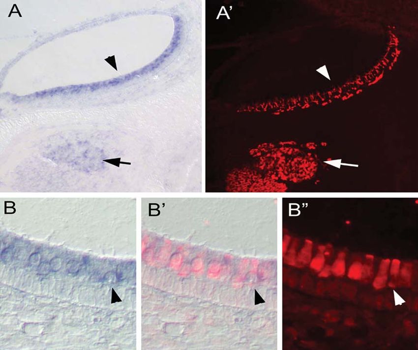
Human Molecular Genetics, 2009, Vol. 18, No. 15         2749

(USH3A), the only member of USH3, codes for a four                  Clrn1 was also expressed in the vestibular hair cells and
transmembrane-domain protein (4) belonging to a large               Scarpa’s ganglion cells. Closer examination of the Clrn1
family of transmembrane proteins which include both the tet-        labeling in the embryonic saccule revealed strong expression
raspanin and the claudin families. Members of this family par-      in the hair cells and a much weaker expression in Scarpa’s
ticipate in a variety of functions including regulating cell        ganglion cells (Fig. 2).
morphology, motility, invasion, fusion and signaling (5 – 7).
Tetraspanins are known to form homo-multimers leading to
the assembly of microdomains that interact and nucleate the         Generation of Clrn1 2/2 mice
congregation of other non-tetraspanin membrane proteins.            To study the function of Clrn1, a transgenic mouse lacking
CLRN1 shares some of the features common to tetraspanin             the first coding exon (exon 1) of this gene was produced by
proteins, including the predicted four transmembrane domain         homologous recombination (Fig. 3A and B). Normal
topology, and very short intracellular loops. This protein          Mendelian segregation of the Clrn1 exon 1 deleted allele
may play a vital role in creating and assembling membrane           (wild-type Clrn1 þ/þ , heterozygous Clrn1 þ/2 and homozy-
microdomains involved in adhesion strengthening and signal-         gous Clrn1 2/2 ) was observed. Lack of Clrn1 expression in
ing (7). However, the precise function of CLRN1 in the inner        the inner ears of Clrn1 2/2 mice was confirmed by RT – PCR
ear is not known.                                                   at various time points from birth well into adulthood (P0 –
   Several different mutations of human CLRN1 have been             P60) (data from time points P30 and P60 are shown in
found that cause progressive hearing loss with variable pene-       Fig. 3C and D). In situ hybridization of cochlear duct sections
trance linked to the N48K mutation, or profound hearing             from Clrn1 2/2 mice confirmed absence of Clrn1 expression

                                                                                                                                      Downloaded from http://hmg.oxfordjournals.org/ by guest on May 14, 2015
impairment linked to the Y176 stop mutation (4). Similarities       in hair cells and SGCs (Fig. 3E). These results demonstrate
between CLRN11 and the calcium channel gamma subunit                that Clrn1 mRNA is not expressed in the inner ear of the
protein 2 (CACNG2, stargazin) have been proposed (4). Star-         Clrn1 2/2 mouse.
gazin has been shown to play a key role in the shaping and             We also confirmed the lack of full-length Clrn1 expression
maintenance of cerebellar synapses (8). However, in vivo            in the retina of the Clrn1 2/2 mouse (data not shown). In
studies are needed to reveal the molecular mechanism that           Clrn1 2/2 mice, though, we failed to discover the deficiencies
underlies CLRN1 function. USH3 is inherited in a recessive          in structural abnormality of photoreceptors and other neurons
pattern, suggesting that the loss of function is the cause of       at the age of 4 months (data not shown). Furthermore, electro-
the disease. Therefore, studies of the Clrn1-null mouse             retinograms (ERG) analyses did not reveal the sign of photo-
should provide insights into the involved pathogenic mechan-        receptor degeneration up to the age of 16 months, as
isms. Here we report a detailed analysis of the Clrn1 2/2           exemplified by the lack of significant differences in the
mouse inner ear phenotype and describe the expression               a-wave amplitudes at various light conditions (Fig. 4).
pattern of Clrn1 in the vestibular and cochlear neuroepithelia.
Our results suggest that Clrn1 plays a novel role in hair cell
development and function.                                           Clrn1 2/2 mice show progressive hearing loss
                                                                    To assess hearing function in Clrn1 2/2 mice, we performed
                                                                    auditory brainstem response (ABR) tests on mice of different
RESULTS                                                             ages starting at P21, the age at which the auditory system in
                                                                    mice becomes fully mature. ABR tests reflect the electrical
Clarin-1 is expressed in hair cells and ganglion cells of the       responses of both the cochlear ganglion neurons and the
inner ear                                                           nuclei of the central auditory pathway to sound stimulation.
To determine the expression pattern of Clrn1 in the cochlear        About 30% of Clrn1 2/2 mice showed elevated ABR
and vestibular hair cells, we carried out mRNA in situ hybrid-      thresholds (Fig. 5A), but the majority (70%) failed to
ization at embryonic (E) stages 16.5, 18.5 and postnatal (P)        produce detectable ABR responses at P21. As expected,
day 0, 3 and 5. Clrn1 was found to be expressed as early as         Clrn1 þ/þ littermates produced characteristic ABR waveforms
E16.5 in the inner ear, in hair cells of the auditory and vestib-   (peaks 1 – 4) at thresholds from 25 to 45 dB peSPL (decibel
ular sensory epithelia and in the spiral ganglion neurons.          peak equivalent Sound Pressure Level) for pure tones 8 –
Expression was most apparent in the spiral ganglion cells           32 kHz (Fig. 5A); Clrn1 þ/2 mice showed similar results
(SGCs) and in the hair cells of the basal turn of the cochlea       (data not shown). Interestingly, in the recordings from
compared with apical turns at early stages, suggesting the          mutant mice, response peak latencies were significantly pro-
time and location of the onset of Clrn1 expression (Fig. 1).        longed compared with controls for all four peaks. For
Hair cell-specific genes typically are initially expressed in       example, at 8 kHz the initial response peak (peak 1) occurred
the more mature hair cells in the basal cochlea and spread to       at 2.0 ms in Clrn1 þ/þ mice, but it was close to 3 ms in the
the apical hair cells with continued development, paralleling       Clrn1 2/2 mice (Fig. 5B). The interpeak latencies P1– P2 and
the gradient in hair cell maturation. By E18.5, all cochlear        P1 –P3 were also prolonged in the mutants compared with
hair cells expressed Clrn1, with a higher level of expression       control siblings (Table 1). By P30, hearing function was not
in the outer hair cells (OHCs) as compared to inner hair            detectable in any Clrn1 2/2 mice tested (data not shown).
cells (IHCs) (Fig. 1). Expression of Clrn1 was detected by          These results suggest that Clrn1 2/2 mice have some auditory
in situ hybridization in the inner ear of all stages analyzed,      function at young ages but lose it rapidly after P21. This
i.e. from both E16.5 to P5, confirming previously reported          prompted us to test mutants at time points earlier than P21.
in situ data (4), and from P30 and P60 by RT–PCR (Fig. 3D).         All of six Clrn1 2/2 mice tested at P14 and P20 showed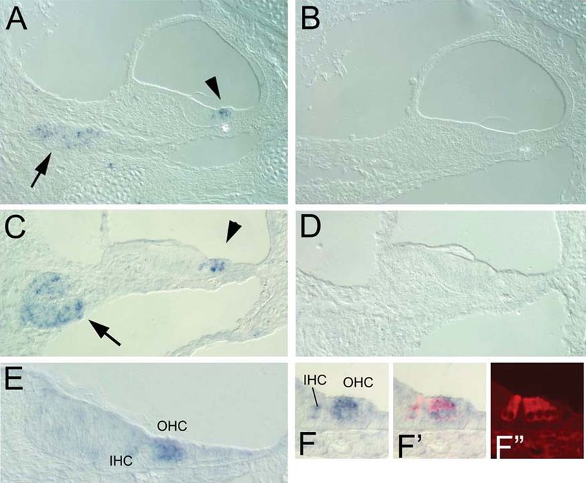
2750       Human Molecular Genetics, 2009, Vol. 18, No. 15

                                                                                                                                                                     Downloaded from http://hmg.oxfordjournals.org/ by guest on May 14, 2015
Figure 1. Expression of Clrn1 in the mouse cochlea detected using in situ hybridization. (A and B) Antisense (A) and sense (B) probes used to localize Clrn1 at
E18 in the middle turn. (C and D) Antisense (C) and sense (D) probes used to localize Clrn1 at P3 in the middle turn. Arrows point to the expression in the spiral
ganglion, while arrowheads point to expression in the hair cells. (E and F –F0 ) Expression of Clrn1 in hair cells was confirmed at E18 by post-in situ antibody
labeling with anti-myosin VI. (F0 , F00 ) Clarin1 is expressed more highly in the outer hair cells (OHC) than in the inner hair cells (IHC).

some hearing function; however, thresholds were significantly                       In summary, our hearing assessments are consistent with a
elevated and peak latencies prolonged (Table 2). The                              cochlear lesion site and a sensorineural hearing loss. Further,
increased intensity of sound needed to elicit a response from                     auditory results show that Clrn1 2/2 mutation affects hair
mutant ears at P14 – 21 suggests diminished hair cell function                    cell function, and either hair cell to afferent nerve communi-
early in the life of Clrn1 2/2 mice and the prolonged P1                          cation or primary afferent neural activation.
latency implies a delay in neural activation.

                                                                                  Clrn1 2/2 mice show progressive loss of balance function
                                                                                  In Clrn1 2/2 mice, balance function was not overtly/severely
Loss of OHC function in Clrn1 2/2 mice
                                                                                  affected by P30. In contrast, circling and head bobbing activity
To determine whether the hearing impairment in Clrn1 2/2                          in the Usher 1F model is evident as early as P12 and becomes
mice specifically involved an OHC defect, we recorded distor-                     more obvious with age (9,10). The head bobbing phenotype in
tion product otoacoustic emissions (DPOAEs) which are                             Clrn1 2/2 mice is mild and variable at young ages (P21 – 40)
indicative of OHC amplification activity. At P21, Clrn1 2/2                       with some mutants indistinguishable from their wild-type sib-
mice produced no detectable DPOAEs above the noise floor                          lings. However, vestibular dysfunction became more apparent
(NF) (Fig. 5C and D). Although, more than 10 Clrn1 2/2                            with age such that some Clrn1 2/2 mice evidenced clearer
mice with high threshold ABR, similar to those tested in                          signs of head bobbing than others by 6 months of age.
Figure 5A and B, were tested at P21, we were unable to                            Young adult (P21– P90) mutants were subjected to swim
record any DPOAEs from these mutants. In contrast, wild-type                      tests along with controls. Generally, Clrn1 2/2 mice appeared
controls showed normal DPOAE responses (Fig. 5C and D).                           less stable in the water compared with controls (Clrn1 þ/þ or
Absence of DPOAEs at a very young age clearly indicates                           Clrn1 þ/2 ), tending to roll from one side to the other, even
lack of OHC function in Clrn1 2/2 mice.                                           though they used their tails effectively to remain prone in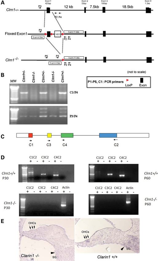
Human Molecular Genetics, 2009, Vol. 18, No. 15 2751
Downloaded from http://hmg.oxfordjournals.org/ by guest on May 14, 2015
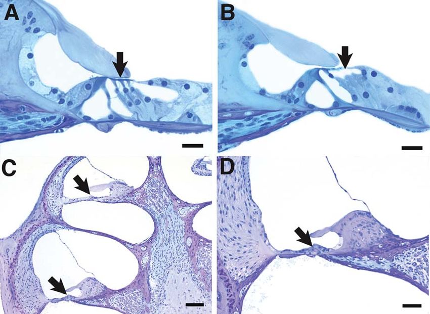
Figure 2. Expression of Clrn1 in the mouse saccule detected using in situ hybridization. (A and A0 ) Clrn1 is expressed in vestibular hair cells (arrowhead) and
ganglion neurons, as shown in (A0 ) using post-in situ immunolabeling with anti-TuJ1 antibody (arrow). (B and B0 ) Higher magnification view of Clrn1 expression
in vestibular hair cells co-labeled with myosin VI. (B0 and B00 ) Arrowhead points to an example of a double-labeled cell.
the water. But abnormal swimming behavior was not discern- communication or primary afferent neural activation.
able in all tested Clrn1 2/2 mice which prompted us to quan- However, no significant change in the vestibular hair cell mor-
tify vestibular function in these knockout (KO) animals. phology was observed within P21 – 30 (data not shown).
To quantitatively examine vestibular function in Clrn1 2/2 Changes in the vestibular system tend to progress much
mice, we recorded vestibular evoked potentials (VsEP) of more slowly than in the cochlea as noted above. Therefore,
Clrn1 2/2 and Clrn1 þ/þ mice at P21 – P30. It should be it will be necessary to examine older (.P30) Clrn1 2/2
noted that VsEP recordings with linear stimulation specifically mice carefully before we come to any conclusion about the
assess the otoconial organs (utricle and saccule). On average, effect of Clrn1 mutation on vestibular hair cells morphology.
VsEP thresholds of Clrn1 2/2 mice (25.5 + 3.6 dB re: 1.0 g/
ms) were significantly higher than those of Clrn1 þ/þ controls
(211.0 + 1.2 dB re: 1.0 g/ms), although the Clrn1 2/2 mice The stereocilia of OHCs are defective in Clrn1 2/2 mice
showed greater variability in their response thresholds To better understand the structure – function relationship in
(t ¼ 23.5, P ¼ 0.012). Some Clrn1 2/2 mice had normal Clrn1 2/2 mice, we examined organs of Corti from young
VsEP thresholds, but all evidenced abnormalities in response animals by scanning electron microscopy (SEM). Stereocilia
peak latencies (Fig. 6A). P1 peak latencies were significantly display a ‘V’ shaped configuration on the OHCs and a crescent
prolonged (t ¼ 2.26, P ¼ 0.00003) (Fig. 6B and Table 2). shaped configuration on the IHCs in normal mice by P10.
In short, albeit more gradually apparent, the overall charac- There were obvious abnormalities in the arrangement of
teristics of the vestibular phenotype are similar to those stereocilia in Clrn1 2/2 mice as compared to controls as
observed in the auditory system of the Clrn1 2/2 mice, thus early as P2 (Fig. 7A and B). Abnormalities similar to those
confirming that the organ of Corti and the saccular and utricu- observed at P2 were also noted in OHC stereocilia at P6
lar sensory receptors of the inner ear are affected. This (Fig. 7C and D). By P10, the derangement of the stereocilia
strengthens our conclusion that disabling mutations in Clrn1 was more apparent compared with the well organized,
affect hair cell function and either hair cell to afferent nerve mature stereocilia bundles typically observed in the control2752 Human Molecular Genetics, 2009, Vol. 18, No. 15
Downloaded from http://hmg.oxfordjournals.org/ by guest on May 14, 2015
Figure 3. Generation Clrn1 transgenic knockout. (A) Map of the targeted exon, targeting construct and excision of exon 1 after exposure to cre recombinase. (B)
PCR-based genotyping to identify mice heterozygous (þ/2) or homozygous (2/2 ) for the KO allele. PCR products amplified using C1/P4 or P3/P4 primers
resolved on agarose gel. (C) Exon-intron map of Clrn-1 and location of RT– PCR primers. (D) RT –PCR analysis of Clrn-1 expression in wild-type and KO
cochlea at P30 and 60. (E) shows in situ hybridization of Clrn1 mRNA in wild-type or Clrn1 2/2 mouse cochlea. Antisense probe used to localize Clrn1 in
the middle turn of the cochlea at P1. Arrows point to the expression in the outer hair cells (OHCs), while arrowheads point to expression in the spiral ganglion
(SG) cells. Clrn1 expression is absent in the KO mouse.Human Molecular Genetics, 2009, Vol. 18, No. 15 2753
Downloaded from http://hmg.oxfordjournals.org/ by guest on May 14, 2015
Figure 4. ERG responses of Clrn1 2/2 mouse. (A and B) ERG responses of 8-month-old mice. (C and D) ERG responses of 16-month-old mice. Under both
scotopic and photopic conditions, there were no significant differences in ERG responses between Clrn1 2/2 and Clrn1 þ/þ mice.
specimen at P10 (Fig. 7E and F). In addition to general disor- porting cells were also degenerated in the basal turn, leading
ganization of the bundles, circular clusters of abnormal stereo- to collapse of the organ of Corti.
cilia were also seen on some OHC (Fig. 7F). Stereocilia
defects of the same type and approximate severity also were
present at P15 and the basal and upper cochlear turns appeared DISCUSSION
to be similarly affected (Fig. 7G and H). There was little pro-
The use of gene knockout technology in the mouse is a power-
gression in the severity of stereocilia defects during the period
ful approach for study of diseases such as USH3A. One of the
from P2 to P15, suggesting that progression of this pathology
most common mutations in USH3A patients is the Y176X
is relatively slow during that interval. In all cases, the stereo-
mutation in CLRN1. The premature stop codon present in
cilia of IHCs appeared normal or only mildly affected. The
Y176X would result in a small truncated protein that is most
severity of stereocilia defects in OHCs varied among
likely functionally inactive or null. Therefore, we hypoth-
Clrn1 2/2 mice, consistent with the variable severity of
esized that the inner ear phenotype in Clrn1-null mouse
hearing loss in different animals.
would be similar to the clinical presentation of patients harbor-
ing the Y176X mutation. Our results show that Clrn1 2/2 mice
display early onset hearing loss that rapidly progresses to a
profound loss by P30. In contrast, vestibular dysfunction
Disabling mutation in Clrn1 leads to cochlear hair cell
was relatively mild in young animals, progressing slowly
death
(relative to hearing loss) to a severe deficit with age. The
The rapidly progressive hearing loss observed in Clrn1 2/2 overall inner ear phenotype in the Clrn1 2/2 mouse is
mice suggests that degeneration of cochlear hair cells may similar to the inner ear dysfunction observed in USH3A
occur at a relatively early age. Accordingly, the cochleas of patients with a presumptive null mutation in CLRN1. As
Clrn1 2/2 and control mice were examined in histological with other Usher mouse models, we failed to detect any
cross sections at P21 and P30. In cochlear cross sections, obvious retinal dysfunctions in Clrn1 2/2 mouse. Therefore,
loss of OHCs was apparent by P21 in Clrn1 2/2 mice as com- the KO mouse reported here should be a good model for the
pared to wild-type specimens (Fig. 8A and B). By P30, almost ear disease occurring in human USH3A patients.
all OHCs and IHCs were missing throughout most of the Clrn1 2/2 mice showed early onset (P14– 21) hearing loss
cochlea in Clrn1 2/2 mice (Fig. 8C and D), whereas these with elevated ABR thresholds of 85– 95 dB peSPL. In
hair cells were well maintained in wild-type mice. The sup- addition, the absolute latencies of all ABR waves and the2754 Human Molecular Genetics, 2009, Vol. 18, No. 15
Downloaded from http://hmg.oxfordjournals.org/ by guest on May 14, 2015
Figure 5. Assessment of hearing impairment in Clrn1 2/2 mice at P21. ABR: (A) shows that the mean ABR thresholds Clrn1 2/2 mice are significantly elevated
compared with Clrn1 þ/þ mice at 8, 16 and 32 kHz. (B) shows significant delay in latency of peaks 1– 4 in Clrn1 2/2 mice compared with Clrn1 þ/þ mice at 8, 16
and 32 kHz. DPOAE: Assessment of OHC functions in Clrn1 2/2 mice. (C and D) Represent input/output (I/O) function test: the input at F1 (x-axis) plotted
against the output, represented as mean (and mean + SD) DPOAE levels (y-axis), at two pairs of frequency: 16–13.3 and 8–6.6 kHz. For I/O test, data
were averaged from five Clrn1 þ/þ and five Clrn1 2/2 mice; results were compared with noise floor average from five trials (n ¼ 5).
interpeak latencies between the waves were significantly expression continues during the postnatal period. While
delayed, suggesting a neural deficit in addition to a hair cell stereocilia bundle morphogenesis is still underway at E16.5
function deficiency. In the vestibular apparatus, gravity recep- (15), this timing also coincides with the onset of mechano-
tor function declined more gradually, but the overall profile transduction in embryonic hair cells of mice (16). In the
was in agreement with the cochlear phenotype. Similar to cochlea, mRNA in situ hybridization shows stronger
the delayed latency in ABR peaks, prolonged VsEP peak expression of Clrn1 in OHCs as compared to IHCs, suggesting
latencies were observed in all Clrn1 2/2 mice tested, sug- a more dominant role for clarin-1 in the OHC, consistent with
gesting a defect in gravity receptor hair cell function and the fact that DPOAEs are absent in hearing Clrn1 2/2 mice.
associated neural activation. Prolonged peak latencies seen SEM studies also provide strong support for early onset
in Clrn1 2/2 mice are reminiscent of mutants with demyelinat- OHC defects in Clrn1 2/2 mice. Our results indicate that the
ing disorders (11,12). These findings suggest that the auditory auditory phenotype is caused at least in part by hair cell
and vestibular deficits in Clrn1 2/2 mice are caused by periph- defects and that Clrn1 is required during hair cell
eral defects that affect sensory transduction, the communi- development.
cation of hair cells with afferent neurons and/or signal Distinct features of the inner ear phenotype in Clrn1 2/2
propagation along the eighth nerve. It has been predicted mice as compared to the phenotype reported in Usher type I
that CLRN1 might play a role in ribbon synapses based on and II mouse models suggests a novel inner ear function for
sequence similarities between the CLRN1-specific motif and CLRN1. Mouse mutants harboring presumptive null mutations
stargazin, a cerebellum synapse protein (4). The expression in Usher type I genes exhibit severe disorganization of stereo-
of Clrn1 in hair cells and the functional deficits observed in cilia during the early stages of IHC and OHC development
Clrn1 2/2 mice support a possible role for CLRN1 in ribbon (17 –19), and the progression of stereocilia pathology from
synapses. The progressive deterioration in cochlear and vestib- P0 to P15 is quite rapid, as exemplified in Usher type 1F
ular function observed in Clrn1 2/2 mice is reminiscent of the models (20,21). In contrast, mice carrying a null mutation in
clinical ear disorder in USH3A (13,14). Clrn1 display relatively less severe stereocilial defects on
In situ mRNA hybridization results confirm the specific OHCs in all turns of the cochlea at early postnatal stages,
expression of Clrn1 in hair cells and ganglion cells of the but the progression of severity seems slow from P2 to P15;
cochlea and saccule as early as E16.5 and indicate that this the stereocilial defects in IHCs are barely detectable at theseHuman Molecular Genetics, 2009, Vol. 18, No. 15 2755
Table 1. Peak latencies and interpeak latencies of ABR response for Clrn1 þ/þ and Clrn1 2/2 shown earlier (Fig. 5A and B). Absolute and interpeak latencies
(measured in ms) were calculated for threshold levels (i.e. 0 dB sensation level). n ¼ 5 for each genotype. Data in this table were generated in the laboratory
of KA at CWRU
ABR latencies at P21
peaks 8 kHz 16 kHz 32 kHz
Clrn1 þ/þ Clrn1 2/2 Clrn1 þ/þ Clrn1 2/2 Clrn1 þ/þ Clrn1 2/2
1 2.14 + 0.09 3.3 + 0.10 1.96 + 0.05 2.87 + 0.14 2.08 + 0.11 3.16 + 0.15
2 3.13 + 0.15 4.52 + 0.31 2.89 + 0.12 4.13 + 0.12 3.07 + 0.20 4.52 + 0.13
3 3.9 + 0.19 5.88 + 0.31 3.78 + 0.23 5.37 + 0.16 3.79 + 0.25 5.8 + 0.21
4 5.05 + 0.28 7.3 + 0.38 4.87 + 0.32 6.95 + 0.36 4.97 + 0.34 7.24 + 0.22
P1–P2a 0.99 1.22 0.93 1.26 0.99 1.36.
P1–P3a 1.77 2.58 1.82 2.50 1.71 2.64
a
Latencies and interpeak latencies in ms.
Table 2. ABR and VsEP response parameters for Clrn1 þ/þ and Clrn1 2/2 mice tested at various ages. Thresholds were measured in dB peSPL for ABR and dB re:
1.0 g/ms for VsEP. Latency was measured in ms and amplitude in mV at equal sensation levels (12 dBSL for ABR and 9 dBSL for VsEPs). The number in par-
entheses indicates the number of animals tested for each measure. Data in this table were generated in the laboratory of SMJ at ECU
Downloaded from http://hmg.oxfordjournals.org/ by guest on May 14, 2015
Parameter Age ABR (8 kHz) VsEP
Clrn1 þ/þ Clrn1 2/2 Clrn1 þ/þ Clrn1 2/2
a
Threshold 14 days 42.2 + 8.4 (4) 76.5 + 6.1 (6) 24.50 (1)
20 days 41.0 + 8.5 (2) 77.0 + 5.0 (6) 210.50 + 0.00 (2) 25.36 + 4.49 (7)
a
25 days NA (10)b 210.50 + 0.00 (3) 23.50 + 4.58 (3)c
30 days 33.6 + 1.2 (4) NA (10)b 211.50 + 1.73 (3) 26.50 + 1.73 (3)c
a
P1 latency 14 days 2.04 + 0.08 (4) 3.26 + 0.11 (6) 1.73 (1)
20 days 2.21 + 0.10 (2) 3.26 + 0.18 (6) 1.48 + 0.12 (2) 1.64 + 0.15 (7)
b
25 days NRa 1.40 + 0.06 (3) 1.64 + 0.04 (3)c
30 days NR 1.36 + 0.05 (3) 1.63 + 0.07 (3)c
a
P1-N1 14 days 3.16 + 1.23 (4) 1.27 + 0.24 (6) 0.66 (1)
Amplitude 20 days 0.96 + 0.07 (2) 0.70 + 0.11 (4) 0.76 + 0.43 (2) 0.54 + 0.07 (7)
a
25 days NR 0.44 + 0.14 (3) 0.46 + 0.12 (3)
30 days NR 0.87 + 0.30 (3) 0.43 + 0.08 (3)c
IPL P1–P2 20 days 0.96 + 0.01 (2) 1.07 + 0.06 (2) 0.74 + 0.11 (2) 1.11 + 0.28 (5)
IPL P1–P3 20 days 2.02 + 0.01 (2) 2.23 + 0.11 (2) 1.76 + 0.14 (2) Insufficient data
IPL,interpeak latencies. anot recorded; bNo ABR at 100 dB peSPL; csignificantly different (P , 0.05).
ages. This is consistent with the hearing loss phenotype of USH3A patients with a single mutant allele of Usher1B exhi-
Clrn1 2/2 mice: 30% of these mutants still have detectable bits USH1 phenotype (26). Myosin VIIa has been previously
hearing (85– 90 dB peSPL) at young ages (P14 – 21), while reported to interact with other proteins, including Usher type
Usher type 1 mutants display profound hearing loss at birth. I proteins (reviewed in 17). Harmonin (Usher 1C) is predicted
In addition, Usher type 1 mutants show early (P10– P15) to interact with Usher type I and type II gene products via the
onset head bobbing and circling behavior, while young PDZ domain and to serve as PDZ domain-based scaffolds to
(P21– P30) Clrn1 2/2 mice exhibit only mild defects, in anchor Usher proteins to F-actin (27,28). A link between
most cases requiring VsEP recordings to confirm vestibular Usher gene products and actin-based organelles also has
dysfunction. Mice carrying mutations in the Usher 2a been established in vivo (reviewed in 17). In this report we
(usherin) gene show hearing loss only at high frequencies show that the F-actin-enriched stereocilia of cochlear hair
and slightly elevated DPOAEs at high frequencies. Concomi- cells are abnormal in the Clrn1 2/2 mice. Regulation and
tantly, usherin mutant mice show hair cell loss only in the homeostasis of actin filaments is a fundamental process affect-
basal turn of the cochlea (22). In contrast, mice carrying ing various developmental and functional processes in a multi-
mutations in the Usher 2d (whirlin) gene are deaf and the cellular organism. Therefore, one possible molecular
stereocilia are stunted (23). Usher 2C (Vlgr1-mutated) mice mechanism for the observed phenotype in Clrn1 2/2 mice
carrying targeted deletion of Vlgr1 are deaf by P21 and stereo- might be linked to defective organization/function of actin
cilia bundles become disorganized soon after birth (24). Evi- filaments in the hair cells and neuronal cells. We plan to test
dence from the recent literature suggests that most, if not this hypothesis in future experiments.
all, Usher proteins interact with each other at some level to In conclusion, the Clrn1 2/2 mouse reported here provides a
ultimately mediate their effects on function in hair cell and useful model for inner ear dysfunction in USH3A and to
photoreceptor cell development and function (3,17,25). Evi- understand the function of CLRN1 in the hair cells and associ-
dence in the literature also suggests possible linkage ated neurons. Disabling mutations in Clrn1 causes clear struc-
between CLRN1 and Myosin VIIa (Usher 1B gene product). tural defects in OHCs and affects IHC and OHC function.2756 Human Molecular Genetics, 2009, Vol. 18, No. 15
Downloaded from http://hmg.oxfordjournals.org/ by guest on May 14, 2015
Figure 6. VsEP recordings in Clrn1 þ/þ and Clrn1 2/2 mice. Mean P1 latencies (A) and peak-to-peak amplitudes for P1-N1 (B) as a function of increasing levels
of jerk. P1-N1 is generated by the peripheral vestibular nerve. Clrn1 2/2 animals showed significantly prolonged latencies and significantly reduced amplitudes
compared with Clrn þ/þ animals. Error bars represent SEM.
Prolonged peak latencies in vestibular and cochlear evoked Genotyping
potentials strongly suggest that Clrn1 is necessary for A PCR-based protocol was used to identify Clrn1 þ/þ ,
normal sensory transduction, hair cell to afferent communi- Clrn1 þ/2 and Clrn1 2/2 mice. Genomic DNA was isolated
cation and/or primary afferent neural activation.
from mouse tails by using the Qiagen DNaeasy Blood &
Tissue Kit. The primers used for genotyping were C1:
50 -TTTACCGAAGCCTTTTCTCG-30 ; P3: 50 -GGAGTAAGA
AGTAGTCAACGG-30 ; and P4: 50 -GCATTTCTCAGCAG
MATERIALS AND METHODS ATCAC. PCR was carried out at an annealing temperature
Transgenic targeting of the Clrn1 gene of 558C for 35 cycles. PCR products were resolved in 1.5%
agarose gels. The genotypes of Clrn1 þ/þ , Clrn1 þ/2 and
A Clrn1 KO mouse was generated by IngenKO, Pty. Ltd.
Clrn1 2/2 mice were distinguished by PCR (Fig. 3B). In
(Clayton, Victoria, Australia). Briefly, the targeting construct
PCR reactions with P3/P4 primers, only a 782 bp band was
was produced by using the ET cloning system (29) with
detected in KO mice, while a 2 kb band was the only band
C57BL/6J genomic DNA. The construct was designed such
present in Clrn1 þ/þ mice; the genotype was further confirmed
that loxP sites flanked part of the Clrn1 upstream promoter,
by PCR with C1/P4 primers. Since the C1 primer sequence is
the 50 -UTR, the coding part of the first exon (exon 1),
located in the first exon, a 1 kb band present in samples from
269 bp of the first intron and a neomycin resistance gene
Clrn1 þ/þ and Clrn1 þ/2 was not detected in Clrn1 2/2 mice.
(NeoR). Standard protocol was used to generate cre-mediated
targeted deletion of exon 1. The targeting construct was sub-
sequently electroporated into C57BL/6J embryonic stem
(ES) cells and recombinant clones were selected using Anatomical analyses
G418. Selected clones were transfected with Cre recombinase For morphological studies of the sequence of postnatal degen-
and screened by PCR for removal of the Clrn1 gene fragment erative changes occurring in the inner ears of Clrn1 2/2
noted above. Recombined ES cells were microinjected into mutants, mice were examined at each of five time points (2,
BALB/c blastocysts and implanted into pseudo-pregnant 5, 10, 20 and 30 days of age). Five Clrn1 2/2 mice plus at
mothers. Chimeric progeny were obtained, and highly chi- least two Clrn1 þ/2 controls per time point were processed
meric animals were subsequently mated with C57BL/6J for histological analysis. In all cases, inner ear tissues were
mice to identify germ-line transmission of the Clrn1 deletion. processed using standard procedure (10).
Founder animals were identified, and heterozygous progeny Methods used for SEM have been described elsewhere
were delivered to the University of California, Berkeley (20,21). Clrn1 2/2 mice with age-matched control specimens
(UCB). Clrn1 2/2 mice derived from mating Clrn1 þ/2 mice (heterozygous littermates) were studied by SEM at each of
were sent to Case Western Reserve University (CWRU) for four time points (P2, P5, P10 and P15).
further evaluation. The new allele described in this report
was maintained by crossing it to the C57BL/6J (B6) strain,
which was used in all parts of this study. The Animal Care
and Use Committee at CWRU approved the care and use of Electroretinograms
the mice included in this investigation. ERGs were recorded as described previously (30).Human Molecular Genetics, 2009, Vol. 18, No. 15 2757
Downloaded from http://hmg.oxfordjournals.org/ by guest on May 14, 2015
Figure 7. Scanning electron micrographs showing surface views of the organ of Corti in Clrn1 þ/þ and Clrn1 2/2 mice aged P2 through P15. Relative to Clrn þ/þ
(shown in left column) the OHCs from the Clrn1 2/2 animals (right column) show abnormally arranged stereocilia. (A and B) P2; (C and D) P6; (E and F) P10;
(G and H) P15. All micrographs taken from basal cochlear turn, expect (G and H) which show mid-basal and lower apical turns, respectively. The arrow in (F)
indicates a circular cluster of stereocilia (a feature occasionally seen on OHC in the mutants). Scale bars in (A –F) indicate 5 mm; scales bars in (G) and (H)
indicate 10 mm.
Auditory-evoked brain stem responses
tested ¼ 20], Clrn1 þ/2 (n ¼ 10) and Clrn1 2/2 (n ¼ 60)
ABR recording was conducted as previously described (31). mice were analyzed. Click stimuli of 100 ms duration were
To test hearing function, Clrn1 2/2 mice were presented presented for at least 500 sweeps to both the left and right
with broadband clicks or pure tone stimuli at 8, 16 or ears (one at a time) through high frequency transducers (a
32 kHz. Clrn1 þ/þ [number (n) of animals with this genotype closed system). ABR thresholds were reported as dB peSPL2758 Human Molecular Genetics, 2009, Vol. 18, No. 15
Downloaded from http://hmg.oxfordjournals.org/ by guest on May 14, 2015
Figure 8. Light micrographs showing cochlear cross sections from animals at P21 (A and B) and P30 (C and D). The P21 wild-type specimen (A) shows three
rows of OHCs (arrow); however, in the mutant (B), the second and third row OHCs are missing (area indicated by arrow). At P30 all cochlear structures appear
normal with the exception of the organ of Corti (arrows in C), which is undergoing degeneration in both the apical and basal turns. (D) Higher power view from
the basal turn area at lower left in (A) demonstrating degeneration and collapse of the organ of Corti (arrow). Scales bars in (A and B) indicate 20 mm bars in (C
and D) indicate 50 mm.
(identifies how the transient tone burst stimuli were calibrated) least 3 dB above the NF, are represented as input/output (I/
were obtained from both ears for each animal by reducing the O) functions: the input at the f1 level (x-axis) is plotted
stimulus intensity from 100 dB peSPL in 10 dB steps; this against the output, represented as the mean (+SD) DPOAE
sequence was repeated in 5 dB steps until the lowest intensity levels (y-axis), at these two frequency pairs, i.e. 16 –13.3
that evoked a reproducible ABR pattern was detected. and 8 – 6.6 kHz. For I/O testing, data were averaged from
5þ/þ and 52/2 mice; results then were compared with the
averaged NF from five trials (n ¼ 5).
DPOAE measurements
The basic protocol used for DPOAE recording has been
described elsewhere (32,33). The main functional measure Vestibular evoked potentials
used in this study was the 2f1 – f2 DPOAE. Briefly, the f1 The use of animals for VsEPs was approved at East Carolina
and f2 primary tones were generated by a synthesizer [Tucker- University. Mice were anesthetized with a ketamine (90 mg/
Davis Technologies (TDT, FL)] and attenuated under compu- kg) and xylazine (10 mg/kg) solution and core body tempera-
ter control by using TDT software. The f1 and f2 primaries ture was maintained at 37.0 + 0.18C with a homoeothermic
(f1/f2 ¼ 1.2) were then presented over two separate transdu- heating blanket system (FHC, Inc.). Recording electrodes
cers with a 10 dB difference in intensity, f1 being 10 dB were placed subcutaneously at the nuchal crest (non-
higher than f2, and delivered to the outer ear canal through inverting), the pinna (inverting) and the hip (ground). VsEPs
an acoustic probe (Etymotic Research, ER-10Bþ, Elk Grove were obtained at P14 (WT, n ¼ 4; KO, n ¼ 1), P21 (WT,
Village, IL), where they were allowed to acoustically mix. n ¼ 2; KO, n ¼ 7), P25 (KO, n ¼ 3; WT, n ¼ 3), P30 (KO,
Ear-canal sound pressure levels were measured by the n ¼ 3; WT, n ¼ 3) and at 3 – 6 months of age (KO, n ¼ 3).
ER-10Bþ emissions microphone assembly embedded in the VsEP recording procedures followed methods described pre-
probe. Corresponding NFs were computed by averaging the viously (34 – 36). Here we used a non-invasive spring clip to
levels of the ear-canal sound pressure for five frequency bins couple the head to a voltage-controlled mechanical shaker
above and below the 2f1 – f2 DPOAE frequency bin (i.e. that delivered stimuli to the head. Linear acceleration pulses
+54 Hz). DPOAEs, considered present when they were at (2 ms duration, 17 pulses/s) were presented to the craniumHuman Molecular Genetics, 2009, Vol. 18, No. 15 2759
in the naso-occipital axis by using two stimulus polarities, examined for each time point. Mouse Clrn1 cDNA (clone
normal (upward) and inverted (initial downward movement). ID: 40130533) was obtained from Open Biosystems Inc. A
Stimulus amplitude ranged from þ6 to 218 dB re: 1.0 g/ms full-length clone containing exons 1 and 4 with 30 and
(where 1.0 g ¼ 9.8 m/s2) adjusted in 3 dB steps. Ongoing elec- 50 -UTRs was used as the template to generate Digoxigenin
troencephalographic activity was amplified (200 000), fil- (DIG)-labeled probes, which were prepared according to the
tered (300– 3000 Hz, 26 dB amplitude points) and digitized manufacturer’s manual for DIG-11-UTP (Roche, Indianapolis,
(1024 points, 10 ms/point). Primary responses (256) were IN); hybridizations were then carried out according to Hayashi
averaged and replicated for each VsEP waveform. VsEP et al. (37). In situ products were visualized by using anti-DIG
recordings began at the maximum stimulus intensity (i.e. alkaline phosphatase-conjugated secondary antibody (Roche)
þ6 dB re: 1.0 g/ms) with and without acoustic masking and the NBT/ BCIP liquid substrate system (Sigma, St
(broadband forward masker 50– 50 000 Hz at 97 dB SPL), Louis, MO). After in situ hybridization, slides were fixed
then the intensity was dropped to 218 dB and subsequently with 4% paraformaldehyde for 1 h and washed in PBS.
raised in 3 dB steps to complete an intensity profile. The Slides then were incubated with 10% fetal bovine serum and
masker was used to verify the absence of auditory components 2% non-fat dry milk in PBS/0.1% Triton X-100 (PBST) for
in the VsEP waveform. The first three positive and negative 30 min. After overnight incubation with the primary antibody
response peaks were scored. Peak latencies (measured in [rabbit anti-Myosin6 (Myo6, Proteus Biosystems)] at 1:2000
ms), peak-to-peak amplitudes (measured in mV) and dilution, or rabbit anti b-tubulin III (TuJ1, Covance, Austin,
thresholds (measured in dB re: 1.0 g/ms) were quantified. TX) at 1:1000 dilution, slides were washed and incubated in
Descriptive statistics were generated and the independent fluorescent-conjugated secondary antibody, rinsed with PBST
Downloaded from http://hmg.oxfordjournals.org/ by guest on May 14, 2015
samples t-test (assuming unequal variances) was used to and cover-slipped in Fluoromount G (Southern Biotechnology,
compare VsEP response parameters between Clrn1 2/2 and Birmingham, AL). Images were captured by a Zeiss Axioplan
Clrn1 þ/þ mice. microscope with a SPOT CCD camera and processed by
using Adobe Photoshop.
Reverse transcriptase-polymerase chain reaction
RT– PCR was used to screen for Clrn1 mRNA in the inner AUTHORS’ CONTRIBUTIONS
ears of the Clrn1 2/2 and Clrn1 þ/þ mice. RT– PCR protocol
R.G., T.H., C.R., T.A.R., O.B.-McD., S.M.J., C.G.W. and
was carried out as described previously (9). RT– PCR was
K.N.A. contributed in experimental design, experiments and
used to amplify various exon combinations of Clrn1
data analysis; S.M. performed DPOAE data analysis; Y.I.
(Fig. 3D). Primers used for this work were C1: 50 -TTTA
and K.P. research design; S.F.G. and J.G.F. designed and gen-
CCGAAGCCTTTTCTCG-30 ; C2: 50 -TATGGACTTCCTTTG
erated the clarin-1 knockout mouse; R.G. and K.N.A. wrote
GCCAC-30 ; C3: 50 - AGGTACTCTCTGTATGAGGACAA
the paper.
-30 ; C4: 50 - TCTTCTCCATGATTCTTGTCGTCT-30 . The fol-
lowing PCR conditions were used: 948C for 2 min followed by
34 cycles of 30 s each, 558C for 30 s and 728C for 1 min. All
ACKNOWLEDGEMENTS
PCR products were resolved on 3% low range ultra agarose
gels (BIO-RAD) and stained with 5% ethidium bromide. The authors thank the technical support provided by Daniel
Three bands of the following sizes were expected: 834, 780 Chen in K.N.A’s laboratory. We thank Dr Kermany of the
and 650 bp. Sequences of RT– PCR products were determined Center for Hearing & Deafness, U. Buffalo for technical
with BigDye Terminator Cycle sequencing reagents and proto- support with DPOAE recordings and analysis. We thank
cols (Applied Biosystems, CA). The ABI Prism 377 DNA Brian McDermott for critically reviewing the manuscript.
sequencer (Applied Biosystems) was used to analyze and We would like thank Guilian Tian (YI’s lab) and Tadao
display the resulting sequence data. Maeda in the Ophthalmology department, CWRU, for techni-
cal support with ERG analysis.
In situ hybridization Conflict of Interest statement. None declared.
Animals were housed in the Department of Comparative
Medicine at the University of Washington and were eutha-
nized according to approved protocols. Timed pregnant FUNDING
female mice were sacrificed and embryos removed at E16.5 This work was supported by the Hope for Vision Foundation
and E18.5. Embryos were fixed in a modified Carnoy’s sol- to K.N.A., Y.I., J.G.F., O.B.-McD. and T.R. and by National
ution (60% ethanol:4% formaldehyde:10% acetic acid) over- Institutes of Health R01 DC006443 to S.M.J.
night at 48C. Specimens were washed and dehydrated in
100% ethanol overnight at 48C and then embedded in paraffin;
6 mm sections were subsequently cut and collected. For the REFERENCES
postnatal mice, we assigned the day of birth as postnatal day
0 (P0) and sacrificed pups at P0, P3 and P5, according to 1. Petit, C. (2001) Usher syndrome: from genetics to pathogenesis. Annu.
Rev. Genomics Hum. Genet., 2, 271– 297.
approved protocols. We then dissected the cochleas from 2. Kremer, H., van Wijk, E., Marker, T., Wolfrum, U. and Roepman, R.
these animals, fixed them and processed them for paraffin (2006) Usher syndrome: molecular links of pathogenesis, proteins and
sectioning as described above. At least three animals were pathways. Hum. Mol. Genet., 15 (Spec no. 2), R262–R270.2760 Human Molecular Genetics, 2009, Vol. 18, No. 15
3. Reiners, J., Nagel-Wolfrum, K., Jurgens, K., Marker, T. and Wolfrum, U. 21. Pawlowski, K.S., Kikkawa, Y.S., Wright, C.G. and Alagramam, K.N.
(2006) Molecular basis of human Usher syndrome: deciphering the (2006) Progression of inner ear pathology in Ames waltzer mice and the
meshes of the Usher protein network provides insights into the role of protocadherin 15 in hair cell development. J. Assoc. Res.
pathomechanisms of the Usher disease. Exp. Eye. Res., 83, 97–119. Otolaryngol., 7, 83–94.
4. Adato, A., Vreugde, S., Joensuu, T., Avidan, N., Hamalainen, R., 22. Liu, X., Bulgakov, O.V., Darrow, K.N., Pawlyk, B., Adamian, M.,
Belenkiy, O., Olender, T., Bonne-Tamir, B., Ben-Asher, E., Espinos, C. Liberman, M.C. and Li, T. (2007) Usherin is required for maintenance of
et al. (2002) USH3A transcripts encode clarin-1, a retinal photoreceptors and normal development of cochlear hair cells.
four-transmembrane-domain protein with a possible role in sensory Proc. Natl. Acad. Sci. USA, 104, 4413– 4418.
synapses. Eur. J. Hum. Genet., 10, 339– 350. 23. Mburu, P., Mustapha, M., Varela, A., Weil, D., El-Amraoui, A., Holme,
5. Hemler, M.E. (2003) Tetraspanin proteins mediate cellular penetration, R.H., Rump, A., Hardisty, R.E., Blanchard, S., Coimbra, R.S. et al. (2003)
invasion, and fusion events and define a novel type of membrane Defects in whirlin, a PDZ domain molecule involved in stereocilia
microdomain. Annu. Rev. Cell. Dev. Biol., 19, 397– 422. elongation, cause deafness in the whirler mouse and families with
6. Hemler, M.E. (2005) Tetraspanin functions and associated microdomains. DFNB31. Nat. Genet., 34, 421– 428.
Nat. Rev. Mol. Cell. Biol., 6, 801–811. 24. McGee, J., Goodyear, R.J., McMillan, D.R., Stauffer, E.A., Holt, J.R.,
7. Hubner, K., Windoffer, R., Hutter, H. and Leube, R.E. (2002) Tetraspan Locke, K.G., Birch, D.G., Legan, P.K., White, P.C., Walsh, E.J. et al.
vesicle membrane proteins: synthesis, subcellular localization, and (2006) The very large G-protein-coupled receptor VLGR1: a component
functional properties. Int. Rev. Cytol., 214, 103–159. of the ankle link complex required for the normal development of auditory
8. Chen, L., Chetkovich, D.M., Petralia, R.S., Sweeney, N.T., Kawasaki, Y., hair bundles. J. Neurosci., 26, 6543– 6553.
Wenthold, R.J., Bredt, D.S. and Nicoll, R.A. (2000) Stargazin regulates 25. Maerker, T., van Wijk, E., Overlack, N., Kersten, F.F., McGee, J.,
synaptic targeting of AMPA receptors by two distinct mechanisms. Goldmann, T., Sehn, E., Roepman, R., Walsh, E.J., Kremer, H. et al.
Nature, 408, 936–943. (2008) A novel Usher protein network at the periciliary reloading point
9. Alagramam, K.N., Murcia, C.L., Kwon, H.Y., Pawlowski, K.S., Wright, between molecular transport machineries in vertebrate photoreceptor
cells. Hum. Mol. Genet., 17, 71– 86.
Downloaded from http://hmg.oxfordjournals.org/ by guest on May 14, 2015
C.G. and Woychik, R.P. (2001) The mouse Ames waltzer hearing-loss
mutant is caused by mutation of Pcdh15, a novel protocadherin gene. Nat. 26. Adato, A., Kalinski, H., Weil, D., Chaib, H., Korostishevsky, M. and
Genet., 27, 99–102. Bonne-Tamir, B. (1999) Possible interaction between USH1B and USH3
10. Alagramam, K.N., Zahorsky-Reeves, J., Wright, C.G., Pawlowski, K.S., gene products as implied by apparent digenic deafness inheritance.
Erway, L.C., Stubbs, L. and Woychik, R.P. (2000) Neuroepithelial defects Am. J. Hum. Genet., 65, 261–265.
of the inner ear in a new allele of the mouse mutation Ames waltzer. Hear. 27. Boeda, B., El-Amraoui, A., Bahloul, A., Goodyear, R., Daviet, L.,
Res., 148, 181– 191. Blanchard, S., Perfettini, I., Fath, K.R., Shorte, S., Reiners, J. et al. (2002)
Myosin VIIa, harmonin and cadherin 23, three Usher I gene products that
11. Zhou, R., Assouline, J.G., Abbas, P.J., Messing, A. and Gantz, B.J. (1995)
cooperate to shape the sensory hair cell bundle. Embo J., 21, 6689–6699.
Anatomical and physiological measures of auditory system in mice with
28. Adato, A., Michel, V., Kikkawa, Y., Reiners, J., Alagramam, K.N., Weil,
peripheral myelin deficiency. Hear. Res., 88, 87–97.
D., Yonekawa, H., Wolfrum, U., El-Amraoui, A. and Petit, C. (2005)
12. Jones, S.M., Johnson, K.R., Yu, H., Erway, L.C., Alagramam, K.N.,
Interactions in the network of Usher syndrome type 1 proteins. Hum. Mol.
Pollak, N. and Jones, T.A. (2005) A quantitative survey of gravity
Genet., 14, 347–356.
receptor function in mutant mouse strains. J. Assoc. Res. Otolaryngol., 6,
29. Angrand, P.O., Daigle, N., van der Hoeven, F., Scholer, H.R. and Stewart,
297– 310.
A.F. (1999) Simplified generation of targeting constructs using ET
13. Pennings, R.J., Fields, R.R., Huygen, P.L., Deutman, A.F., Kimberling, recombination. Nucleic Acids Res., 27, e16.
W.J. and Cremers, C.W. (2003) Usher syndrome type III can mimic other 30. Maeda, A., Maeda, T., Imanishi, Y., Kuksa, V., Alekseev, A., Bronson,
types of Usher syndrome. Ann. Otol. Rhinol. Laryngol., 112, 525– 530. J.D., Zhang, H., Zhu, L., Sun, W., Saperstein, D.A. et al. (2005) Role of
14. Ness, S.L., Ben-Yosef, T., Bar-Lev, A., Madeo, A.C., Brewer, C.C., photoreceptor-specific retinol dehydrogenase in the retinoid cycle in vivo.
Avraham, K.B., Kornreich, R., Desnick, R.J., Willner, J.P., Friedman, J. Biol. Chem., 280, 18822–18832.
T.B. et al. (2003) Genetic homogeneity and phenotypic variability 31. Megerian, C.A., Semaan, M.T., Aftab, S., Kisley, L.B., Zheng, Q.Y.,
among Ashkenazi Jews with Usher syndrome type III. J. Med. Genet., 40, Pawlowski, K.S., Wright, C.G. and Alagramam, K.N. (2008) A mouse
767– 772. model with postnatal endolymphatic hydrops and hearing loss. Hear. Res.,
15. Nayak, G.D., Ratnayaka, H.S., Goodyear, R.J. and Richardson, G.P. 237, 90–105.
(2007) Development of the hair bundle and mechanotransduction. 32. Jimenez, A.M., Stagner, B.B., Martin, G.K. and Lonsbury-Martin, B.L.
Int. J. Dev. Biol., 51, 597–608. (1999) Age-related loss of distortion product otoacoustic emissions in four
16. Geleoc, G.S., Risner, J.R. and Holt, J.R. (2004) Developmental mouse strains. Hear. Res., 138, 91– 105.
acquisition of voltage-dependent conductances and sensory signaling 33. Martin, G.K., Vazquez, A.E., Jimenez, A.M., Stagner, B.B., Howard,
in hair cells of the embryonic mouse inner ear. J. Neurosci., 24, M.A. and Lonsbury-Martin, B.L. (2007) Comparison of distortion product
11148–11159. otoacoustic emissions in 28 inbred strains of mice. Hear. Res., 234,
17. Brown, S.D., Hardisty-Hughes, R.E. and Mburu, P. (2008) Quiet as a 59– 72.
mouse: dissecting the molecular and genetic basis of hearing. Nat. Rev. 34. Jones, S.M., Erway, L.C., Johnson, K.R., Yu, H. and Jones, T.A. (2004)
Genet., 9, 277–290. Gravity receptor function in mice with graded otoconial deficiencies.
18. El-Amraoui, A. and Petit, C. (2005) Usher I syndrome: unravelling the Hear. Res., 191, 34–40.
mechanisms that underlie the cohesion of the growing hair bundle in inner 35. Jones, T.A. and Jones, S.M. (1999) Short latency compound action
ear sensory cells. J. Cell Sci., 118, 4593–4603. potentials from mammalian gravity receptor organs. Hear. Res., 136,
19. Lefevre, G., Michel, V., Weil, D., Lepelletier, L., Bizard, E., Wolfrum, U., 75– 85.
Hardelin, J.P. and Petit, C. (2008) A core cochlear phenotype in USH1 36. Jones, S.M., Subramanian, G., Avniel, W., Guo, Y., Burkard, R.F. and
mouse mutants implicates fibrous links of the hair bundle in its cohesion, Jones, T.A. (2002) Stimulus and recording variables and their effects
orientation and differential growth. Development, 135, 1427–1437. on mammalian vestibular evoked potentials. J. Neurosci. Methods, 118,
20. Kikkawa, Y.S., Pawlowski, K.S., Wright, C.G. and Alagramam, K.N. 23– 31.
(2008) Development of outer hair cells in Ames waltzer mice: mutation in 37. Hayashi, T., Cunningham, D. and Bermingham-McDonogh, O. (2007)
protocadherin 15 affects development of cuticular plate and associated Loss of Fgfr3 leads to excess hair cell development in the mouse organ of
structures. Anat. Rec. (Hoboken), 291, 224– 232. Corti. Dev. Dyn., 236, 525– 533.You can also read