Circular dumbbell miR 34a 3p and 5p suppresses pancreatic tumor cell induced angiogenesis and activates macrophages
←
→
Page content transcription
If your browser does not render page correctly, please read the page content below
ONCOLOGY LETTERS 21: 75, 2021
Circular dumbbell miR‑34a‑3p and ‑5p suppresses pancreatic
tumor cell‑induced angiogenesis and activates macrophages
MANU GNANAMONY1, LUSINE DEMIRKHANYAN2, LIANG GE3, PARESH SOJITRA4,
SNEHA BAPANA2, JOSEPH A. NORTON2 and CHRISTOPHER S. GONDI2,5,6
Departments of 1Pediatrics and 2Internal Medicine, University of Illinois College of Medicine Peoria, Peoria,
IL 61605; 3University of Pittsburgh Medical Center, Presbyterian University Hospital, Pittsburgh, PA 15213;
4
Sanford Center for Digestive Health, Sioux Falls, SD 57105; Departments of 5Surgery and 6Pathology,
University of Illinois College of Medicine Peoria, Peoria, IL 61605, USA
Received November 6, 2019; Accepted October 2, 2020
DOI: 10.3892/ol.2020.12336
Abstract. Angiogenesis is a tightly regulated biological Introduction
process by which new blood vessels are formed from
pre‑existing blood vessels. This process is also critical in Metastatic tumors are usually highly vascularized, and this
diseases such as cancer. Therefore, angiogenesis has been increased vascularity aids the dissemination of tumor cells to
explored as a drug target for cancer therapy. The future promote metastatic events. This high vascularity is induced
of effective anti‑a ngiogenic therapy lies in the intelligent via the expression of pro‑angiogenic cytokines, which
combination of multiple targeting agents with novel modes of promote the development of abnormal tumor vasculature. The
delivery to maximize therapeutic effects. Therefore, a novel abnormal vasculature is characterized by hyperpermeable
approach is proposed that utilizes dumbbell RNA (dbRNA) to vessels, increased vessel diameter and abnormally thickened
target pathological angiogenesis by simultaneously targeting basement membranes. All these factors contribute to tumor
multiple molecules and processes that contribute to angiogen‑ growth (1‑4). Several anti‑angiogenic agents, such as bevaci‑
esis. In the present study, a plasmid expressing miR‑34a‑3p zumab, sunitinib and vatalanib have been developed to target
and ‑5p dbRNA (db34a) was constructed using the permuted increased tumor vascularity (5). The goal of anti‑angiogenic
intron‑exon method. A simple protocol to purify dbRNA therapy is the obliteration of tumor‑induced vasculature with
from bacterial culture with high purity was also developed the aim of decreasing vascular permeability and the perfusion
by modification of the RNASwift method. To test the efficacy of oxygen and nutrients to tumor cells. This approach has
of db34a, pancreatic cancer cell lines PANC‑1 and MIA often been termed as ‘tumor starvation’. A converse approach
PaCa‑2 were used. Functional validation of the effect of db34a to anti‑angiogenic therapy is the use of agents to stabilize
on angiogenesis was performed on human umbilical vein abnormal tumor vasculature by reducing blood vessel diam‑
endothelial cells using a tube formation assay, in which cells eter and permeability, controlling vessel perfusion, reducing
transfected with db34a exhibited a significant reduction in tube tumor interstitial pressure and improving tumor oxygenation
formation compared with cells transfected with scrambled with the aim of reducing tumor hypoxia and thereby
dbRNA. These results were further validated in vivo using a controlling metastasis (6‑9). Researchers have even suggested
zebrafish angiogenesis model. In conclusion, the present study that anti‑angiogenic therapy may transiently normalize tumor
demonstrates an approach for blocking angiogenesis using vasculature and its microenvironment, thus enhancing the
db34a. The data also show that this approach may be used to efficacy of chemoradiotherapy (10). Unfortunately, both these
targeting multiple molecules and pathways. approaches have failed to achieve a clinically relevant standing
despite the vast amount of research being conducted. The main
drawback of these approaches is that they have been directed at
single targets and have not produced clinically consistent desir‑
able outcomes. In the present study, the targeting of multiple
pro‑angiogenic cytokines to achieve a complete anti‑angio‑
Correspondence to: Dr Christopher S. Gondi, Department of
genic outcome is proposed. This multipronged approach aims
Internal Medicine, University of Illinois College of Medicine Peoria,
1 Illini Drive, Peoria, IL 61605, USA to exploit the microRNA (miRNA, miR) machinery to attain a
E‑mail: gondi@uic.edu clinically desirable outcome. The present study may pave the
way for the development of a multi‑targeted strategy, as well
Key words: circular RNA, miR‑34a, dumbbell RNA, db34a, as methodologies that could be applicable to numerous other
tumor‑associated macrophage, angiogenesis, pancreatic cancer, disease conditions.
inflammation The use of RNA molecules to target angiogenesis is not
new (11‑14). Various approaches have been developed to target
angiogenesis, including the use of RNA aptamers, miRNAs,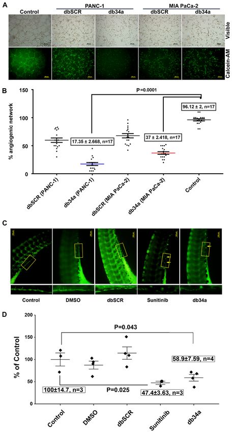
2 GNANAMONY et al: db34a MEDIATES ANGIOGENIC SUPPRESSION AND MACROPHAGE ACTIVATION
small interfering RNAs (siRNAs) and combinations thereof. for 25 min at 4˚C to precipitate the small RNAs, which included
Although promising, the half‑life of RNA molecules is very the dbRNA product. The small RNA was washed twice with
short. In vitro, natural RNA exhibits a half‑life of a few seconds ice‑cold 75% ethanol and then dissolved in sterile water. The
to a few minutes in various biological fluids, including human purity of the small RNA was verified in a 1% agarose gel.
serum, whereas RNAs partially modified at the 20th position The quality of RNA was determined using a NanoDrop™
have extended half‑lives of 5 sec to 15 h (15). An RNA‑based spectrophotometer.
anti‑angiogenesis drug called Macugen has these modifica‑
tions; however, although somewhat successful, it is limited Purification of dbRNAs. The isolated small RNAs were
in addressing the problem of angiogenesis (16). The approach separated on a 15% urea‑polyacrylamide gel (PAGE). The gel
used in the present study is the development of a closed circular was stained with ethidium bromide (0.5 mg/ml) for 15 min at
RNA having multiple double‑stranded regions coding for room temperature and then destained by washing with distilled
miRNAs, which are interrupted by loops or dumbbells. Using water twice. The gel portion containing the dbRNA was excised
a circular RNA should eliminate exonuclease activity while with a sharp scalpel and the RNA was purified using the
double‑stranded RNA (dsRNA), being inherently more stable ZR Small‑RNA PAGE Recovery Kit (Zymo Research Corp.)
than single‑stranded RNA (ssRNA), should contribute to an according to the manufacturer's instructions. RNA quality and
increased half‑life. To the best of our knowledge, this is the quantity were measured using a NanoDrop spectrophotometer.
first attempt to use closed circular dumbbell RNAs (dbRNAs)
to code for miRNAs/siRNAs with a therapeutic intention. The Verification of dbRNA specificity. The isolated dbRNAs were
study uses miR‑34a‑3p and ‑5p in a circular form. separated on a 15% urea‑PAG (TBE is the running buffer) at
20 mA followed by blotting using the iBlot™ Gel Transfer
Materials and methods Device onto a Novex™ iBlot™ DNA Transfer Stack with a
nylon membrane (both Invitrogen; Thermo Fisher Scientific,
Construction of dbRNAs. The permuted intron‑exon (PIE) Inc.). Northern blot analysis was conducted using a miR‑34a
method was used, which is an enzyme‑free RNA circular‑ specific biotinylated probe (sequence: /5Biosg/AGGGCAGTA
ization method based on group I intron self‑splicing (17). TACTTGCTGAT) after membrane was incubated with prehy‑
Circularization is triggered by the presence of magne‑ bridization buffer (6X SSC buffer, 10X Denhardt's solution,
sium ions and guanine nucleotides, and the production of 0.2% SDS; MilliporeSigma) for 1 h at 42˚C on rotating shaker
circular RNA can be conducted using essentially any type following 3 day incubation with hybridization buffer (6X SSC
of cell (17‑19). A previously described strategy was used, buffer, 5X Denhardt's solution, 0.2% SDS; MilliporeSigma)
in which recombinant RNA is disguised as a natural RNA with probe at the same conditions. The chemiluminescent signal
and thus appropriates the host machinery to evade cellular was detected using a Pierce™ Chemiluminescent Nucleic
RNases (20), but with an added RNA circularization step using Acid Detection Module kit (Thermo Fisher Scientific, Inc.)
the rrnC terminator (21). Sequences coding for miR‑34a‑3p according to the manufacturer's recommendations.
and ‑5p (db34a) and its scrambled sequence (dbSCR) were
synthesized with the lpp promoter and the rrnC terminator Determining the RNase A resistance of db34a RNA. To deter‑
sequences (Fig. 1A and B), and inserted in pUC57‑Kan mine the resistance of db34a to RNase A, 1 µg total RNA
plasmids (GenScript Biotech). The JM101Tr E. coli strain from bacteria transformed with db34a‑expressing plasmid was
[Δ (lac pro), supE, thi, recA56, srl‑300.:Tn10, (F', traD36, isolated and incubated with or without 0.7x10‑5 U/µl RNase‑A
proAB, lacIq, lacZ, ΔM15)] (20) was used for the isolation of for 24 h in four independent treatments at room temperature.
bacterial RNA. Modeling analysis using the on line mfold tool Since the bacterial lipoprotein (lpp) promoter (18,24) is a
(http://unafold.rna.albany.edu/?q=mfold/RNA‑Folding‑Form) constitutive bacterial promoter that drives the expression of
was done to determine the secondary structure of db34a (22). bacterial lpp, bacterial lpp RNA was used as the linear control
for db34a. First, cDNA was synthesized using miScript II RT
Isolation of small RNAs. Purification of small RNA was kit (Qiagen GmbH) using conditions: 37˚C for 60 min and 95˚C
performed using a modified RNASwift protocol (23) and for 5 min from untreated and 24 h RNase A treated samples
compared with purification performed using RNAzol reagent followed by quantitative PCR (qPCR) with the following
(Sigma‑Aldrich; Merck KGaA). In the modified RNASwift primers: lpp1 forward, CTGTCTTCTGACGTTCAGACTC
method, bacteria expressing the db34a or dbSCR plasmid were and lpp reverse, ACGAGCTGCGTCATCTTTAG. Expression
grown in Luria‑Bertani (LB) medium (BD Biosciences) with of db34a was measured by qPCR with miR‑34a specific primers
50 µg/ml of kanamycin (MilliporeSigma) for 24 h at 37˚C. [5'UGGCAGUGUC UUAGCUGGU UGU, cat. no. 218300;
The cultured cells (1.5x10¹⁰ cells) were spun down (3,000 x g Hs_miR‑34a_1 miScript Primer Assay (forward primer)
for 20 min at 26˚C), suspended in 10 ml of LB1 lysis reagent and miScript Universal Primer (reverse)] using the miScript
(4% sodium dodecyl sulfate, pH 7.5, 0.5 M NaCl), and lysed SYBR Green PCR Kit (Qiagen GmbH). The qPCR conditions
by incubation at 90˚C for 4 min. Then, 5 ml of 5 M NaCl was used were as follows: Initial heat activation 95˚C for 15 min,
added to the homogenate and the mixture was centrifuged at followed by denaturation at 94˚C for 15 sec, annealing at 55˚C
16,000 x g for 25 min at 4˚C. The supernatant was transferred to for 30 sec and extension at 70˚C for 30 sec, for 40 cycles. Four
a new tube, and 6 ml of isopropanol was added and the mixture independent experiments with three replicates in each experi‑
was centrifuged at 16,000 x g for 25 min at 4˚C to precipitate out ment were performed. ΔCq was calculating by subtracting
the large RNA. To the remaining supernatant, 10.5 ml of isopro‑ the average Cq values of lpp from db34a for both untreated
panol was added and the mixture was centrifuged 20,000 x g (control) and RNase A treated cells. ΔΔCq values wereONCOLOGY LETTERS 21: 75, 2021 3
obtained by subtracting the average ΔCq of the control group manufacturer's instructions. In brief, MIA PaCa‑2 and PANC‑1
from both the control and treatment ΔCq data followed by the cells were seeded (1.2x10⁶/well; 6‑well plate) and cultured
calculation of 2‑ΔΔCq (25). The difference between treated and overnight in RPMI‑1640 medium (Thermo Fisher Scientific
control groups with reference to db34a/lpp RNA was analyzed Inc.) supplemented with 10% FBS (VWR Corporation) and
using one‑way ANOVA. 1% penicillin‑streptomycin (Thermo Fisher Scientific Inc.) in
a humidified 5% CO2 atmosphere at 37˚C. The next day, cells
Cell culture conditions. The MIA PaCa‑2 and PANC‑1 were transfected with 4 µg of db34a or dbSCR at 37˚C using
pancreatic cancer cell lines were obtained from American jetPRIME transfection reagent or remained untransfected
Type Culture Collection (ATCC) and maintained under the (control). After 20 h, the medium was replaced with serum‑free
conditions recommended by the supplier. Cells were grown medium RPMI‑1640 (Thermo Fisher Scientific Inc.) and the
in tissue culture‑treated Petri plates in RPMI‑1640 medium plates were incubated for another 16 h. Conditioned medium
(Thermo Fisher Scientific, Inc.) with 10% FBS (VWR was then collected after centrifugation at 16,000 x g at 4˚C for
Corporation) and 1% penicillin‑streptomycin (Thermo Fisher 10 min and transferred to freshly grown macrophages. After
Scientific Inc.) in a humidified 5% CO2 atmosphere at 37˚C. overnight incubation at 37˚C, the macrophages were lysed
To evaluate immune activation, J774A.1 mouse macrophages with lysis buffer provided with kit and equal samples from
obtained from ATCC were used. The macrophages were grown each condition with regard to total protein quantity (100 µg
in tissue culture‑treated Petri plates in DMEM (Thermo Fisher per sample; total protein concentration was determined by
Scientific Inc.) with 10% FBS and 1% penicillin‑streptomycin. Pierce 660 nm Protein Assay (Thermo Fisher Scientific Inc.)
Human umbilical vein endothelial cells (HUVECs) were were applied to the antibody‑spotted membranes. Following
obtained from Thermo Fisher Scientific, Inc. and cultured overnight incubation at 4˚C, the membranes were washed and
in Medium 200 with 1X low serum growth supplement 1 ml diluted biotin‑conjugated antibody mix was added to the
(Thermo Fisher Scientific Inc.). HUVECs with a low passage membranes, which were kept at room temperature for 2 h. After
number (3 passages) were used for the tube formation assay as several washes, the membranes were incubated with diluted
described in a previous study (26). HRP‑conjugated streptavidin for 2 h. Detection was performed
using ECL protocols in which the membranes were exposed
Angiogenesis antibody array. An angiogenic antibody array to X‑ray films to detect the binding of inflammatory factors.
analysis was performed using a RayBio® Human Angiogenesis Spot intensities were quantified using ImageJ version 1.52a
Array C1 kit (AAH‑ANG‑1; RayBiotech, Inc.) according to software. Significant differences between macrophages grown
the manufacturer's instructions. In brief, MIA PaCa‑2 and in dbSCR and db34a conditioned media were identified using
PANC‑1 cells were seeded (1.2x10 6/well, 6‑well plate) in one‑way ANOVA.
tissue culture‑treated Petri plates overnight in RPMI‑1640
medium (Thermo Fisher Scientific Inc.) with 10% FBS (VWR In vitro angiogenic assay. To determine the anti‑angiogenic
Corporation) and 1% penicillin‑streptomycin (Thermo Fisher effect of the dbRNA molecules, an endothelial cell tube
Scientific Inc.) in a humidified 5% CO2 atmosphere at 37˚C. formation assay was conducted using HUVECs as previously
The cells were transfected with 4 µg of db34a or dbSCR using described (26). Briefly, HUVECs were stained with 2 µg/ml
jetPRIME® transfection reagent (Polyplus‑transfection SA cell‑permeant fluorescent dye (calcein‑AM; Thermo Fisher
at 37˚C) or remained untransfected (control). After a 24‑h Scientific, Inc.) with incubation at 37˚C in the dark for ≥30 min.
incubation period, the medium was replaced with serum‑free A Geltrex basement membrane matrix (Thermo Fisher
RPMI‑1640 medium (Thermo Fisher Scientific Inc.) and the Scientific, Inc; 40 µl) was applied to 96‑well plates and allowed
plates were incubated for another 24 h. Conditioned medium to solidify for 30 min at room temperature. The stained
was then collected, and the antibody‑spotted membranes from HUVECs were trypsinized, re‑suspended in serum‑free
the array kit were incubated with the conditioned medium medium (Gibco Medium 200PR; Gibco; Thermo Fisher
according to the manufacturer's protocol. Following incubation, Scientific Inc.) and plated at a density of 20,000 cells/well in
the membranes were washed, 1 ml diluted biotin‑conjugated 100 µl conditioned medium from the db34a and dbSCR trans‑
antibody mix was added and the membranes were incubated at fected cells. HUVECs in complete medium (Gibco Medium
room temperature for 2 h, followed by washing and the addition 200PRF supplemented with Gibco Low Serum Growth
of diluted HRP‑conjugated streptavidin at room temperature Supplement; Gibco; Thermo Fisher Scientific Inc.) were used
for 2 h. Detection was performed using ECL protocols in as controls. The plates were incubated at 37˚C in a humidified
which the membranes were exposed to X‑ray films to visualize incubator for 3 h. The formation of angiogenic network was
the binding of angiogenic factors. The intensities of the spots visualized at x10 magnification using an inverted fluorescence
were quantified using ImageJ version 1.52a software (National microscope and quantified by ImageJ version 1.52a (n=17).
Institutes of Health), and angiogenic molecules showing a The significance of difference of each HUVEC treatment
significant difference in expression levels between the db34a group compared to control was determined using one‑way
and dbSCR conditioned media were identified. Significant ANOVA.
differences between dbSCR and db34a conditioned media
were detected using one‑way ANOVA. Zebrafish aquaculture. Transgenic VEGFR2:green‑reef
coral fluorescent protein (G‑RCFP) zebrafish (Danio rerio)
Inflammation array. A mouse inflammation array analysis that express G‑RCFP under the control of a VEGFR2/KDR
was performed using a RayBio Mouse Inflammation Array C1 promoter were obtained from ZFIN (y1Tg). The fish were main‑
kit (cat. no. AAM‑INF‑1; RayBiotech, Inc.) according to the tained under standard aquaculture conditions in a circulating4 GNANAMONY et al: db34a MEDIATES ANGIOGENIC SUPPRESSION AND MACROPHAGE ACTIVATION
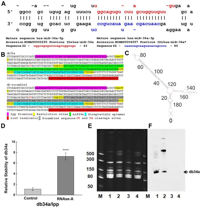
Figure 1. Designing and purification of db34a. (A) Sequences of miR‑34a‑3p and miR‑34a‑5p used in the study. (B) Linear sequence of db34a and its scrambled
version, dbSCR. (C) Modeling analysis using the mfold web server, showing the predicted structure of db34a. (D) Stability assay as performed by reverse
transcription‑quantitative PCR showing increased stability of the circular db34a compared with linear lpp RNA in the presence of RNase A. The signifi‑
cance of the difference between the control and RNase A‑treated groups was evaluated by one‑way ANOVA (****P=1.41627x10 ‑4). (E) TBE‑urea acrylamide
gel electrophoresis of db34a in small RNA samples purified from db34a‑expressing E. coli (lanes 1 and 2) and dbSCR‑expressing control (lanes 3 and 4).
Samples 2 and 4 were purified using RNAzol reagent, and samples 1 and 3 were purified using a modified RNASwift protocol. Lane M, dsRNA ladder
(BioLabs). (F) Northern blotting analysis using a miR‑34a‑specific biotinylated probe showing the presence of db34a in lanes 1 and 2 but not in lanes 3 and 4.
miR, microRNA; db34a, miR‑34a‑3p and ‑5p dumbbell RNA; dbSCR, scrambled dumbbell RNA; lpp, lipoprotein.
tank system constructed as previously described (27), and eggs were collected soon after fertilization and rinsed at 28˚C
according to protocols approved by the Institutional Animal in embryo water (Milli‑Q water with 60 mg/l Instant Ocean‑
and Use Committee (IACUC) of University of Illinois Spectrum Brands) and immediately followed by incubation in
College of Medicine at Peoria. Water was purified by a 1% methylene blue in embryo water at 28˚C for 24 h. After
reverse osmosis system and supplemented with sea salts at which the embryos were dechorionated and allowed to develop
concentration of 60 mg/l in order to provide the trace minerals in embryo water alone for 24 h at 28˚C (48 h post fertilization).
that the fish required. The pH of the water was maintained Following this the embryos were incubated in embryo water
between 7.6 and 8.5 by the addition of sodium bicarbonate. All alone or embryo water supplemented with 10 pmol db34a or
experimental procedures using zebrafish were performed with dbSCR RNA, 5 µM sunitinib or a 1:1,000 dilution of DMSO
approval from the IACUC of University of Illinois College of as vehicle control at 28˚C. After 24 h treatment, the 72 h old
Medicine at Peoria. embryos were fixed in 10% buffered formaldehyde at room
temperature for 10 min. The fixed embryos were mounted on
In vivo zebrafish angiogenic assay. Adult male and female glass slides and the completeness of sub‑intestinal vein (SIV)
zebrafish were placed in spawning baskets. Newly fertilized vasculature was observed by the detection of G‑RCFP usingONCOLOGY LETTERS 21: 75, 2021 5 fluorescent inverted microscopy. The significance of differ‑ ences of each treatment group compared to control was determined using one‑way ANOVA. Statistical analysis. P
6                   GNANAMONY et al: db34a MEDIATES ANGIOGENIC SUPPRESSION AND MACROPHAGE ACTIVATION

Figure 3. Inhibition of angiogenesis by db34a in in vitro and in vivo models. (A) and (B) Endothelial cell tube formation assay was performed using HUVECs as
a measure of angiogenesis. Phase contrast and calcein AM stained fluorescent microscope images showing reduced angiogenic tubule formation in HUVECs
incubated with conditioned media from MIA PaCa‑2 and PANC‑1 cell lines transfected with db34a when compared with dbSCR control for 3 h. Scale bar,
200 µm. (C) In vivo angiogenesis assay was performed in zebrafish embryos with analysis using a fluorescent microscope. Dechorionated embryos were
allowed to develop for 72 h then placed in embryo water alone (control), or supplemented with 10 pmol db34a or dbSCR RNA, 5 µM sunitinib or a 1:1,000 dilu‑
tion of DMSO. The db34a‑treated zebrafish embryos exhibited a reduction in the completeness of the SIV when compared with dbSCR and the controls.
(D) Measurement of SIV completeness by the quantification of fluorescent intensity values of identical SIV areas and presented as a percentage of the control
(control vs. db34a, P= 0.043; control vs. sunitinib; P= 0.025). HUVECs, human umbilical vein endothelial cells; db34a, miR‑34a‑3p and ‑5p dumbbell RNA;
dbSCR, scrambled dumbbell RNA; SIV, sub‑intestinal vein.
ONCOLOGY LETTERS 21: 75, 2021                                                       7

Table I. Cytokines significantly different between macrophages treated with conditioned media from PANC‑1 and MIA PaCa‑2
cell lines transfected with db34a compared with dbSCR.

                                        PANC‑1		                                                MIA PaCa‑2
Cytokine                        (fold change vs. control) P‑value                        (fold changes vs. control)           P‑value

CD30 ligand (TNFSF8)                    1.543449                   4.61297x10‑5                  1.808521                        ‑
Fractalkine (CX3CL1)                    1.591752                      0.00309                    0.798687                        ‑
IFN‑γ                                   5.434441                      0.02768                    1.484371                        ‑
IL‑1α (IL‑1 F1)                         1.793997                      0.02553                    1.287388                        ‑
IL‑1β (IL‑1 F2)                         5.106111                      0.0092                     0.943795                        ‑
IL‑2                                    2.601422                      0.04669                    0.909556                        ‑
IL‑6                                    3.973113                      0.02473                    1.081643                        ‑
IL‑10                                   7.051837                      0.00978                    0.605812                        ‑
IL‑12 p70                               6.513021                      0.02201                    0.673057                        ‑
IL‑13                                   2.100802                      0.00251                    1.849357                     0.00994
IL‑17A                                  3.668945                      0.00847                    3.655409                     0.03355
I‑TAC (CXCL11)                          1.996696                      0.02114                    2.297556                        ‑
KC (CXCL1)                              1.596054                       0.0123                     2.07834                        ‑
Leptin                                  4.536913                       0.0375                    2.313414                        ‑
LIX (CXCL5)                             1.252327                          ‑                      1.334515                     0.02446
M‑CSF                                   1.339565                      0.02349                     1.40063                     0.03974
RANTES (CCL5)                           2.056593                      0.00633                    2.646111                     0.00422
SDF‑1α (CXCL12α)                        2.493808                      0.00911                    1.820613                     0.01686
I‑309 (TCA‑3/CCL1)                      1.689148                      0.00128                    1.319467                     0.00263
TECK (CCL25)                            1.633533                      0.02981                    1.637408                     0.02115
TIMP‑2                                  1.091021                          ‑                      1.089587                     0.04152
TNF‑α                                   1.387676                   4.64829x10‑4                  2.003669                     0.02406
TNF RI (TNFRSF1A)                       1.355374                      0.02447                    1.223076                     0.02037
TNF RII (TNFRSF1B)                      1.114493                          ‑                      1.962633                     0.00873

db34a, miR‑34a‑3p and ‑5p dumbbell RNA; dbSCR, scrambled dumbbell RNA; IL, interleukin; CCL5, C‑C motif chemokine ligand 5;
CXCL12α, C‑X‑C motif chemokine ligand 12α.

Zebrafish strain y1tg that expresses a green fluorescent           significantly induced the expression of interleukin (IL)‑13,
protein in the vasculature was used as a model organism. SIV       CCL5 and C‑X‑C motif chemokine ligand 12 α (CXCL12α)
vasculature was observed 24 h after treatment with 10 pmol         among other inflammatory response genes (Table I and Fig. 4).
db34a or dbSCR RNA, 5 µM sunitinib or a 1:1,000 dilution
of DMSO, and images were captured using a fluorescent              Discussion
microscope (Fig. 3C). It was observed that the embryos treated
with db34a showed reduced SIV integrity compared with              Despite decades of research, the treatment of pancreatic ductal
the controls (58.9±7.59% of the untreated control; P= 0.043),      adenocarcinoma continues to be a major challenge. According
which confirmed the functionality of db34a and indicated the       to the American Cancer Society, for all stages of pancreatic
potential of dbRNA as a therapeutic agent. Positive control        cancer combined, the 1‑year survival rate is ~20% and the
embryos treated with sunitinib also exhibited decreased SIV        5‑year survival rate is 9% (28). One of the major factors that
vasculature (47.4±3.63% of the untreated control; P=0.025).        contributes to this poor prognosis is late detection, as early‑stage
                                                                   pancreatic cancer is usually asymptomatic (29). New surgical
db34a RNA harboring miR‑34a‑3p and miR‑34a‑5p induces              techniques and evolving therapeutics have achieved only
the PANC‑1 and MIA PaCa‑2 pancreatic cancer cell‑mediated          modest outcomes (30). A clearer understanding of targetable
activation of macrophage inflammatory markers. To determine        molecular pathways is required to design therapeutic agents
whether db34a induces immune activation, conditioned media         and achieve a desired clinical outcome. Non‑coding RNAs
from PANC‑1 and MIA PaCa‑2 cells transfected with db34a            such as miRNAs play important roles in the regulation of
were collected and the ability of the conditioned media to acti‑   gene expression. Unlike siRNAs, miRNAs target multiple
vate an inflammatory response in a mouse macrophage cell           genes and are usually process specific (31). Most biological
line was determined. It was observed that conditioned media        processes are in some way regulated by miRNAs, which are
from PANC‑1 and MIA PaCa‑2 cells transfected with db34a            small RNA molecules ~22 nucleotides in length that probably
8 GNANAMONY et al: db34a MEDIATES ANGIOGENIC SUPPRESSION AND MACROPHAGE ACTIVATION
Figure 4. Inflammatory proteins secreted by macrophages in response to conditioned medium from pancreatic cancer cells expressing db34a as detected using
a mouse inflammation sp16 array. Array results for (A) PANC‑1 and (B) MIA PaCa‑2 cells. Expression levels were plotted as relative density ratios compared
with wild‑type controls. The analysis shows significant alterations in the expression of several key inflammatory cytokines and proteins in macrophages treated
with db34a conditioned medium compared with dbSCR conditioned medium. *P≤0.05, **P≤0.01, ***P≤0.001 vs. dbSCR. db34a, miR‑34a‑3p and ‑5p dumbbell
RNA; dbSCR, scrambled dumbbell RNA; IL, interleukin; CCL5, C‑C motif chemokine ligand 5; CXCL12α, C‑X‑C motif chemokine ligand 12α.
function as antisense regulators of other RNAs (32). The the limitations of low half‑life and questionable in vivo effi‑
mechanisms of miRNA action have been elucidated, although cacy (36,37), which is mainly due to the effects of exonuclease
not completely (33). Their capacity to regulate mRNAs and RNases. The present study circumvents the problem of a low
other miRNAs is being reported with increasing frequency, half‑life by designing dbRNAs that should not be degraded
and they have become powerful tools for gene regulation. by the exonuclease activity of RNases. A previous study
The use of RNA molecules to target angiogenesis continues demonstrated the stability of a 35mer FITC‑labeled RNA in
to be explored (11‑16). However, the approach described in the presence of circular dbRNA/DNA chimeric oligonucle‑
the present study, utilizing a circular RNA molecule with two otides to RNase H. This induced stability is most likely due
miRs is novel and, to the best of our knowledge, has not been to the competition of RNA/DNA chimeric with the 35mer
described previously. The approach was to develop a closed FITC‑labeled RNA (38). The stability experiments conducted
circular RNA coding for miRNAs. A circular RNA is not in the present study demonstrate that circular RNA exposed
readily degraded by exonucleases and the inherent higher to RNase A for 24 h was more stable than linear RNA
stability of dsRNAs compared with ssRNAs may contribute driven by the same lpp promoter (18,24). Gene silencing by
to an increased half‑life (34). Increased stability of db34a siRNA/miRNAs can be used as a therapeutic approach for
compared with linear RNA was observed in the present study. the treatment of un‑druggable targets. Researchers have also
To the best of our knowledge, the present study is the first to shown that dumbbell‑shaped DNA minimal vectors can be
use closed circular dbRNAs to code for both miR34‑3p and ‑5p used for small hairpin RNA expression (39,40). These studies
simultaneously with a therapeutic intention. also demonstrated the use of a dumbbell DNA vector to produce
The present study aimed to demonstrate that circular RNA hairpin RNAs that can be used therapeutically. Another study
harboring miRs are functional and have significant therapeutic described the generation of dumbbell‑shaped nano‑circular
potential using dbRNA encoding miR‑34a‑3p and ‑5p. To RNAs for RNA interference, using a chemical method of
construct the circular RNA expressing miR‑34a‑3p and ‑5p, synthesis involving the enzyme‑based circularization of small
the PIE method was used, which is an enzyme‑free RNA circu‑ RNA molecules (41). By contrast, the present study reveals the
larization method based on group I intron self‑splicing (17), development of a larger dbRNA and is unique in that it uses a
which can be conducted using essentially any type of cell. bacterial system for large‑scale dbRNA production.
Cells transcribe more RNA than they accumulate (35). This Although not much is known about miR‑34a‑3p, miR‑34a‑5p
indicates that RNA is constantly being degraded and that a is well studied and has been shown to be involved in tumor
dynamic process occurs to balance RNA synthesis and degra‑ suppression, angiogenic suppression and cancer stem cell
dation. The use of siRNA therapeutically is not new but has suppression (42‑49); hsa‑miR‑34a‑3p was previously knownONCOLOGY LETTERS 21: 75, 2021 9
as hsa‑miR‑34a* and hsa‑miR‑34a‑5p was previously known of activated natural killer cells to the tumor microenviron‑
hsa‑miR‑34a. It was recently demonstrated that normal cells do ment (61). Another study highlighted the importance of CCL5,
show detectable expression levels of miR34a (50). Normal cell demonstrating that the blockade of 1,4‑ α‑glucan‑branching
lines were not used in the present study and this is a limitation enzyme in lung cancer cells promoted the secretion of
of the study. Since normal cells do show detectable levels of CCL5, which induced the recruitment of CD8+ T lympho‑
miR34a, the addition of db34a to normal cells should not, in cytes to the tumor microenvironment (62). CCL5 expression
theory, induce a notable perturbance in the existing molecular has also been shown to restore the immune surveillance in
pathways of normal cells; this however requires further study. antigen‑expressing MYC;CTNNB1 hepatocellular carcinoma
To demonstrate the possibility of using circular RNA therapeu‑ tumors (63). This indicates that CCL5 is necessary for tumor
tically, in the present study circular miR‑34a‑3p and ‑5p were cells to be visible to the immune system; especially, CCL5
transfected into pancreatic cancer cells and the expression of expression is necessary for immune recognition. However,
pro‑angiogenic molecules was determined. It was observed that it is also important to note that CCL5 was recently identi‑
in the two pancreatic cancer cell lines transfected with db34a, fied as a signature of poor prognosis for oral squamous cell
the expression levels of bFGF and CCL5 were significantly carcinoma, indicating its multifaceted role (64). Interestingly,
increased. miR‑34a‑5p expression is known to be increased by another study observed that the expression of CCL5 in breast
the activation of the PI3K signaling pathway (51) and, notably, cancer cells was associated with lymph node status and tumor
mir‑34a‑5p has been shown to suppress the PI3K signaling node‑metastasis stage (65). The conflicting roles of CCL5, and
pathway (52‑56). These contradictory observations indicates the whether its overexpression is a predisposing factor for poor
possibility of the existence of a tight regulatory apparatus that is prognosis or a response to poor prognosis remain unclear. The
not yet clearly understood. Interestingly, TNF is predicted as the association of CCL5 with other immune‑associated molecules
target of hsa‑miR‑34a‑3p by the miRDB online database, and may provide clearer insight, and CCL5 expression alone should
TNF is a strong promoter of angiogenesis (57). This indicates not be used as a measure of prognosis.
that hsa‑miR‑34a‑3p may be more therapeutically relevant in Furthermore, the present study investigated whether the
angiogenic suppression. It should be noted that both miR‑34a‑3p transfection of pancreatic cancer cells with db34a induces
and miR‑34a‑5p are processed from the same pre‑miR. the activation of immune cells. It was observed that the
This is consistent with the observation that angiogenesis was conditioned medium from db34a‑transfected pancreatic
suppressed significantly in the in vitro and in vivo angiogenic cancer cells significantly induced the expression of IL‑13,
assays of the present study. This observation appears contrary to CCL5 and CXCL12α among other inflammatory response
the expected outcome; however, miR‑34a‑5p is a known angio‑ genes in mouse macrophage cell lysates. Tumor‑associated
genic suppressor (43) and the quantification of other relevant macrophages (TAMs) are present in high numbers in the
angiogenesis‑associated molecules will provide a clearer mech‑ microenvironment of solid tumors and have been studied
anistic profile of this angiogenesis modulation. Every biological for their potential as targets for cancer therapy (66). TAMs
process in an oncogenic system is tightly regulated; therefore, suppress host immune responses and also promote tumor
targeting a process rather than a single molecule would not be cell proliferation, angiogenesis, invasion and metastasis (67).
clinically relevant as compensatory pathways can be activated The role of TAMs remains unclear; however, most evidence
to achieve the oncogenic phenotype. The overexpression of suggests that TAMs promote tumor progression. From the
bFGF may be one such compensatory response, which is seen data in the present study, especially the overexpression of
in both pancreatic cell lines. IL‑13, it appears that the J774A.1 macrophages exposed
The present study also demonstrated that CCL5 is to conditioned medium from db34a‑transfected pancreatic
significantly upregulated in PANC‑1 and MIA PaCa‑2 cells cancer cells appeared more closely associated with the M1
transfected with db34a. CCL5, also known as regulated on phenotype than the M2 phenotype (data not shown) (68).
activation, normal T cell expressed and secreted (RANTES), Further investigation is required to decipher the influence of
is a chemokine that is involved in T‑cell activation. RANTES db34a on macrophages.
has been identified as a major HIV‑suppressive factor. Studies In conclusion, the present data suggest that the supplemen‑
have shown that recombinant human RANTES induces tation of miR‑34a‑3p and ‑5p as a circular pre‑miR form is a
a dose‑dependent inhibition of different strains of HIV‑1, viable method of miR replacement therapy.
HIV‑2 and simian immunodeficiency virus (SIV) (58,59).
The expression of CCL5 in MIA PaCa‑2 and PANC‑1 cells Acknowledgements
transfected with db34a RNA indicates the involvement of
a conserved pathway. Additional research is necessary to Assistance for the maintenance of Zebrafish culture at the
determine whether transfection with db34a can also induce Laboratory Animal Care Facility, UICOMP, LACF‑Peoria IL,
the overexpression of CCL5 in other types of cells. In a recent by Laboratory Animal Care Supervisor Ms. Angela Daniels and,
study, it was shown that CCL5 and CCR5 expression levels Laboratory Animal Care Technician Ms. Stephanie Sampson
are increased in pancreatic cancer tissue sections, and the is acknowledged.
overexpression of CCL5 and CCR5 increases the invasiveness
and metastatic potential of pancreatic cancer cells (60). It has Funding
also been reported that progranulin (PGRN) expression levels
correlate with a poor prognosis for melanoma patients, and The Department of Internal Medicine, University of Illinois
PGRN inhibits CCL5 gene expression at the transcriptional College of Medicine at Peoria; McElroy Foundation
level, showing that CCL5 is responsible for the recruitment (Springfield, IL, USA) (grant no, 096726) and gift from The10 GNANAMONY et al: db34a MEDIATES ANGIOGENIC SUPPRESSION AND MACROPHAGE ACTIVATION
Theresa Tracy Strive to Survive Foundation (East Peoria, IL, 8. Liu JF, Tolaney SM, Birrer M, Fleming GF, Buss MK, Dahlberg SE,
Lee H, Whalen C, Tyburski K, Winer E, et al: A phase 1 trial of
USA) supported the present study. the poly(ADP‑ribose) polymerase inhibitor olaparib (AZD2281)
in combination with the anti‑angiogenic cediranib (AZD2171) in
Availability of data and materials recurrent epithelial ovarian or triple‑negative breast cancer. Eur J
Cancer 49: 2972‑2978, 2013.
9. Climent‑Salarich M, Quintavalle M, Miragoli M, Chen J,
The plasmid constructs db34a and dbSCR are available Condorelli G and Elia L: TGFβ triggers miR‑143/145 transfer
from the corresponding author upon reasonable request from smooth muscle cells to endothelial cells, thereby modulating
vessel stabilization. Circ Res 116: 1753‑1764, 2015.
in accordance with institutional MTA approvals and 10. Huang G and Chen L: Tumor vasculature and microenviron‑
all applicable rules and regulations. The datasets used ment normalization: A possible mechanism of antiangiogenesis
and/or analyzed during the current study are available from therapy. Cancer Biother Radiopharm 23: 661‑667, 2008.
11. Barbas AS and White RR: The development and testing of
the corresponding author on reasonable request. aptamers for cancer. Curr Opin Investig Drugs 10: 572‑578, 2009.
12. Guglielmelli T, Bringhen S and Palumbo A: Update on the use of
Authors' contributions defibrotide. Expert Opin Biol Ther 12: 353‑361, 2012.
13. Gatto B and Cavalli M: From proteins to nucleic acid‑based
drugs: The role of biotech in anti‑VEGF therapy. Anticancer
MG designed and performed experiments and reviewed the Agents Med Chem 6: 287‑301, 2006.
manuscript. LD validated the constructs, designed the RNA 14. Kanwar JR, Mahidhara G and Kanwar RK: Antiangiogenic
therapy using nanotechnological‑based delivery system. Drug
stability experiments, confirmed dbRNA stability and quanti‑ Discov Today 16: 188‑202, 2011.
fied data. LG and PS were involved in designing the concept 15. Friedman AD, Kim D and Liu R: Highly stable aptamers selected
of the study. SB was involved in RNA experiments and data from a 20‑fully modified fGmH RNA library for targeting
biomaterials. Biomaterials 36: 110‑23, 2015.
collection. JAN was involved in zebrafish data collection. CSG 16. Inoue M, Kadonosono K, Arakawa A, Yamane S and Ishibashi T:
designed, and developed the concept and experiments, and Long‑term outcome of intravitreal pegaptanib sodium as mainte‑
wrote the manuscript. All authors read and approved the final nance therapy in japanese patients with neovascular age‑related
macular degeneration. Jpn J Ophthalmol 59: 173‑178, 2015.
manuscript. 17. Puttaraju M and Been MD: Circular ribozymes generated in
Escherichia coli using group I self‑splicing permuted intron‑exon
Ethics approval and consent to participate sequences. J Biol Chem 271: 26081‑26087, 1996.
18. Umekage S and Kikuchi Y: In vivo circular RNA production
using a constitutive promoter for high‑level expression. J Biosci
All experimental procedures using zebrafish were performed Bioeng 108: 354‑356, 2009.
with approval from the Institutional Animal and Use Committee 19. Ford E and Ares M Jr: Synthesis of circular RNA in bacteria
and yeast using RNA cyclase ribozymes derived from a group I
of University of Illinois College of Medicine at Peoria. intron of phage T4. Proc Natl Acad Sci USA 91: 3117‑3121, 1994.
20. Ponchon L and Dardel F: Recombinant RNA technology: The
Patient consent for publication tRNA scaffold. Nat Methods 4: 571‑576, 2007.
21. Umekage S and Kikuchi Y: In vitro and in vivo production and
purification of circular RNA aptamer. J Biotechnol 139: 265‑272,
Not applicable. 2009.
22. Zuker M: Mfold web server for nucleic acid folding and
hybridization prediction. Nucleic Acids Res 31: 3406‑3415,
Competing interests 2003.
23. Nwokeoji AO, Kilby PM, Portwood DE and Dickman MJ:
The authors declare that they have no competing interests. RNASwift: A rapid, versatile RNA extraction method free from
phenol and chloroform. Anal Biochem 512: 36‑46, 2016.
24. Chen QX, Wang WP, Zeng S, Urayama S and Yu AM: A general
References approach to high‑yield biosynthesis of chimeric RNAs bearing
various types of functional small RNAs for broad applications.
Nucleic Acids Res 43: 3857‑3869, 2015.
1. Boehm T, Folkman J, Browder T and O'Reilly MS: Antiangiogenic 25. Livak KJ, Schmittgen TD: Analysis of relative gene expression
therapy of experimental cancer does not induce acquired drug data using real‑time quantitative PCR and the 2(‑delta delta
resistance. Nature 390: 404‑407, 1997. C(T)) method. Methods 25: 402‑408, 2001.
2. Zetter BR: Angiogenesis and tumor metastasis. Annu Rev 26. Gnanamony M, Antony R, Fernandez KS, Jaime L, Lin J,
Med 49: 407‑424, 1998. Joseph PA and Gondi CS: Chronic radiation exposure of neuro‑
3. Folkman J: Antiangiogenic gene therapy. Proc Natl Acad Sci blastoma cells reduces nMYC copy number. Oncol Lett 14:
USA 95: 9064‑9066, 1998. 3363‑3370, 2017.
4. Donmez G, Sullu Y, Baris S, Yildiz L, Aydin O, Karagoz F and 27. Kim S, Carlson R, Zafreen L, Rajpurohit SK and Jagadeeswaran P:
Kandemir B: Vascular endothelial growth factor (VEGF), matrix Modular, easy‑to‑assemble, low‑cost zebrafish facility.
metalloproteinase‑9 (MMP‑9), and thrombospondin‑1 (TSP‑1) Zebrafish 6: 269‑274, 2009.
expression in urothelial carcinomas. Pathol Res Pract 205: 854‑7, 28. Siegel RL, Miller KD and Jemal A: Cancer statistics, 2019. CA
2009. Cancer J Clin 69: 7‑34, 2019.
5. Neves KB, Montezano AC, Lang NN and Touyz RM: Vascular 29. Jelski W and Mroczko B: Biochemical diagnostics of pancreatic
toxicity associated with anti‑angiogenic drugs. Clin Sci cancer‑present and future. Clin Chim Acta 498: 47‑51, 2019.
(Lond) 134: 2503‑20, 2020. 30. McGuigan A, Kelly P, Turkington RC, Jones C, Coleman HG and
6. Mattison R, Jumonville A, Flynn PJ, Moreno‑Aspitia A, McCain RS: Pancreatic cancer: A review of clinical diagnosis,
Erlichman C, LaPlant B and Juckett MB: A phase II study of epidemiology, treatment and outcomes. World J Gastroenterol 24:
AZD2171 (cediranib) in the treatment of patients with acute 4846‑4861, 2018.
myeloid leukemia or high‑risk myelodysplastic syndrome. Leuk 31. Zhou LY, Qin Z, Zhu YH, He ZY and Xu T: Current RNA‑based
Lymphoma 56: 2061‑2066, 2015. therapeutics in clinical trials. Curr Gene Ther 19: 172‑196, 2019.
7. Hong DS, Garrido‑Laguna I, Ekmekcioglu S, Falchook GS, 32. Ambros V: microRNAs: Tiny regulators with great potential.
Naing A, Wheler JJ, Fu S, Moulder SL, Piha‑Paul S, Cell 107: 823‑826, 2001.
Tsimberidou AM, et al: Dual inhibition of the vascular endothelial 33. Toscano‑Garibay JD and Aquino‑Jarquin G: Transcriptional
growth factor pathway: A phase 1 trial evaluating bevacizumab regulation mechanism mediated by miRNA‑DNA•DNA triplex
and AZD2171 (cediranib) in patients with advanced solid tumors. structure stabilized by Argonaute. Biochim Biophys Acta 1839:
Cancer 120: 2164‑2173, 2014. 1079‑1083, 2014.ONCOLOGY LETTERS 21: 75, 2021 11
34. Li J, Mohammed‑Elsabagh M, Paczkowski F and Li Y: 53. Jiang C, Cheng Z, Jiang T, Xu Y and Wang B: MicroRNA‑34a
Circular nucleic acids: Discovery, functions and applications. inhibits cell invasion and epithelial‑mesenchymal transition via
Chembiochem 21: 1547‑1566, 2020. targeting AXL/PI3K/AKT/snail signaling in nasopharyngeal
35. Houseley J and Tollervey D: The many pathways of RNA carcinoma. Genes Genomics 42: 971‑978, 2020.
degradation. Cell 136: 763‑776, 2009. 54. Wang S, Tang Q, Ge F and Guo Q: Typhae pollen polysac‑
36. Ryther RC, Flynt AS, Phillips JA III and Patton JG: siRNA charides protect hypoxia‑induced PC12 cell injury via
therapeutics: Big potential from small RNAs. Gene Ther 12: regulation of miR‑34a/SIRT1. Int J Immunopathol Pharmacol 34:
5‑11, 2005. 2058738420910005, 2020.
37. Bakhtiyari S, Haghani K, Basati G and Karimfar MH: siRNA 55. Zhang YM, Wu QM, Chang LY and Liu JC: miR‑34a and
therapeutics in the treatment of diseases. Ther Deliv 4: 45‑57, miR‑125a‑5p inhibit proliferation and metastasis but induce
2013. apoptosis in hepatocellular carcinoma cells via repressing the
38. Park WS, Miyano‑Kurosaki N, Abe T, Takai K and Takaku H: MACC1‑mediated PI3K/AKT/mTOR pathway. Neoplasma 67:
Properties of circular dumbbell RNA/DNA chimeric oligonucle‑ 1042‑1053, 2020.
otides containing antisense phosphodiester oligonucleotides. 56. Tao H, Cheng L and Yang R: Downregulation of miR‑34a
Nucleic Acids Symp Ser: 225‑226, 1999. promotes proliferation and inhibits apoptosis of rat osteoarthritic
39. Yu H, Jiang X, Tan KT, Hang L and Patzel V: Efficient production cartilage cells by activating PI3K/akt pathway. Clin Interv
of superior dumbbell‑shaped DNA minimal vectors for small Aging 15: 373‑385, 2020.
hairpin RNA expression. Nucleic Acids Res 43: e120, 2015. 57. Mahdavi Sharif P, Jabbari P, Razi S, Keshavarz‑Fathi M and
40. Jiang X, Yu H, Teo CR, Tan GS, Goh SC, Patel P, Chua YK, Rezaei N: Importance of TNF‑alpha and its alterations in the
Hameed NB, Bertoletti A and Patzel V: Advanced design of development of cancers. Cytokine 130: 155066, 2020.
dumbbell‑shaped genetic minimal vectors improves non‑coding 58. Scarlatti G, Tresoldi E, Bjorndal A, Fredriksson R, Colognesi C,
and coding RNA expression. Mol Ther 24: 1581‑1591, 2016. Deng HK, Malnati MS, Plebani A, Siccardi AG, Littman DR, et al:
41. Abe N, Abe H, Nagai C, Harada M, Hatakeyama H, Harashima H, In vivo evolution of HIV‑1 co‑receptor usage and sensitivity to
Ohshiro T, Nishihara M, Furukawa K, Maeda M, et al: Synthesis, chemokine‑mediated suppression. Nat Med 3: 1259‑1265, 1997.
structure, and biological activity of dumbbell‑shaped nano‑ 59. Cocchi F, DeVico AL, Garzino‑Demo A, Arya SK, Gallo RC
circular RNAs for RNA interference. Bioconjug Chem 22: and Lusso P: Identification of RANTES, MIP‑1 alpha, and
2082‑2092, 2011. MIP‑1 beta as the major HIV‑suppressive factors produced by
42. Misso G, Di Martino MT, De Rosa G, Farooqi AA, Lombardi A, CD8+ T cells. Science 270: 1811‑5, 1995.
Campani V, Zarone MR, Gullà A, Tagliaferri P, Tassone P and 60. Singh SK, Mishra MK, Eltoum IA, Bae S, Lillard JW Jr and
Caraglia M: Mir‑34: A new weapon against cancer? Mol Ther Singh R: CCR5/CCL5 axis interaction promotes migratory and
Nucleic Acids 3: e194, 2014. invasiveness of pancreatic cancer cells. Sci Rep 8: 1323, 2018.
43. Li XJ, Ren ZJ and Tang JH: MicroRNA‑34a: A potential 61. Voshtani R, Song M, Wang H, Li X, Zhang W, Tavallaie MS,
therapeutic target in human cancer. Cell Death Dis 5: e1327, 2014. Yan W, Sun J, Wei F and Ma X: Progranulin promotes melanoma
44. Kofman AV, Kim J, Park SY, Dupart E, Letson C, Bao Y, Ding K, progression by inhibiting natural killer cell recruitment to the
Chen Q, Schiff D, Larner J and Abounader R: microRNA‑34a tumor microenvironment. Cancer Lett 465: 24‑35, 2019.
promotes DNA damage and mitotic catastrophe. Cell Cycle 12: 62. Li L, Yang L, Cheng S, Fan Z, Shen Z, Xue W, Zheng Y, Li F,
3500‑3511, 2013. Wang D, Zhang K, et al: Lung adenocarcinoma‑intrinsic GBE1
45. Kumar B, Yadav A, Lang J, Teknos TN and Kumar P: signaling inhibits anti‑tumor immunity. Mol Cancer 18: 108,
Dysregulation of microRNA‑34a expression in head and neck 2019.
squamous cell carcinoma promotes tumor growth and tumor 63. Ruiz de Galarreta M, Bresnahan E, Molina‑Sanchez P,
angiogenesis. PLoS One 7: e37601, 2012. Lindblad KE, Maier B, Sia D, Puigvehi M, Miguela V,
46. Tivnan A, Tracey L, Buckley PG, Alcock LC, Davidoff AM Casanova‑Acebes M, Dhainaut M, et al: β ‑catenin activation
and Stallings RL: MicroRNA‑34a is a potent tumor suppressor promotes immune escape and resistance to anti‑PD‑1 therapy in
molecule in vivo in neuroblastoma. BMC Cancer 11: 33, 2011. hepatocellular carcinoma. Cancer Discov 9: 1124‑1141, 2019.
47. Gallardo E, Navarro A, Vinolas N, Marrades RM, Diaz T, 64. Wang J, Wang Y, Kong F, Han R, Song W, Chen D, Bu L, Wang S,
Gel B, Quera A, Bandres E, Garcia‑Foncillas J, Ramirez J and Yue J and Ma L: Identification of a six‑gene prognostic signature
Monzo M: miR‑34a as a prognostic marker of relapse in surgi‑ for oral squamous cell carcinoma. J Cell Physiol 235: 3056‑3068,
cally resected non‑small‑cell lung cancer. Carcinogenesis 30: 2020.
1903‑1909, 2009. 65. An G, Wu F, Huang S, Feng L, Bai J, Gu S and Zhao X: Effects of
48. Wei JS, Song YK, Durinck S, Chen QR, Cheuk AT, Tsang P, CCL5 on the biological behavior of breast cancer and the mecha‑
Zhang Q, Thiele CJ, Slack A, Shohet J and Khan J: The MYCN nisms of its interaction with tumorassociated macrophages.
oncogene is a direct target of miR‑34a. Oncogene 27: 5204‑5213, Oncol Rep 42: 2499‑2511, 2019.
2008. 66. Komohara Y, Fujiwara Y, Ohnishi K and Takeya M:
49. Bommer GT, Gerin I, Feng Y, Kaczorowski AJ, Kuick R, Love RE, Tumor‑associated macrophages: Potential therapeutic targets for
Zhai Y, Giordano TJ, Qin ZS, Moore BB, et al: p53‑mediated anti‑cancer therapy. Adv Drug Deliv Rev 99: 180‑185, 2016.
activation of miRNA34 candidate tumor‑suppressor genes. Curr 67. Cui R, Yue W, Lattime EC, Stein MN, Xu Q and Tan XL:
Biol 17: 1298‑1307, 2007. Targeting tumor‑associated macrophages to combat pancreatic
50. Bozgeyik E, Tepe NB and Bozdag Z: Identification of microRNA cancer. Oncotarget 7: 50735‑50754, 2016.
expression signature for the diagnosis and prognosis of cervical 68. Bosurgi L, Cao YG, Cabeza‑Cabrerizo M, Tucci A, Hughes LD,
squamous cell carcinoma. Pathol Res Pract 216: 153159, 2020. Kong Y, Weinstein JS, Licona‑Limon P, Schmid ET,
51. Barnes PJ, Baker J and Donnelly LE: Cellular senescence as a Pelorosso F, et al: Macrophage function in tissue repair
mechanism and target in chronic lung diseases. Am J Respir Crit and remodeling requires IL‑4 or IL‑13 with apoptotic cells.
Care Med 200: 556‑564, 2019. Science 356: 1072‑1076, 2017.
52. Zhai L, Zhao Y, Liu Z, Wu J and Lin L: mRNA expression
profile analysis reveals a C‑MYC/miR‑34a pathway involved in This work is licensed under a Creative Commons
the apoptosis of diffuse large B‑cell lymphoma cells induced by Attribution-NonCommercial-NoDerivatives 4.0
yiqichutan treatment. Exp Ther Med 20: 2157‑2165, 2020. International (CC BY-NC-ND 4.0) License.You can also read