Endothelial Insulin Receptors Promote VEGF-A Signaling via ERK1/2 and Sprouting Angiogenesis
←
→
Page content transcription
If your browser does not render page correctly, please read the page content below
Endocrinology, 2021, Vol. 162, No. 8, 1–15
                                                                                          doi:10.1210/endocr/bqab104
                                                                                                     Research Article

Research Article

Endothelial Insulin Receptors Promote
VEGF-A Signaling via ERK1/2 and Sprouting

                                                                                                                                                               Downloaded from https://academic.oup.com/endo/article/162/8/bqab104/6284300 by guest on 12 October 2021
Angiogenesis
Andrew M. N. Walker,1,* Nele Warmke,1,* Ben Mercer,1,* Nicole T. Watt,1
Romana Mughal,1 Jessica Smith,1 Stacey Galloway,1Natalie J. Haywood,1
Taha Soomro,1,2 Kathryn J. Griffin,1 Stephen B. Wheatcroft,1
Nadira Y. Yuldasheva,1 David J. Beech,1 Peter Carmeliet,3 Mark T. Kearney,1
and Richard M. Cubbon1
1
 Leeds Institute of Cardiovascular and Metabolic Medicine, The University of Leeds, Leeds LS2 9JT, UK;
2
 Imperial College Ophthalmology Research Group, Western Eye Hospital, London NW1 5QH, UK; and
3
 Laboratory of Angiogenesis and Vascular Metabolism, Center for Cancer Biology, Vlaams Instituut voor
Biotechnologie (VIB), Department of Oncology, University of Leuven, Leuven 3000, Belgium
ORCiD numbers: 0000-0002-6741-9012 (S. B. Wheatcroft); 0000-0001-7844-3600 (R. M. Cubbon).

*These authors contributed equally.

Abbreviations: Con, control; EC, endothelial cell; EdU, 5-ethynyl-2′-deoxyuridine; eNOS, endothelial nitric oxide synthase;
ERK, extracellular signal-regulated kinase; FCS, fetal calf serum; HUVEC, human umbilical vein endothelial cell; Insr, insulin
receptor; PEC, pulmonary endothelial cell; VEGF, vascular endothelial growth factor; VEGFR2, vascular endothelial growth
factor receptor 2; WT, wild-type
Received: 7 April 2021; Editorial Decision: 20 May 2021; First Published Online: 25 May 2021; Corrected and Typeset:
24 June 2021.

Abstract
Endothelial insulin receptors (Insr) promote sprouting angiogenesis, although the under-
pinning cellular and molecular mechanisms are unknown. Comparing mice with whole-
body insulin receptor haploinsufficiency (Insr+/-) against littermate controls, we found
impaired limb perfusion and muscle capillary density after inducing hind-limb ischemia;
this was in spite of increased expression of the proangiogenic growth factor Vegfa. Insr+/-
neonatal retinas exhibited reduced tip cell number and branching complexity during
developmental angiogenesis, which was also found in separate studies of mice with
endothelium-restricted Insr haploinsufficiency. Functional responses to vascular endo-
thelial growth factor A (VEGF-A), including in vitro angiogenesis, were also impaired
in aortic rings and pulmonary endothelial cells from Insr+/- mice. Human umbilical vein
endothelial cells with shRNA-mediated knockdown of Insr also demonstrated impaired
functional angiogenic responses to VEGF-A. VEGF-A signaling to Akt and endothelial ni-
tric oxide synthase was intact, but downstream signaling to extracellular signal-reduced

ISSN Online 1945-7170
© The Author(s) 2021. Published by Oxford University Press on behalf of the Endocrine Society.
This is an Open Access article distributed under the terms of the Creative Commons Attribution License (http://
creativecommons.org/licenses/by/4.0/), which permits unrestricted reuse, distribution, and reproduction in any medium,     https://academic.oup.com/endo   1
provided the original work is properly cited.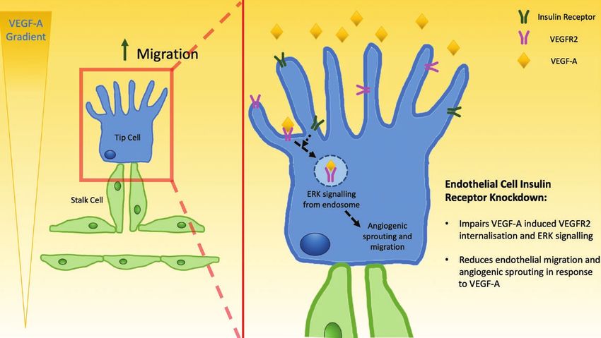
2                                                                                               Endocrinology, 2021, Vol. 162, No. 8

kinase 1/2 (ERK1/2) was impaired, as was VEGF receptor-2 (VEGFR-2) internalization,
which is required specifically for signaling to ERK1/2. Hence, endothelial insulin recep-
tors facilitate the functional response to VEGF-A during angiogenic sprouting and are
required for appropriate signal transduction from VEGFR-2 to ERK1/2.
Key Words: angiogenesis, endothelial, ERK, insulin, vascular, VEGF

Insulin is a primary regulator of systemic carbohydrate              Insulin receptor (Insr) haploinsufficient (Insr+/-) mice
and lipid metabolism (1), but also has an important role             As we have previously described (15), Insr+/- mice (also
in vascular function, for example promoting vasodilation             known as IRKO) were obtained from the Medical Research
and tissue perfusion (2). Indeed, loss of endothelial insulin        Council Mammalian Genetics Unit (Harwell, Oxfordshire,

                                                                                                                                           Downloaded from https://academic.oup.com/endo/article/162/8/bqab104/6284300 by guest on 12 October 2021
receptors, or perturbation of their signaling function, in-          UK), and were maintained as heterozygotes on a C57BL/6J
duces endothelial dysfunction, hypertension, and athero-             background. Insr+/- were compared with age-matched wild-
sclerosis (3-5). Sprouting angiogenesis, the phenomenon              type (WT) littermates.
of new capillary formation, is another fundamental ele-
ment of vascular biology that is intrinsically linked to             Endothelial cell-specific Insr haploinsufficient (ECInsr+/-)
metabolism (6). In this highly orchestrated and conserved            mice
process, endothelial “tip cells” emerge from existing ves-           ECInsr+/- mice were generated by crossing mice that have
sels, followed by proliferating stalk cells that extend the          loxP sites flanking exon 4 of the insulin receptor (Line
sprout and form a lumenized vessel; these neo-vessels then           006955, The Jackson Laboratory, Bar Harbor, ME) (16)
anastomose into an immature network that remodels to                 with mice possessing a Cre transgene driven by the Tie2
meet local demands for oxygen and metabolite transport               promoter/enhancer (Line 004128, The Jackson Laboratory)
(7). Insulin has been reported to promote angiogenesis               (17) and were maintained on a C57BL/6J background.
in vitro and in vivo; (8-12) these studies found pro-                ECInsr+/- were compared with age-matched Cre-negative
angiogenic effects in nanomolar concentrations in vitro,             Insrlox/+ littermates, which are referred to as WT.
but did not explore more physiological picomolar concen-
trations. In vivo, insulin receptor expression is known to
                                                                     Assessment of retinal developmental
be enriched in human tumor endothelial tip cells (13), and
                                                                     angiogenesis
loss of endothelial insulin receptors has been shown to
impair angiogenesis in murine retinopathy (14). However,             Tissue collection and processing
it remains unclear how endothelial insulin receptors influ-          Retinal angiogenesis was assessed in postnatal day 5 pups by
ence the cellular and molecular processes of angiogenesis            precisely following the protocol of Pitulescu et al (18). In brief,
and so we set out to define this.                                    all pups from at least 3 litters were included in each experi-
                                                                     ment, with analysis blinded to the results of genotyping data.
                                                                     Both eyes were processed identically with a mean value from
Materials and Methods                                                these to represent that pup. Vascular endothelium was stained
Animal models                                                        with Isolectin B4 conjugated with Alexa Fluor 488 (I21411;
                                                                     Thermo Fisher Scientific, Warrington, UK). Costaining with
All experimental mice were kept in a conventional
                                                                     a rabbit anti-mouse anti-Collagen IV antibody (19) followed
animal facility with a 12-hour light/dark cycle and
                                                                     by an Alexa Fluor-647 conjugated goat anti-rabbit antibody
received a standard chow diet. Genotyping was per-
                                                                     (20) was used to visualize the vascular basement membrane.
formed using PCR of ear notch (or tail-tip for pups)
                                                                     To define cell proliferation, pups were injected with 125 μg
genomic DNA by Transnetyx (Cordova, TN). All pro-
                                                                     5-ethynyl-2′-deoxyuridine (EdU) 2 hours before tissue col-
cedures were approved by the Animal Welfare and
                                                                     lection; this was stained with Alexa Fluor 647 azide using
Ethical Review Committee at the University of Leeds
                                                                     Click-iT technology (C10640; Thermo Fisher Scientific).
and were conducted in accordance with The Animals
(Scientific Procedures) Act of 1986 Amendment
Regulations 2012 (SI 2012/3039) under United                         Confocal microscopy and image analysis
Kingdom Home Office project licenses PL40/3523                       Microscopy was performed using a Zeiss LSM880 up-
and P144DD0D6.                                                       right confocal microscope with 10×/0.3NA, 20×/0.8NA,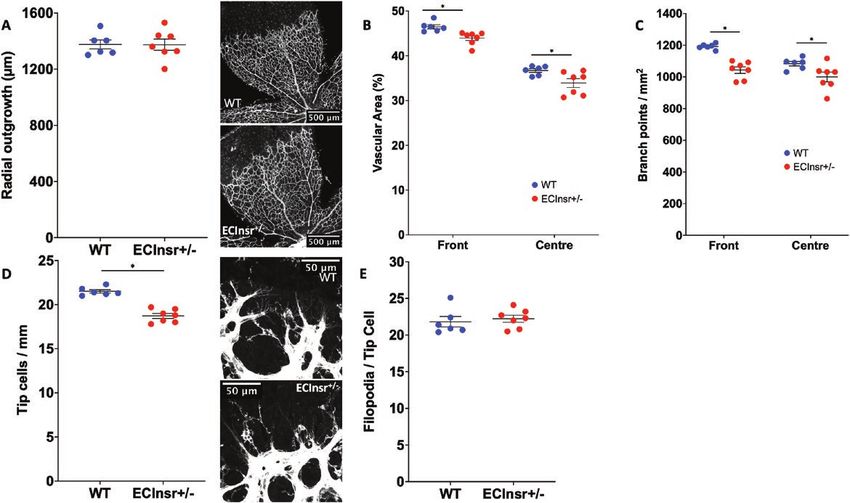
Endocrinology, 2021, Vol. 162, No. 8                                                                                     3

and 40×/1.4NA objectives and Zen software (Carl Zeiss            embedded in optimal cutting temperature media (Tissue-
Microscopy Ltd, Cambridge, UK). Tile scanning was used           Tek OCT compound, Sakura, Netherlands) before snap
to image entire retinal segments with the 20× objective and      freezing in liquid nitrogen and cryosectioning at 10-µm
maximum intensity projection of 5 consecutive 1 Airy unit        thickness. Vascular endothelium was stained with Isolectin
thickness z-slices was used with the 40× to define tip cells     B4 conjugated with Alexa Fluor 488 (I21411; Thermo
and filopodia. Image analysis used ImageJ (NIH, Bethesda,        Fisher Scientific) and slides were mounted with DAPI-
MD). Radial outgrowth was defined as the distance from           Fluoromount-G (Southern Biotech, AL) to define nuclei.
the optic disc periphery to the emerging vascular front
measured at 12 points in each retina. Vascular area was de-      Confocal microscopy and image analysis
fined by binary thresholding of the Isolectin B4 signal and      Microscopy was performed using a Zeiss LSM880 up-
expressed as a percentage of the region of interest, bounded     right confocal microscope with 20×/0.8NA objective and

                                                                                                                               Downloaded from https://academic.oup.com/endo/article/162/8/bqab104/6284300 by guest on 12 October 2021
either by the peripheral or central half of the vascularized     Zen software (Carl Zeiss Microscopy Ltd). Image analysis
area. Vascular branching was quantified in multiple 200 ×        used ImageJ (NIH). Vascular area was defined by binary
200-μm regions of interest placed between arteries and           thresholding of the Isolectin B4 signal and expressed as a
veins, in the peripheral or central vascular plexus. Tip cell    percentage of the image area.
abundance was normalized to the perimeter of the con-
tiguous vascular front in each image and filopodia were
normalized to tip cell number. Capillary regression was de-      RT-PCR
fined as Collagen IV staining without colocalized Isolectin      Snap frozen ischemic adductor muscle was mechanic-
B4 staining, and expressed as total length per mm2 in com-       ally lysed in Trizol reagent (Sigma Aldrich, Gillingham,
plete retinal segments, as per the method of Franco et al        UK) to isolate RNA. After reverse transcription to gen-
(21). Endothelial proliferation, defined by EdU+ nuclei          erate cDNA (kit), quantitative PCR was performed (ABI
costaining with Isolectin B4, was quantified in multiple         Prism 7900HT, Applied Biosystems) using Taqman probes
200 × 200-μm regions of interest placed between arteries         against murine vegfa (Mm01281449-m1), murine insr
and veins, in the peripheral vascular plexus.                    (Mm00439688_m1), murine actb (Mm00607939_s1), 18s
                                                                 (Mm01281449-m1); 18s or actb were used to normalize
                                                                 gene expression using the equation
Assessment of pathological angiogenesis after
hind-limb ischemia                                                                    2−deltaCT × 100.
Surgical procedure
Following the protocol we have published (22), 9- to
13-week-old male Insr+/- mice were anesthetized with             Ex vivo aortic ring angiogenesis
isoflurane before dissecting the left femoral artery, ligating   Aortae were harvested from 8- to 12-week-old Insr+/- mice
it proximally at the inguinal ligament and distally at the bi-   under terminal isoflurane anesthesia and then processed
furcation to saphenous and popliteal vessels, and excising       according to the protocol of Baker et al (23). In brief,
the intervening segment.                                         after dissection of perivascular fat and overnight storage
                                                                 in serum free OptiMEM media (Thermo Fisher Scientific),
Laser Doppler perfusion imaging                                  aortae were cut in to 1-mm-thick rings that were then em-
Laser Doppler analysis (Moor LDI2-HR, Moor Systems,              bedded in rat type I collagen. Rings were incubated for
UK) of ischemic and nonischemic limbs was performed              5 days at 37°C in 5% CO2 in Opti-MEM media containing
postoperatively in a temperature-controlled environment,         2.5% fetal calf serum (FCS), 50 ng/mL VEGF-A165 (R&D
to confirm induction of ischemia, and repeated weekly            Systems, Abingdon, UK) and penicillin-streptomycin, with
until day 21. Images were analyzed (MoorLDI software,            a media change on day 3. Rings were then fixed with 4%
Version 5.3, Moor Systems, UK) to derive an ischemic to          paraformaldehyde, stained with BS-1 lectin-fluorescein
nonischemic limb perfusion ratio, based upon flux below          (Sigma Aldrich) to define endothelium, and then imaged
the level of the inguinal ligament.                              with an inverted confocal microscope (LSM700, Carl
                                                                 Zeiss Microscopy Ltd.); tiled images were collected using
Tissue collection and processing                                 a 10×/0.2NA objective and stitched using Zen software.
Ischemic and contralateral gastrocnemius muscle was har-         Image analysis was performed with Image J (NIH), defining
vested and fixed in 4% paraformaldehyde for 48 hours,            the number of fluorescein staining sprouts per ring and the
whereas adductor muscles were snap frozen with liquid            mean length of these sprouts; mean data were then pro-
nitrogen for RNA isolation. Fixed muscle specimens were          duced for each experimental animal from at least 4 rings.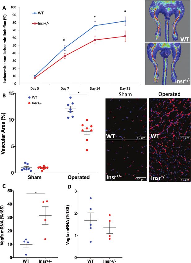
4                                                                                    Endocrinology, 2021, Vol. 162, No. 8

Mouse pulmonary endothelial cell studies                     Human umbilical vein endothelial cell studies
Isolation and functional analysis                            Cell culture and lentiviral manipulation
Pulmonary endothelial cells (PECs) were isolated from        Human umbilical vein endothelial cells (HUVECs;
both lungs of 8- to 12-week-old Insr+/- mice, precisely      PromoCell, Heidelberg, Germany) were cultured at 37°C in
following the protocol of Sobczak et al (24). This uses      5% CO2 in EGM2 media on 1% gelatin-coated plasticware
immuno-magnetic selection of CD31+ cells, which are          and used between passages 3 and 6. Silencing of the in-
then cultured in EGM2 media (Lonza, Slough, UK)              sulin receptor was induced using insulin receptor shRNA
for 10 to 14 days before a second round of immuno-           introduced by lentiviral particles (SHCLNV-NM_00208,
magnetic selection from ICAM2+ endothelial cells that        TRCN0000196786; MISSION, Sigma Aldrich), with GFP-
were cultured for a further 5 to 7 days in EGM2 before       targeting shRNA lentivirus particles (SHC002H; MISSION,
functional assays.                                           Sigma Aldrich) serving as control. Both lentiviruses were

                                                                                                                             Downloaded from https://academic.oup.com/endo/article/162/8/bqab104/6284300 by guest on 12 October 2021
                                                             applied at 15 multiplicity of infection and HUVECs were
Matrigel sprouting assay                                     used in downstream experiments 4 days after transduction.
Twenty-four well plates were coated with growth factor
reduced Matrigel (BD Biosciences, Wokingham, UK) be-         Bead sprouting assay
fore seeding each well with 2 × 10⁵ PEC suspended in         Following the protocol of Nakatsu et al (26), HUVECs
EBM2 media (Lonza) containing 1% FCS and 50 ng/mL of         were coated on to the surface of Cytodex-3 microcarrier
VEGF-A165. After 24 hours, phase contrast microscopy was     beads (Sigma Aldrich) and then embedded in a fibrin matrix
used to image each well and count tubule-like structures     that was overlaid with EGM2 media (Lonza) without the
according to our published protocol (22). Each sample was    supplemental bullet kit, but containing 50 ng/mL human
run in triplicate, with a single mean datapoint calculated   VEGF-A165 (PeproTech, NJ) and 5ng/ml human basic FGF
for each experimental animal.                                (PeproTech). After 48 hours of incubation at 37°C in 5%
                                                             CO2, 25 beads per condition were imaged with phase con-
Scratch wound assay                                          trast microscopy (Olympus CX41, Olympus Life Sciences,
PECs were grown to confluence in EGM2 media on 1%            Southend-On-Sea, UK) and analyzed with Image J (NIH),
gelatin coated 96-well plates before forming a scratch       defining sprouts per bead and the mean length of these
wound using the WoundMaker tool (Essen Bioscience,           sprouts; mean data were then produced for each experi-
Royston, UK) and imaging wound closure hourly in a live      mental condition.
cell imaging system (Incucyte, Essen Bioscience) to define
residual wound area.                                         Scratch wound assay
                                                             HUVECs were grown to confluence in EGM2 media on
Boyden chamber                                               1% gelatin-coated 96-well plates before forming a scratch
Following our published protocol (25), 5 × 10⁴ PECs          wound using the WoundMaker tool (Essen Bioscience) and
were seeded in 1% gelatin coated Boyden chamber ap-          imaging wound closure 8 hours later to define percentage
paratus to define migration toward 50 ng/mL VEGF-A165.       wound closure from baseline.
The number of migrating cells per microscopic field
was counted using standard light microscopy and pre-         Adhesion assay
sented as net migration by subtracting the number of         HUVECs were seeded on to 1% gelatin-coated 24-well
cells migrating in paired control experiments without        plates in EBM2 media with 1% FCS, with or without 50 ng/
VEGF-A165 gradient.                                          mL VEGA-A165, at a density of 4 × 10⁴ cells per well and left
                                                             for 1 hour before washing 3 times with PBS and fixing with
Cell proliferation                                           4% paraformaldehyde. Cells were counterstained with
Sparsely seeded PECs on 1% gelatin coated plastic, cul-      Hoechst and Phalloidin Alexa Fluor 488 conjugate and
tured in EGM2 media, were exposed to 10 µM EdU 2             imaged with confocal microscopy (LSM700, Carl Zeiss
hours before fixation with 4% paraformaldehyde and           Microscopy Ltd.) to count adherent cells per mm2.
processing with the Click-iT EdU cell proliferation assay
(Thermo Fisher Scientific) to label nuclei containing ac-
tively forming DNA with Alexa Fluor 488 and a Hoechst        Assessment of VEGF signaling
nuclear counterstain. Confocal microscopy (LSM700, Carl      Western blotting
Zeiss Microscopy Ltd.) was used to define the proportion     HUVECs were lysed in cell extraction buffer (FNN0011;
of EdU+ nuclei.                                              Thermo Fisher Scientific; containing, in mmol/L, 10 mM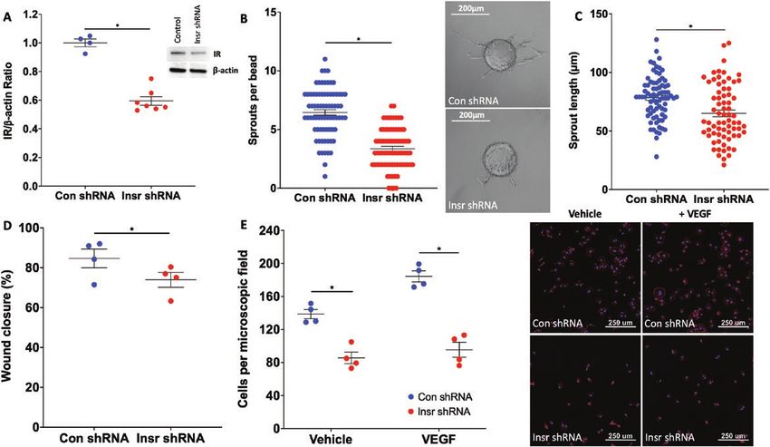
Endocrinology, 2021, Vol. 162, No. 8                                                                                   5

Tris, pH 7.4, 100 mM NaCl, 1 mM EDTA, 1 mM EGTA,                were washed with radio-immunoprecipitation assay buffer
1 mM NaF, 20 mM Na4P2O7, 2 mM Na3VO4, 1% Triton                 to remove any unbound antibody before incubating with
X-100, 10% glycerol, 0.1% SDS, 0.5% deoxycholate, 2             cell lysate for 1 hour with rotation at room temperature
sodium orthovanadate, 0.5 μg/mL leupeptin, 0.2 PMSF,            to allow the biotinylated antigen-antibody complexes to
0.5 μg/mL aprotinin). Cell extracts were centrifuged for 15     form. At the end of the pull-down period, the beads were
minutes before protein measurement using the biocinochinic      washed 5 times with radio-immunoprecipitation assay
acid assay (Thermo Fisher Scientific). Equal amounts            buffer (50 mmol/L Tris-HCl [pH 8.0], 150 mmol/L NaCl,
of protein were resolved on SDS-polyacrylamide gels             0.5% [w/v] sodium deoxycholate, 0.1% [w/v] SDS, and
(Thermo Fisher Scientific) and transferred to polyvinyldine     1% [v/v] Igepal) to remove any nonspecific binding. The
difluoride membranes. Immunoblotting was carried out            immunoprecipitated, biotinylated VEGFR2 complexes
with primary antibodies against beta-actin (27), insulin        were mixed with dissociation buffer and boiled to release

                                                                                                                             Downloaded from https://academic.oup.com/endo/article/162/8/bqab104/6284300 by guest on 12 October 2021
receptor-beta (28), Akt (29), phospho-S473 Akt (30), endo-      the complexes from the beads. The proteins were resolved
thelial nitric oxide synthase (eNOS) (31), phospho-S1177        by electrophoresis through 4% to 12% polyacrylamide
eNOS (32), extracellular signal-regulated kinase 1/2            gels and then transferred to nitrocellulose membrane. The
(ERK1/2) (33), phospho-T202/Y204 ERK1/2 (34), vas-              membrane was blocked for 1 hour in PBS (1.5 mmol/L
cular endothelial growth factor receptor 2 (VEGFR2) (35),       KH2PO4, 2.7 mmol/L Na2HPO4, 150 mmol/L NaCl [pH
and phospho-Y951 VEGFR2 (36). Blots were incubated              7.4]) containing 5% (w/v) dried milk powder and 0.1%
with appropriate peroxidase-conjugated secondary anti-          (v/v) Tween-20, followed by incubation with peroxidase-
bodies and developed with enhanced chemiluminescence            conjugated streptavidin (1:1000 dilution in PBS containing
(37, 38), and imaged with SynGene chemiluminescence             0.1% [v/v] Tween-20) for 1 hour. Bound peroxidase conju-
imaging system (SynGene, Cambridge, UK). Densitometry           gates were visualized using an enhanced chemiluminescence
of phospho-proteins was normalized to respective total          detection system (Amersham Biosciences). Quantification
proteins from the same sample and then these data were          of immunoblots was performed using ImageJ software.
normalized to the value of unstimulated control shRNA-
transduced cells in each experiment (which included paired
control and insulin receptor shRNA-transduced cells, with       Statistics
samples run on 1 membrane). VEGF-A induced signaling            All data are presented as mean (SEM). Comparison be-
was calculated by subtracting the data from unstimulated        tween groups was performed using Student t tests, or
(control or insulin receptor shRNA transduced) cells from       2-way ANOVA for time series data. All tests were 2-sided
their respective VEGF-A–exposed cells in each experiment.       and statistical significance was defined as P < 0.05.

Surface biotinylation and immunoprecipitation
of VEGFR2
                                                                Results
Surface VEGFR2 biotinylation, immunoprecipitation, and
VEGFR2 Western blotting were performed according to             Pathological angiogenesis is impaired in
our previously published protocol (39). Briefly, HUVECs         Insr+/- mice and is associated with impaired
were incubated for 1 hour at 4°C with 0.5 mg/mL biotin          responsiveness to VEGF
sulfo-NHS (Sigma-Aldrich, Gillingham, UK), before being         To study pathological angiogenesis, we induced hind-
stimulated with 50 ng/mL VEGF at 37°C for either 5 or           limb ischemia in Insr+/- mice and quantified limb perfu-
15 minutes. At the end of the exposure period, the cells        sion recovery every 7 days using laser Doppler imaging.
were washed 3 times with PBS with calcium and magne-            This revealed that, in spite of similar reductions in limb
sium before either: immediate lysis with cell extraction        perfusion immediately postoperatively, Insr+/- exhibited
buffer to enable the measurement of total VEGFR2 in the         lower ischemic limb perfusion at all timepoints there-
sample; or, treated with 0.5 mL of 0.05% trypsin/EDTA to        after (Fig. 1A), being 82% (5.6) in WT and 62% (7.1)
cleave and remove any remaining biotin-labelled, cell sur-      in Insr+/- (P < 0.05) at day 21. Histological analysis of
face VEGFR2, meaning detected biotin-labelled VEFGR2            gastrocnemius muscle also revealed a lower capillary
would define only internalized protein. The trypsinized         density in the ischemic limb of Insr+/- at day 21 (vas-
cell pellet was lysed using cell extraction buffer as before.   cular area 12% [0.5] vs 8% [0.6] in WT and Insr+/-, re-
Immunoprecipitation of VEGFR2 was carried out using             spectively, P < 0.05 Fig. 1B), in spite of greater VEGFA
protein A Dynabeads (Thermo Fisher Scientific) loaded           mRNA in the ischemic limb adductor muscle of Insr+/-
with anti-VEGFR2 antibody (diluted 1:100) (35) for 30           (31.4% [6.7] of 18S mRNA in Insr+/- vs 9.8% [2.5] WT
minutes with rotation at room temperature. The beads            P < 0.05; Fig. 1C). Notably, Vegfa mRNA was similar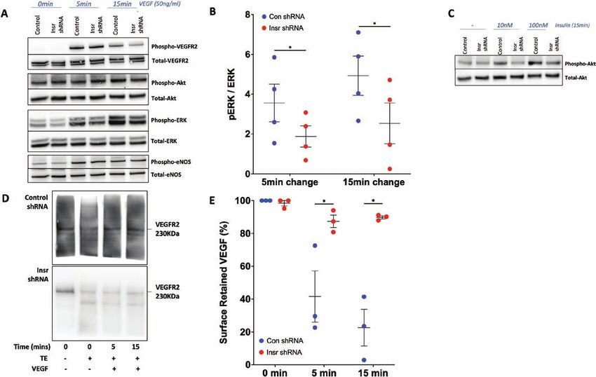
6                                                                                                      Endocrinology, 2021, Vol. 162, No. 8

                                                                                                                                                    Downloaded from https://academic.oup.com/endo/article/162/8/bqab104/6284300 by guest on 12 October 2021

Figure 1. Angiogenesis is impaired in Insr+/- mice with hindlimb ischemia. (A) Ischemic to nonischemic limb perfusion defined by laser Doppler
imaging, with representative day 21 images, showing impaired recovery from hindlimb ischemia in Insr+/- vs WT (n = 14, 13). (B) Confocal immuno-
fluorescence of ischemic and nonischemic gastrocnemius muscle reveals reduced capillary density in ischemic Insr+/- vs WT muscle. Representative
images of ischemic muscle show isolectin B4 stained capillaries in red and nuclei in blue. Scale bars denote 50 μm. (n = 8, 7). (C) Vegfa mRNA
normalized to 18S mRNA is higher in the ischemic limb adductor muscle of Insr+/- vs WT (n = 4, 4). D) Vegfa mRNA normalized to 18S mRNA is higher
in the nonischemic limb adductor muscle of Insr+/- vs WT (n = 4, 4). *P < 0.05. Insr, insulin receptor; WT, wild-type.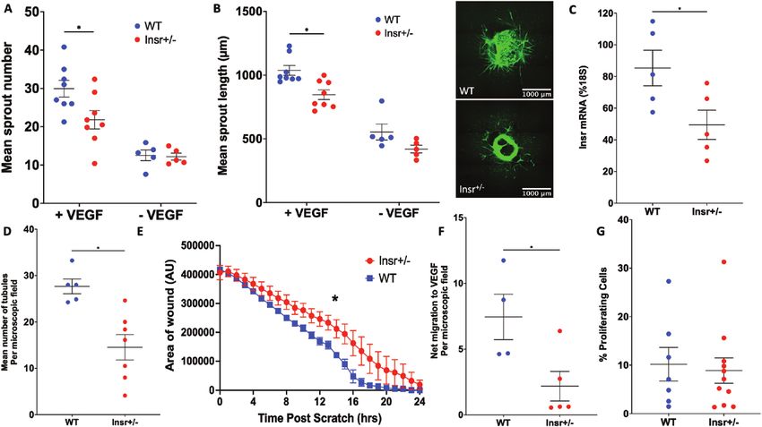
Endocrinology, 2021, Vol. 162, No. 8 7
in the nonischemic limb adductor muscle or WT and P < 0.05; Fig. 3B) and reduced vascular branching com-
Insr+/- (1.7% [0.3] of 18S mRNA in WT vs. 1.4% [0.3] plexity (front 48.0 [0.4] WT vs 41.9 [0.7] Insr+/- branches
in Insr+/-; Fig. 1D). The increased ischemic muscle expres- per microscopic field, P < 0.05; center 52.0 [1.3] WT vs
sion of Vegfa is indicative of significant residual ischemia 46.4 [1.1] Insr+/- branches per microscopic field, P < 0.05;
and could also imply an inadequate functional response Fig. 3C) in both central and peripheral vascular plex-
to this central regulator of angiogenesis. To address this uses of Insr+/-. High-resolution images of the emerging
possibility, we explanted aortae from a separate group vascular plexus revealed a reduction in the number of
of Insr+/- mice to embed rings in a collagen matrix con- sprouting tip cells of Insr+/- (21.2 [0.3] WT vs 18.3 [0.2]
taining VEGF-A165, which induces sprouting angiogen- Insr+/- tip cells per mm, P < 0.05; Fig. 3D), fewer filo-
esis. This revealed fewer capillary sprouts emerging from podia per length of vascular forefront (22.0 [0.5] WT vs
Insr+/- aortae in the presence of VEGF (30 [2.2] in WT vs 18.1 [0.3] Insr+/- filopodia per 100 µm, P < 0.05), and
Downloaded from https://academic.oup.com/endo/article/162/8/bqab104/6284300 by guest on 12 October 2021
22 [2.4] in Insr+/-; P < 0.05; Fig. 2A), and the length of fewer filipodia per tip cell (22.1 [0.3] WT vs 20.6 [0.4]
sprouts was also reduced (1037 µm [39] WT vs 847 µm Insr+/- filopodia per tip cell, P < 0.05; Fig. 3E). Because
[38] Insr+/-; P < 0.05; Fig. 2B). Next, we isolated PECs reduced vascularity could also be explained by increased
for functional studies; these exhibited appropriate reduc- vessel regression, we quantified the number of “empty”
tion in Insr mRNA (85% [5] of 18S in WT vs 49% [4] in collagen IV sleeves (ie, collagen IV basement membrane
Insr+/-; P < 0.05; Fig. 2C). We then conducted a Matrigel without overlying Isolectin B4, linking 2 regions of estab-
in vitro angiogenesis assay, which demonstrated reduced lished vasculature) in the retinal periphery; we observed
tubule formation in Insr+/- (28 [2] tubules per microscopic fewer in Insr+/- vs WT, indicating regression was not ex-
field in WT vs 14.5 [2.4] in Insr+/-; P < 0.05; Fig. 2D). aggerated (0.46 [0.02] WT vs 0.40 [0.02] Insr+/- regressed
Similarly, a scratch wound assay performed on confluent vessels/100 µm2; P = 0.028; Fig. 3F). Next, we asked if
PECs revealed significantly slower closure of the wound endothelial cell (EC) proliferation was reduced in Insr+/-,
formed in Insr+/- PECs (area under curve 4,430,670 arbi- but found similar numbers of EdU+ ECs in the peripheral
trary units WT [154,516] vs 5,085,825 [126,748] arbi- retinal vasculature of both genotypes (781 [44] WT vs
trary units Insr+/-; P < 0.05; Fig. 2E). Because the Matrigel 814 [24] Insr+/- EdU+ EC per mm2; P = 0.53; Fig. 3G).
and scratch wound assays define responses to VEGF-A Overall, these data are compatible with a reduction in
with other stimulatory factors, we then performed as- vascular sprouting in the emerging vasculature of Insr+/-,
says to more specifically define functional responses to resulting in a less branched neovasculature.
VEGF-A. First, we conducted a migration assay using To discern whether loss of insulin receptors expressed
Boyden chamber apparatus and found fewer Insr+/- PECs by ECs contribute to the retinal vascular phenotype of
migrated toward VEGF-A165 (7.5 [1.7] WT vs 2.2 [1.1] Insr+/-, we then studied mice with ECInsr+/-. Again, the ra-
Insr+/- net cell migration to VEGF per microscopic field; dial outgrowth of their retinal vascular plexus was similar
P < 0.05; Fig. 2F). Second, we studied VEGF-A165–in- to controls (1377 µm [32] WT vs 1375 µm [40] ECInsr+/-;
duced PEC proliferation using EdU incorporation and P = 0.96; Fig. 4A), although there was reduced vascular
elicited no difference between Insr+/- and WT (10.2% area (front 46.5% [0.5] WT vs 44.0 [0.5] ECInsr+/-,
[3.5] WT vs 8.9% [2.6] Insr+/- EdU+ cells; P = 0.77; Fig. P < 0.05; center 36.7% [0.4] WT vs 33.9% [1.0] ECInsr+/-,
2G). Collectively, these data imply that Insr+/- endothe- P < 0.05; Fig. 4B) and branching complexity (front
lial cells have selectively impaired migratory responses 1192.5 [7.0] WT vs 1043.3 [20.4] ECInsr+/- branch points
to VEGF-A. per mm2, P < 0.05; center 1083.5 [14.3] WT vs 1000.0
[30.5] ECInsr+/-, P < 0.05; Fig. 4C) in both the peripheral
and central vascular plexuses of ECInsr+/-. Also mirroring
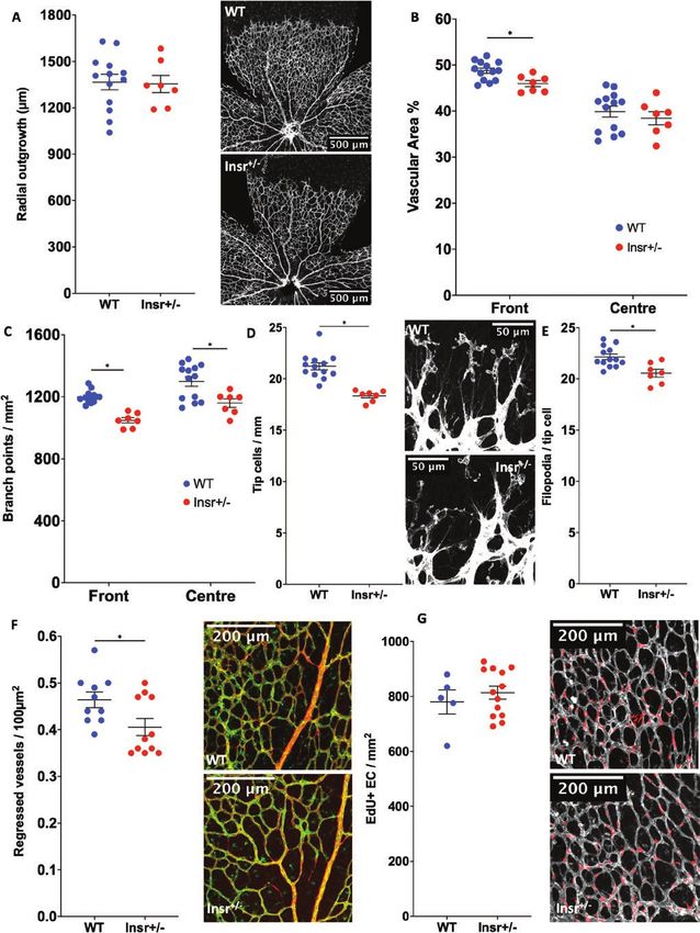
Developmental angiogenesis is impaired in Insr+/- Insr+/- phenotype, there was a reduction in the number of
and ECInsr+/- mice sprouting tip cells of ECInsr+/- (21.5 [0.2] WT vs 18.7 [0.3]
Next, we asked whether the abnormalities of patho- ECInsr+/- tip cells/mm; P < 0.05; Fig. 4D), along with fewer
logical angiogenesis in Insr+/- were recapitulated during filopodia per length of vascular forefront (21.2 [0.4] WT
developmental angiogenesis, which we assessed using vs 18.7 [0.3] ECInsr+/- filopodia per 100 µm; P < 0.05), but
whole-mounted retinas, at P5 when the vasculature is still with no difference in filopodia per tip cell between geno-
developing. The radial outgrowth of the retinal vascular types (21.8 [0.7] WT vs 22.2 [0.5] ECInsr+/- filopodia per tip
plexus was similar in Insr+/- and WT (1367 µm [50] WT cell; P = 0.65; Fig. 4E). Collectively, these data suggest that
vs 1354 µm [56] Insr+/-, P = 0.87; Fig. 3A), although there endothelial cell insulin receptor expression is important in
was reduced vascular area in the peripheral vascular the generation of vascular sprouts, and the branching struc-
plexus of Insr+/- (48.8% [0.6] WT vs 46.0% [0.7] Insr+/-, ture of the nascent vasculature.8 Endocrinology, 2021, Vol. 162, No. 8
Downloaded from https://academic.oup.com/endo/article/162/8/bqab104/6284300 by guest on 12 October 2021
Figure 2. Insr+/- exhibits impaired in vitro functional responses to VEGF. Capillary sprouting from aortic rings embedded in a collagen matrix with
VEGF-A165 is reduced in Insr+/- vs WT (A), as is mean sprout length (B); representative images show isolectin B4 staining of endothelium in green, with
scale bars denoting 1000 μm (n = 5, 5). (C) Insulin receptor (Insr) mRNA normalized to 18S mRNA is reduced in Insr+/- vs WT PEC (n = 5, 5). (D) In vitro
angiogenesis in Matrigel is impaired in Insr+/- vs WT PEC (n = 7, 4). (E) Scratch wound closure is impaired in Insr+/- vs WT PEC (n = 5, 6). (F) Migration
toward VEGF-A165 in Boyden chamber apparatus is impaired in Insr+/- vs WT PEC (n = 5, 4). (G) Proliferation defined by nuclear EdU incorporation is
similar in Insr+/- and WT PEC (n = 11, 7). *P < 0.05. EdU, 5-ethynyl-2′-deoxyuridine; Insr, insulin receptor; PEC, pulmonary endothelial cell; VEGF, vas-
cular endothelial growth factor; WT, wild-type.
Insulin receptor silencing impairs human EC shRNA vs 95.4 [9.1] Insr shRNA cells per microscopic
functional responsiveness to VEGF field; P < 0.05; Fig. 5E). Overall, these data suggest that
To explore the relevance of these data in human ECs, the insulin receptor is also important for VEGF-A–induced
we transduced HUVECs with lentivirus particles to de- angiogenic sprouting and cell motility in human EC.
liver insulin receptor targeting shRNA (referred to as Insr
shRNA), or control GFP-targeting shRNA (referred to
as control [Con] shRNA), reducing insulin receptor pro- Endothelial insulin receptors are required for
tein by 40% (Fig. 5A). Transduced HUVECs were then VEGFR2 internalization and subsequent ERK
coated onto Cytodex-3 carrier beads and embedded in signaling
a fibrin matrix to study VEGF-A165–induced sprouting Because we had demonstrated that insulin receptor expres-
angiogenesis in vitro. Mirroring data from Insr+/- aortic sion influenced functional responses to VEGF-A in human
ring sprouting experiments (Fig. 2A-C), we observed that and murine ECs, we then asked if this was associated with
Insr shRNA HUVECs produced fewer sprouts (6.5 [1.2] altered VEGF-A signaling. VEGF-A promotes angiogen-
Con shRNA vs 3.4 [1.0] Insr shRNA sprouts per bead; esis by binding to VEGFR2, a cell membrane-bound re-
P < 0.05; Fig. 5B), although the mean length of sprouts was ceptor tyrosine kinase that initiates a complex intracellular
unaffected (78.3 µm [10.7] Con shRNA vs 65.0 µm [14.0] signaling cascade. We therefore stimulated Insr shRNA and
Insr shRNA, P > 0.05; Fig. 5C). Insr shRNA HUVECs ex- control shRNA HUVECs with 50 ng/mL VEGF-A165 and
hibited impaired scratch wound closure (84.7% [4.7] Con studied major VEGF-A signaling nodes 5 and 15 minutes
shRNA vs 74.0% [3.7] Insr shRNA; P < 0.05; Fig. 5D) later, along with unstimulated cells (Fig. 6A). Insr shRNA
and impaired adhesion to gelatin coated plates, which HUVECs exhibited unaffected activation of VEGFR2
was more marked in the presence of VEGF-A165 (vehicle (measured by phosphorylation at Y951), or the down-
138.7 [5.5] Con shRNA vs 85.6 [6.9] Insr shRNA cells per stream nodes Akt (measured by phosphorylation at S473)
microscopic field; P < 0.05; VEGF-A165 184.3 [6.6] Con and eNOS (measured by phosphorylation at S1177) (dataEndocrinology, 2021, Vol. 162, No. 8 9
Downloaded from https://academic.oup.com/endo/article/162/8/bqab104/6284300 by guest on 12 October 2021
Figure 3. Developmental angiogenesis is impaired in the neonatal P5 retina of Insr+/- mice. (A) Radial outgrowth of the developing retinal vascula-
ture is comparable in Insr+/- and WT, with representative images showing white isolectin B4 staining of endothelium and scale bars denoting 500 μm
(n = 7, 13). (B) Vascular endothelial area is reduced in the peripheral half of the retinal vasculature in Insr+/- vs WT (n = 7, 13). (C) Vascular branching
is reduced in the peripheral and central zones of the retinal vasculature in Insr+/- vs WT (n = 7, 13). (D) Emerging tip cells per millimeter of vascular
front perimeter are reduced in Insr+/- vs WT, with representative images showing white isolectin B4 staining of endothelium and scale bars denoting
50 μm (n = 7, 13). (E) The number of filopodia per tip cell is similar in Insr+/- vs WT (n = 7, 13). (F) The number of regressed vessels, defined as Collagen
IV sleeves (red) without overlying isolectin B4 (green) in representative images, is lower in Insr+/- than WT (n = 11, 10). (G) The number of proliferating
endothelial cells, defined as EdU+ nuclei (red) overlying isolectin B4 (white) in representative images, is similar in Insr+/- and WT (n = 13, 5). *P < 0.05.
EdU, 5-ethynyl-2′-deoxyuridine; Insr, insulin receptor; WT, wild-type.10 Endocrinology, 2021, Vol. 162, No. 8
Downloaded from https://academic.oup.com/endo/article/162/8/bqab104/6284300 by guest on 12 October 2021
Figure 4. Developmental angiogenesis is impaired in the neonatal P5 retina of ECInsr+/- mice. (A) Radial outgrowth of the developing retinal vascu-
lature is comparable in ECInsr+/- and WT, with representative images showing white isolectin B4 staining of endothelium and scale bars denoting
500 μm (n = 7, 6). (B) Vascular endothelial area is reduced in the peripheral and central zones of the retinal vasculature in ECInsr+/- vs WT (n = 7, 6).
(C) Vascular branching is reduced in the peripheral and central zones of the retinal vasculature in ECInsr+/- vs WT (n = 7, 6). (D) Emerging tip cells per
millimeter of vascular front perimeter are reduced in ECInsr+/- vs WT, with representative images showing white isolectin B4 staining of endothelium
and scale bars denoting 50 μm (n = 7, 6). (E) The number of filopodia per tip cell is similar in ECInsr+/- vs WT (n = 7, 6). ECInsr, endothelial cell insulin
receptor; WT, wild-type.
not shown). However, downstream activation of ERK1/2 [15.6] Con shRNA vs 87.4% [3.8] Insr shRNA surface-
(measured by phosphorylation at T202/Y204) was im- retained VEFGR2; P < 0.05; 15 minutes: 20.6% [12.9]
paired in Insr shRNA HUVECs at 5 and 15 minutes after Con shRNA vs 89.8% [0.9] Insr shRNA surface-retained
stimulation (5-minute change: 3.6 [0.95] Con shRNA vs 1.9 VEFGR2; P < 0.05; Fig. 6D,E), which is known to select-
[0.53] Insr shRNA pERK/ERK ratio; P < 0.05; 15-minute ively impede ERK1/2 activation. Overall, these data reveal
change: 4.9 [0.98] Con shRNA vs 2.5 [1.02] Insr shRNA a selective deficit in VEGF-A signal transduction in Insr
pERK/ERK ratio; P < 0.05; Fig. 6B). Control experiments shRNA HUVECs, which is likely to result from impaired
defining the signaling response to insulin revealed clear re- internalization of activated VEGFR2.
duction in Akt activation after 15 minutes, confirming that
Insr shRNA induced the expected changes in activation of
Discussion
this major insulin signaling node (Fig. 6C). Importantly,
the activation of Akt and ERK1/2 downstream of VEGFR2 Major findings and implications
follows highly distinct pathways, with internalization of Our data reveal for the first time that endothelial insulin
VEGFR2 being essential for only the latter (40); more- receptors are required for appropriate migration and
over, integrins are known to influence this process (41). angiogenic sprouting in response to VEGF-A, both in vitro
Hence, we then asked whether VEGF-A induced internal- and in vivo. At a molecular level, we found that insulin re-
ization of VEGFR2 was impaired in Insr shRNA HUVEC ceptor expression promotes the internalization of VEGF-A–
by performing a surface biotinylation assay to quantify activated VEGFR2, allowing signaling to ERK1/2. Our data
surface-resident and internalized VEGFR2 protein. This suggest that the proangiogenic effects of insulin receptors
revealed that, although baseline surface located VEGFR2 relate to crosstalk with VEGF-A signaling, although the na-
was similar in both groups, there was reduced VEGFR2 ture of this interaction, and whether insulin participates in
internalization in Insr shRNA HUVECs (5 minutes: 41.6% the process, requires further study (Fig. 7). This previouslyEndocrinology, 2021, Vol. 162, No. 8 11
Downloaded from https://academic.oup.com/endo/article/162/8/bqab104/6284300 by guest on 12 October 2021
Figure 5. Insr knockdown in HUVECs impairs functional responses to VEGF. (A) Insulin receptor protein knockdown of 40% was achieved in Insr
shRNA HUVECs vs control shRNA HUVECs, with representative gel (n = 7, 7). (B) Angiogenic sprout numbers were reduced from Cytodex beads
coated with Insr shRNA HUVECs vs control shRNA HUVECs, with representative microscopy images (n = 3, 3). (C) Angiogenic sprout length was
similar from Cytodex beads coated with Insr shRNA HUVECs vs control shRNA HUVECs (n = 3, 3). (D) Scratch wound closure was impaired in Insr
shRNA HUVECs vs control shRNA HUVECs (n = 4, 4). (E) Adhesion to gelatin was impaired in Insr shRNA HUVECs vs control shRNA HUVECs, espe-
cially in context of media supplemented with VEGF-A165; representative microscopy images show DAPI-defined nuclei in blue and phalloidin-defined
filamentous actin in red, with scale bars denoting 250 μm (n = 4, 4). *P < 0.05. HUVEC, human umbilical vein endothelial cell; Insr, insulin receptor;
VEGF, vascular endothelial growth factor.
unappreciated crosstalk establishes a further link between Ronald Kahn’s group indicate a ligand-independent
systems regulating metabolism and angiogenesis. role of insulin receptors in the membrane trafficking of
brown pre-adipocytes (46), potentially aligning with our
findings. However, although the concentrations of in-
Insulin and angiogenesis sulin experienced by sprouting endothelial cells in vivo
A number of studies have shown insulin exerts are unknown, our in vitro data are likely to reflect low
proangiogenic effects, although they did not dissect picomolar concentrations of insulin because of its pres-
the role of endothelial insulin receptors (8-12). These ence in fetal calf serum. This may suggest a role for in-
mainly in vitro studies revealed proangiogenic effects sulin in promoting VEGF-induced ERK signaling, as has
of nanomolar range insulin, but did not explore more been shown for epidermal growth factor signaling (47).
physiological picomolar concentrations. The extent to Another explanation may be that insulin regulates a
which picomolar insulin augments ERK signaling in common endocytic mechanism for its own receptor and
endothelial cells is a source of disagreement in the litera- VEGFR2, as discussed later.
ture (11, 42-44), probably reflecting known heterogen- The only existing data describing the role of endothe-
eity between endothelial populations, including in their lial insulin receptors in angiogenesis were published by
insulin receptor expression. Notably, insulin receptor Kahn’s group in 2003. Using mice with complete deletion
expression is reported to be enriched in endothelial tip of endothelial insulin receptors, they found a 57% reduc-
cells of human tumors (13), yet tip cells generally lack a tion in retinal neovascularization during oxygen-induced
lumen (45), so may be exposed to lower concentrations retinopathy (14). However, they did not study the indi-
of insulin than other endothelial cells. Hence, one pos- vidual cellular processes contributing to angiogenesis, or
sible explanation for our data is a ligand-independent examine VEGF signaling. Our in vivo data suggest that
role of tip cell insulin receptors. Indeed, recent data from insulin receptors regulate endothelial tip cell emergence12 Endocrinology, 2021, Vol. 162, No. 8
Downloaded from https://academic.oup.com/endo/article/162/8/bqab104/6284300 by guest on 12 October 2021
Figure 6. Insr knockdown in HUVECs impairs signaling responses to VEGF. (A) Representative blots illustrating major VEGF signaling nodes in
Insr shRNA HUVECs and control shRNA HUVECs grown on gelatin at baseline, 5 minutes, and 15 minutes after stimulation with VEGF-A165 50 ng/
mL, (B) with quantification of VEGF-induced phosphorylation of ERK1/2 at 5 and 15 minutes (n = 4, 4). (C) Representative blot illustrating impaired
insulin-stimulated Akt phosphorylation in Insr shRNA HUVECs vs control shRNA HUVECs after 15 minutes’ exposure to 10 and 100 nm insulin. (D)
Representative blots and (E) quantification from surface biotinylation experiment illustrating impaired internalization of VEGFR2 in Insr shRNA
HUVECs vs control shRNA HUVECs (n = 4, 4). (D) Detected biotin-labelled VEGFR2 in the presence or absence of TE and so is not directly represented
in €, which presents derived internalization data. HUVEC, human umbilical vein endothelial cell; Insr, insulin receptor; TE, trypsin exposure; VEGF,
vascular endothelial growth factor; VEGFR2, vascular endothelial growth factor receptor 2.
and migration, although with no major impact on endo- studied, ERK1/2 activation has recently emerged as regu-
thelial cell proliferation. Notably, we found no reduction lating endothelial tip cell function and sprouting angiogen-
in nonischemic muscle tissue vascularity of Insr+/- mice, esis; (54, 55) importantly, tip cells are exposed to the highest
implying that impaired vascularization ultimately catches VEGF-A concentrations during angiogenesis, and tip cell
up; this is seen in many published examples of impaired ERK phosphorylation is prevented in vivo by VEGF inhib-
angiogenesis (48-50), presumably reflecting persistent acti- ition (7, 55). There are many known interacting partners
vation of proangiogenic programmes. of VEGFR2, which can modify its signaling and internal-
ization (41, 56). The insulin receptor signaling adaptor, in-
sulin receptor substrate-1, has been implicated in receptor
VEGFR2 signaling endocytosis (57), and is reported to interact with VEGFR2
VEGF-A binding to VEGFR2 induces a complex intracel- (58). Interestingly, recent data show that insulin signaling
lular signaling cascade (51), a crucial element of which is to ERK1/2 (and Src homology phosphatase 2) feeds back
internalization (endocytosis) of ligand-bound VEGFR2. via serine phosphorylation of insulin receptor substrate-1
This moves the phosphorylated receptor to a domain to induce insulin receptor internalization, which aug-
where it is less susceptible to the phosphatase PTP1B, ments ERK1/2 signaling (59). Therefore, it is possible that
hence sustaining signal transmission, which is particularly VEGFR2 internalization is similarly affected, and this pu-
important for ERK1/2 signaling (40). VEGFR2 internaliza- tative insulin-dependent mechanism also warrants further
tion, and subsequent ERK1/2 signaling, is known to be cru- exploration. Integrins are also well established to modulate
cial to vascular biology (eg, during arterial morphogenesis the propagation of ERK1/2 signals downstream of many
and lymphatic specification) (52, 53). Although less well growth factor receptors, including during angiogenesis, soEndocrinology, 2021, Vol. 162, No. 8 13
Downloaded from https://academic.oup.com/endo/article/162/8/bqab104/6284300 by guest on 12 October 2021
Figure 7. Schematic illustration of the proposed role of Insr during sprouting angiogenesis. Insr expression is known to be enriched in tip ECs, which
migrate along VEGF gradients, leading emerging sprouts during angiogenesis. Knockdown of Insr in ECs impairs VEGF signaling to ERK1/2 as a result
of impaired VEGFR2 internalization, which manifests as diminished sprout formation and EC migration. EC, endothelial cell; ERK, extracellular signal-
regulated kinase; Insr, insulin receptor; VEGF, vascular endothelial growth factor; VEGFR2, vascular endothelial growth factor receptor 2.
warrant future assessment (60). Finally, the insulin receptor in this setting. Finally, our ECInsr+/- control data come
can regulate cytoskeletal actin remodeling (61), another from littermates expressing Tie2-Cre, and recent data
process that influences endocytosis (62), warranting further show that Cre is not biologically inert (64); however, the
exploration in future. similar retinal vascular phenotype of Insr+/- and ECInsr+/-
mice provides some reassurance that off-target Cre ef-
fects do not underpin our findings.
Limitations
Although we show that internalization of activated Conclusions
VEGFR2 is impaired, the underlying mechanism of this
We show that endothelial insulin receptors are required
requires further investigation. It will also be interesting
for appropriate migration and angiogenic sprouting in re-
to explore signal transduction downstream of other re-
sponse to VEGF-A, along with internalization of activated
ceptor tyrosine kinases to assess the generalizability of
VEGFR2 and downstream signaling to ERK1/2. This novel
this phenomenon. As alluded to earlier, it is also im-
link between major regulators of systemic metabolism and
portant to acknowledge that impaired VEGFR2 intern-
angiogenesis warrants further mechanistic exploration to
alization may not be the only mechanism by which Insr
understand its wider relevance.
silencing impairs VEGF-A responses; indeed, intracellular
signaling networks are highly complex, as is their per-
turbation. Moreover, our work only sought to describe Acknowledgments
the fundamental links between insulin receptors and Financial Support: This work was funded by a British Heart
VEGF signaling during angiogenesis, and so we cannot Foundation Intermediate Clinical Fellowship awarded to R.M.C.
comment on disease relevance. However, obesity-induced (FS/12/80/29821). A.M.N.W., B.M., and K.J.G. were supported by
British Heart Foundation Clinical Research Training Fellowships.
insulin resistance is associated with reduced vascular in-
N.W. was supported by a University of Leeds Alumni PhD fellow-
sulin receptor expression and impaired angiogenesis ship. M.T..K is a British Heart Foundation professor. The Bioimaging
(63), so it would be interesting to explore endothelial Facility has received equipment grants from the Wellcome Trust to
VEGFR2 internalization and ERK signal transduction purchase confocal microscopes used in this project.14 Endocrinology, 2021, Vol. 162, No. 8
Author Contributions: A.M.N.W. conducted experiments, ana- 10. Wei X, Song H, Semenkovich CF. Insulin-regulated protein
lyzed data, and drafted manuscript. N.W. conducted experiments, palmitoylation impacts endothelial cell function. Arterioscler
analyzed data, and drafted manuscript. B.M. conducted experi- Thromb Vasc Biol. 2014;34(2):346-354.
ments, analyzed data, and drafted manuscript. N.T.W. conducted ex- 11. Lassance L, Miedl H, Absenger M, et al. Hyperinsulinemia
periments, analyzed data, and drafted manuscript. R.M. conducted stimulates angiogenesis of human fetoplacental endothelial
experiments, analyzed data, and drafted manuscript. J.S. conducted cells: a possible role of insulin in placental hypervascularization
experiments, analyzed data, and critically revised the manuscript. in diabetes mellitus. J Clin Endocrinol Metab. 2013;98(9):
S.G. conducted experiments, analyzed data, and critically revised E1438-E1447.
the manuscript. N.J.H. conducted experiments, analyzed data, and 12. Liu Y, Petreaca M, Martins-Green M. Cell and molecular
critically the revised manuscript. K.J.G. conducted experiments, ana-
mechanisms of insulin-induced angiogenesis. J Cell Mol Med.
lyzed data, and critically revised the manuscript. S.B.W. acquired
2009;13(11-12):4492-4504.
ethical approvals and critically revised the manuscript. N.Y.Y. con-
13. Goveia J, Rohlenova K, Taverna F, et al. An integrated gene ex-
ducted experiments, analyzed data, and critically revised the manu-
pression landscape profiling approach to identify lung tumor
Downloaded from https://academic.oup.com/endo/article/162/8/bqab104/6284300 by guest on 12 October 2021
script. D.J.B. critically revised the manuscript. P.C. critically revised
endothelial cell heterogeneity and angiogenic candidates.
the manuscript. M.T.K. critically revised the manuscript. R.M.C. de-
Cancer Cell. 2020;37(3):421.
signed the study, acquired funding, conducted experiments, analyzed
data, and drafted the manuscript. 14. Kondo T, Vicent D, Suzuma K, et al. Knockout of insulin and
IGF-1 receptors on vascular endothelial cells protects against ret-
inal neovascularization. J Clin Invest. 2003;111(12):1835-1842.
15. Kahn MB, Yuldasheva NY, Cubbon RM, et al. Insulin resistance
Additional Information impairs circulating angiogenic progenitor cell function and de-
Correspondence: Richard M. Cubbon, PhD, LIGHT Laborator- lays endothelial regeneration. Diabetes. 2011;60(4):1295-1303.
ies 7.04, The University of Leeds, Clarendon Way, Leeds, LS2 9JT, 16. Brüning JC, Michael MD, Winnay JN, et al. A muscle-specific
United Kingdom. E-mail: r.cubbon@leeds.ac.uk. insulin receptor knockout exhibits features of the metabolic
Disclosures: The authors have nothing to disclose. syndrome of NIDDM without altering glucose tolerance. Mol
Data Availability: All datasets generated during and/or analyzed Cell. 1998;2(5):559-569.
during the current study are not publicly available but are available 17. Koni PA, Joshi SK, Temann UA, Olson D, Burkly L, Flavell RA.
from the corresponding author on reasonable request. Conditional vascular cell adhesion molecule 1 deletion in mice:
impaired lymphocyte migration to bone marrow. J Exp Med.
2001;193(6):741-754.
References 18. Pitulescu ME, Schmidt I, Benedito R, Adams RH. Inducible
1. Belfiore A, Frasca F, Pandini G, Sciacca L, Vigneri R. Insulin gene targeting in the neonatal vasculature and analysis of ret-
receptor isoforms and insulin receptor/insulin-like growth inal angiogenesis in mice. Nat Protoc. 2010;5(9):1518-1534.
factor receptor hybrids in physiology and disease. Endocr Rev. 19. RRID:AB_2082660.
2009;30(6):586-623. 20. RRID:AB_2535814.
2. Manrique C, Lastra G, Sowers JR. New insights into insulin 21. Franco CA, Jones ML, Bernabeu MO, et al. Dynamic endothe-
action and resistance in the vasculature. Ann N Y Acad Sci. lial cell rearrangements drive developmental vessel regression.
2014;1311:138-150. PLoS Biol. 2015;13(4):1–19.
3. Duncan ER, Walker SJ, Ezzat VA, et al. Accelerated endothe- 22. Cubbon RM, Yuldasheva NY, Viswambharan H, et al.
lial dysfunction in mild prediabetic insulin resistance: the early Restoring Akt1 activity in outgrowth endothelial cells from
role of reactive oxygen species. Am J Physiol Endocrinol Metab. South Asian men rescues vascular reparative potential. Stem
2007;293(5):E1311-E1319. Cells. 2014;32(10):2714-2723.
4. Gage MC, Yuldasheva NY, Viswambharan H, et al. Endothelium- 23. Baker M, Robinson SD, Lechertier T, et al. Use of the mouse aortic
specific insulin resistance leads to accelerated atherosclerosis in ring assay to study angiogenesis. Nat Protoc. 2011;7(1):89-104.
areas with disturbed flow patterns: a role for reactive oxygen 24. Sobczak M, Dargatz J, Chrzanowska-Wodnicka M. Isolation
species. Atherosclerosis. 2013;230(1):131-139. and culture of pulmonary endothelial cells from neonatal mice.
5. Rask-Madsen C, Li Q, Freund B, et al. Loss of insulin signaling J Vis Exp. 2010;(46):e2316.
in vascular endothelial cells accelerates atherosclerosis in 25. Sengupta A, Patel PA, Yuldasheva NY, et al. Endothelial in-
apolipoprotein E null mice. Cell Metab. 2010;11(5):379-389. sulin receptor restoration rescues vascular function in male
6. Eelen G, de Zeeuw P, Treps L, Harjes U, Wong BW, Carmeliet P. insulin receptor haploinsufficient mice. Endocrinology.
Endothelial cell metabolism. Physiol Rev. 2018;98(1):3-58. 2018;159(8):2917-2925.
7. Potente M, Gerhardt H, Carmeliet P. Basic and therapeutic 26. Nakatsu MN, Davis J, Hughes CCW. Optimized fibrin gel bead
aspects of angiogenesis. Cell. 2011;146(6):873-887. assay for the study of angiogenesis. J Vis Exp. 2007;(3):e186.
8. Rensing KL, Houttuijn Bloemendaal FM, Weijers EM, et al. 27. RRID:AB_2714189.
Could recombinant insulin compounds contribute to adeno- 28. RRID:AB_2280448.
carcinoma progression by stimulating local angiogenesis? 29. RRID:AB_329827.
Diabetologia. 2010;53(5):966-970. 30. RRID:AB_329825.
9. Pellegatta F, Brambilla C, Reduzzi A, Bragheri M, Zerbini G, 31. RRID:AB_397690.
Catapano AL. Endothelin-1 does not impair insulin-induced 32. RRID:AB_399751.
angiogenesis in vitro. Int J Mol Med. 2011;28(3):443-448. 33. RRID:AB_330744.Endocrinology, 2021, Vol. 162, No. 8 15
34. RRID:AB_331646. 50. Saab S, Buteau B, Leclère L, et al. Involvement of plasmalogens
35. RRID:AB_2212507. in post-natal retinal vascular development. Plos One.
36. RRID:AB_331367. 2014;9(6):e101076.
37. RRID:AB_772210. 51. Koch S, Claesson-Welsh L. Signal transduction by vascular
38. RRID:AB_772206. endothelial growth factor receptors. Cold Spring Harb Perspect
39. McNally BD, Moran A, Watt NT, et al. Inorganic nitrate pro- Med. 2012;2(7):a006502.
motes glucose uptake and oxidative catabolism in white adi- 52. Deng Y, Atri D, Eichmann A, Simons M. Endothelial ERK
pose tissue through the XOR-catalyzed nitric oxide pathway. signaling controls lymphatic fate specification. J Clin Invest.
Diabetes. 2020;69(5):893-901. 2013;123(3):1202-1215.
40. Simons M, Gordon E, Claesson-Welsh L. Mechanisms and regu- 53. Simons M, Eichmann A. Molecular controls of arterial morpho-
lation of endothelial VEGF receptor signalling. Nat Rev Mol genesis. Circ Res. 2015;116(10):1712-1724.
Cell Biol. 2016;17(10):611-625. 54. Mavria G, Vercoulen Y, Yeo M, et al. ERK-MAPK signaling
41. Jones MC, Caswell PT, Norman JC. Endocytic recycling path- opposes Rho-kinase to promote endothelial cell survival and
Downloaded from https://academic.oup.com/endo/article/162/8/bqab104/6284300 by guest on 12 October 2021
ways: emerging regulators of cell migration. Curr Opin Cell sprouting during angiogenesis. Cancer Cell. 2006;9(1):33-44.
Biol. 2006;18(5):549-557. 55. Shin M, Beane TJ, Quillien A, Male I, Zhu LJ, Lawson ND.
42. Johansson GS, Chisalita SI, Arnqvist HJ. Human microvascular Vegfa signals through ERK to promote angiogenesis, but not
endothelial cells are sensitive to IGF-I but resistant to insulin at artery differentiation. Development. 2016;143(20):3796-3805.
the receptor level. Mol Cell Endocrinol. 2008;296(1-2):58-63. 56. Simons M. An inside view: VEGF receptor trafficking and
43. De Nigris V, Pujadas G, La Sala L, Testa R, Genovese S, signaling. Physiology (Bethesda). 2012;27(4):213-222.
Ceriello A. Short-term high glucose exposure impairs in- 57. Yoneyama Y, Lanzerstorfer P, Niwa H, et al. IRS-1 acts as an
sulin signaling in endothelial cells. Cardiovasc Diabetol. endocytic regulator of IGF-I receptor to facilitate sustained IGF
2015;14:114. signaling. Elife. 2018;7:e32893.
44. Li G, Barrett EJ, Wang H, Chai W, Liu Z. Insulin at physiological 58. Senthil D, Ghosh Choudhury G, Bhandari BK, Kasinath BS. The
concentrations selectively activates insulin but not insulin-like type 2 vascular endothelial growth factor receptor recruits in-
growth factor I (IGF-I) or insulin/IGF-I hybrid receptors in sulin receptor substrate-1 in its signalling pathway. Biochem J.
endothelial cells. Endocrinology. 2005;146(11):4690-4696. 2002;368(Pt 1):49-56.
45. Iruela-Arispe ML, Davis GE. Cellular and molecular mechanisms 59. Choi E, Kikuchi S, Gao H, et al. Mitotic regulators and the
of vascular lumen formation. Dev Cell. 2009;16(2):222-231. SHP2-MAPK pathway promote IR endocytosis and feedback
46. Nagao H, Cai W, Wewer Albrechtsen NJ, et al. Distinct regulation of insulin signaling. Nat Commun. 2019;10(1):1473.
signaling by insulin and IGF-1 receptors and their extra- 60. Schwartz MA, Ginsberg MH. Networks and crosstalk: integrin
and intracellular domains. Proc Natl Acad Sci USA. signalling spreads. Nat Cell Biol. 2002;4(4):E65-E68.
2021;118(17):e2019474118. 61. Hu H, Juvekar A, Lyssiotis CA, et al. Phosphoinositide 3-kinase
47. Borisov N, Aksamitiene E, Kiyatkin A, et al. Systems-level regulates glycolysis through mobilization of aldolase from the
interactions between insulin-EGF networks amplify mitogenic actin cytoskeleton. Cell. 2016;164(3):433-446.
signaling. Mol Syst Biol. 2009;5:256. 62. Smythe E, Ayscough KR. Actin regulation in endocytosis. J Cell
48. Ngo MH, Borowska-Fielding J, Heathcote G, et al. Fzd4 Sci. 2006;119(Pt 22):4589-4598.
haploinsufficiency delays retinal revascularization in the 63. Cubbon RM, Ali N, Sengupta A, Kearney MT. Insulin- and
mouse model of oxygen induced retinopathy. Plos One. growth factor-resistance impairs vascular regeneration in dia-
2016;11(8):e0158320. betes mellitus. Curr Vasc Pharmacol. 2012;10(3):271-284.
49. Napp LC, Jabs O, Höckelmann A, et al. Normal endothelial but 64. Brash JT, Bolton RL, Rashbrook VS, Denti L, Kubota Y, Ruhrberg C.
impaired arterial development in MAP-kinase activated protein Tamoxifen-activated CreERT impairs retinal angiogenesis inde-
kinase 2 (MK2) deficient mice. Vasc Cell. 2016;8:4. pendently of gene deletion. Circ Res. 2020;127(6):849-850.You can also read