COVID-19: ORF8 Synthesizes Nitric Oxide to Break the Blood-Brain/Testi Barrier and Damage the Reproductive System
←
→
Page content transcription
If your browser does not render page correctly, please read the page content below
COVID-19: ORF8 Synthesizes Nitric Oxide to Break the

Blood-Brain/Testi Barrier and Damage the Reproductive System
                                  Wenzhong Liu 1,2,*, Hualan Li2

 1
     School of Computer Science and Engineering, Sichuan University of Science & Engineering,
     Zigong, 643002, China;
 2
     School of Life Science and Food Engineering, Yibin University, Yibin, 644000, China;
 *
     Correspondence.Wenzhong Liu, liuwz@suse.edu.cn.

Abstract
     Studies have reveal that the SARS-CoV-2 virus can break through the blood-brain/testi
barrier and invade the human reproductive system, causing infertility or complications in patients.
Excessive nitric oxide (NO) is a fundamental reason for breaking the Blood-Brain/Testi Barrier.
Nitric oxide often relates the spread or replication of many viruses in the body. The nitric oxide
synthase (NOS) that synthesizes NO in the human by binding heme to complete the oxygenase
reaction. This study focused on the relationship between heme, NO, and the novel coronavirus
using bioinformatics methods such as domain search and molecular docking. The results showed
ORF8 had three domains similar to nitric oxide synthase : oxygenase, reductase, and calmodulin
(CaM). ORF6 could bind to these three domains. The dimer of ORF8 was identical to the dimer of
NOS enzyme. The oxygenase domain was in the core, and the reductase domain was on both sides.
ORF8 could capture Heme, H4B, L-arginine, FAD, FMN, and NADPH, and bind with CaM
protein to catalyze NO production. The heme bound by ORF8 mainly came from the attacked
hemoglobin. ORF8 also attached to E protein and synthesized NO through the heme hunted by E
protein. We believed NO synthesized by ORF8 inhibited SARS-CoV-2 from reinfecting infected
cells and controlled the virus replication speed to avoid cell collapse because of exhaustion of
resources. After the SARS-CoV-2 virus combined with an extensive ORF8, the produced NO
stream permeably expanded blood vessels and broke the blood-brain/testi barrier. The
SARS-CoV-2 virus spread to nearby tissues through small blood vessel holes created by NO
stream. It would increase the tendency to bleed and the blood clotting of the tissue: blood clotting
and viral infections severely damaged organs such as the respiratory, heart, nerve, reproductive.
Short NO could not open vascular permeability dilation, causing high viral load in asymptomatic
patients' blood. Excessive NO stimulated the reproductive organs and generated abnormal
functions. It also made abnormal hormone regulation, such as excessive secretion of luteinizing
hormone (LH). Much LH would hurt the reproductive organs, leading to infertility. Extreme NO
also interfered with the human NO signaling pathway and damaged the immune nerve,
metabolism, cardiovascular and other systems. This theory is for academic discussion only. We
hope that this discovery will help block the virus’s transmission through the human circulatory
system and help reproductive health management during the pandemic.
Keywords: Nitric oxide; Nitric Oxide Synthase(NOS); Luteinizing hormone (LH) ; Oxygenase;
Heme; Dimer.

                                                 1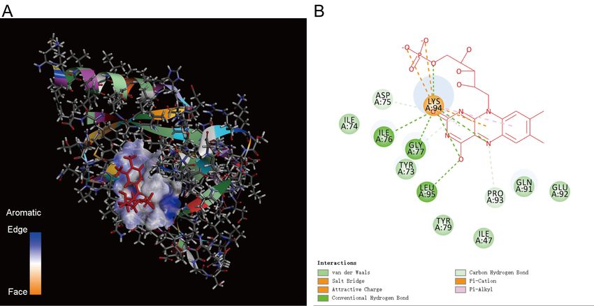
1. Introduction
      The SARS-CoV-2 virus targets cells through the S protein binding to the human ACE2
receptor. The cellular serine protease transmembrane protease serine 2 (TMPRSS2) triggers a
cascade event, leading to a fusion between the cell membrane and the viral membrane to complete
the cell infection process. The testis is one of the body tissues with the highest expression of
ACE2 mRNA and protein, which shows that SARS-CoV-2 infection may threaten male fertility(1).
ACE2 is also widely expressed in the ovaries, uterus, vagina, and placenta, so SARS-CoV-2 may
also interfere with female reproductive functions(2). Statistics indicate that the incidence and
mortality of COVID-19 in men are higher than those in women(3), then the reproductive system
of men is more vulnerable to damage. But there are contradictory reports in the literature about
SARS-CoV-2 infection of the human reproductive system. Therefore, studying the damage
mechanism of the SARS-CoV-2 virus to the reproductive system is of great significance for
reproductive health management during the pandemic.
      There are reports that SARS-CoV-2 is not detected in the vaginal fluid of women with severe
COVID-19 infection, and the results of vaginal swabs are negative to SARS-CoV-2(4). Another
report shows that women’s vaginal secretion test is positive(5). There is no epidemiological
investigation describing evidence that COVID-19 is a venereal disease(6). Because of viremia in
SARS-CoV-2 patients, invasive operations such as vaginal oocyte retrieval increase the chance of
infection(7). When assisted reproductive technology induces pregnancy, the risk of SARS-CoV-2
disease also increases. ACE2 receptors have been detected in the ovaries of childbearing women
and postmenopausal women. ACE2 is abundantly expressed in ovaries and oocytes, and
SARS-CoV-2 may target ovaries and oocytes(2). ACE2 is more expressed in oocytes than
TMPRSS2, which suggests that the latter may be a potential limiting factor for female gamete
infection, and cumulus-encapsulated oocytes are unlikely to be infected by SARS-CoV-2(8). But
In novel coronavirus pneumonia patients not related to pregnancy, pelvic illness, or surgery, right
ovarian vein thrombosis (OVT) also are observed(9). Ovarian vein thrombosis is a rare disease
related to postpartum conditions, pelvic infection, gynecological surgery, and other thrombotic
conditions. This situation highlights that the SARS-CoV-2 virus induces a hypercoagulable state in
female reproductive organs. In OVT, the use of new oral anticoagulants causes premature delivery
in COVID-19-positive women(10). SARS-CoV-2 was also detected in the blood samples of
mothers and their newborns, and the placental SARS-CoV-2 gene was also positive(11). It
represents pregnant women who were severely infected with COVID-19, meets a vertical
transmission of the virus(12), then placental transmission causes neonatal viremia and
inflammation of the placenta. Besides, RT-PCR’s detection of breast milk samples reveals that
virus particles are positive(13), which is not suitable for breastfeeding of COVID-19-positive
women.
      Compared with ovarian cells, testicular ACE2 expression is significantly higher, and male
gonadal functions (including steroid production) are more susceptible to SARS-CoV-2 induced
damage(14). The testis also showed almost the most elevated expression levels of ACE2 mRNA
and protein among different body tissues(15). There are four main cell types at the testicular cells
(1, 16): vas deferens, spermatogonia, stromal cells, and supporting cells. For the evidence of
testicular infection, current literature reports are also inconsistent. It reports that using qRT-PCR,
no SARS-CoV-2 virus is found in semen samples of healthy male COVID-19 patients(17). The

                                                 2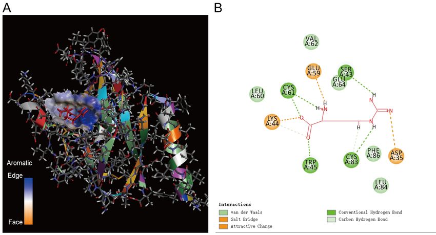
blood-testis barrier protects it, and the incidence of testicular infections in COVID-19 patients is
low. All are plausible reasons(18). It also reports that the SARS-CoV-2 virus is contained in semen
samples of acute and recovered male COVID-19 patients (19). Using RT-qPCR technology, it
finds SARS-CoV-2 nuclei protein in infected men’s testicular tissue, showing that the virus
directly invaded the male reproductive system(20). SARS-CoV-2 may cause male reproductive
diseases by regulating the level of autophagy in male germ cells(21). It finds seminiferous tubule
damage reduces Leydig cell number and then causes lymphocytic inflammation in the testes of
COVID-19 patients(22). It also reveals that COVID-19 positive cases have lymphocyte and
macrophage infiltration, coronavirus-like particles, and abnormal sperm morphology(23). In
SARS coronavirus-infected testes, leukocyte infiltration shows the virus disrupts the blood-testis
barrier, and subsequently, the body loses immune protection(24). Like other SARS viruses,
SARS-COV-2 may damage tissues and blood testicular barriers(25) and hurt high-risk testicular
cells(26, 27). If research confirms viruses in semen, assisted reproductive technology will also be
affected(28).
      Orchitis is a complication of SARS(24). The crucial question is whether COVID-19 causes
testicular damage and infertility in male patients. So far, there is no definite answer(29). In
COVID-19 cases, testicular degeneration is because of the indirect effects of inflammation leading
to an increase in testicular temperature(25), affecting testicular function(30, 31). Oxidative
stress(OS) is widely recognized as an essential cause of male infertility(32). Testicular pathology
under inflammation and oxidative stress may lead to infertility in a large proportion of COVID-19
survivors(33). SARS-CoV-2 can activate pathways sensitive to oxidants through the innate
immune pathway’s inflammatory response, inducing oxidative stress(34, 35). Oxidative stress by
SARS-CoV-2 has long-term harmful effects on male fertility, especially on developing sperm(36).
In the epididymis, sperm are more susceptible to oxidative damage(37). SARS-CoV-2 induces
impaired spermatogenesis and delayed sperm maturation, which may be the PT-mediated
depletion of testosterone levels(38). A recent study shows that because Leydig cells express ACE2,
there may be a direct correlation between lower serum testosterone and inflammatory cytokines
and poor clinical outcomes in male patients infected with SARS-CoV-2 (39).
      Most viruses enter the human body through the nasal cavity and oral cavity, and virus
particles may destroy the blood-brain barrier. According to reports, brain cells (glial cells and
neurons) also express ACE 2 receptors, making them a target for inducing neurons’ death. Notably,
the central nervous system plays a crucial role in endocrine control and sperm formation (40). The
central nervous system’s disease in patients with COVID-19, includes increased antidiuretic
hormone secretion, hypothalamic-pituitary disorders, and abnormal LH secretion rhythm(41).
Although the impact of COVID-19 on the hypothalamic-pituitary-adrenal (HPA) axis abnormality
has been speculated, the mechanism of SARS-CoV-2 mediated hypothalamic-pituitary-gonad
(HPG) axis dysregulation remains unclear(42). SARS-CoV-2 infection may produce an active
inflammatory response in the hypothalamus-pituitary-testis (HPT) axis, leading to reduced LH,
stimulating follicle hormone (FSH), and testosterone levels (30, 34). However, there is a
contradiction with this theory. Because it has recently been reported that compared with healthy
men, patients with COVID-19 have lower serum testosterone levels, higher LH levels, and a lower
ratio of testosterone to LH(43). It shows that SARS-CoV-2 has subclinical damage to male
gonadal function. Another report indicates that the serum LH level of COVID-19 patients is
significantly higher than that of healthy men, but testosterone/LH and FSH levels are lower,

                                                3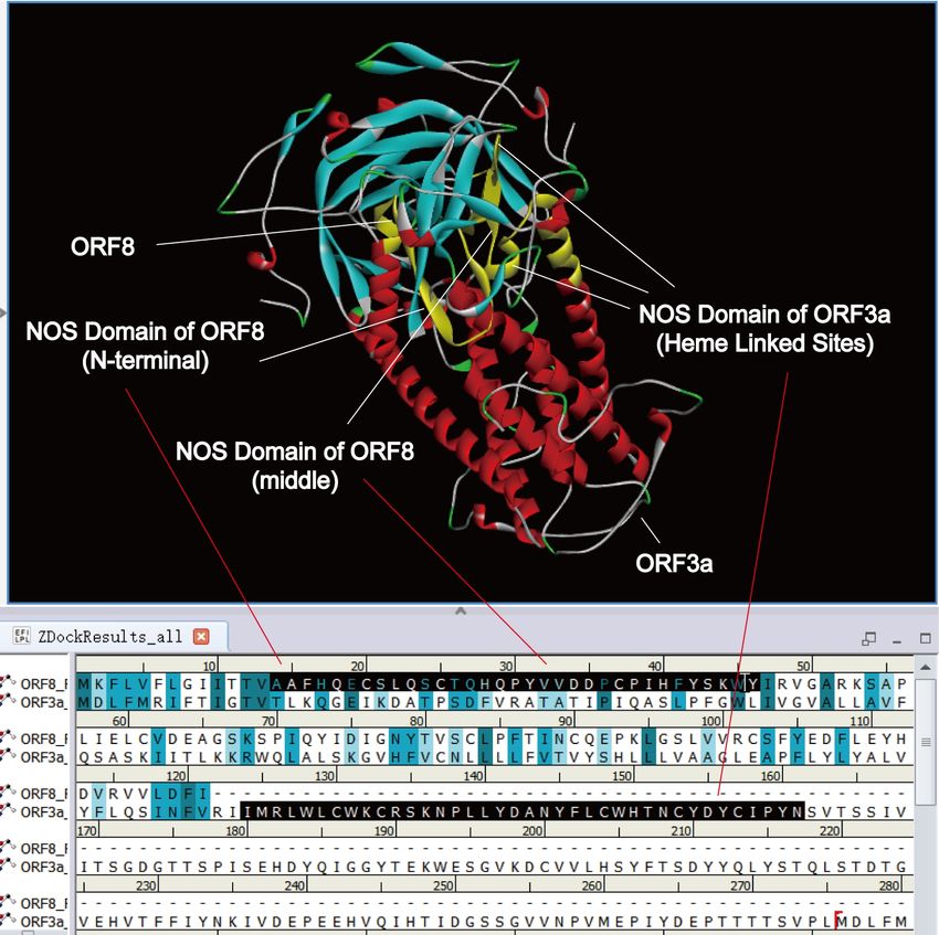
showing potential hypogonadism(44). In the COVID-19 group, more elevated serum luteinizing
hormone (LH) and lower testosterone(T) are observed, and there is no significant difference in the
ratio of serum FSH, serum E2 and T:E2(45). It negatively correlated the LH ratio with the WBC
count and CPR level of COVID-19 patients. The evidence reveals the complex relationship
between SARS-CoV-2 infection and dysregulation of sex hormones.
      The hypothalamic-pituitary-gonadal axis (HPGa) plays a vital role in reproduction. HPGa can
inhibit the body’s reproductive function through hormones. The activation of HPGa and
subsequent changes in hormone concentration holds a key role in sperm quality. Sperm
count/sperm volume/semen volume, and a higher sperm DNA fragmentation, also induce sexual
dysfunction(46). GnRH neurons in the hypothalamus secretes Gonadotropin-releasing
hormone(GnRH). Then GnRH activates follicle-stimulating hormone (FSH) and luteinizing
hormone (LH) secreted by the pituitary gland. Low GnRH levels reduce FSH and LH, leading to
impaired Sertoli and Leydig cell function(43). GnRH neurons' cell bodies are in the endplate
microvascular apparatus (OVLT) area, and the cell bodies of NOS neurons overlap with GnRH
neuron fibers (47). The NOS enzyme of NOS neurons synthesize NO, and the released NO
stimulates GnRH neurons to secrete GnRH. Both endogenous and exogenous NO can stimulate
the secretion and release of GnRH (48). High and enough NO stimulates the high secretion of
GnRH(49). The high secretion of GnRH causes the level of LH to increase (50).
      NO is also an effective vasodilator that promotes vasodilation. NO mainly acts on sGC to
regulate cerebral vascular endothelial cells’ permeability under physiological conditions (51).
Then control the transport of ions, nutrients, and other molecules across the blood-brain barrier
through cGMP. Glutamate closely related the synthesis/release of NO to the permeability changes
in the blood-brain barrier(52). Angiotensin-converting enzyme 2(ACE2) is the receptor of
SARS-CoV-2, which regulates vascular function by controlling the release of nitric oxide (NO)
and oxidative stress. In severe SARS patients, iNO can significantly improve ARDS patients’
oxygenation and reduce pulmonary vascular resistance, reducing the need for invasive mechanical
ventilation(53). But Excessive NO may damage tissue and cell. NO rapidly reacts with Hb in red
blood cells, inactivating NO(53). The free heme in the blood binds to nitric oxide (NO), further
aggravating the organ’s ischemia. Also, because nitric oxide induces blood vessel relaxation and
dilation, reducing blood pressure, the increase in nitric oxide content may develop the disease into
a severe bleeding. During focal cerebral ischemia, much NO is neurotoxic to hurt nerve cells(54).
NO directly increases the blood’s permeability and breaks the blood-brain barrier(55). The
increased synthesis and release of NO during acute hypertension is essential for damaging the
blood-brain barrier(56). Therefore, for COVID19 patients, excessive NO may osmotically expand
blood vessels and break the blood-brain barrier.
      It relates nitric oxide to many viruses’ pathogenic mechanism (such as human rhinovirus,
hepatitis C, dengue fever virus, herpes simplex virus 1))(57). Nitric oxide synthase in the body
needs to bind heme when synthesizing NO. So heme is essential for NO synthesis. In the theory of
heme (or hemoglobin), we found that ORF3a could attack hemoglobin and dissociate heme into
porphyrin and iron(58). At the same time, viral proteins could bind porphyrin to inhibit heme
metabolism. Why was doxyhemoglobin more vulnerable to attack? After ORF3a attacked
hemoglobin, much heme was fallen off. What was the significance of this dissociated heme to the
virus? The biological mechanism of which was still unknown. In the ROS damage theory, we
found that E protein has the functions of cytochrome C oxidase, catalase, peroxidase, and

                                                4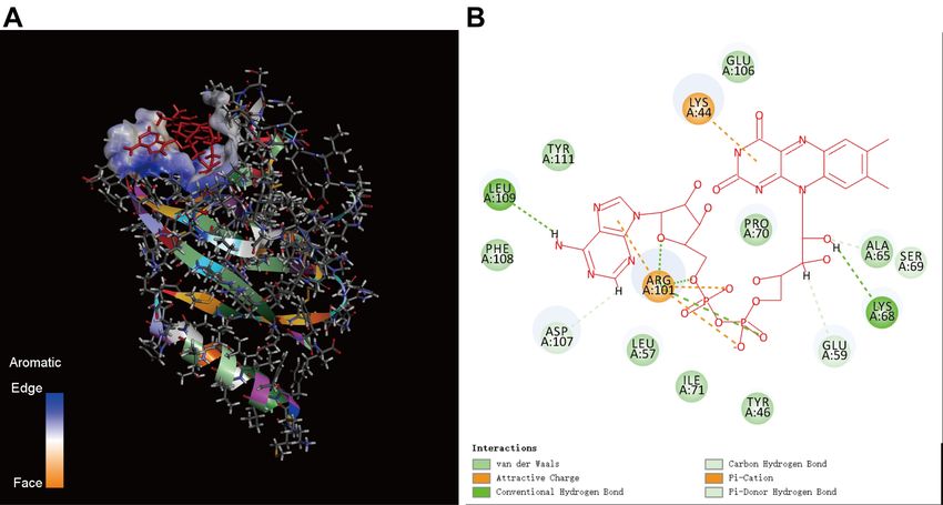
superoxide dismutase by binding heme or iron(59). E protein catalyzed the production of ROS
free radicals to damage immune cells. The virus escaped the attack of immune cells by breaking
down ROS free radicals through E protein. Is there another viral protein that bound heme to
produce NO? This research tried to discover the relationship between heme, nitric oxide, and viral
proteins.
     This study adopted bioinformatics methods such as domain search and molecular docking to
study the relationship between the novel coronavirus proteins and nitric oxide. The results showed
ORF8 had three functional domains of NOS synthase: oxygenase domain, reductase domain and
calmodulin (CaM) binding domain. It must combine NOS synthase into a dimer to be catalytically
active. Interestingly, ORF8 also has into a dimer structure(60), a rapidly evolving protein structure
may help viruses achieve immune escape(61) and interfere with signal transduction pathways(62).
Therefore, the ORF8 antibody has also become the primary clinical marker of SARS-CoV-2
infection(63). We found ORF8 could capture heme, H4B, L-arginine, FAD, FMN, NADPH,
combine with CaM to form a NO generator, and synthesize nitric oxide. The heme bound by
ORF8 mainly came from the attacked hemoglobin. SARs-COV-2 (E protein) also could combined
with ORF8 to generate NO stream to break the blood-brain (testi) barrier and damage reproductive
system and immune cells.

2. Data sets and methods

2.1. Analysis flow
     It shows the data analysis process of this research in Figure 1. It divides the process into
seven steps:The first step is to download the protein sequence data set; The second step is
homology modeling of viral proteins; The third step is to search for the conserved domains of viral
proteins; The fourth step is the protein docking of ORF8 and CaM binding protein; In the fifth
step, it is docked ORF8 with heme, H4B, L-arginine, FAD, FMN, NADPH molecules; The sixth
step is protein docking between viral proteins; The seventh step is the analysis of dimer structure
of ORF8.

                                     Figure 1. Analysis flow chart

                                                 5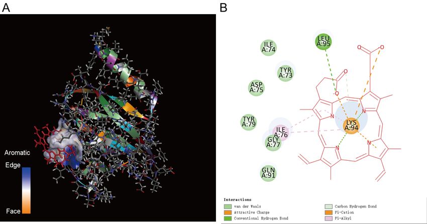
2.2. Data set
      1. The sequences of SARS-COV-2 proteins. The SARS-COV-2 protein sequences came
from the NCBI database. Including: S, E, N, M, ORF3a, ORF8, ORF7a, ORF7b, ORF6, ORF10,
orf1ab, orf1a. Among them, the orf1ab and orf1a sequences also included corresponding
subsequences. Since the sequences corresponding to these structures in PDB database are missing
some fragments, homology modeling of these proteins is required.
      We also downloaded the two isoforms sequences of human CaM 3.
      2. Bacterial nitric oxide synthase sequence. We downloaded 3,094 protein sequences of
bacterial nitric oxide synthase from the UniProt data set and searched keyword was "Bacterial +
nitric oxide synthase". The nitric oxide synthase sequences were compared with the viral proteins
to search for the NOS conserved domains.
      3. Crystal structures of S and ORF8. we downloaded the S (PDB ID: 6xlu) protein and
ORF8 (PDB ID: 7JTL) protein (SARS-COV-2) from the PDB database.
      4. Crystal structures of ligand molecules. We downloaded the ligand molecular structure
files of heme, H4B, L-arginine, FAD, FMN, and NADPH from the PDB database.

2.3. Molecular structure modeling
     1. Robetta modeling.The modeling method is the same as that of the heme theory(58).
Modeling E, ORF3a, ORF8, ORF7a, ORF7b, ORF6, ORF10, and CaM 3.
     2. Discovery Studio 2016 Modeling. Before using Discovery-Studio (DS) to model
homology of an unknown protein (such as S), the pdb structure file of the template protein, such
as 6xlu Structure of SARS-CoV-2 spike at pH 4.0, should be downloaded from the PDB database.
Next, the sequence alignment tool of Discovery-Studio is utilized to align homologous sequences
between 6xlu and S. Then the spatial structure file of S was constructed based on the template
protein 6xlu.

2.4. Localized MEME tool to scan for conserved domains.
    The steps for using LibDock molecular docking with Discovery-Studio are same as the ROS
damage theory study(59).

2.5. Docking of Discovery Studio 2016
     Molecular and Protein docking technology. The steps for using LibDock molecular or
protein docking with Discovery-Studio are same as the heme theory study(58).

3. Results

3.1 Homologous modeling of virus protein and CaM protein
     We modeled the structural and non-structural proteins of the virus (except N, M, orf1ab and
orf1a), and the two isoforms of human CaM. The resulting files are in the URL attached at the end
of the paper. Due to limitation of the paper space, the structure file pictures are not posted here.

                                                 6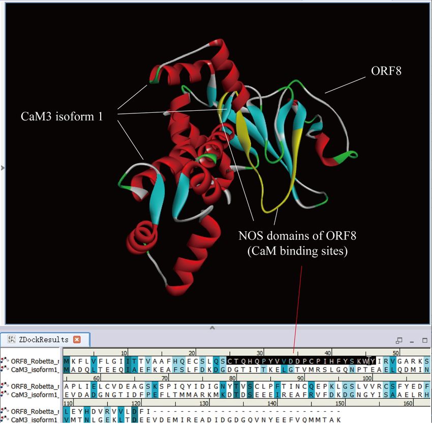
3.2 ORF8 protein had NOS conserved domains
      The human has three kinds of NOS isoenzymes, which catalyze L-arginine’s oxidation to
produce endogenous NO. One is nNOS in nerve cells, the other is eNOS in endothelial cells, and
the third is inducible iNOS in platelets, macrophages, and glial cells. The three NOS isoenzymes
have the exact three conserved domains(64, 65). The first is the N-terminal oxygenase domain
(66). The domain combines heme(67), tetrahydrobiopterin (H4B), and L-arginine. The second is
the C-terminal reductase domain. The domain binds to flavin mononucleotide (FMN), flavin
adenine dinucleotide (FAD), and reduced nicotinamide adenine dinucleotide phosphate (NADPH).
The third is the calmodulin (CaM) binding area. The domain is between the oxygenase and
reductase domains(68). The third domain regulates the electron transfer from the reductase domain
to oxygenase domain. NADPH provides electrons, and the electron transport route is:
NADPH->FAD->FMN->Heme. If the viral protein has NOS enzyme function, it needs to have
three positional domains simultaneously. And the three domains should be on the same protein and
not on different proteins.
      We downloaded all sequences of bacterial NOS synthase from the UniProt database. We then
employed the local MEME’s domain search tool to find the NOS conserved domains of all
SARS-COV-2 viral proteins (Table 1). Table 1 shows that among all SARS-COV-2 proteins, only
ORF8 has NOS domains in three directions: N-terminal, middle, and C-terminal. The three
domains are AAFHQECSLQSCTQHQPY(14-31), CTQHQPYVVDDPCPIHFYSKWY(25-46),
QYIDIGNYTVSCLPFTINCQ(72-91), respectively. CTQHQPY(25-31) belongs to both
N-terminal and middle domains. ORF7b, N, and M proteins have no NOS domain. Through the
analysis of the crystal structure of the ORF8 modeling, we found that
CTQHQPYVVDDPCPIHFYSKWY (25-46) and QYIDIGNYTVSCLPFTINCQ(72-91) can form
functional pockets, while AAFHQECSLQS(14-24) cannot form functional pockets. The
N-terminal NOS domain is a region that binds to the L-Arg etc. substrates and generates NO, so it
is impossible to develop an active pocket that can stabilize the binding molecules. After the pocket
formed near the middle NOS domain hunts the substrate (ligand) molecule, ORF8 rapidly changes
its conformation and sends the substrate to the N-terminus or C-terminus. And the molecular
substrate is unstable on N-terminal domain, so the bound substrate such as L-Arg will fall off after
the NO synthesis reaction.
      We tried to use a similar method of the heme theory to determine the heme iron site of
ORF8. We only found that the NCBI protein WP_008179633.1 corresponding to A0A1H8NYM7
(UniProt id) has “Related Structures (Summary)”. But the URL of the corresponding structure
file could not be opened. The ORF8’s motif corresponding to A0A1H8NYM7 is
“HQPYVVDDPCPIHFYSKW”, which is in the N-terminus and middle NOS domain. Therefore,
the existing data and methods cannot determine the iron linked site of ORF8.
      Other viral proteins only have NOS domains in one or two directions. For orf1ab,
"CCREHEHEIAWYTERSE" is in the middle of “YP_009742609.1 nsp2” protein, and
“RRLISMMGFKMNYQVNGYPNMFITREEAIRH” is in the middle of "YP_009725309.1
3'-to-5' exonuclease" protein too. Therefore, only ORF8 protein had NOS synthase function, and
other proteins had no NOS synthase function.

                                                 7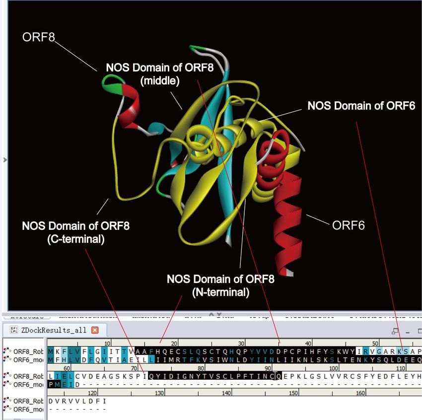
Table 1. NOS Domains of SARS-COV-2 proteins

Protein                          NOS Domains                     Count         Direction

ORF8                                                              25

          AAFHQECSLQSCTQHQPY                                      1       N-terminal

          CPIHFYSK                                                1       middle

          CTQHQPYVVDDPCPIHFY                                      6       middle

          CTQHQPYVVDDPCPIHFYSKW                                   5       middle

          HQPYVVDDPCPIHFYSKW                                      1       middle

          HQPYVVDDPCPIHFYSKWY                                     10      middle

          QYIDIGNYTVSCLPFTINCQ                                    1       C-terminal

ORF7a                                                             7

          FHPLADNKFALTCFSTQ                                       4       middle

          FHPLADNKFALTCFSTQFAFACPD                                2       middle

          YHYQECV                                                 1       N-terminal

ORF7b                                 -                               -            -

ORF3a                                                             9

          CWHTNC                                                  1       middle

          CWHTNCYDYC                                              1       middle

          CWKCRSKNPLLYDANYFLCWHTNC                                1       middle

          MRLWLCWKCR                                              2       middle

          MRLWLCWKCRSKNPLLYDANYFLCWHTNCYDYCIPY                    3       middle

          MRLWLCWKCRSKNPLLYDANYFLCWHTNCYDYCIPYN                   1       middle

ORF10                                                             51

          CRMNSRN                                                 3       C-terminal

          CRMNSRNYIAQ                                             47      C-terminal

          CRMNSRNYIAQVDVVNF                                       1       C-terminal

ORF6                                                              4

          IIMRTFKVSIWNLDYIINLIIKNLSKSLTENKYSQ                     1       middle

          MRTFKVSIWNLDYIINLIIKNLSKS                               1       middle

          MRTFKVSIWNLDYIINLIIKNLSKSLTENKYSQLDEEQP                 1       middle

          MRTFKVSIWNLDYIINLIIKNLSKSLTENKYSQLDEEQPM                1       middle

orf1ab                                                            2

          CCREHEHEIAWYTERSE                                       1       N-terminal

          RRLISMMGFKMNYQVNGYPNMFITREEAIRH                         1       middle

  S                                                               14

          EQYIKWPWYIW                                             7       C-terminal

          EQYIKWPWYIWLGFIAGLIAIVMVTIMLCCM                         1       C-terminal

          KYEQYIKWPWYIWLGFIAGLIAIVMVTIMLCCMTSCCSCLKGCC            1       C-terminal

          WPWYIW                                                  4       C-terminal

          YEQYIKWPWYIW                                            1       C-terminal

  E       CAYCCNIVNVSLVKPSFYVYSRVK                                1       middle

  N                                   -                               -            -

  M                                   -                               -            -

                                                8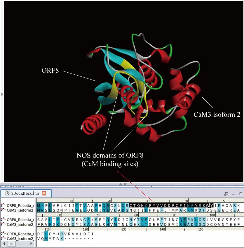
3.3 ORF8 protein could bind human CaM protein
     We first determined whether the NOS domain in the middle of ORF8 had the function of
binding CaM. We download the human CaM protein from NCBI. It had two isoforms
(NP_005175.2 calmodulin-3 isoform 1 and NP_001316854.1 calmodulin-3 isoform 2). Since the
CaM structure files on the PDB database were mostly combined with other molecules, we used
Robetta to model the two isoforms separately. Then adopted the DS 's protein docking tool
(ZDOCK) to dock the two isoforms with ORF8, respectively.
     Figure 2 shows the docking result of NP_005175.2 calmodulin-3 isoform 1 (CaM3 isoform 1,
the same below) and ORF8. Ser24 (S) of ORF8 is the intersection of two proteins. The middle
NOS domain, 'CTQHQPYVVDDPCPIHFYSKWY' (marked yellow area), behind Ser24 is close
to calmodulin-3 isoform 1. It indicates that the NOS domain in the middle of ORF8
('CTQHQPYVVDDPCPIHFYSKWY') can bind CaM protein.

     Figure 2. Docking result between calmodulin-3 isoform 1 and ORF8. calmodulin-3 isoform 1
is embedded in ORF8. Ser24 of ORF8 is the crossover point. The middle NOS domain,
'CTQHQPYVVDDPCPIHFYSKWY' ( marked yellow area),behind Ser24 is close to calmodulin-3
isoform 1.

     Figure 3 shows the docking result of NP_001316854.1 calmodulin-3 isoform 2 (CaM3
isoform 2, the same below) and ORF8. Asp35 (D) and Pro36 (P) of ORF8 is the intersection of
two proteins. The Asp35 (D) and Pro36 (P) are in sites 'CTQHQPYVVDDPCPIHFYSKWY'
(middle NOS domain, marked yellow area). It indicates that the NOS domain in the middle of
ORF8 ('CTQHQPYVVDDPCPIHFYSKWY') can bind CaM protein too.

                                             9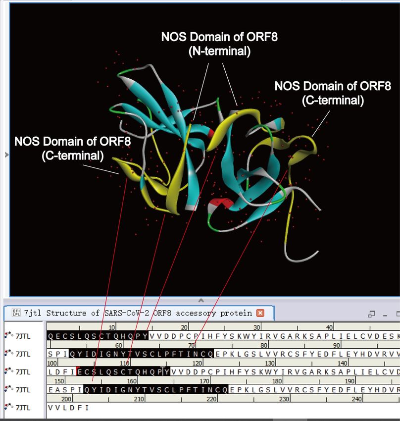
Figure 3. Docking result between calmodulin-3 isoform 2 and ORF8. calmodulin-3 isoform 2
is embedded in ORF8. Asp35 and Pro36 of ORF8 are the crossover point. The Asp35 and Pro36
are in sites 'CTQHQPYVVDDPCPIHFYSKWY' (middle NOS domain, marked yellow area).

3.4 ORF8 could capture heme, H4B, L-arginine
     We determined whether ORF8 could capture HEME, H4B, and L-arginine. We downloaded
the corresponding structure files of the three ligands of HEM, H4B, and ARG from the PDB
database. Then adopted DS's molecular docking technology (LibDock) to dock these three ligands
to ORF8, respectively.
     According to the technical documentation requirements of DS, FE( iron ions) should be
deleted before ORF8 and heme were docked. There are three docking results between ORF8 and
heme. The binding energy of SITE 1 and SITE 2 are both -88.5114 kcal/mol (selected), and the
positions are the same. The binding energy of SITE3 is both -73.9493 kcal/mol, which is slightly
higher than the binding energy of SITE 1 and SITE2. The docking results of SITE2 and SITE 1
are shown in Figure 4. TYR73, ILE74, ASP75, ILE76, GLY77, TYR79, GLN91 amino acids are
in the C-terminal NOS domain. Therefore, ORF8 captures heme through the active pocket
generated by the C-terminal NOS domain and rapidly transfers heme to the N-terminal NOS
domain by changing the conformation. The N-terminal NOS domain does not form an active
pocket for molecular binding. Therefore, heme may drop off after participating in the NO
synthesis reaction.

                                              10
Figure 4. The results of molecular docking between ORF8 and heme. A. Schematic diagram
of the docking result. B. Amino acids of ORF8 that bind heme. TYR73, ILE74, ASP75, ILE76,
GLY77, TYR79, GLN91 amino acids are in the C-terminal NOS domain.
ORF8 and H4B also have three docking results. The binding energy of SITE3 and SITE2 is
both -69.9598kcal/mol (selected), and the binding position is the same. The binding energy of
SITE1 is -6.7802 kcal/mol, which is higher than the binding energy of SITE 3 and SITE2. The
docking result of SITE3 and SITE2 is shown in Figure 5. The LYS44, TRP45, and TYR46 of
binding pockets are in the middle NOS domain of ORF8. Therefore, ORF8 directly captures H4B
through the middle NOS domain. The N-terminal NOS domain does not form an active pocket for
molecular binding. Thus, H4B may also drop off after participating in the NO synthesis reaction.
Figure 5. Molecular docking results of ORF8 and H4B. A. Schematic diagram of the docking
result. B. Amino acids of ORF8 that bind H4B. LYS44, TRP45, TYR46 are in the middle NOS
domain of ORF8
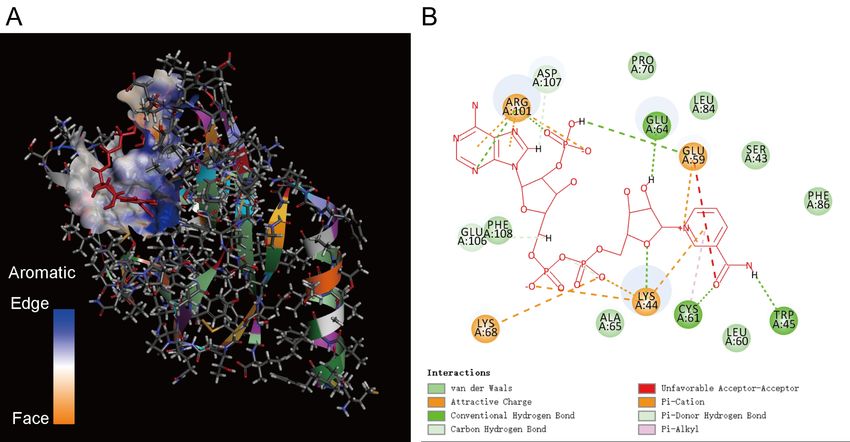
There are also three docking results for ORF8 and L-Arg. The binding energy of SITE1,
11SITE2, and SITE3 are all -124.315 kcal/mol, and the binding position is the same. The docking
result is shown in Figure 6. ASP35, SER43, LYS44, and TRP45 are in the middle NOS domain of
ORF8. CYS83, PH86, and LEU84 are in the C-terminal NOS domain of ORF8. Therefore, ORF8
captures L-Arg through the middle NOS domain and the C-terminal NOS domain. Then quickly
change the conformation and move L-Arg to the N-terminal NOS domain. The N-terminal NOS
domain does not form an active pocket for molecular binding. Therefore, L-Arg may fall off
quickly after participating in the NO synthesis reaction.
Figure 6. The results of molecular docking between ORF8 and L-Arg. A. Schematic diagram
of the docking result. B. The amino acid of ORF8 binds L-Arg. ASP35, SER43, LYS44, TRP45
are in the middle NOS domain ORF8, CYS83. PH86, and LEU84 are in the C-terminal NOS
domain ofORF8.
3.5 ORF8 could capture NADPH, FAD and FMN
We determine whether ORF8 can capture NADPH, FAD and FMN. We downloaded the
corresponding NAP, FAD and FMN three ligand structure files from the PDB database. Then
employed DS's molecular docking technology (LibDock) to dock these three ligands to ORF8,
respectively.
There are also three docking results for ORF8 and NADPH. The binding energy of SITE1,
SITE2, and SITE3 is all -256.329 kcal/mol, and the binding position is the same. It shows the
docking result in Figure 7. SER43, LYS44, TRP45 are in the middle NOS domain of ORF8.
LEU84 and PHE86 are in the C-terminal NOS domain of ORF8. Therefore, ORF8 captures
NADPH through the middle NOS domain and the C-terminal NOS domain. Then it quickly
changes the conformation and moves NADPH to the C-terminal NOS domain.
Docking results for ORF8 and FAD is as shown in Figure 8. The binding energy of SITE1,
SITE2, and SITE3 is all -144.458 kcal/mol, and the binding position is the same. LYS44,
TYR46 are in the middle NOS domain of ORF8. So, ORF8 captures FAD through the middle
NOS domain. Then it quickly changes the conformation and sends FAD to the C-terminal NOS
domain.
12Figure 7. The results of molecular docking between ORF8 and NADPH. A. Schematic
diagram of the docking result. B. Amino acids of ORF8 that bind NADPH. SER43, LYS44,
TRP45 are in the middle NOS domain of ORF8. LEU84 and PHE86 are in the C-terminal NOS
domain of ORF8.
Figure 8. The results of molecular docking between ORF8 and FAD. A. Schematic diagram
of the docking result. B. Amino acids of ORF8 that bind FAD. LYS44, TYR46 are in the middle
NOS domain of ORF8.
Docking results for ORF8 and FMN is as shown in Figure 9. The binding energy of SITE1,
SITE2 is all -227.695 kcal/mol (selected), and the binding position is the same. The binding
energy of SITE3 is -125.086 kcal/mol. TYR73, ILE74, ASP75, ILE76, GLY77, TYR79, GLN91
are in the C-terminal NOS domain of ORF8. So, ORF8 captures FMN through C-terminal NOS
domain.
13Figure 9. The results of molecular docking between ORF8 and FMN. A. Schematic diagram
of the docking result. B. Amino acids of ORF8 that bind FMN. TYR73, ILE74, ASP75, ILE76,
GLY77, TYR79, GLN91 are in the C-terminal NOS domain of ORF8.
3.6 ORF8 could capture heme through ORF3a and E protein
Molecular docking shows that ORF8 captures heme through the C-terminal NOS domain and
then sends it to the N-terminal NOS domain. It may be just one of the less efficient ways. In Table
1, ORF7a, ORF3a, ORF10, ORF6, orf1ab, E, S have partial NOS domains. In the heme theory
study, we found that ORF3a and ORF10 can attack hemoglobin, while ORF3a and E could bind
heme. Then we applied protein molecular docking technology of DS to study whether ORF8
interacted with these proteins and determined whether there was the possibility of heme
transmission.
According to Table 1 and the heme theory, the heme linked site “CWKCR” overlaps with
the NOS domain “MRLWLCWKCRSKNPLLYDANYFLCWHTNCYDYCIPYN” of ORF3a .
It showed the docking result of ORF8 and ORF3a in Figure 10. Ser24, Tyr42, and Ser43 of
ORF8 are the three intersections with ORF3a. Tyr42 and Ser43 belong to the middle NOS domain
of ORF8 and Ser24 belong to the N-terminal NOS domain of ORF8. From the heme theory, we
know ORF3a can attack hemoglobin and hunt heme. Therefore, the heme hunted by ORF3a may
be transferred to the N-terminal NOS domain of ORF8 through the middle NOS domain of ORF8.
From the heme theory, ORF10 also assisted ORF3a in attacking hemoglobin. But the docking
result of ORF8 and ORF10 (Figure 11) shows that the two proteins are not embedded together. It
indicates that ORF10 will not directly send ligand molecules such as heme to ORF8.
14Figure 10. Docking results of ORF8 and ORF3a. The NOS domain of ORF3a includes the
heme linked site “CWKCR”, and it crosses the middle NOS domain of ORF8. ORF3a crosses the
N-terminal NOS domain of ORF8 too.
Figure 11. Docking result of ORF8 and ORF10. ORF10 and ORF8 are not embedded in each
other.
15The existing literature shows that ORF8 and ORF6 have an interference effect on the immune
response. Docking result of ORF8 and ORF6 (Figure 12) reveals that it embedded the two proteins
in each other. It connected the NOS domain of ORF6 to the three NOS domains of ORF8. Perhaps
ORF6 has a combined effect on the three structures of ORF8, and is an essential auxiliary protein
in the process of NO synthesis.
From the heme theory, E protein has a heme-binding site, “CAYCC”. The site is in the NOS
domain of the E protein “CAYCCNIVNVSLVKPSFYVYSRVK”. The docking result of ORF8
and E protein (Figure 13) represents that ORF8 and E are embedded in each other. The heme
attachment site of E is close to the N-terminal NOS domain of ORF8. It indicates that the
N-terminal NOS domain of ORF8 can use the heme hunted by E in the NOS synthesis reaction.
Docking results on ORF8 and S protein, ORF8 and ORF7a protein show that ORF8 can not
be embedded with S and ORF7a. Orf1ab is composed of a series of sub-proteins; there is no
heme-binding point found in heme theory. Therefore, ORF8 does not directly cooperate with S,
ORF7a, and orflab proteins to synthesize NO.
Figure 12. Docking results of ORF8 and ORF6. ORF8 and ORF6 are embedded together. The
NOS domain of ORF6 is connected to the three NOS domains of ORF8.
16Figure 13. Docking result of ORF8 and E. ORF8 and E are embedded together. The NOS
domain of E (heme connection site) is close to the N-terminal NOS domain of ORF8.
3.7 Dimer structure of ORF8 was similar to NOS
Two nitric oxide synthase form a dimer to have the activity of catalyzing the production of
NO. On both sides of the dimer is a reductase structure, and the center of the dimer is a dimer core
synthesized by two oxygenase domains(66). Interestingly, ORF8 also exhibits a unique dimer
structure(60) and has a similar dimer core. We downloaded the ORF8 structure file 7JTL from the
PDB database. The sequence corresponding to this structure is missing the N-terminal fragment
“MKFLVFLGIITTVAAFH”. But the N-terminal NOS domain of ORF8 still exists. One ORF8 is
“QECSLQSCTQHQPY” and the other ORF8 is “ECSLQSCTQHQPY”. As shown in Figure 14,
the two N-terminal NOS domains (oxygenase) are both in the core of the ORF8 dimer, forming a
paired posture. It may be the role of clamping substrates such as heme and ARG and catalyzing
NO production. The two C-terminal NOS domains (reductase) are on both sides of the ORF8
dimer. The crystal structure diagram also shows that the N-terminal NOS domain nears the
C-terminal NOS domain in the space structure. Therefore, it is possible that ORF8 hunts heme
through the C-terminal NOS domain and sends heme to the N-terminal NOS domain through
conformational changes.
17Figure 14. Dimer structure of ORF8 (PDB ID: 7JTL). The two N-terminal NOS domains
(oxygenase) are in the core of the ORF8 dimer, and the two C-terminal NOS domains (reductase)
are on both sides of the ORF8 dimer. The ORF8 dimer structure conforms to the dimer structure
characteristics of the NOS enzyme.
4. Discussion
4.1 SARs-COV-2 combined with ORF8 to generate NO to break the blood-brain
(testi) barrier and damage immune cells
From the above results, the N-terminal NOS domain of ORF8 directly binds heme with low
efficiency. This study found that ORF8 could bound ORF3a and E protein. The heme theory found
that ORF3a attacked hemoglobin, and deoxyhemoglobin was more likely to be attacked. Therefore,
heme (divalent iron) shed from attacking deoxyhemoglobin became the direct source of ORF8’s
heme.The conjugation of E protein to ORF8 may not be good news. This study found that E
protein's close position to the N-terminal NOS domain of ORF8 is exactly the heme linked site
“CAYCC” (the last C is the iron linked site). It meant that ORF8 used the heme hunted by the E
protein to produce NO. In the heme theory and ROS damage theory, we found that E protein had
the functions of cytochrome C oxidase, catalase, peroxidase, and superoxide dismutase. The heme
iron bound by the E protein was oxidized and reduced through these enzymes' processes. After
ORF8 bound to E protein, ORF8 did not need to solve the source of heme. But it is not clear that
ORF8 also needed CaM, FAD、FMN、NADPH, and ORF6.
Confusingly, ORF8 is highlighted by structural plasticity and high diversity, and its gene
transcripts are expressed in higher amounts(69, 70). Although ORF8 catalyzed NO production, it
was necessary to continuously produce NO to expand blood vessels or open the blood-brain (testi)
barrier. NO had a short lifespan and was easily bound to the iron on the heme of hemoglobin.
Therefore, NO was not suitable for continuous long-distance transmission in the body. It was also
18impossible for the SARS-COV-2 virus to passively expand blood vessels through NO produced by
ORF8 of a long distance. E protein was the structural protein of SARS-COV-2. When E protein
was combined with ORF8, the SARS-COV-2 virus was equivalent to have a weapon that
continuously produces NO. Many E proteins of the SARS-COV-2 virus could bound ORF8 so that
the virus could constantly create a tremendous amount of NO. The SARS-COV-2 virus achieved
an osmotic expansion of blood vessels or opened the blood-brain (testi) barrier through a steady
NO stream. Then the SARS-CoV-2 virus spread to nearby tissues through small blood vessel holes
created by NO stream.
Of course, these NO interfered with immune cells. The free radicals converted from these NO
also damaged immune cells. After the cell synthesized ORF8, ORF8 directly bound E protein. The
membrane vesicle transport system transported the ORF8-E complex to the cell membrane surface.
Therefore, through the ORF8-E complex, infected cells also had abnormal NO synthesis function.
For immune cells (such as NK cells, lymphocytes) in contact with infected cells, the NO stream
interfered with the immune cells’ NO signaling pathway. Besides, the NO rapidly decayed and
converted into nitrogen free radicals, which damaged the cell membrane of immune cells, making
immune cells death or abnormal function.
4.2 NO made the virus effectively spread and replicate in the body
Existing evidence shows that endogenous and exogenous NO can inhibit SARS-CoV-2 virus
replication(71).The target proteins of NO antiviral activity include protease, reverse transcriptase,
ribonucleotide reductase, zinc fingers, and related domains(72). One mechanism is to reduce the
spike (S) protein’s palmitoylation, which is crucial in anchoring the S protein to the lipid
bilayer(57). So nitric oxide interferes with the interaction between S protein and its cognate host
receptor ACE-2. Another mechanism is the nitrosation of viral proteins. S protein and 3CL
cysteine protease may be targets of nitrosation(73). Because of the potential of NO in the
treatment of COVID-19 and the anti-coronavirus infection, medical regulatory authorities in some
countries have approved inhaled NO as a safe treatment(74). We believed ORF8 synthesized NO
to inhibit 3C-Like protein was the virus's rate-limiting behavior on its replication, avoiding the cell
collapse of insufficient replication resources. ORF8 synthesized NO to inhibit S protein,
preventing the SARS-COV-2 virus from re-infecting already infected cells to make the infection
more efficient.
4.3 The high viral load in asymptomatic patients was associated with insufficient
NO to open the vascular permeability
An enormous amount of NO also expanded the permeability of blood vessels and opened
blood-brain barriers. It promoted the spread of virus particles to tissue cells near blood vessels.
For the asymptomatic patient, the virus was lurking in the phagocyte vesicles or lysosomes(59),
and the viral protein did not attack the heme proteins on a large scale. Therefore, there was less
heme in the blood , and then the ORF8 proteins that bound heme to catalyze NO were less too.
The small amount of NO combined with its brief life span made it challenging to open the
vascular permeability. It created a strange phenomenon: the virus particles spread through
phagocytes are in the incubation period. However, more virus particles in the blood were
difficult to spread in the tissue cells because the vascular permeability was not opened. Therefore,
Viral loads of asymptomatic patients were found to be significantly higher when compared with
19symptomatic patients(75). So, the high viral load in asymptomatic patients was associated with
insufficient NO to open the vascular permeability.
4.4 Excessive NO hiddenly damaged the female reproductive system
The inflammatory factors produced by the cytokine storm will stimulate inflammatory cells,
phagocytes, and neutrophils to produce oxygen free radicals and NO.There have been reports in
the literature that large amounts of NO metabolites (nitrite and nitrate) are found in the plasma and
urine of COVID-19 patients. Reactive oxygen species (ROS) and reactive nitrogen species (RNS)
are by-products of various cellular processes, including aerobic metabolism. These reactive
oxygen/nitrogen species (RONS), namely hydroxyl, superoxide anion, nitric oxide, and nitrosyl
anion, are highly reactive molecules because of their unpaired valence electrons. RONS plays a
critical biological role in cell signaling (redox signaling pathway): thiol switch, regulation of
inflammatory cytokines, growth factors, etc. When the redox balance is disrupted, these powerful
oxidants (free radicals) have harmful effects.
It synthesizes nitric oxide in the breast and triggers human lactation(76). Nitric oxide
metabolites and adrenomedullin are detected in breast milk(77). Luteinizing hormone (LH) is a
type of gonadotropin secreted by the pituitary gland’s anterior lobe. LH acts on the corpus luteum
or corpus luteum cells of the ovary to promote progesterone’s secretion. High luteinizing hormone
is a kind of endocrine disorder that quickly affects normal follicular development, causing
abnormal ovulation, and unnatural conception. The high luteinizing hormone also produces more
obvious premature ovarian failure or polycystic ovary syndrome in the human body. We found
ORF8 synthesized an enormous amount of NO, and these NO could trigger the abnormal secretion
of milk and LH, which hiddenly damaged the breast and ovaries.
4.5 ORF8 protein interfered with the NO signaling pathway
Nitric oxide (NO) is a free gas molecule, which plays a vital role as a signal molecule in
human physiology. In KEGG's signal pathway database, after inputting "Human" and "Nitric
Oxide", 33 relevant and important signal pathways are retrieved (Table 2). NO has essential
effects on the immune system, cardiovascular system, metabolic system, nervous system, motor
system, hormone control, rhythm control, cancer occurrence, and other signaling pathways. So,
ORF8 protein could catalyze the synthesis of NO, interfering with signaling pathways and
damaging immune, nervous, cardiovascular, metabolic, and reproductive systems.
5. Conclusion
It relates nitric oxide to many viruses’ pathogenic mechanisms. We believe that the novel
coronavirus is also closely related to nitric oxide. This study adopted bioinformatics methods such
as domain search and molecular docking to study the relationship between the novel coronavirus
proteins and nitric oxide. The results showed ORF8 had three functional domains of NOS
synthase: oxygenase domain, reductase domain and calmodulin (CaM) binding domain. ORF8
also is integrated into a dimer structure to be catalytically active. ORF8 could capture heme, H4B,
L-arginine, FAD, FMN, NADPH molecules, combine with CaM to form a NO generator, and
synthesize nitric oxide. The heme bound by ORF8 mainly came from the attacked hemoglobin.
The high viral load in asymptomatic patients was associated with insufficient NO to open the
20vascular permeability as insufficient heme. SARs-COV-2 (E protein) also could combined with
ORF8 to generate NO stream to break the blood-brain (testi) barrier and damage reproductive,
nervous and immune system. Then the SARS-CoV-2 virus spread to nearby tissues through small
blood vessel holes created by NO stream.
Table 2. Nitric oxide is involved in human signaling pathways
Entry Pathway Name Entry Pathway Name
hsa04022 cGMP-PKG signaling pathway hsa04940 Type I diabetes mellitus
hsa04066 HIF-1 signaling pathway hsa00330 Arginine and proline metabolism
hsa04926 Relaxin signaling pathway hsa04071 Sphingolipid signaling pathway
hsa05332 Graft-versus-host disease hsa04151 PI3K-Akt signaling pathway
hsa04062 Chemokine signaling pathway hsa04270 Vascular smooth muscle contraction
hsa01100 Metabolic pathways hsa04370 VEGF signaling pathway
hsa05014 Amyotrophic lateral sclerosis hsa04540 Gap junction
hsa05022 Pathways of neurodegeneration hsa04611 Platelet activation
hsa05415 Diabetic cardiomyopathy hsa04621 NOD-like receptor signaling pathway
hsa00910 Nitrogen metabolism hsa04730 Long-term depression
hsa05418 Fluid shear stress and atherosclerosis hsa04915 Estrogen signaling pathway
hsa04371 Apelin signaling pathway hsa04921 Oxytocin signaling pathway
hsa04713 Circadian entrainment hsa04924 Renin secretion
hsa04931 Insulin resistance hsa04970 Salivary secretion
hsa05010 Alzheimer disease hsa05330 Allograft rejection
hsa05200 Pathways in cancer hsa04115 p53 signaling pathway
hsa05225 Hepatocellular carcinoma
The life cycle of NO is short. If the NO content was low, it could not break the blood-brain
(testis) barrier of some patients. So the virus could not infect the reproductive organs. Much NO
dilates blood vessels in the reproductive system for a long time, causing severe congestion and
abnormal function. Much NO also stimulated the gonads to secrete hormones abnormally and
affects sperm and ovum production quality. Extreme NO osmotically expanded blood vessels and
aggravate bleeding tendency. Bleeding caused tissue congestion or coagulation. Excessive NO
destroyed the blood-brain (testis) barrier, allowing the SARS-COV-2 virus to penetrate the tissues
and cells around the blood vessels, and infect the nerves, reproductive and gonadal organs.
The NO produced could protect infected cells and weaken attack NK cells or lymphocytes. It
also inhibited S protein not to reinfect infected cells by SARS-CoV-2 viruses. NO inhibited
3C-like protein from limiting the virus replication process's speed and preventing the host cell
from crashing due to the insufficient replication resources. Viruses may hijack the cell's nitric
oxide signal pathways and cause diseases such as immunity, nerves, metabolism, and
cardiovascular diseases. In short, this research has important guiding significance for disease
prevention and treatment, medicine, and vaccine research.
21Declarations
Ethics approval and consent to participate
Not applicable.
Consent for publication
Not applicable.
Availability of data and material
The datasets and results supporting the conclusions of this article are available at
https://pan.baidu.com/s/1e-pPwWH86HMOglznzNS7mQ , code: 9zza.
Or: https://mega.nz/folder/gjgzTa7T#6J9n8CqHd70QUNSm3wXM6w
Competing interests
The authors declare that they have no competing interests.
Funding
This work was funded by a grant from the National Natural Science Foundation for the Talent
Introduction Project of Sichuan University of Science and Engineering (award number: 2018RCL20,
grant recipient: WZL).
Author’s contribution
Funding was obtained by WZL. Besides, design, analysis and writing are finished by WZL, while
data curation and manuscript check are undertaken by HLL. Both authors have read and agreed to the
published version of the manuscript.
Acknowledgements
Thanks readers for free review and suggestions.
Author details
1
School of Computer Science and Engineering, Sichuan University of Science & Engineering, Zigong, 643002,
China.
2
School of Life Science and Food Engineering, Yibin University, Yibin, 644000, China.
References
1. Wang, Z., and X. Xu. 2020. scRNA-seq profiling of human testes reveals the presence of the
ACE2 receptor, a target for SARS-CoV-2 infection in spermatogonia, Leydig and Sertoli cells.
Cells 9: 920.
2. Jing, Y., L. Run-Qian, W. Hao-Ran, C. Hao-Ran, L. Ya-Bin, G. Yang, and C. Fei. 2020. Potential
influence of COVID-19/ACE2 on the female reproductive system. Molecular human reproduction
26: 367-373.
223. Peckham, H., N. M. de Gruijter, C. Raine, A. Radziszewska, C. Ciurtin, L. R. Wedderburn, E. C.
Rosser, K. Webb, and C. T. Deakin. 2020. Male sex identified by global COVID-19 meta-analysis
as a risk factor for death and ITU admission. Nature communications 11: 1-10.
4. Qiu, L., X. Liu, M. Xiao, J. Xie, W. Cao, Z. Liu, A. Morse, Y. Xie, T. Li, and L. Zhu. 2020.
SARS-CoV-2 is not detectable in the vaginal fluid of women with severe COVID-19 infection.
Clinical Infectious Diseases 71: 813-817.
5. Cui, P., Z. Chen, T. Wang, J. Dai, J. Zhang, T. Ding, J. Jiang, J. Liu, C. Zhang, and W. Shan. 2020.
Severe acute respiratory syndrome coronavirus 2 detection in the female lower genital tract.
American Journal of Obstetrics & Gynecology 223: 131-134.
6. Tur-Kaspa, I., T. Tur-Kaspa, G. Hildebrand, and D. Cohen. 2021. COVID-19 May Affect Male
Fertility but is Not Sexually Transmitted: A Systematic Review. F&s Reviews.
7. Sadeghi, M. R. 2020. Implications of assisted human reproduction during coronavirus disease
2019 (COVID-19) pandemic. Journal of Reproduction & Infertility 21: 155.
8. Stanley, K. E., E. Thomas, M. Leaver, and D. Wells. 2020. Coronavirus disease-19 and fertility:
viral host entry protein expression in male and female reproductive tissues. Fertility and sterility
114: 33-43.
9. DeBoer, R. E., O. O. Oladunjoye, and R. Herb. 2021. Right Ovarian Vein Thrombosis in the
Setting of COVID-19 Infection. In Cureus. e12796.
10. Abdelazim, I. A., M. AbuFaza, and S. Al-Munaifi. 2021. COVID-19 Positive Woman Presented
with Preterm Labor: Case Report. Gynecology Obstetrics & Reproductive Medicine: 1-3.
11. Vivanti, A. J., C. Vauloup-Fellous, S. Prevot, V. Zupan, C. Suffee, J. Do Cao, A. Benachi, and D.
De Luca. 2020. Transplacental transmission of SARS-CoV-2 infection. Nature Communications
11: 3572.
12. Alzamora, M. C., T. Paredes, D. Caceres, C. M. Webb, L. M. Valdez, and M. La Rosa. 2020.
Severe COVID-19 during pregnancy and possible vertical transmission. American journal of
perinatology 37: 861.
13. Wu, Y., C. Liu, L. Dong, C. Zhang, Y. Chen, J. Liu, C. Zhang, C. Duan, H. Zhang, B. Mol, C.-L.
Dennis, T. Yin, J. Yang, and H. Huang. 2020. Coronavirus disease 2019 among pregnant Chinese
women: case series data on the safety of vaginal birth and breastfeeding. BJOG: An International
Journal of Obstetrics & Gynaecology 127: 1109-1115.
14. Shastri, A., J. Wheat, S. Agrawal, N. Chaterjee, K. Pradhan, M. Goldfinger, N. Kornblum, U.
Steidl, A. Verma, and J. Shastri. 2020. Delayed clearance of SARS-CoV2 in male compared to
female patients: high ACE2 expression in testes suggests possible existence of gender-specific
viral reservoirs. MedRxiv.
15. Shen, Q., X. Xiao, A. Aierken, W. Yue, X. Wu, M. Liao, and J. Hua. 2020. The ACE2 expression
in Sertoli cells and germ cells may cause male reproductive disorder after SARS‐CoV‐2 infection.
Journal of Cellular and Molecular Medicine 24: 9472-9477.
16. Fan, C., W. Lu, K. Li, Y. Ding, and J. Wang. 2021. ACE2 Expression in Kidney and Testis May
Cause Kidney and Testis Infection in COVID-19 Patients. Frontiers in medicine 7: 1045.
17. Song, C., Y. Wang, W. Li, B. Hu, G. Chen, P. Xia, W. Wang, C. Li, X. Yang, and B. Yao. 2020.
Detection of 2019 novel coronavirus in semen and testicular biopsy specimen of COVID-19
patients. MedRxiv.
18. Wang, W., Y. Xu, R. Gao, R. Lu, K. Han, G. Wu, and W. Tan. 2020. Detection of SARS-CoV-2 in
different types of clinical specimens. Jama 323: 1843-1844.
2319. Li, D., M. Jin, P. Bao, W. Zhao, and S. Zhang. 2020. Clinical characteristics and results of semen
tests among men with coronavirus disease 2019. JAMA network open 3: e208292-e208292.
20. Ma, X., C. Guan, R. Chen, Y. Wang, S. Feng, R. Wang, G. Qu, S. Zhao, F. Wang, and X. Wang.
2021. Pathological and molecular examinations of postmortem testis biopsies reveal SARS-CoV-2
infection in the testis and spermatogenesis damage in COVID-19 patients. Cellular & molecular
immunology 18: 487-489.
21. Sun, J. 2020. The hypothesis that SARS-CoV-2 affects male reproductive ability by regulating
autophagy. Medical hypotheses 143: 110083.
22. Yang, M., S. Chen, B. Huang, J.-M. Zhong, H. Su, Y.-J. Chen, Q. Cao, L. Ma, J. He, and X.-F. Li.
2020. Pathological findings in the testes of COVID-19 patients: clinical implications. European
urology focus 6: 1124-1129.
23. Achua, J. K., K. Y. Chu, E. Ibrahim, K. Khodamoradi, K. S. Delma, O. A. Iakymenko, O. N.
Kryvenko, H. Arora, and R. Ramasamy. 2021. Histopathology and ultrastructural findings of fatal
COVID-19 infections on testis. The world journal of men's health 39: 65.
24. Xu, J., L. Qi, X. Chi, J. Yang, X. Wei, E. Gong, S. Peh, and J. Gu. 2006. Orchitis: a complication
of severe acute respiratory syndrome (SARS). Biology of reproduction 74: 410-416.
25. Youssef, K., and K. Abdelhak. 2020. Male genital damage in COVID-19 patients: Are available
data relevant? Asian Journal of Urology.
26. Özveri, H., M. T. Eren, C. E. Kırışoğlu, and N. Sarıgüzel. 2020. Atypical presentation of
SARS-CoV-2 infection in male genitalia. Urology Case Reports 33: 101349.
27. Chen, F., and D. Lou. 2020. Rising concern on damaged testis of COVID-19 patients. Urology
142: 42.
28. de Carvalho, R. C., M. F. Groner, J. Camillo, P. R. A. Ferreira, and R. Fraietta. 2020. The
interference of COVID-19 in the male reproductive system: Important questions and the future of
assisted reproduction techniques. Clinics 75.
29. Abobaker, A., and A. A. Raba. 2020. Does COVID-19 affect male fertility? World journal of
urology: 1-2.
30. Dutta, S., and P. Sengupta. 2020. SARS-CoV-2 infection, oxidative stress and male reproductive
hormones: can testicular-adrenal crosstalk be ruled-out? Journal of basic and clinical physiology
and pharmacology 1.
31. Sengupta, P., and S. Dutta. 2020. Does SARS-CoV-2 infection cause sperm DNA fragmentation?
Possible link with oxidative stress. Eur J Contracept Reprod Health Care 25: 405-406.
32. Alahmar, A. T. 2019. Role of oxidative stress in male infertility: An updated review. Journal of
human reproductive sciences 12: 4.
33. Selvaraj, K., S. Ravichandran, S. Krishnan, R. K. Radhakrishnan, N. Manickam, and M.
Kandasamy. 2021. Testicular atrophy and hypothalamic pathology in COVID-19: possibility of
the incidence of male infertility and HPG axis abnormalities. Reproductive Sciences: 1-8.
34. Dutta, S., and P. Sengupta. 2021. SARS-CoV-2 and male infertility: possible multifaceted
pathology. Reproductive Sciences 28: 23-26.
35. Delgado-Roche, L., and F. Mesta. 2020. Oxidative stress as key player in severe acute respiratory
syndrome coronavirus (SARS-CoV) infection. Archives of medical research.
36. Roychoudhury, S., A. Das, N. K. Jha, K. K. Kesari, S. Roychoudhury, S. K. Jha, R. Kosgi, A. P.
Choudhury, N. Lukac, and N. R. Madhu. 2021. Viral pathogenesis of SARS-CoV-2 infection and
male reproductive health. Open biology 11: 200347.
2437. Wu, P. Y., E. Scarlata, and C. O’Flaherty. 2020. Long-term adverse effects of oxidative stress on
rat epididymis and spermatozoa. Antioxidants 9: 170.
38. Sengupta, P., and S. Dutta. 2021. COVID-19 and hypogonadism: secondary immune responses
rule-over endocrine mechanisms. Human Fertility: 1-6.
39. Hussain, A. N., F. Hussain, and S. K. Hashmi. 2020. Role of testosterone in COVID-19 patients–A
double-edged sword? Medical Hypotheses 144: 110287.
40. Vishvkarma, R., and S. Rajender. 2020. Could SARS‐CoV‐2 affect male fertility? Andrologia 52:
e13712.
41. Yousaf, Z., S. D. Al-Shokri, H. Al-Soub, and M. F. Mohamed. 2020. COVID-19-associated
SIADH: a clue in the times of pandemic! American Journal of Physiology-Endocrinology and
Metabolism 318: E882-E885.
42. Pal, R. 2020. COVID-19, hypothalamo-pituitary-adrenal axis and clinical implications. Endocrine
68: 251-252.
43. Ma, L., W. Xie, D. Li, L. Shi, Y. Mao, Y. Xiong, Y. Zhang, and M. Zhang. 2020. Effect of
SARS-CoV-2 infection upon male gonadal function: A single center-based study. MedRxiv.
44. Seymen, C. M. 2021. The other side of COVID‐19 pandemic: Effects on male fertility. Journal of
medical virology 93: 1396-1402.
45. Ma, L., W. Xie, D. Li, L. Shi, G. Ye, Y. Mao, Y. Xiong, H. Sun, F. Zheng, and Z. Chen. 2021.
Evaluation of sex‐related hormones and semen characteristics in reproductive‐aged male
COVID‐19 patients. Journal of medical virology 93: 456-462.
46. Li, R., T. Yin, F. Fang, Q. Li, J. Chen, Y. Wang, Y. Hao, G. Wu, P. Duan, and Y. Wang. 2020.
Potential risks of SARS-Cov-2 infection on reproductive health. Reproductive biomedicine online.
47. Bhat, G. K., V. B. Mahesh, C. A. Lamar, L. Ping, K. Aguan, and D. W. Brann. 1995.
Histochemical Localization of Nitric Oxide Neurons in the Hypothalamus: Association with
Gonadotropin-Releasing Hormone Neurons and Co-Localization with N-Methyl-D-Aspartate
Receptors. Neuroendocrinology 62: 187-197.
48. Mahachoklertwattana, P., S. M. Black, S. L. Kaplan, J. D. Bristow, and M. M. Grumbach. 1994.
Nitric oxide synthesized by gonadotropin-releasing hormone neurons is a mediator of
N-methyl-D-aspartate (NMDA)-induced GnRH secretion. Endocrinology 135: 1709-1712.
49. Kalra, S. P. 1993. Mandatory Neuropeptide-Steroid Signaling for the Preovulatory Luteinizing
Hormone-Releasing Hormone Discharge*. Endocrine Reviews 14: 507-538.
50. Maeda, K.-i., S. Ohkura, Y. Uenoyama, Y. Wakabayashi, Y. Oka, H. Tsukamura, and H. Okamura.
2010. Neurobiological mechanisms underlying GnRH pulse generation by the hypothalamus.
Brain Research 1364: 103-115.
51. Janigro, D., G. A. West, T.-S. Nguyen, and H. R. Winn. 1994. Regulation of blood-brain barrier
endothelial cells by nitric oxide. Circulation Research 75: 528-538.
52. Mayhan, W. G., and S. P. Didion. 1996. Glutamate-induced disruption of the blood-brain barrier in
rats: role of nitric oxide. Stroke 27: 965-970.
53. Griffiths, M. J., and T. W. Evans. 2005. Inhaled nitric oxide therapy in adults. New England
Journal of Medicine 353: 2683-2695.
54. Zhang, J., H. Benveniste, B. Klitzman, and C. A. Piantadosi. 1995. Nitric oxide synthase
inhibition and extracellular glutamate concentration after cerebral ischemia/reperfusion. Stroke 26:
298-304.
25You can also read