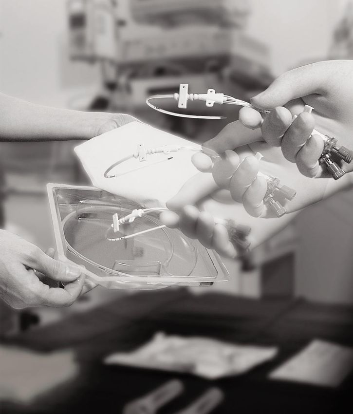
Cvc-partner 1 Guide for Central Venous Catheterization
←
→
Page content transcription
If your browser does not render page correctly, please read the page content below
cvc-partner 1
                           Guide for Central Venous Catheterization
Central Venous Catheters
The handbook series “cvc-partner” deals with the use and application of
central venous catheters. Arterial or pulmonary catheters, hemodialysis
catheters, tunneled or implanted catheters are not included in the
category of central venous catheters in this series.

All information corresponds to the current standard of knowledge in
the field.

The absence of trademarks does not indicate that product names are
not protected.

This series has been prepared in consultation with many users to
whom we wish to express our heartfelt gratitude for their various
contributions. It is the intention of this series to assist the various
users which needs a continuos dialogue with our readers. Any comment
or tip is welcome and should be sent to info@cvc-partner.com or
placed at the homepage www.cvc-partner.com.
Guide for Central Venous Catheterization
Preface

The present handbook is part of a new, unique concept where           The techniques of central venous catheterisation and catheter
medical specialists demonstrate the technique of central              placement via Seldinger wire can be learned easily by each
venipuncture for other medical staff.                                 medical specialist who is interested to do so. The present
The manual contains a concise summary of the skills necessary         handbook on central venipuncture is a concise summary of the
for central venipuncture, and in combination with the corre-          essential practical skills necessary for this intervention. The
sponding video tape “Introduction of Central Venous Catheters         medical who is willing to learn these techniques can profit well
by the Seldinger Technique” all practical aspects of this interven-   from this practice-oriented manual. Tips from daily experiences
tion are described and shown in detail.                               will help to build on his own experiences and to quickly gain
Central venous catheterisation allows for an adequate therapy of      practical competence in this technique. To the experienced, this
critically ill patients during complex therapeutic interventions,     handbook gives a survey on the current technical improvements
especially in anaesthesia, intensive care and emergency medi-         in catheter materials and puncture techniques. He will find
cine. A successful venipuncture requires profound knowledge of        information on how to further improve his technique as well as
the indication and anatomic conditions, comprehensive experi-         suggestions how to pass his knowledge and practical experiences
ences, a precise technique as well as high quality instruments        on to medical assistants. I do hope that this excellent and
(puncture set and catheter). Continuous technical developments        practice-oriented manual will find many readers, eager to
and the resulting improvements led to a significant increase in       improve their knowledge.
patient safety. Today´s medical professionals can choose from a
variety of catheters and puncture techniques to match the indi-
vidual requirements of each patient. In case of elective insertion
of a central venous catheter the method of choice should be the
Seldinger technique due to a reduced trauma and a larger vari-
ety of catheters available. After exact catheter positioning and      Heidelberg, in March 2002
verification of the correct catheter tip position in the vena cava
(right in front of the atrium) by ECG-control via the Seldinger       Prof. Dr. Johann Motsch
wire, an additional x-ray control is usually no more necessary.       Medical Director
This significantly reduces the costs as well as the exposure to       Department of Anaesthesiology
x-rays for both, patients and medical staff.                          University Hospital Heidelberg
Contents

 1 When Is Central Venous Catheterization Indicated    7

 2 Criteria for the Selecting of a Puncture Site      10

 3 From venesection to the Seldinger technique        14

 4 Selecting the proper catheter                      18

 5 Preperation for Catheterization                    23

 6 Catheter Placement with the Seldinger Method       26

 7 Catheter Management                                32

 8 What To Do When Complications Occur                34

 9 Glossary                                           38
When Is Central Venous Catheterization Indicated

                     The increased rate of morbidity
                     among patients in critical care
                     medicine often necessitates complex
                     anesthesiological interventions where
                     a central venous catheter can be
                     essential. For each patient the
                     reasons for catheterization must be
                     given careful consideration.

                        7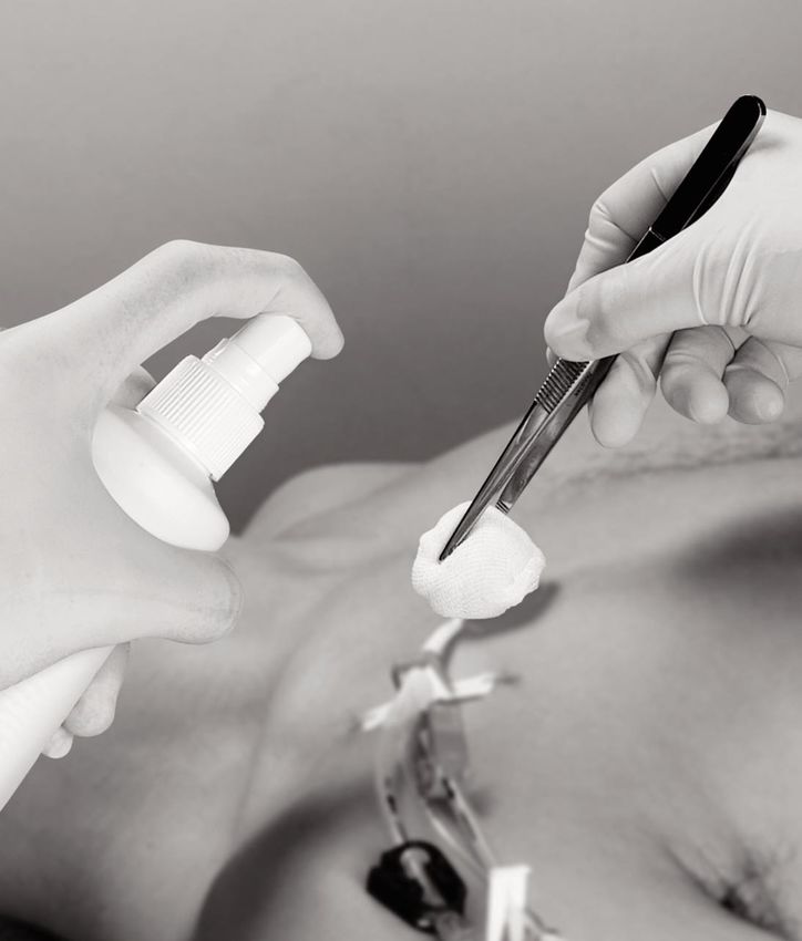
When Is Central Venous Catheterization Indicated

The history of central venous cannulation starts in 1929 when
Forssmann described the advance of a plastic tube to the heart
by puncturing his own arm vein (1). At the beginning of the
1950s Aubaniac reported about the puncture of the subclavian
vein. This puncture technique helped to broaden the use of this
technically demanding procedure (2). Since this time central
venous catheterization has developed to a standard procedure in
routine clinical practice. In critical care and emergency medicine
as well as for long-term therapies such as chemotherapy or dial-
ysis, the use of central venous catheters or central lines has
developed into an essential element of medical practice. The
ongoing technical development of these medical products has
resulted in a continual improvement of the therapeutic options
for patients.

A central venous catheter is selected (3), when an i.v. catheter
is not sufficient for the intended clinical therapy and it is
necessary to have access to a large volume blood vessel for:

  Quick administration of large volume substitution and/or
  drugs
  Administration of i.v. solutions or drugs in the event of the
  collapse of peripheral vessels (shock)
  Administration of irritating or toxic drugs
  (e.g. catecholamines, chemotherapeutic agents)
  Administration of high-osmolarity solutions (> 800 mosm/l),
  e.g. for parenteral nutrition
  Therapies lasting several days or weeks which require
  a venous access
  Vein-venous hemofiltration (dialysis)
  Measurement of central venous pressure during or after
  an operation

                                                                     8
Central Venous Catheters

    The catheter tip of the central line is always in the superior
    or inferior vena cava thus guaranteeing the rapid distribution of
    infused solutions in the vascular system. So-called midline
    catheters are not advanced to the vena cava but are positioned
    in one of the large veins in the vicinity of the heart (e.g. sub-
    clavian vein).

    For patients with clotting disturbances, particular attention must
    be given to using a gentle puncture technique (e.g. the Seldinger
    method, Section 3 “From Venesection to the Seldinger
    Technique”). The puncture location must be carefully
    selected when the patient has skin abnormalities such as scars
    or burns or unusual anatomical features, e.g. a large goiter in
    the puncture area. This is also the case when the operation field
    is in close vicinity to the puncture site.

    The decision to make use of a central venous catheter must
    always be made on the basis of a strict risk-benefit analysis.
    The key point in making this decision is the following: A central
    venous catheter should only be used when other access routes or
    procedures are not appropriate. The catheter should be
    removed promptly as soon as it is no longer required.

    Literature
    (1) Forssmann, W.:
        Die Sondierung des rechten Herzens.
        Klin. Wschr. 1929, 8: 2080
    (2) Aubaniac, R.:
        L’injection intraveineuse sosclaviculaire,
        advantages et technique.
        Presse Médicale 1952, 60: 1456
    (3) Kirby, R. R.:
        Clinical Anesthesia Practice.
        W.B. Saunders Philadelphia 2002,
        2nd edition: 531–541

9
Criteria for the Selection of a Puncture Site

   A correct assessment of one’s own
   experience, the patient’s condition and
   the purpose for which the central venous
   catheter will be used are the main factors
   determining the selection of a puncture
   site. Six different access sites have
   become widely used in clinical practice
   owing to their favorable risk-benefit
   profile.

                                       10
Central Venous Catheters
The six most frequently used access routes for central venous Physicians with less extensive experience should choose an
catheters are: access route where a puncture mistake cannot result in life-
the internal jugular vein threatening complications. A puncture location that fits this cri-
the subclavian vein terion is the basilic vein. This venous access is also used for
the basilic vein long-term therapies or for inserting catheters which are not
the external jugular vein advanced all the way to the heart, e.g. midline catheters or
the brachiocephalic vein peripherally inserted central venous catheters (PICC).
the femoral vein. If the circulatory condition of the patient is severely disturbed,
A range of other puncture sites including locations such as the then peripheral puncture locations are not suitable since the
cephalic vein or the brachial vein in the upper arm are used less veins will be collapsed. In such cases, the subclavian vein or the
frequently because of their anatomical variability (1). brachiocephalic vein are possible choices because the lumens of
For most of the access routes there are at least two different these veins always remain open as a result of their placement in
puncture directions which may be employed. For the subclavian connective tissue. Risks associated with the puncture of these
vein, for example, there is an infraclavicular and also a sub- veins can be found in the table presented below (2).
clavicular puncture approach. A detailed description of the If infusions are to be administered to a conscious patient via a
various puncture approaches is to be found in Latto et al. (1). central venous catheter over a period of several weeks, then an
The most important factors determining the selection of the access point should be selected that can be well tolerated by the
puncture site are: patient and easily maintained. The basilic vein or the subclavian
the experience of the user vein is generally preferred in such cases.
the condition of the patient, particularly the pressure The femoral vein is only used when other access routes have
conditions in the venous system been rejected. Typical indications for the puncture of the
the eventual use to which the central line will be put and femoral vein are burn injuries on the upper body or patients who
the situation in which the catheter is inserted (e.g. the are undergoing long-term therapy that requires a rotation of
availability of sterile material for draping the patient and puncture locations .
inserting the catheter).
The decision tree presented below provides assistance in selecting a puncture location depending on the specific situation.
Internal and external jugular vein
Subclavian vein
Basilic vein
Brachiocephalic vein
Femoral vein
Some experience No
Basilic vein
with central venous cannulation
No Subclavian vein
Almost normal blood pressure Brachiocephalic vein
Reanimation/State of shock Femoral vein
No Internal jugular vein
Head injuries or Neck/Spine syndrome External jugular vein
Subclavian vein
Brachiocephalic vein
Basilic vein
Subclavian vein Femoral vein
Femoral vein
11Criteria for the Selection of a Puncture Site
Knowledge Success rate Location Remark regarding puncture
Internal jugular vein
Beginner, Almost 95 % Hospital Preferred: Internal
experienced jugular vein dextra (straight
vein course)
Subclavian vein Experienced Almost 95 % Hospital, Lumen is always open even
Particularly well-suited for shock patients, because
for emergency medicine vein is fixed in mediastinal
connective tissue
Basilic vein Beginner, About 80 % Hospital, Easy to puncture, comparable
experienced Particularly well suited for with i.v. cannula
non-sterile surroundings
External jugular vein Beginner, 60 %–90 % Hospital, Thrusting puncture of the
experienced particularly well suited for vessel
emergency medicine
Brachiocephalic vein
Experienced About 85 % Hospital, Lumen is always open even
(= Innominate vein)
particularly well suited for for shock patients, because
emergency medicine vein is fixed in mediastinal
connective tissue
Femoral vein Experienced Almost 95 % Hospital, The puncture is done approx.
Selected patients 1 cm medial of the artery in a
(burn cases, cardiology) slightly diagonal direction
towards proximal, in a depth
of 2–4 cm
12Central Venous Catheters
Special Features Complications
1
Trendelenburg position, Complication rate: 0–2 %;
head turned away from Puncture of the carotid
puncture site artery; Pneumothorax,
Hemothorax, Air embolism
2
Trendelenburg position, head Complication rate: 2–5 %;
turned slightly to the side; Pneumothorax, Hemothorax,
As catheter is advanced, Infusion thorax; Injury of the
4
head must be turned back cranially positioned veins
towards puncture site; and arteries; 1
Valsalva maneuver can Damage of the brachial
improve the filling of the vein 5
2
3
Difficulties in advancing the Complication rate up to 17 %
catheter can be avoided by Incorrect catheter placement
overstretching the patient’s
arm
4
Trendelenburg position, head Complication rate: 2–11 %;
turned away from puncture Unsuccessful puncture of the
site; for better filling of the vein; Difficulty in advancing 3
vein, apply pressure a finger’s catheter; Incorrect catheter
width above the clavicle placement
5
Trendelenburg position, head Complication rate: not avail-
turned away from puncture able; Pneumothorax, Infusion
site; not suitable for cervical thorax; Injury of the cranially
spine patients positioned subclavian artery
6
6
Place a cushion under the Complication rate: 5–15 %;
patient’s buttock when Thrombosis, lung embolism,
puncturing the vena femoralis ascending infections
Literature
(1) Latto, I. P. et al. (2000):
Percutaneous central venous and arterial catheterization. W.
B. Saunders London 3rd edition
(2) Malatinsky, J. et al.:
Misplacement and Loopformation of central venous
catheters. Acta Anaesth. Scan b. 1976, 20:
237–247
13From venesection to the Seldinger technique
Over the last 60 odd years, physicians
have gradually been improving the
technique for inserting central venous
catheters – beginning first with self-
constructed devices and later using
industrially produced items – so that the
risks for patients have steadily declined.
Today, the Seldinger technique is the
method of choice in many countries for
placing central venous catheters.
14Central Venous Catheters
Surgical venous incision (venesection)
Prior to the invention of percutaneous kits to place a central
venous catheter it was always necessary to surgically expose the
vessel in order to introduce a venous catheter. Today this tech-
nique is only rarely used, for example when implanting a long-
term catheter or as last resort when other puncture techniques
cannot be employed.
To place the central venous catheter using this technique, the
vessel is surgically exposed, clamped at two points and then
opened with a small incision. The proximal vein clamp is opened
and the catheter is then introduced into the vessel lumen
through the opening. Following this, the vessel and surrounding
tissue are surgically closed. This placement technique can only
be used for large-bore veins. Careful maintenance of aseptic
conditions is essential. The catheter placement requires a large
amount of time and is therefore only suitable for special indica-
tions such as long-term catheterization.
The technique should only be performed by experienced
specialists and should not be employed on a routine basis.
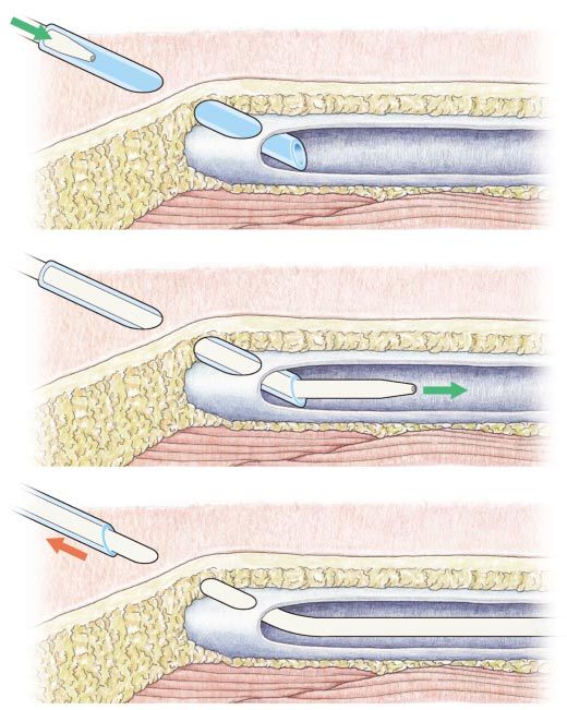
(A) Catheter-through-needle technique
A significant improvement to the venesection was the first
percutaneous method using a metal needle. After successful
puncture of the vessel (A) the catheter is advanced through the
needle to the vena cava (B). As soon as the intended position has
been reached, the placement is checked by means of a chest
radiograph. Then the steel needle is withdrawn and fixed at the
distal hub of the catheter (C). To avoid injuring the patient, the
sharp bevel of the needle must be secured, for example with a
(B) needle guard that is placed over the distal end of the catheter
and the needle (1). This procedure represents a significant
improvement over venesection. However, the juncture between
the catheter and the puncture hole in the vessel wall is too loose
which often results in hematoma formation. Another serious dis-
advantage is the fact that the plastic catheter is inside a metal
needle. Withdrawal of the catheter through the needle must be
avoided in all situations because this can result in the shearing
off of the plastic catheter tubing. In the worst case, the sharp
(C) needle bevel cuts through the catheter. The resultant fragments
can then enter the venous blood system and cause serious
catheter embolisms (see chapter 8 “What To Do When
Complications Occur”).
This puncture technique puts the patient at unnecessary risk, as
there are other procedures that allow a safe placement of a cen-
tral venous catheter. The through-the-needle technique is not to
be performed on a routine basis.
15From venesection to the Seldinger technique
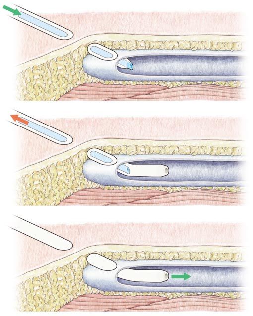
Catheter-over-needle technique (A)
Catheter-over needle kits quickly replaced the former puncture
technique due to distinct technical improvements. For this
method, a needle surrounded by a plastic cannula until close to
the needle tip is used to perform the puncture (A). Distal to the
patient, the plastic cannula gives way to a catheter, which is
surrounded by a protective sheath. After puncture of the vein,
the needle is withdrawn out of the catheter and the sheath via a
fine wire (B). The catheter is then advanced into the blood vessel
(C) (1). In contrast to the catheter-through-needle technique, (B)
there is almost no hematoma formation since the catheter over
the needle completely fills the puncture hole created by the
needle. A negative aspect of this method is the fact that a
large-diameter puncture needle must be used, which makes the
puncture of the vessel sometimes difficult. In addition, there is
no interior guidewire along which the catheter can be advanced
in the vein. This makes it difficult to successfully place the
catheter along a venous course that is not straight, for example (C)
when puncturing the subclavian vein. This puncture technique is
principally suited for routine applications and in emergency
situations. However, it requires high manual dexterity and much
experience.
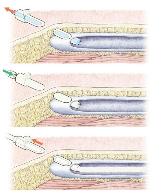
Catheter-through-cannula technique (A)
The introduction of the catheter-through cannula technique
in the Sixties greatly improved the placement security and the
patient safety. With this technique, the blood vessel is pre-
punctured with an i.v. catheter. The i.v. catheter consists of a
needle surrounded by a plastic cannula. After puncture of the
vessel, the needle is withdrawn (A) and the cannula remains in
the blood stream. The central venous catheter, which usually is
contained in a protective sheath, is connected to the cannula by (B)
an airtight coupling (B). The catheter is then advanced through
the cannula into the blood vessel. Positioning is facilitated by
means of a mandrin inside the catheter. The cannula is
removed distally after the correct catheter position has been
reached (C) (1).
As the catheter is advanced it slides over the smooth plastic
walls of the cannula and not over a sharp needle edge. The
shearing off or separation of fragments from the central venous
catheter is clearly avoided. The through-cannula technique (C)
presents fewer risks for the patient and provides significantly
better handling for the user, who is able to change the position
of the central venous catheter at any time during the placement
procedure.
The catheter-through cannula technique is part of a physician’s
standard repertoire to be used in the hospital or in emergency
situations for central venous puncture.
16Central Venous Catheters
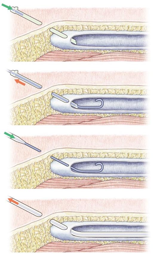
(A) Guidewire technique = Seldinger technique
The Seldinger technique was first described in 1953 for an
arterial approach (2). In the field of anesthesiology and critical
care, the puncture technique quickly acquired a leading role.
When puncturing the blood vessel, the user may choose between
a steel needle or an i.v. catheter. For safety reasons, the i.v.
catheter is preferred. When using the i.v. catheter, the steel
needle is removed so that the plastic cannula remains in the
vein. Through this cannula or alternatively a steel needle, a
(B) flexible guidewire is advanced into the vein (A). Then the needle
or cannula is removed (B). The diameter of the puncture needle
is always smaller than the central venous catheter. To facilitate
the entry of the catheter through the tissue, a dilator made of
plastic is put over the guidewire and advanced into the tissue.
Then the central venous catheter is threaded over the wire and
advanced into the vein (C). The guidewire stabilizes the plastic
catheter and facilitates its positioning. After the placement of
the catheter has been checked, the wire is removed (D).
(C)
The puncture hole in the blood vessel can be kept very small
using the Seldinger technique. This is a significant advantage
with patients suffering from clotting disorders. The central
venous catheter, which always has a larger diameter than the
puncture needle, completely fills the original puncture hole.
Hematoma formation is therefore almost entirely ruled out.
Positioning of the central venous catheter is made much easier
by the presence of the guidewire. The central venous catheter is
(D) much more readily advanced and directed through the vein
thanks to the metal guidewire.
Despite the exacting requirements for maintaining sterility and
the complexity of the puncture procedure, the Seldinger tech-
nique has come to be very widely used. It is suitable for hospital
use for all indications.
Literature
(1) Latto, I. P. et al.:
Percutaneous central venous and arterial catheterisation.
W. B. Saunders London 2000, 3rd edition: 13–31
(2) Seldinger, S. I.:
Catheter replacement of needle in percutaneous
arteriography: new technique.
Acta Radiologica 1953, 39: 368
17Selecting the proper catheter
Technical advances have made central
venous catheterization safe and easy, greatly
expanding the application of
central venous catheters. The wide range of
catheters offered by different
companies makes it possible to select an
optimal product for the particular therapy
requirements.
18Central Venous Catheters
Material choice
Central venous catheters intended for short-term use up to
30 days are usually made of polyurethane. Due to its low throm-
bosis rate this plastic material is clearly superior to polyvinyl
chloride or polyethylene, which were commonly used in former
times (1). At room temperature a catheter made of polyurethane
is sufficiently stiff to easily push it forward into the vein. After a
short time exposed to the 37° C temperature of the bloodstream,
the polyurethane becomes softer and more flexible, thus reduc-
ing the risk of irritating the venous wall. Polyvinyl chloride and
polyethylene catheters do not possess this “softening character-
istic” and therefore should no longer be used (2).
Therefore central venous catheters are used for the short-term
application espescially because of there mechanical charac-
teristics (4).
For the long-term application a lot of special catheters are
available.
They are made of silicon (3) witch is known for its high biocom-
patibilty and well proven mechanical characteristics.
19Selecting the proper catheter
Soft tip Surface quality
The quality of the catheter tip The surface and workmanship of the plastic catheter represents
is of particular importance for an important quality criterion that affects the rate of complica-
catheter placement. If the tip tions (5). Depending on the roughness of the catheter, blood
has sharp edges or uneven cells and plasma components such as fibrinogen are deposited
polymer outcroppings, these on the catheter surface. The deposited blood platelets and
product faults can injure the plasma proteins act as an initiator and center of thrombus
sensitive venous wall during formation. A smooth catheter surface, in particular at the
advancing of the catheter. lumen apertures, is therefore a crucial factor in determining
Injuries of the venous wall whether there will be rapid thrombus formation. Specialized
might lead to thrombosis for- cardiological catheters (e.g. angiography catheters) often
mation. A faulty catheter tip of display special surface modifications – such as hydrophilic
this sort also creates risks after polymers or heparin coatings – that should reduce
placement because the thrombus formation. In the anesthesiology field the
catheter moves in the blood importance of such modifications is a matter of
vessel in conjunction with the dispute.
heartbeat and might erode the
venous wall. A rounded and
readily malleable soft tip pro-
vides safety during placement
and also while the catheter is
in use.
Placement control
A modern central venous catheter should be visible along its full
length in a radiograph. To make the catheter visible in most
cases heavy metals are mixed into the plastic material. Should
some portion of the catheter tubing be cut off inside the patient
or should the catheter form a loop, the radiographic contrast
allows easy recovery of the catheter.
In many cases, a radiograph procedure is used to check the
correct placement of the catheter. In recent years, however, the
use of an ECG lead to check the position of the catheter tip has
become increasingly widespread. This technique provides a
reliable indication of catheter position even during catheter
placement (see Handbook 2). Various manufacturers offer sets
that allow the ECG signal to be conducted via a saline solution.
A simpler and more elegant method is to conduct the ECG
signal via a conductive wire like the Seldinger guidewire. In
selecting a catheter set, this aspect of checking catheter position
without additional x-ray exposure should be taken into account.
20Central Venous Catheters
Guidewire
Another important component of Seldinger systems in addition
to the catheter is the metallic guidewire. Following puncture,
this wire is advanced into the blood vessel and then serves as a
guide for the placement of the catheter (see Section 6). The
guidewire must be at once sturdy (so as to withstand high
tensile force when being pulled) and highly flexible to facilitate
the advance of the catheter. These characteristics are obtained
when special hardened steel thread is closely wrapped
around a core. In addition, many manufacturers
offer guidewires either with straight or so-called J-
tips. The J-tip gives way as soon as it encounters an
obstacle and is therefore preferred so as to protect
the venous wall. Due to this high pliability, however, it
is in rare instances difficult to find the access route to
the vena cava. In such cases, a second attempt should be
made using a straight tip. Both types of tip should, of course,
have a rounded end and not have any outcroppings.
Needles
Other components of the catheter set differ depending on the
manufacturer, the intended use and the preferences of the
user. General recommendations are therefore difficult to make.
B. Braun offers three different introduction needles for the
puncture of the vein: a Seldinger needle, an i.v. catheter or a
valve needle. When using the i.v. catheter, the catheter is
advanced through the plastic cannula that remains in the blood-
stream, making it highly unlikely that the catheter could be
sheared off by mistake. The Seldinger needle and valve needle
each consist of a steel needle; the valve needle, however, Literature
provides a second access port in a Y-fixture. Both needles are (1) Curelaru, I. et al.:
used for the Seldinger method. The guidewire is advanced Thrombogenicity in Central Venous Catheterization III.
through the steel needle. This must be performed with great care A Comparison Between Soft Polyvinylchloride and Soft
because the sharp bevel of the needle can damage the guidewire Polyurethane Elastomer, Long, Antebrachial Catheters.
(see Section 6). Both of these needle types should therefore only Acta Anaesth. Scan. 1984, 28: 204–208
be used by experienced physicians. The second port of the valve Pottecher et al.:
needle allows to advance the Seldinger wire into the blood Thrombogenicity of central venous catheters.
vessel while a syringe is attached to the needle. Europ J Anaesth 1984, 1: 361–365
(2) Pearson, M. L. and the Hospital Infection Control Practices
Advisory Committee (HICPAC): Guidelines for prevention of
intravascular-device-related infections. Infect Control Hosp
Epidemiol 1996, 17: 438–473.
(3) Moss, A. H. et al.:
Use of a silicone catheter with a Dacron cuff for dialysis
short-term vascular access.
Am J Kidney Dis 1988, 12: 492–498.
(4) Lind, T.: Stability of intravenous catheter in long term use.
Lancet 1981: 673
(5) Hecker, J. F., Scandrett, L. A.:
Roughness and thrombogenicity of the outer surfaces
of intravascular catheters.
J Biomed Mat Res 1985, 19: 381–395
21Selecting the proper catheter
Leading manufacturer offer a broad range of catheters suited to the age of the patient, the puncture site and the puncture
technique. The following table show the product range of B. Braun Melsungen indicative of the wide variety of catheters available
on the market. A summary of catheters available from B. Braun Melsungen is attached to this handbook at the end.
Certofix® Cavafix®
Seldinger guidewire with J-tip Catheter with transparent protective sheath
Catheter with soft tip, transparent extension tubing and Safsite Catheter with plastic mandrin or ECG J-wire as
valves, available with various puncture sets mandrin, available with various puncture sets
Adults Children Adults
Diameter: 4F (18G) Diameter: 3F (22G) Diameter: 3F (22G)
5F (16G) 4F (18G) 4F (18G)
6F (14G) 5F (16G)
6F (14G)
Length: 15 cm, 20 cm, 30 cm Length: 10 cm, 15 cm, 20 cm Length: 32 cm, 45 cm, 70 cm
Diameter: 7F: 16G/16G Diameter: 4F: 22G/22G Diameter: 4F: 18G/20G
14G/18G 5F: 18G/20G 6F: 16G/18G
9F: 13G/13G
12F: 11G/11G
Length: 15 cm, 20 cm, 25 cm, 30 cm Length: 8 cm, 13 cm, 20 cm Length: 20 cm, 32 cm, 45 cm, 60 cm, 70 cm
Diameter: 7F: 16G/18G/18G Diameter: 5,5F: 20G/22G/22G
12F: 16G/12G/12G
Length: 15 cm, 20 cm, 25 cm, 30 cm Length: 8 cm, 13 cm, 20 cm
Diameter: 9F: 16G/18G/18G/14G
Length: 15 cm, 20 cm, 30 cm
Diameter: 12F:
16G/18G/18G/18G/12G
Length: 15 cm, 20 cm, 30 cm
22Preparation for Catheterization
Like all anesthesiological procedures,
central venous catheterization demands
good knowledge of the patient. Preventive
measures – such as positioning of the head
or aseptic technique during inserting of the
catheter – as well as follow-up
activities such as checking the catheter
lumens for obstruction help to avoid
complications.
23Preparation for Catheterization
Anamnesis / Reviewing medical records Length measurement
The following subjects should be addressed with particular After selection of the puncture location, the necessary catheter
attention: length is determined by use of a measurement tape. When punc-
• Medication intake, in particular anti-coagulant therapy turing the right subclavian or jugular vein the correct
• Previous infectious, pulmonary or cardiac illnesses catheter position immediately before the right atrium is
• Known allergic reactions reached in 13–16 cm. The approach from the left side of the
Visual inspection of the intended puncture site and the body requires 15–20 cm. If the anatomical landmarks are
ausculation of the lungs and heart are essential elements of the unclear, it is advisable to conduct an ultrasound examination of
patient examination. If the intended puncture site is not usable the course of the vein so as to make an accurate estimate
owing to a skin ailment or if it is located in the operating area, of the required catheter length (2).
then a more suitable point of access should be
selected. An ongoing anti-coagulation therapy necessitates a Ultrasound examination of the vein
careful risk/benefit analysis and the selection of a puncture It may be advisable to conduct an ultrasound examination
site where a bleeding incident could be kept under control of the course of the vein depending on the experience of the
(e.g. jugular vein, basilic vein). user or the anatomical situation of the patient (3). If it is not
possible to get a clear imaging of the course of the vein at
Clotting status the planned puncture location, then it is better to select a
Prior to the insertion of a central venous catheter, the clotting different puncture site.
status of the patient must be known. The following clinical
parameters are taken into consideration (1):
Thrombocyte count: Normal range 150–400 x 109/l.
Thrombocytes are essential for blood clotting.
Thrombopathy begins at ≤ 30 x 109 thrombocytes/l.
With an elevated thrombocyte count, the patient must be
closely monitored following the procedure so as to quickly
recognize any developing infection.
Fibrinogen concentration: Normal range 2–4 g/l.
Fibrinogen is essential for hemostasis. At ≤ 1,20 g/l the
fibrinogen concentration is no longer sufficient for
hemostasis during an operation.
Partial thromboplastin time (PTT): Normal range:
26–40 seconds, longer with anti-coagulation therapy.
Measure for the speed of blood clotting.
Prolonged PTT times and a reduced thromboplastin time
(see below) are indicative of serious disorders in the clotting sys-
tem (e.g. consumptive coagulopathy, liver damage,
anti-coagulation therapy).
Thromboplastin time or INR: Normal range: 0.7–1 (70–100%).
Anti-coagulation therapy reduces the value to 0.15.
Measure for the speed of blood clotting. INR value of ≤ of 0.5
(delayed blood clotting) requires a drug therapy to increase the
value before a central venous catheter may be inserted.
Thrombin time (TT): Normal range 18–22 seconds.
This time becomes longer when the patient undergoes heparin
therapy or when there is a high concentration of fibrinogen
breakdown products. Measure for the speed of blood clotting.
24Central Venous Catheters
Positioning of the patient Sterile catheter placement technique
In the neck and shoulder region, the liquid pressure in the large The central venous catheter forms a sort of bridge between
veins is lower than the atmospheric pressure. Unimpeded air the outer world and the venous blood system, creating a
entry through an 18G puncture needle could therefore allow as possible pathway for the infiltration of germs.
much as 100 ml of air to enter the venous system in a single To avoid infection, strict aseptic practices should be observed
second. This can result in an air embolism and the death of the when placing the catheter. Skin disinfection of the patient and
patient. of the physician inserting the catheter is essential (5). If a
When puncturing the internal or external jugular veins as well as central line must be inserted outside of the hospital, for example
the subclavian or brachiocephalic veins, it is advisable to put the in an emergency situation, then it is necessary to use a puncture
patient’s head in the Trendelenburg position. This entails lower- technique that rules out the possibility of a contamination of
ing the head by 15°–30° with the patient in a supine position so the catheter. Commercially available catheter-through-cannula
as to increase the venous pressure (caution: cranio-cerebral systems provide safety sheaths that prevent direct contact
injuries with increased cranial pressure). between the catheter and the person inserting it. Care should be
No special positioning of the patient is necessary for puncture in taken that all other components of the system are also handled
the region of the arm. For the most frequently used puncture under aseptic conditions.
techniques the patient is placed in a dorsal position (4). In the hospital, there are no restrictions as to the employed
puncture technique arising from the surrounding sterile
conditions. Central venous puncture is performed using a
maximum sterile barrier practice. The physician performing the
puncture wears a mask, cap, gloves and gown. In the puncture
area, the patient is covered with a large, sterile drape.
Literature
(1) Hope, R. A. et al.:
Oxford Handbook of clinical medicine. Bern 1990
3rd edition: 700–701
(2) Kirby, R. R. et al.:
Clinical Anesthesia Practice.
W. B. Saunders Philadelphia 2002, 2nd edition: 531–540
(3) Fry, W. R. et al.:
Ultrasound guided central venous access. Arch Surg. 1999,
134: 738–741
(4) Latto, I. P. et al.:
Percutaneous central venous and arterial catheterization.
W. B. Saunders London 2000, 3rd edition
(5) Pearson, M. L. and the Hospital Infection Control Practices
Advisory Committee (HICPAC):
Guidelines for prevention of intravascular-device-related
infections.
Infect Control Hosp Epidemiol 1996, 17: 438–473.
25Catheter Placement with the Seldinger Method
The placement of a central venous
catheter using the Seldinger method is
easy to learn but requires some manual
dexterity. Thanks to modern catheter
technology, it is possible to prevent
some complications such as incorrect
positioning of the catheter already
during the placement procedure.
26Central Venous Catheters
1 2
Central venous puncture usually occurs in the context of a
comprehensive anesthesiological intervention with the accompa-
nying preparation of the patient (e.g. Trendelenburg position,
sedation, intubation, etc.). Because of the better accessibility it
affords, the right internal jugular vein is recommended for
right-handed physicians.
2
5 ml of a local anesthetic is injected into the
puncture area. With an attached syringe the
puncture needle is inserted in a caudal direction
at an angle of 30° to the skin between the two
bellies of the sternocleidomastoid muscle toward
the ipsilateral nipple. The vein is reached at a
depth of 2.5–4.5 cm.
1
The patient is disinfected in the puncture
area and amply covered with sterile drapes.
The head is turned to the opposite side and
slightly extended dorsally. The puncture site
is located lateral to the easily felt carotid
artery and between the two heads of the
sternocleidomastoid muscle.
27Catheter Placement with the Seldinger Method
3 4 5
3
If the blood flowing back
into the syringe is mostly
dark red and not flowing
with a pulsing rhythm
(indicative of arterial blood),
then the guidewire can be
advanced via the puncture
needle. Be sure that there is
a secure connection between
the needle and the dispenser
unit of the guidewire.
5
The central venous catheter is advanced
4 into the vein over the guidewire. A length
The guidewire is at first inserted only 5–6 cm. The puncture needle marking on the guidewire indicate when
is removed; the venous position of the guidewire must not be altered the catheter tip has almost reached the
during this procedure. The skin directly at the puncture site can be tip of the wire but the flexible J-tip
widened with a scalpel (caution: do not damage the guidewire). remains outside of the catheter. When
A dilator that can be threaded over the guidewire and advanced this point has been reached, the catheter
downward to the vein is a safer way of facilitating the subsequent and the guidewire are then advanced
introduction of the catheter. The dilator is then removed. together further into the vein.
28Central Venous Catheters
6 7 8
7
When the catheter is advanced
into the right atrium, a pro-
nounced elevation of the
P-wave occurs in the electro-
cardiogram. It must be re-
tracted approximately 2 cm
and is now positioned correctly
6 in the superior vena cava.
A universal adapter for conducting an
electrical signal from the guidewire is
attached to the distal end of the guidewire.
The ECG signal is switched over to the
8
guidewire lead. The advancement of the
All catheter lumens are
catheter (with the guidewire inside) is
checked for possible
continually monitored on the ECG screen.
obstructions using physio-
logical saline solution.
29Catheter Placement with the Seldinger Method
9 10
10
Blood on the skin at the puncture site is cleaned
away and the site is covered with a transparent
dressing. The type of catheter and any compli-
cations that may have occurred are noted in the
patient’s file.
Checking the position of the central venous catheter
The correct position of the catheter is in the vena cava directly
before the right atrium. If the catheter is too deeply inserted the
cardiac muscle can be damaged, which in the worst case can
result in the death of the patient (cardiac tamponade).
Commonly a chest radiograph is made directly after placement
of the catheter. Modern catheter sets, however, make it
possible to spare the patient this x-ray exposure by conducting
an ECG during the placement procedure. The catheter is
9 initially advanced to the point where an elevated P-wave is
The sliding fixation wing is brought into visible in the electrocardiogram; then it is retracted 2 cm.
position and the clip for catheter fixation The ECG reading returns to normal. The elimination of an
is attached. Unintended slippage of the elevated P-wave is a clear signal of the catheter’s position
catheter out of the vena cava is ruled out before the right atrium. In some circumstances, a chest
as far as possible by this arrangement. radiograph may still be necessary to rule out the occurrence
The fixation wing is attached to the skin of puncturing errors (e.g. a puncture of the pleural cavity).
with purse-string suture.
30Central Venous Catheters
Asepsis during catheter care
The puncture site must be examined daily for signs of infection
(redness), effluence and pain when pressed, so that any local
infection will be quickly recognized. A transparent dressing over
the puncture site facilitates this inspection. The dressing should
be changed in accordance with the hospital policy for catheter
care and if there is any indication of a local infection or conta-
mination of the site. A local infection which is not recognized in
time facilitates entry of bacteria and might damage the skin
around the puncture site. Depending on the degree of infection,
it may be necessary to remove and replace the entire catheter.
Testing catheter function
Despite having checked the catheter position using the ECG lead,
it is still essential to test that all the catheter lumens are free of
obstructions. A syringe filled with physiological saline solution is
connected to each of the lumens and blood is briefly aspirated.
The aspirated solution is reinjected. If the aspiration or injection
is obstructed, then the position of the catheter must be verified
with a chest radiograph and corrected if necessary. If a Seldinger
system has been used, the repositioning can be done easily.
Catheters of the over-the-needle and through-the-cannula types
can only be manipulated to a limited degree. If it is not possible
to free the catheter lumens, then the catheter must be removed.
Idle catheter lumen
Depending on the policy of the particular hospital, unused
lumens may be filled with a so-called lock solution. The lock
solution is composed of saline solution together with a heparin
and/or an antibiotic. The solution in the catheter lumen prevents
blood flowing back into the lumen. The heparin additive should
help to prevent the deposit of blood platelets and resultant clot
Literature
formation.
Kirby, R. R. et al.:
Clinical Anesthesia Practice.
W. B. Saunders Philadelphia 2002, 2nd edition: 531–534
Latto, I. P. et al.:
Percutaneous central venous and arterial catheterization.
W. B. Saunders London 2000, 3rd edition
31Catheter Management
Meticulous aseptic technique during
catheter placement and catheter care
is a prerequisite to avoid catheter-
associated infections. The infusion
system must be checked in the same
careful way as the catheter because of
the numerous possibilities for pathogen
germs to enter the catheter via the
luminal pathway.
32Central Venous Catheters
Depending on the indication central venous catheter can be used patient to become septic if the antibiotic lock method has not
short-term, e.g. for 1–2 days or up to several weeks. A longer worked.
indwelling time increases the risk for the patient to acquire a
catheter-associated infection which is one of the most serious
complications related to central venous catheterization (1). Careful catheter care includes all measures related to the
If clinical signs of a local infection at the puncture site (redness, infusion line. Any position in the infusion line that can be
tenderness, pain, heat) or of a systemic infection (fever, chilling, opened to the exterior, e.g. a stopcock or any change of the
low blood pressure) occur and blood culture from two different infusion line opens the possibility for bacterial colonization if
sites show bacteria whereas no second source for a bacteremia aseptic techniques are not adhered to. Bacteria, which once have
is obvious then a catheter-associated infection is proposed (2). entered the lumen of an infusion line, will migrate into the
catheter lumen and proliferate on it.
A scheduled change of infusion line has shown some promise to
Top priority for the catheter management is to reduce the avoid catheter-associated infections. Infusion lines which are
number of bacteria settling on the catheter's outer surface or used for infusing lipid containing solutions a change after 24 h
invading the bloodstream via the infusion lines. is recommended. Infusion lines for application of medicines or
Bacteria can attach to the catheter surface during placement if other infusion solutions can be exchanged after 48 h (3).
aseptic technique has not been properly adhered to e.g. during
emergency placement. Improper disinfection of the patient’s skin
opens the possibility for bacteria to enter the bloodstream by The importance of using a good aseptic technique will help to
migrating along the catheter. Clinical studies indicate that reduce the incidence of catheter-associated infections.
meticulous aseptic technique during catheter placement lowers
the infection risk. This means mask, cap, glove and gown for the
physician and a large sterile dressing around the puncture site
(3).
After catheter placement the puncture site is covered by a
wound dressing. In principle bacteria can quickly proliferate
beneath this dressing and migrate along the catheter into the
bloodstream if the catheter surface does not prevent this
invasion pathway. Careful, daily control of the puncture site and
the wound dressing is necessary to prevent bacterial invasion.
Clotted blood or wound secretion at the puncture site must be Literature
removed using sterile saline solution. If clinical signs of a local
infection are obvious the puncture site must be disinfected. (1) Raad, I. I. :
Experts do not recommend the use of topical antibiotics (4). Intravascular-catheter related infections.
Lancet 1998, 351: 893–898
(2) Garner, J. S.:
Depending on the recommendations of the hospital the central CDC definitions for nosocomial infections.
venous catheter is immediately removed if a catheter-associated Am J Infect Control 1988, 16: 128–140
infection has been recognized. The central venous catheter can (3) Pearson, M. L. and the Hospital Infection Control Practices
easily be exchanged if a Seldinger guidewire is used. Advisory Committee (HICPAC):
Replacing an infected catheter with a new one at the same Guidelines for prevention of intravascular-device-related
site has provoked some discussion because of the risk to infections.
contaminate the new catheter (5). Infect Control Hosp Epidemiol 1996, 17: 438–473
(4) Raad, I. I. et al.:
Prevention of central venous catheter-related infections
Instead of an immediate replacement one could try to sanitize by using maximal sterile barrier precautions during insertion.
the infected catheter. A highly concentrated antibiotic lock Infect Control Hosp Epidemiol 1994, 15: 231–238
solution is filled into the lumen for several hours. The antibiotic (5) Bach, A. et al.:
should kill the bacteria on the catheter surface. The success Infections risk of replacing venous catheters by the
rate for this method greatly differs leaving a high risk for the guidewire technique. Zbl Hyg 1992, 193: 150–159
33What To Do When Complications Occur
Central venous catheterization requires
repeated practice to minimize the risk
of complications. A correct estimation
of one’s own skills and the selection of
an appropriate puncture technique for
that skill level help to avoid unwanted
difficulties for the patient.
34Central Venous Catheters
Each puncture technique has its own risk profile – independent
of the user’s experience. The most important complications of
each access method are described in section 2: “Criteria for the
Selecting of a Puncture Site“.
The table on the following pages lists the complications that
occur most frequently or that may be life threatening (see
Literature 1–7). The second column shows when the first signs of
the complication normally become evident (Onset Time).
A strict division between early, late and long-term complications
is not possible and therefore has not been made.
If there are no symptoms of a complication in the first 15
minutes following the puncture, it cannot be assumed that
the catheterization is necessarily complication-free. Many
injuries that are caused directly during the placement of the
central venous catheter (e.g. damage to the inner wall of the
vein) first become clinically recognizable some days later.
The third column lists the clinical observations that will be
made in the event of the respective complications. The fourth
column indicates counter-measures that may be taken to limit
the effects of the complication.
Complications which can be fatal when diagnosed too late are
highlighted in red in the table.
Literature
(1) Dailey, R. H.:
Late vascular perforations by cvp catheter tips.
J Emergency Med 1988, 6: 137–140
(2) Gravenstein, N.:
In vitro evaluation of relative perforating potential of central
venous catheters: Comparison of materials, selected models,
number of lumens, and angles of incidence to simulated
membrane. J Clin Mat. 1991, 7: 1–6
(3) Fletcher, S. J. et al.:
Safe placement of central venous catheters: where should
the tip of the catheter lie?
Br. J Anaesth. 2000, 85: 188–191
(4) Timsit, J.-F.:
Central vein catheter-related thrombosis in intensive
care patients.
Chest 1998, 114: 207–213
(5) Malatinsky, J. et al.:
Misplacement and Loopformation of central venous
catheters. Acta Anaesth. Scand 1976, 20: 237–247
(6) Hennessey, B.:
Venous Air Embolism: Keep Your Patient out of Danger.
Americ. J Nurs. 1993, 93: 54–56
(7) Thomas, C. J., Butler, C. S.:
Delayed pneumothorax and hydrothorax with central venous
catheter migration. Anaesthesia 1999, 54: 987–998
35What To Do When Complications Occur
Complication Onset time
Incorrect Puncture
- Into tissue Immediately
- With perforation of the vessel Immediately
- With arterial damage Immediately
- With puncture of pleural cavity In the first 15 minutes, on the same day
- With nerve damage Immediately, in the first 15 minutes, on the same day
Incorrect catheter position
- in another vein On the same day
- single lumen openings outside Immediately, in the first 15 minutes, on the same day
the vein
- too deeply inserted in the right Immediately
atrium
- with puncture of the cardiac On the same day, within one week
muscle
Embolism
- Catheter embolism Immediately
- Guidewire embolism Immediately
- Air embolism Immediately, in the first 15 minutes
Other Disorders
- Dysrhythmia Immediately, on the same day, within one week
- Thrombosis On the same day, within one week
Infection
- Local infection Within one week
- Catheter associated infection to Within one week
the point of sepsis
36Central Venous Catheters
Observation Counter-Measures
No reflux of blood, No other observable damage New puncture attempt at the same location (up to 3 times)
or at a new location
Initially no reflux of blood; when needle is withdrawn reflux of Compression bandage, Change of puncture location
blood. Swift hematoma formation
Blood reflux in synch with pulse, brightly colored blood Compression bandage, surgical closure of vessel
No reflux of blood through lumen after infusion Removal of catheter, Pleura drainage if pneumothorax
Breathing problems, Pneumothorax occures
Absent or delayed effect of administered drugs
Paresis “wait and see”
Check with chest radiograph, with ECG
Usually coincidental chest radiograph finding If possible, repositioning of catheter; if not, removal of
catheter, new puncture
No reflux of blood through lumen, lumen obstructed for infusion If possible, repositioning of catheter; if not, removal of
After infusion: tissue tender to touch (Hydrothorax) catheter, new puncture
Absent or delayed effect of administered drugs
Arrhythmia, extrasystole Repositioning if possible
Pericardium tamponade, falling blood pressure, asystole, Pericardiocentesis, Resuscitation
cardiac arrest
Portions of the puncture needle are missing Radiographic inspection, surgical removal or “wait and see”
Portions of the catheter are missing when retracted
Portions of the guidewire are missing when retracted Radiographic inspection, surgical removal or “wait and see”
Oxygen deficiency, gasping breathing Check of all medical items in the infusion system for air
Stop of circulation tightness, respiration
Arrhythmia, extrasystole ECG examination, Drug therapy, Defibrillation
Ventricular fillibration from disturbance of cardiac impulse
propogation
Vein sensitive to pressure Sonography, Application of anticoagulant drugs, Removal
of catheter
Redness, effluence, puncture site sensitive to pressure Inspection and disinfection of puncture site
Fever or shivering, blood culture with detection of bacteria, low Broad-spectrum antibiotic therapy, Removal of catheter
blood pressure, oliguria
37Glossar
Antibiotic lock technique Instillation of an highly concentrated antibiotic solution in the catheter lumen to eradicate
bacteria on the catheter surface
Catheter Semi-rigid or soft plastic tubing of longer length used for central venous catheterization
Catheter-through-needle Technique for cvc placement: catheter is pushed through a needle
Catheter-over-needle Technique for cvc placement: needle is surrounded by catheter. After puncture needle is
retracted and catheter remains in place
Catheter-through-cannula Technique for cvc placement: needle is surrounded by cannula and retracted after puncture.
Catheter is pushed through the cannula
Cannula Short and rigid plastic tube, mainly used as intravenous catheter for short-term use
Central venous catheter Venous catheter which has been placed either via peripheral veins or via large bore veins close
to the heart; its tip lies in the vena cava or close to the heart
Hemothorax Accumulation of blood beneath the pleura due to simultaneous puncturing of a large blood
vessel and the pleura
Hydrothorax Accumulation of infusion solution beneath the pleura due to malposition of a catheter tip
Intravenous catheter Short venous catheter which is always placed via a peripheral vein
Lock solution Physiological saline solution with or without heparin which is instilled in an idle catheter lumen
to prevent clot formation
Midline Peripherally inserted venous catheter whose tip doesn’t lie in the vena cava superior but more
peripherally
Needle Metal tube with bevel to puncture tissue and blood vessels
Pneumothorax Collapse of one or both lungs due to puncturing of the pleura and loss of pressure
PICC Peripherally inserted central venous catheter whose tip lies in the superior vena cava
Seldinger technique Technique for cvc placement: a metal guidewire is advanced through the puncture needle
or i.v. catheter into the vein; the central venous catheter is threaded over the wire and after
correct placement of the catheter just before the atrium the wire is retracted
Trendelenburg position About 15° inclined position of head and chest to increase blood volume in abdominal veins
Tunneled catheter Exit site of central venous catheter is remote to the venipuncture site in order to prevent fast
migration of skin bacteria through the puncture site into the blood vessel
Valsalva maneuver Expiration of patient through nose with closed lips, increases blood volume in subclavian veinCentral Venous Catheters
B. Braun Melsungen AG
                               P.O.Box 11 20
                               D-34209 Melsungen
                               Tel (0 56 61) 71- 0
                               www.bbraun.com
B. 03. 03. 05/1 Nr. 606 2686   www.cvc-partner.com
You can also read